Proceeding Paper

Examination of Bcl-2 and Bax Protein Levels for Determining the Apoptotic Changes in Placentas with Gestational Diabetes and Preeclampsia †

1
Department of Histology and Embryology, Faculty of Medicine, Dicle University, 20100 Diyarbakır, Turkey
2
Department of Gynecology and Obstetrics, Faculty of Medicine, Dicle University, 20100 Diyarbakır, Turkey
*
Author to whom correspondence should be addressed.
Presented at the 2nd International Cell Death Research Congress, Izmir, Turkey, 1–4 November 2018.
Proceedings 2018, 2(25), 1548; https://doi.org/10.3390/proceedings2251548
Published: 5 December 2018
(This article belongs to the Proceedings of The 2nd International Cell Death Research Congress)

Abstract

:
Anti-apoptotic Bcl-2 and proapoptotic Bax genes are the most significant genes that are involved in the regulation of apoptosis. Abnormal apoptotic activity in preeclampsia and gestational diabetes is caused by dysregulation of these genes. In this study; we examined Bcl-2 and Bax protein expressions using immunohistochemical techniques in human placental tissue samples from cases of gestational diabetes (n: 20) and preeclampsia (n: 20). It was observed that Bax expression showed positive reaction compared to Bcl-2 expression so; Bax protein was thought to be an effective marker in determining apoptotic changes in placentas with gestational diabetes and preeclampsia.

1. Introduction

Programmed cell death (apoptosis) plays a key role in the physiology of human placenta [1]. Anti-apoptotic Bcl-2 and proapoptotic Bax genes are the most significant genes that are involved in the regulation of apoptosis [2]. One of the proapoptotic members of the Bcl-2 family is Bax and it is expressed in the villous trophoblast in the first and third trimester. It is localized in cytoplasm of the cytotrophoblast in the first trimester and also expressed in syncytiotrophoblast in the third trimester. Researches indicated that Bcl-2 was expressed at low levels during the entire gestational period but, Bax was low during the first trimester and increased by the end of gestation means that Bcl-2 and Bax genes perform in harmony to maintain programmed cell death in placenta [3]. Abnormal apoptotic activity in placental dysfunctions was shown to be caused by dysregulation of these genes [4]. The role of apoptotic turnover in the development of placental pathology such as preeclampsia was reported in previous studies [5,6]. Apoptosis increases in complicated pregnancies such as preeclampsia, diabetes and fetal growth restriction [6]. A theory based on the idea that cell kinetics change in placentas with pregnancy disorders such as preeclampsia is reported by another study showing an increase of Bax expression in all the placental sections [7]. Studies indicated that there is a relationship between hyperglycemia and apoptosis in congenital malformations and pregnancy loss in diabetic women. The aim of this present study was to examine Bcl-2 and Bax protein expressions using immunohistochemical techniques in human placental tissue samples from cases of both gestational diabetes and preeclampsia.

2. Experimental Procedure

2.1. Samples

Placenta samples with gestational diabetes (n: 20) and preeclempsia (n: 20) were collected from Dicle University Hospitals Department of Gynecology and Obstetrics Clinics. All experimental protocols were approved by the Human Research Ethics Committee of Dicle University and the consent form was obtained from each pregnant.

2.2. Immunohistochemistry
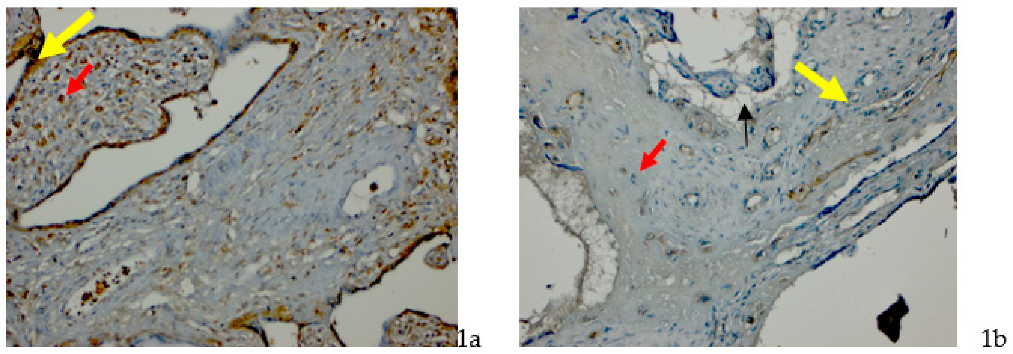
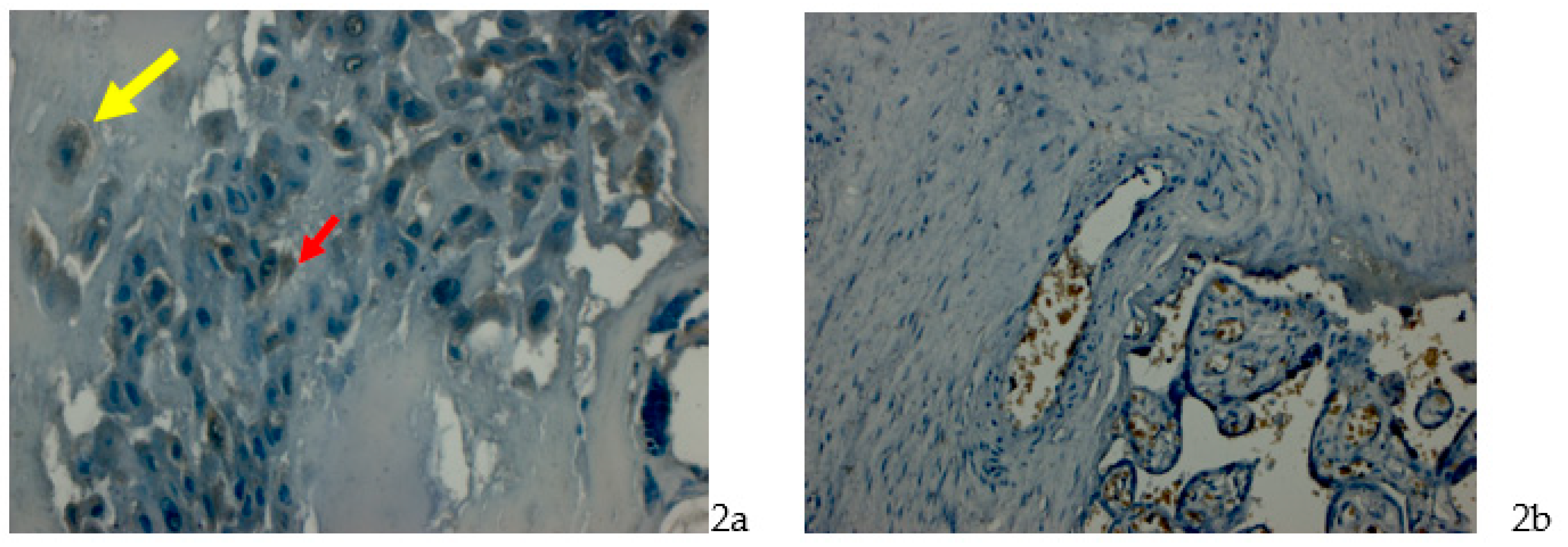
Samples taken from the maternal area of placentas were placed to 10% neutral formaldehyde solution. Following the routine paraffin protocol, 4–6 µm paraffin sections were cut with a microtome (Leica, Germany). Antigen retrieval process was performed in citrate buffer solution (pH: 6.0) two times (5 min and 3 min, distinctly) in microwave oven at 700 W. The sections were allowed to cool at room temperature for 20 min and washed in distilled two times water for 4 min. Endogenous peroxidase activity was blocked in 10% Hydrogen peroxide solution for 7 min. Ultra V block (Histostain-Plus Kit, Novex Life Tech., Frederick, MD, USA) was applied for 8 min prior to the application of primary antibodies (Bax Antibody Kit, Santa Cruz Biotech., Santa Cruz, CA, USA), (Bcl-2 antibody, Santa Cruz Biotech., Santa Cruz, CA, USA) for overnight. Secondary antibody (Histostain-Plus Kit, Novex Life Tech., Waltham, MA, USA) was applied for 20 min. The sections were then exposed to streptavidin-peroxidase for 20 min. Diaminobenzidine (DAB-Plus Substrate Kit, Novex Life Tech., Waltham, MA, USA) was used as a chromogen. Control slides were prepared as mentioned above but omitting only the primary antibodies step. After counterstaining with Hematoxylin, washing in tap water for 3 min and in distilled water for 2 × 3 min, the slides were mounted. Sections were examined under light microscope (Carl Zeiss Imager A2, Oberkochen, Germany).

3. Results

In maternal face of preeclampsia group; in some decidual cells, root villi, syncytial nodes and capillary endothelial cells, Bax protein expression was evaluated as positive (Figure 1a,b). In gestational diabetes group; Bax protein expression showed positive reaction in decidual and cytotrophoblast cells. In preeclampsia group; a weak Bcl-2 expression was seen in decidual cells, syncytial nodes and cytotrophoblast cells. Cells on maternal and fetal faces of gestational diabetes group showed negative Bcl-2 expression (Figure 2a,b).

4. Discussion

Apoptosis increases during normal pregnancy as a process of normal placental development and thereby provide placental homeostasis [1]. It was demonstrated that apoptosis also increases in pregnancies complicated by certain pathologies such as preeclampsia, fetal growth restriction, and diabetes. A number of studies indicated a significant increase in placental apoptosis, which may be the underlying cause in the pathophysiology of preeclampsia but a study of Kos and Matkovic did not confirmed these findings [5]. A study of Magee et al. (2014) also suggested that apoptosis is reduced in gestational diabetic placentas according to the dysregulation of apoptotic and inflammatory genes [4]. Eventually, there is a disagreement on apoptotic process leading to diseases of placenta. Bcl-2 protein family is known to be responsible for the regulation of apoptosis [1].
Bcl-2 inhibits programmed cell death while Bax induces and cellular sensitivity is arranged according to their balance. Cobellis et al. [6] observed a strong increase of Bax expression in the cytotrophoblast, stroma, endothelial cells and decidua of aborted placentas of the first trimester. They also reported an increase of Bax expression in preeclamptic placentas compared to the normal full-term placentas. Besides, they noted a moderate Bax expression in diabetic placentas that is lower than the normal full-term placentas. A study of Daher et al. on Bcl-2 and Bax expressions in pre-term, term and post-term placentas demonstrated that immunostaining results for Bcl-2 and Bax was the same in all placenta samples with different intensities. They indicated an intense staining so a high Bax/Bcl-2 ratio in pre-term and post-term placentas compared with term placentas and for Bax in pre-term and post-term placentas also and, a decrease for Bcl-2 in pre-term placentas [2]. In this study we also observed positive reaction of Bax protein in placentas with both gestational diabetes and preeclampsia. Sgarbosa et al., compared Bcl-2 expression in normoglycemic and diabetic placentas and showed a lower expression of Bcl-2 protein in hyperglycemic placentas that may lead to apoptosis causing pathologies [1]. Bcl-2 protein was localized in the cytoplasm of syncytiotrophoblast to maintain syncytial integrity in normal pregnancy. We observed a weak reaction of Bcl-2 protein in syncytial nodes, decidual and cytotrophoblast cells in preeclampsia group. But, in gestational diabetes group Bcl2 expression was negative in cells located on maternal and fetal faces. In another study, Bax protein was expressed mainly in connective tissue and perivascular cells within the villous core, showed a weak reaction in cytotrophoblast and negative reaction in syncytiotrophoblast [8]. We also observed Bax positive reaction in decidua cells, root villi, syncytial nodes and also in endothelial cells of capillaries in preeclampsia group. Meanwhile in gestational diabetes group, a positive reaction of Bax was observed in descidual and cytotrophoblast cells. As a consequence, we found positive Bax expression but, weak Bcl-2 expression in both preeclampsia and gestational diabetes groups so, when compared to each other it can be suggested that Bax protein may be an effective marker in determining apoptotic changes in placentas with gestational diabetes and preeclampsia.

Author Contributions

E.G.-O. designed the experiments and wrote the paper, E.D. designed the experiments and analyzed the data, F.A. and others performed the experiments.

Conflicts of Interest

The authors declare no conflict of interest.

References

  1. Sgarbossa, F.; Barbisan, L.F.; Brasil, M.A.; Costa, E.; Calderon, I.M.; Gonçalves, C.R.; Bevilacqua, E.; Rudge, M.V. Changes in apoptosis and Bcl-2 expression in human hyperglycemic, term placental trophoblast. Diabetes Res. Clin. Pract. 2006, 73, 143–149. [Google Scholar] [CrossRef] [PubMed]
  2. Daher, S.; Guimarães, A.J.; Mattar, R.; Ishigai, M.M.; Barreiro, E.G.; Bevilacqua, E. Bcl-2 and Bax expressions in pre-term, term and post-term placentas. Am. J. Reprod. Immunol. 2008, 60, 172–178. [Google Scholar] [CrossRef] [PubMed]
  3. De Falco, M.; De Luca, L.; Acanfora, F.; Cavallotti, I.; Cottone, G.; Laforgia, V.; De Luca, B.; Baldi, A.; De Luca, A. Alteration of the Bcl-2:BAX ratio in the placenta as pregnancy proceeds. Histochem. J. 2001, 33, 421–425. [Google Scholar] [CrossRef] [PubMed]
  4. Magee, T.; Ross, M.G.; Wedekind, L.; Desai, M.; Kjos, S.; Belkacemi, L. Gestational diabetes mellitus alters apoptotic and inflammatory gene expression of trophobasts from human term placenta. J. Diabetes Complicat. 2014, 28, 448–459. [Google Scholar] [CrossRef] [PubMed]
  5. Kos, M.; Matkovich, E. Bcl-2 and Bax immunoreactivity in placentas from pregnancies complicated with intrauterine growth restriction and hypertension. Period. Biol. 2014, 116, 167–172. [Google Scholar]
  6. Cobellis, L.; De Falco, M.; Torella, M.; Trabucco, E.; Caprio1, F.; Federico, E.; Manente, F.; Coppola, G.; Laforgia, V.; Cassandro, R.; et al. Modulation of Bax Expression in Physiological and Pathological Human Placentas Throughout Pregnancy. In Vivo 2007, 21, 777–784. [Google Scholar] [PubMed]
  7. Crocker, I.P.; Cooper, S.; Ong, S.C.; Baker, P.N. Differences in apoptotic susceptibility of cytotrophoblasts and syncytiotrophoblasts in normal pregnancy to those complicated with preeclampsia and intrauterine growth restriction. Am. J. Pathol. 2003, 162, 637–643. [Google Scholar] [CrossRef]
  8. Ratts, V.S.; Tao, X.J.; Webster, C.B.; Swanson, P.E.; Smith, S.D.; Brownbill, P.; Krajewski, S.; Reed, J.C.; Tilly, J.L.; Nelson, D.M. Expression of Bcl-2, Bax and Bak in the trophoblast layer of the term human placenta: A unique model of apoptosis within a syncytium. Placenta 2000, 21, 361–366. [Google Scholar] [CrossRef] [PubMed]
Figure 1. (a) Preeclampsia group, an increased positive expression in decidual cells (red arrow), syncytitial nodes (yellow arrow) and endothelial cells; Bax immunohistochemistry stain, Bar 100 µm. (b) Preeclampsia group, a weak Bcl-2 expression in decidual cells (red arrow), syncytial nodes (arrow) and cytotrophoblast cells (yellow arrow); Bcl-2 immunohistochemistry stain, Bar 100 µm.
Proceedings 02 01548 g001
Figure 2. (a) Gestational diabetes mellitus group, positive Bax protein expression in decidual cells (yellow arrow) and cytotrophoblast cells (red arrow); Bax immunohistochemistry stain, Bar 50 µm. (b) Gestational diabetes mellitus group, negative Bcl-2 expression in maternal and fetal faces; Bcl2 immunohistochemistry stain, Bar 100 µm.
Proceedings 02 01548 g002
Publisher’s Note: MDPI stays neutral with regard to jurisdictional claims in published maps and institutional affiliations.

