Article

Changes in Endothelial Function after Acute Resistance Exercise Using Free Weights

Cardiovascular Dynamics Laboratory, Exercise Physiology, 161F MACC Annex, Kent State University, Kent, OH 44242, USA
*
Author to whom correspondence should be addressed.
J. Funct. Morphol. Kinesiol. 2018, 3(2), 32; https://doi.org/10.3390/jfmk3020032
Submission received: 9 May 2018 / Revised: 27 May 2018 / Accepted: 29 May 2018 / Published: 1 June 2018
(This article belongs to the Special Issue Resistance Training for Performance and Health)

Abstract

:
We determined the effects of an acute bout of free-weight resistance exercise (ARE) on cardiovascular hemodynamics and endothelial function in resistance-trained individuals. Nineteen young, healthy, resistance-trained individuals performed two randomized sessions consisting of ARE or a quiet control (CON). The ARE consisted of three sets of 10 repetitions at 75% 1-repetition maximum for the squat, bench press, and deadlift. Cardiovascular hemodynamics was assessed using finger photoplethysmography. Forearm blood flow (FBF), and vasodilatory capacity markers, were assessed using venous occlusion plethysmography. Forearm vascular conductance was calculated by the division of mean FBF by mean arterial pressure. A two-way ANOVA was used to compare the effects of condition (ARE, CON) across time (rest, recovery). There were significant (p ≤ 0.05) decreases in mean arterial pressure and total peripheral resistance across conditions and time. There were significant condition-by-time interactions (p ≤ 0.05) for heart rate, stroke volume, and cardiac output after the ARE compared to the CON and rest. FBF was significantly (p = 0.001) increased during the recovery from ARE, as well as vasodilatory capacity markers such as peak blood flow (p = 0.05) and reactive hyperemia-induced blood flow (p = 0.0001). These data suggest that whole-body free-weight exercises acutely reduced blood pressure while simultaneously augmenting FBF, and vasodilatory capacity markers.

1. Introduction

Resistance training is currently recommended by the American College of Sports Medicine due to the innumerable effects it has on health and functional capacity [1]. While the effects of resistance exercise are well known in terms of muscle strength and endurance [1,2], prevention of osteoporosis [3], and sarcopenia [4], the effects on the vasculature are less clear. Previous work that has evaluated changes in endothelial function after resistance exercise has demonstrated positive effects [5]. This particular study also investigated how heart rate (HR), cardiac output (CO), and total peripheral resistance (TPR) play a role as physiological modulators of endothelial function [5].
Furthermore, while endothelial function has been examined after an acute bout of resistance exercise (ARE) in healthy individuals, these studies have primarily focused on weight machines [5,6,7], with a few exceptions [8]. While Fahs et al. (2009) utilized free weights, the exercise regime consisted of the bench press and the biceps curl, not a whole-body routine [8]. The differences in responses of the vasculature after an acute bout of free-weight exercises compared to weight machines may be attributed to greater activation of primary muscles [9], agonist muscles [10], and/or stabilizing muscles [11]. The difference in muscle activation may make it difficult to compare the responses of free-weight exercises to those of weight machines. Because whole-body free-weight exercises are task-specific, and require some degree of familiarity and skill, the use of resistance-trained participants is a must. To our knowledge, no studies have examined cardiovascular hemodynamics or endothelial function markers such as forearm blood flow (FBF), and vasodilatory capacity (i.e., peak blood flow (BFpeak) and reactive hyperemia (RH)-induced flow) in resistance-trained individuals using solely free-weight exercises for the whole body such as the squat, bench press, and deadlift.
Accordingly, the purpose of the present study was to evaluate the effects of whole-body free-weight ARE on hemodynamics, and endothelial function markers in young, healthy, resistance-trained individuals. We hypothesized that there would be decreases in mean arterial pressure (MAP) and total peripheral resistance (TPR) with increases in HR, stroke volume (SV), and CO after ARE. In addition, we also hypothesized that there would be significant increases in FBF, BFpeak, and RH-induced flow during recovery from ARE.

2. Materials and Methods

2.1. Experimental Approach to the Problem

This study was a randomized, crossover study. Nineteen young, healthy, resistance-trained individuals volunteered to participate. Every participant completed both ARE and a quiet control (CON) session. Anthropometrics, body composition, and maximal strength data were also collected prior to the ARE and CON sessions. Participants were assessed for cardiovascular hemodynamics, FBF, vasodilatory capacity, and RH-induced flow at rest and during recovery from ARE and CON.

2.2. Subjects

Nineteen, young (18–28 years of age), healthy individuals (13 men, 6 women) self-reported that they had been engaging in resistance exercise for at least 1 year. The average time of resistance training was 8 ± 4 (mean ± standard deviation) years. Participants were 22 ± 3 years, had a height of 1.71 ± 0.11 m, and weighed 78.7 ± 17.7 kg. The men had a body weight of 86.9 ± 16.2 kg, a body fat percentage of 13.7 ± 6.2%, which resulted in 74.2 ± 8.9 kg of lean mass and 12.7 ± 8.5 kg of fat mass, while the women had a body weight of 67.0 ± 12.5 kg, with a body fat percentage of 25.3 ± 10.1%, 51.5 ± 3.9 kg of lean mass, and 15.5 ± 11.1 kg of fat mass. The 1-repetition maximum (1RM) on the squat, bench press, and deadlift were 125 ± 34, 88 ± 34, and 142 ± 38 kg, respectively. Exclusion criteria included a smoking history (within < 6 months), obesity (body mass index (BMI) ≥ 30 kg/m2), orthopedic problems, cancer, known cardiovascular disease, metabolic disease, hypertension (resting brachial blood pressure (BP) ≥ 140/90 mm Hg), taking any medications or supplements known to affect HR, BP, or endothelial function as assessed via a medical questionnaire, and/or competing in international or national competitive lifting meets. All women were tested during the mid-follicular phase of their menstrual cycle (Days 1–9). This research was approved by the Kent State University Institutional Review Board and was completed in accordance with the Declaration of Helsinki. Informed consent was obtained from each participant included in the study.

2.3. Procedures

Participants came to the laboratory a total of four times. The initial visit consisted of anthropometric measurements and assessment of maximal strength. The second visit was separated by at least 72 h and consisted of verification of muscular strength. For the third and fourth visits, the participants reported to the Cardiovascular Dynamics Laboratory for either the ARE or CON, which were performed in a randomized design based on an online randomization software. All testing occurred between the hours of 6–11 AM in order to control for diurnal variation. All participants were ≥3 h postprandial and had avoided caffeine, alcohol, and strenuous exercise for at least 24 h prior to testing. The modalities were separated by a minimum of one week, a maximum of ten days, and were completed at the same time of day (±1 h). The temperature of the room was constant at approximately 22 °C. Upon arriving at the laboratory, participants rested quietly in the supine position for a period of ten minutes. Cardiovascular hemodynamics, FBF, BFpeak, and RH-induced flow were assessed over the ensuing 10 min of rest. The resistance exercise bout consisted of three sets of 10 repetitions at 75% of the 1RM on the squat, bench press, and deadlift, with 2 min of rest between sets and exercises, which was completed in 30 min. Within 3 min of completing the ARE, participants returned to the supine position during which all instrumentation was reapplied to assess recovery. The CON was matched for time with the ARE and consisted of 30 min of supine rest. Cardiovascular hemodynamics and all measurements of blood flow were assessed between 15–25 min during the recovery.

2.3.1. Anthropometric Measurement

Height and weight were measured using a stadiometer and a beam balance platform scale, respectively. Body composition was determined using 7-site skinfold analysis [12]. Each site was measured twice, and if the measurements revealed differences greater than 1 mm, a third measurement was conducted. Body density was calculated using the Brozek equation [13].

2.3.2. Muscle Strength

Muscle strength was assessed by the 1RM test for three different resistance exercises. The resistance exercises were comprised of the squat, bench press, and deadlift, in that order. Each 1RM was assessed within five attempts following a warm-up with 50% of the participant’s body weight, based on recommendations from the National Strength and Conditioning Association [14]. The highest resistance that was able to be lifted between the initial assessment and verification was used for the ARE and subsequent data analysis. The intraclass coefficient (ICC) for the squat, bench press, and deadlift were 0.95, 0.94, and 0.96, respectively.

2.3.3. Hemodynamics

Beat-to-beat BP was recorded during all measurements of blood flow via finger photoplethysmography (NexfinCC, BMEYE, Amsterdam, The Netherlands), which has been shown to be valid [15]. From the pressure wave, we were able to assess not only MAP, but also HR, SV, and CO. The Modelflow technique allows the pressure on the index finger to compute an aortic waveform to calculate SV [16]. The aortic waveform per beat provides measurement of left ventricular SV and thus CO through the multiplication of SV and HR. TPR was then calculated through the division of MAP by CO.

2.3.4. Forearm Blood Flow

Resting FBF was assessed using a mercury-in-silastic strain gauge plethysmography (EC-6; DE Hokanson Inc., Bellevue, WA, USA). For a more detailed explanation of this method, see Higashi et al. [17]. Briefly, the arm is elevated above heart level while the participant is supine. The circumference of the widest portion of the left forearm was quantified and the appropriate strain gauge was attached and then connected to the plethysmography. A cuff was wrapped around the wrist and inflated to 220 mm Hg 1 min prior to and throughout the measurements of FBF. Another cuff was placed at the most proximal portion of the left arm over the brachial artery and was inflated to 50 mm Hg for 7 s of each 15-s measurement cycle using a rapid cuff inflator (EC-20; DE Hokanson Inc., Bellevue, WA, USA) in order to occlude venous flow. All data were saved by the program and analyzed using a Noninvasive Vascular Program 3 Software Package (DE Hokanson Inc., Bellevue, WA, USA). Six measurements were averaged from the plethysmograph in order to determine resting FBF both at rest and during recovery. Forearm vascular conductance (FVC) of the arm was calculated by the division of mean forearm blood flow by MAP. Immediately after resting FBF was determined, the circulatory occlusion was induced by inflating the brachial cuff to 220 mm Hg for 5 min in order to assess vasodilatory capacity markers. One minute prior to release of the brachial cuff, the wrist cuff was inflated to 220 mm Hg for the subsequent measurements. At the end of the 5 min, the brachial cuff was released to induce RH. Blood flow was measured for the next 3 min (13 total readings). The highest reading was recorded as BFPeak. The measurements taken after the first minute until the third minute, measurements 4 through 13, were graphed into a curve, and the area under the curve was taken as a measure of RH (AUCRH). This segment, the mid-to-late phase, of the RH response has been shown to be dependent on the endothelium (nitric oxide (NO) dependent) [18].

2.3.5. Acute Bout of Resistance Exercise

Participants warmed up on a cycle ergometer for 5 min before resistance exercise. The ARE consisted of three sets of 10 repetitions at 75% 1RM on the squat, bench press, and deadlift, in that order. Two minutes rest was given between sets and exercises. Sessions were supervised by strength and conditioning specialists, providing a spotter for the squat and bench press, if needed. If participants could not complete all 10 repetitions for the given set, then the weight was reduced for the next set. Each session lasted approximately 30 min.

2.4. Data Analysis

Our sample size was based on data in our laboratory that was collected under identical conditions using seven healthy, resistance-trained participants. From our pilot data, an effect size for the outcome variable of FBF estimated a sample size of 15 participants giving us an effect size of 1.2, with an alpha of 0.05 and a power of 80%. A 2 × 2 repeated measures ANOVA was used to test the effects of condition (ARE and CON) across time (rest and recovery) on hemodynamics (MAP, HR, SV, CO, and TPR) and endothelial function (FBF, BFPeak, AUCRH, and FVC) parameters. AUCRH was calculated using GraphPad Prism 5.0 (GraphPad, La Jolla, CA, USA) using the trapezoidal rule. If the interactions were significant using the ANOVA, paired t-tests were used to determine significant differences. Partial eta squared ( η p 2 ) was used to assess the effects size of each variable [19]. Significance was set a priori at p ≤ 0.05. Values are presented as mean ± standard deviation (SD). All statistical analyses were completed using IBM SPSS version 21 (IBM, Armonk, NY, USA).

3. Results

3.1. Hemodynamics

Hemodynamics are presented in Table 1. There were significant condition-by-time interactions for MAP (F1,37 = 8.8, p = 0.005, η p 2 = 0.19) and TPR (F1,37 = 101.3, p = 0.0001, η p 2 = 0.73), such that they were decreased during recovery from the ARE compared to the CON. There were also significant condition-by-time interactions for HR (F1,37 = 309.2, p = 0.0001, η p 2 = 0.89), SV (F1,37 = 9.3, p = 0.004, η p 2 = 0.20), and CO (F1,37 = 48.8, p = 0.0001, η p 2 = 0.56), such that they were augmented after the ARE but not after the CON from rest to recovery.

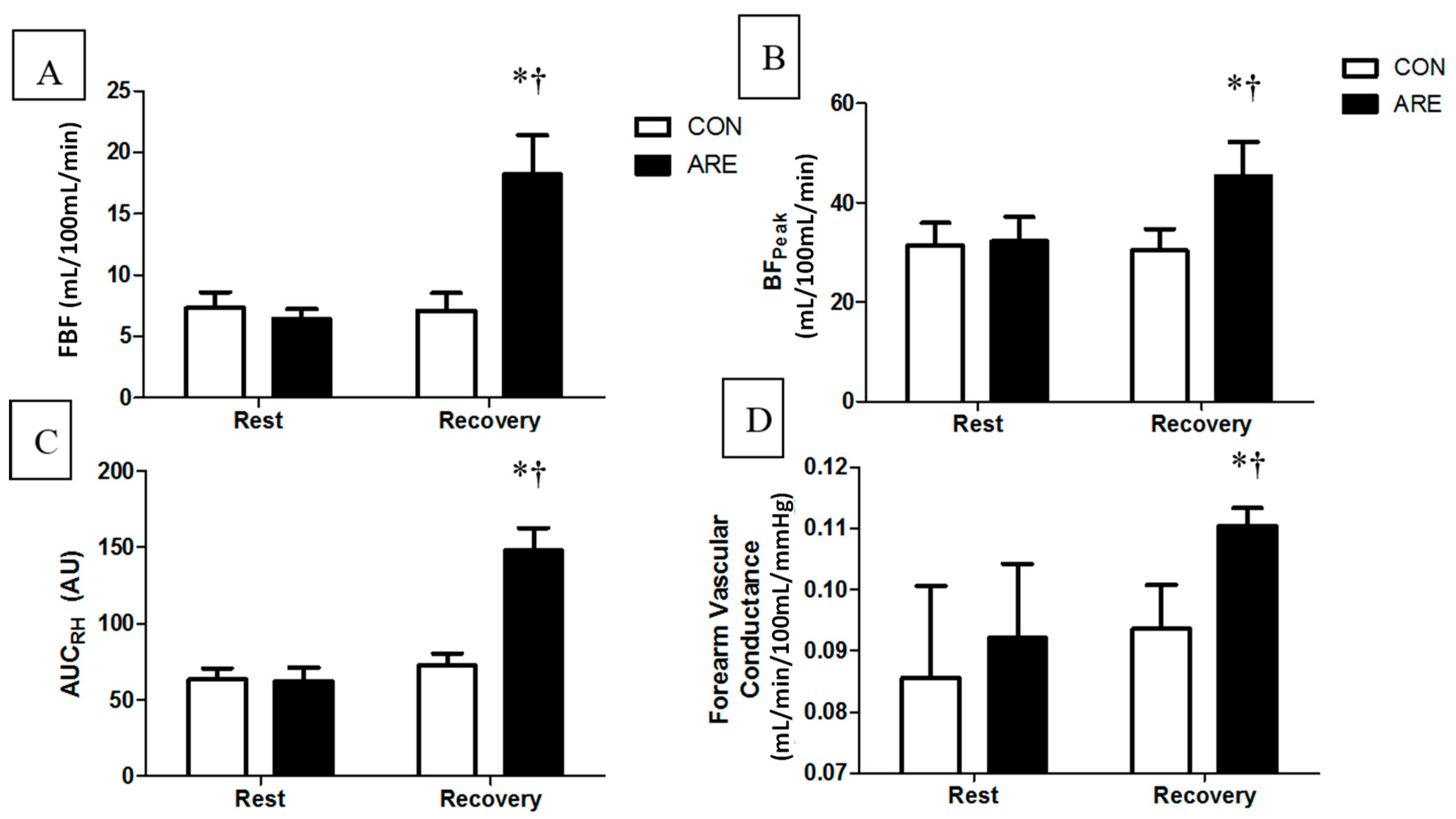
3.2. Endothelial Function

Blood flow data are presented in Figure 1. There was a significant condition-by-time interaction for FBF (F1,37 = 23.1, p = 0.0001, η p 2 = 0.38) and BFPeak (F1,37 = 8.94, p = 0.006, η p 2 = 0.18), such that they were increased after ARE during the recovery. There was also a significant condition-by-time interaction for AUCRH (F1,37 = 19.3, p = 0.0001, η p 2 = 0.34), as well as FVC (F1,37 = 21.8, p = 0.0001, η p 2 = 0.38), such that they were increased after ARE and demonstrated no change after the CON.

4. Discussion

This investigation examined the acute effects of free-weight resistance exercises on hemodynamics and endothelial function in resistance-trained individuals. The primary findings of the present study are: (1) that ARE decreases MAP and TPR with concomitant increases in HR, decreases in SV, and increases in CO; (2) that measures of endothelial function are significantly increased after ARE. Our findings suggest that an acute bout of resistance exercise has a positive impact on brachial blood pressure and endothelial function in resistance-trained individuals.
In agreement with previous research, our data demonstrate that ARE has a significant effect on hemodynamics [20]. Our results show that ARE decreased MAP in resistance-trained individuals, which is consistent with a previous study performing whole-body free-weight resistance exercise in resistance-trained men [20]. Halliwill et al. (1996) reported increased sympathoinhibiton and decreased vascular responsiveness as primary factors that influence reductions in MAP after exercise [21]. While Halliwill et al. (1996) utilized aerobic, not resistance exercise, the same physiological alterations may explain the results of the current study. In a previous study, we reported significant decreases in baroreflex sensitivity using the same protocol as the present study [22], which is supported by other researchers [23]. While it is not known if baroreflex resetting occurs after acute resistance exercise, similar to aerobic exercise, it has been shown to occur after a period of resistance training [24], thereby reducing sympathetic outflow to the central nervous system. However, this is speculation, as we did not measure resetting of the baroreflex or sympathetic outflow in the present study. In regards to changes that are specific for resistance exercise, a previous study has argued that a large total volume of resistance exercise is essential to the onset of post-exercise hypotension [25]. However, our study, with a low total exercise volume and a higher muscle mass recruitment due to the involvement of the primary [9], agonist [10], and stabilizing [11] muscles of free-weight exercises, may have displaced the importance of the total volume to a secondary role. In this sense, the muscle mass involved is another important co-factor in the onset of post-exercise hypotension [26]. Additionally, it was previously pointed out that designs such as ours, with intensities around 70–80% 1RM, are very effective at producing a reduction in blood pressure in resistance-trained individuals, comparatively to lower intensities [20,27]. The decrease in MAP was manifested by a decrease in TPR that was not fully compensated by the increases observed in CO. Such hemodynamic changes were previously shown in the supine position after a resistance exercise bout with a comparable intensity and a combination of machines and free weights [5]. However, it is contrary to data reported in the seated position after a resistance exercise bout with a similar intensity, resulting in an increase in TPR and a reduction in CO due to a decrease in SV [28]. This suggests that the hemodynamic responses after resistance exercise may be posture-dependent, which is different to the previously believed notion that any resistance exercise produces a decrease in SV, and thus a decrease in CO [28]. These differences may be due to the facilitation of the venous return that occurs in the supine position, which helps to increase cardiac preload and thus augment SV in comparison with the seated position. In this sense, Collier et al. (2010) observed no differences while we observed a slight decrease in SV.
The novelty of our methods suggests that whole-body resistance exercise has a positive, transient effect on endothelial function in resistance-trained individuals. In agreement with the present study, Collier et al. (2010) reported significant increases in FBF, and vasodilatory capacity markers such as BFpeak and AUCRH, after an acute bout of resistance exercise [5]. In their study, participants underwent three sets of 10 repetitions at the 10RM on eight different exercises and 90-s rest breaks, which included a combination of resistance machines and free weights. Our data also support the findings of Fahs et al. (2009), which reported an increase in FBF, vasodilatory capacity markers (BFPeak and AUCRH) after an ARE [8]. Their intervention consisted of participants performing 80% 1RM on the bench press for four sets of five repetitions, followed by four sets of 10 repetitions on the biceps curl at 70% 1RM. The increases in vasodilatory capacity reported in the present study, Collier et al. (2010), and Fahs et al. (2009), may be explained by augmented endothelial function. The increase in endothelial function may be further attributed to increased blood flow and the concomitant increase in the transient shear stress [29]. Preceding the increased blood flow to the working muscle, CO must increase to allow the distribution of blood to the peripheries. Henceforth, a redistribution of blood flow to the exercising muscles occurs, which causes the working muscles to dilate, thus reducing MAP and TPR. It has been shown that shear stress during exercise acts to increase NO signaling and may also increase other endothelium-dependent vasodilatory signals such as acetylcholine, prostaglandins, and endothelial-derived hyperpolarizing factors [30,31,32]. This would explain increases in vasodilatory capacity markers [18,29,33], which is supported by our data. On the contrary, a previous study observed reduced FBF while maintaining AUCRH in resistance-trained individuals after a resistance exercise consisting of bilateral knee extension [25]. This may indicate that the capacity to increase blood flow after resistance exercise may be more local than systemic, being restricted only to the limbs actively involved during the resistance exercise session. This local capacity may suggest the importance of performing whole-body routines when the objective is an increase in endothelial function in all extremities, as was previously reported in a whole-body design with a mixture of machines and free weights [5]. Nevertheless, we did not measure lower limb endothelial function (e.g., calf blood flow) in our study, so this inference remains speculative at this time. With six weeks of resistance training, a previous study reported an improvement in endothelial function in healthy individuals [34], putting into consideration a possible relationship between the acute and chronic responses to resistance exercise that should be analyzed in the future. In brief, the present study supports the notion that ARE has a favorable effect on FBF in young, healthy, resistance-trained individuals. In addition, we noted increases in vasodilatory capacity markers, which demonstrates that different aspects of the microvasculature are responding as expected (NO only plays a role in the mid-to-late phase of reactive hyperemia) [18,35].
The present study is not without limitations. Both men and women were used in this study, and that could be a confounding factor. Indeed, sex differences were previously observed after resistance training in FBF [36]. However, that was a training study that did not examine acute alterations and utilized pre-hypertensive and stage 1 hypertensive participants. Nevertheless, to the best of our knowledge, there are no studies that have analyzed the possible sex differences that occur after an ARE. In addition, with our sample size of six women, the ability to draw conclusions based specifically on sex is limited. To try to reduce these potential effects, since despite not being conclusive [37], we did control for the menstrual cycle, as it has been pointed out that different phases of the menstrual have a direct effect on the vasculature [38,39]. Finally, we did not measure lower limb endothelial function (e.g., calf blood flow). Therefore, the responses in the leg blood flow to a whole-body free-weight design of resistance exercise is unknown.

5. Conclusions

In conclusion, ARE is associated with post-exercise hypotension manifested by decreases in total peripheral resistance. This, in turn, significantly increased FBF, and vasodilatory capacity markers such as BFpeak and AUCRH. Future studies should investigate physiological mechanisms regarding endothelial function to further determine the relationship between post-exercise hypotension, changes in hemodynamics, and increases in endothelial function after an acute bout of resistance exercise, and the relationship between acute and chronic responses in endothelial function with resistance training.

Author Contributions

Conceptualization: Y.L.T. and J.D.K.; Formal analysis: J.D.K.; Investigation: Y.L.T.; Methodology: E.M., J.C.P., X.M. and A.G.; Project administration: Y.L.T.; Supervision: J.D.K.; Writing of original draft: Y.L.T. and J.D.K.; Review and editing: E.M., J.C.P., X.M. and A.G.

Acknowledgments

We acknowledge the support of the Inditex-UDC Predoctoral Grant for Xian Mayo. This grant did not cover costs associated with the study. It only paid for his ability to work in the laboratory as a Visiting Scholar.

Conflicts of Interest

The authors declare no conflict of interest.

References

  1. Positon-Stand, American College of Sports Medicine Position Stand. Progression models in resistance training for healthy adults. Med. Sci. Sports Exerc. 2009, 41, 687–708. [Google Scholar]
  2. Kraemer, W.J.; Ratamess, N.A.; French, D.N. Resistance training for health and performance. Curr. Sports Med. Rep. 2002, 1, 165–171. [Google Scholar] [CrossRef] [PubMed]
  3. Milliken, L.A.; Going, S.B.; Houtkooper, L.B.; Flint-Wagner, H.G.; Figueroa, A.; Metcalfe, L.L.; Blew, R.M.; Sharp, S.C.; Lohman, T.G. Effects of exercise training on bone remodeling, insulin-like growth factors, and bone mineral density in postmenopausal women with and without hormone replacement therapy. Calcif. Tissue Int. 2003, 72, 478–484. [Google Scholar] [CrossRef] [PubMed]
  4. Hurley, B.F.; Roth, S.M. Strength training in the elderly: Effects on risk factors for age-related diseases. Sports Med. 2000, 30, 249–268. [Google Scholar] [CrossRef] [PubMed]
  5. Collier, S.R.; Diggle, M.D.; Heffernan, K.S.; Kelly, E.E.; Tobin, M.M.; Fernhall, B. Changes in arterial distensibility and flow-mediated dilation after acute resistance vs. aerobic exercise. J. Strength Cond. Res. 2010, 24, 2846–2852. [Google Scholar] [CrossRef] [PubMed]
  6. Yoon, E.S.; Jung, S.J.; Cheun, S.K.; Oh, Y.S.; Kim, S.H.; Jae, S.Y. Effects of acute resistance exercise on arterial stiffness in young men. Korean Circ. J. 2010, 40, 16–22. [Google Scholar] [CrossRef] [PubMed]
  7. DeVan, A.E.; Anton, M.M.; Cook, J.N.; Neidre, D.B.; Cortez-Cooper, M.Y.; Tanaka, H. Acute effects of resistance exercise on arterial compliance. J. Appl. Physiol. 2005, 98, 2287–2291. [Google Scholar] [CrossRef] [PubMed] [Green Version]
  8. Fahs, C.A.; Heffernan, K.S.; Fernhall, B. Hemodynamic and vascular response to resistance exercise with l-arginine. Med. Sci. Sports Exerc. 2009, 41, 773–779. [Google Scholar] [CrossRef] [PubMed]
  9. Escamilla, R.F.; Fleisig, G.S.; Zheng, N.; Lander, J.E.; Barrentine, S.W.; Andrews, J.R.; Bergemann, B.W.; Moorman, C.T. Effects of technique variations on knee biomechanics during the squat and leg press. Med. Sci. Sports Exerc. 2001, 33, 1552–1566. [Google Scholar] [CrossRef] [PubMed] [Green Version]
  10. McCaw, S.T.; Friday, J.J. A comparison of muscle activity between a free weight and machine bench press. J. Strength Cond. Res. 1994, 8, 259. [Google Scholar]
  11. Santana, J.C.; Vera-Garcia, F.J.; McGill, S.M. A kinetic and electromyographic comparison of the standing cable press and bench press. J. Strength Cond. Res. 2007, 21, 1271–1277. [Google Scholar] [PubMed]
  12. Harrison, G.G.; Buskirk, E.R.; Lindsay Carter, J.E.; Johnston, F.E.; Lohman, T.G.; Pollock, M.L.; Roche, A.F.; Wilmore, J.H. Skinfold thickness and measurement technique. In Athropommetric Standardization Reference Manual; Lohman, T.G., Roche, A.F., Martorell, R., Eds.; Human Kinetics: Champaign, IL, USA, 1988; pp. 55–70. [Google Scholar]
  13. Brozek, J.; Grande, F.; Anderson, J.T.; Keys, D.A. Densitometric analysis of body composition: Revision of some quantative assumptions. Ann. N. Y. Acad. Sci. 1963, 110, 113–140. [Google Scholar] [CrossRef] [PubMed]
  14. Haff, G.G.; Triplett, N.T. Essentials of Strength Training and Conditioning, 4th ed.; Human Kinetics: Champaign, IL, USA, 2016. [Google Scholar]
  15. Eeftinck Schattenkerk, D.W.; van Lieshout, J.J.; van den Meiracker, A.H.; Wesseling, K.R.; Blanc, S.; Wieling, W.; van Montfrans, G.A.; Settels, J.J.; Wesseling, K.H.; Westerhof, B.E. Nexfin noninvasive continuous blood pressure validated against Riva-Rocci/Korotkoff. Am. J. Hypertens. 2009, 22, 378–383. [Google Scholar] [CrossRef] [PubMed]
  16. Sugawara, J.; Tanabe, T.; Miyachi, M.; Yamamoto, K.; Takahashi, K.; Iemitsu, M.; Otsuki, T.; Homma, S.; Maeda, S.; Ajisaka, R.; et al. Non-invasive assessment of cardiac output during exercise in healthy young humans: Comparison between Modelflow method and Doppler echocardiography method. Acta Physiol. Scand. 2003, 179, 361–366. [Google Scholar] [CrossRef] [PubMed]
  17. Higashi, Y.; Sasaki, S.; Nakagawa, K.; Matsuura, H.; Kajiyama, G.; Oshima, T. A noninvasive measurement of reactive hyperemia that can be used to assess resistance artery endothelial function in humans. Am. J. Cardiol. 2001, 87, 121–125. [Google Scholar] [CrossRef]
  18. Tagawa, T.; Imaizumi, T.; Endo, T.; Shiramoto, M.; Harasawa, Y.; Takeshita, A. Role of nitric oxide in reactive hyperemia in human forearm vessels. Circulation 1994, 90, 2285–2290. [Google Scholar] [CrossRef] [PubMed]
  19. Richardson, J.T.E. Eta squared and partial eta squared as measures of effect size in educational research. Educ. Res. Rev. 2011, 6, 135–147. [Google Scholar] [CrossRef]
  20. Duncan, M.J.; Birch, S.L.; Oxford, S.W. The effect of exercise intensity on postresistance exercise hypotension in trained men. J. Strength Cond. Res. 2014, 28, 1706–1713. [Google Scholar] [CrossRef] [PubMed]
  21. Halliwill, J.R.; Taylor, J.A.; Eckberg, D.L. Impaired sympathetic vascular regulation in humans after acute dynamic exercise. J. Physiol. 1996, 495, 279–288. [Google Scholar] [CrossRef] [PubMed]
  22. Kingsley, J.D.; Mayo, X.; Tai, Y.L.; Fennell, C. Arterial stiffness and autonomic modulation following free-weight resistance exercises in resistance trained individuals. J. Strength Cond. Res. 2016, 30, 3373–3380. [Google Scholar] [CrossRef] [PubMed]
  23. Mayo, X.; Iglesias-Soler, E.; Carballeira-Fernandez, E.; Fernandez-Del-Olmo, M. A shorter set reduces the loss of cardiac autonomic and baroreflex control after resistance exercise. Eur. J. Sport Sci. 2015, 15, 1–9. [Google Scholar] [CrossRef] [PubMed]
  24. Tatro, D.L.; Dudley, G.A.; Convertino, V.A. Carotid-cardiac baroreflex response and LBNP tolerance following resistance training. Med. Sci. Sports Exerc. 1992, 24, 789–796. [Google Scholar] [CrossRef] [PubMed]
  25. Polito, M.D.; da Nobrega, A.C.; Farinatti, P. Blood pressure and forearm blood flow after multiple sets of a resistive exercise for the lower limbs. Blood Press. Monit. 2011, 16, 180–185. [Google Scholar] [CrossRef] [PubMed]
  26. Polito, M.D.; Farinatti, P.T. The effects of muscle mass and number of sets during resistance exercise on postexercise hypotension. J. Strength Cond. Res. 2009, 23, 2351–2357. [Google Scholar] [CrossRef] [PubMed]
  27. Figueiredo, T.; Willardson, J.M.; Miranda, H.; Bentes, C.M.; Reis, V.M.; Simao, R. Influence of Load Intensity on Postexercise Hypotension and Heart Rate Variability after a Strength Training Session. J. Strength Cond. Res. 2015, 29, 2941–2948. [Google Scholar] [CrossRef] [PubMed]
  28. Rezk, C.C.; Marrache, R.C.; Tinucci, T.; Mion, D., Jr.; Forjaz, C.L. Post-resistance exercise hypotension, hemodynamics, and heart rate variability: Influence of exercise intensity. Eur. J. Appl. Physiol. 2006, 98, 105–112. [Google Scholar] [CrossRef] [PubMed]
  29. Hutcheson, I.R.; Griffith, T.M. Release of endothelium-derived relaxing factor is modulated both by frequency and amplitude of pulsatile flow. Am. J. Physiol. 1991, 261, H257–H262. [Google Scholar] [CrossRef] [PubMed]
  30. Busse, R.; Edwards, G.; Feletou, M.; Fleming, I.; Vanhoutte, P.M.; Weston, A.H. EDHF: Bringing the concepts together. Trends Pharmacol. Sci. 2002, 23, 374–380. [Google Scholar] [CrossRef]
  31. Koller, A.; Huang, A.; Sun, D.; Kaley, G. Exercise training augments flow-dependent dilation in rat skeletal muscle arterioles. Role of endothelial nitric oxide and prostaglandins. Circ. Res. 1995, 76, 544–550. [Google Scholar] [CrossRef] [PubMed]
  32. Martin, C.M.; Beltran-Del-Rio, A.; Albrecht, A.; Lorenz, R.R.; Joyner, M.J. Local cholinergic mechanisms mediate nitric oxide-dependent flow-induced vasorelaxation in vitro. Am. J. Physiol. 1996, 270, H442–H446. [Google Scholar] [CrossRef] [PubMed]
  33. Rakobowchuk, M.; McGowan, C.L.; de Groot, P.C.; Hartman, J.W.; Phillips, S.M.; MacDonald, M.J. Endothelial function of young healthy males following whole body resistance training. J. Appl. Physiol. 2005, 98, 2185–2190. [Google Scholar] [CrossRef] [PubMed] [Green Version]
  34. Heffernan, K.S.; Fahs, C.A.; Iwamoto, G.A.; Jae, S.Y.; Wilund, K.R.; Woods, J.A.; Fernhall, B. Resistance exercise training reduces central blood pressure and improves microvascular function in African American and white men. Atherosclerosis 2009, 207, 220–226. [Google Scholar] [CrossRef] [PubMed]
  35. Engelke, K.A.; Halliwill, J.R.; Proctor, D.N.; Dietz, N.M.; Joyner, M.J. Contribution of nitric oxide and prostaglandins to reactive hyperemia in human forearm. J. Appl. Physiol. 1996, 81, 1807–1814. [Google Scholar] [CrossRef] [PubMed]
  36. Collier, S.R.; Frechette, V.; Sandberg, K.; Schafer, P.; Ji, H.; Smulyan, H.; Fernhall, B. Sex differences in resting hemodynamics and arterial stiffness following 4 weeks of resistance versus aerobic exercise training in individuals with pre-hypertension to stage 1 hypertension. Biol. Sex Differ. 2011, 2, 9. [Google Scholar] [CrossRef] [PubMed]
  37. Ketel, I.J.; Stehouwer, C.D.; Serne, E.H.; Poel, D.M.; Groot, L.; Kager, C.; Hompes, P.G.; Homburg, R.; Twisk, J.W.; Smulders, Y.M.; et al. Microvascular function has no menstrual-cycle-dependent variation in healthy ovulatory women. Microcirculation 2009, 16, 714–724. [Google Scholar] [CrossRef] [PubMed]
  38. Adkisson, E.J.; Casey, D.P.; Beck, D.T.; Gurovich, A.N.; Martin, J.S.; Braith, R.W. Central, peripheral and resistance arterial reactivity: Fluctuates during the phases of the menstrual cycle. Exp. Biol. Med. 2010, 235, 111–118. [Google Scholar] [CrossRef] [PubMed]
  39. Su, S.Y.; Wei, C.C.; Hsieh, C.L.; Tsao, J.Y.; Li, T.C.; Lin, T.H.; Chang, H.H.; Lo, L.C. Influence of menstrual cycle on pulse pressure waveforms measured from the radial artery in biphasic healthy women. J. Altern. Complement. Med. 2009, 15, 645–652. [Google Scholar] [CrossRef] [PubMed]
Figure 1. Change in resting (A) forearm blood flow (FBF), (B) vasodilatory capacity (BFpeak), and (C) area under the curve after reactive hyperemia (AUCRH), and (D) forearm vascular conductance measured at rest and during recovery from the CON and an acute bout of resistance exercise (ARE) in young, healthy, resistance trained individuals (n = 19). Data are mean ± standard deviations. * p ≤ 0.05, significantly different from rest; p ≤ 0.05, significantly different from CON.
Jfmk 03 00032 g001
Table 1. Hemodynamic parameters at rest and during recovery form a control session or an acute bout of whole-body free-weight resistance exercise in young, healthy resistance-trained individuals (n = 19).
VariableCONARE
RestRecoveryRestRecovery
MAP (mm Hg)80 ± 584 ± 781 ± 779 ± 7 *,†
TPR (mm Hg/mL/min)0.12 ± 0.0130.13 ± 0.0140.13 ± 0.030.10 ± 0.13 *,†
HR (bpm)61 ± 1058 ± 960 ± 989 ± 12 *,†
SV (mL/beat)86 ± 786 ± 786 ± 683 ± 3.3 *,†
CO (L/min)6.8 ± 0.96.5 ± 0.76.7 ± 0.97.8 ± 0.7 *,†
CON: quiet control; ARE: acute bout of resistance exercise; CO: cardiac output; HR: heart rate; MAP: mean arterial pressure; SV: stroke volume; TPR: total peripheral resistance. Data are mean ± standard deviation. * p ≤ 0.05, different from rest; p ≤ 0.05, significant from CON.

