Article

Antimicrobial Peptides Are Expressed during Early Development of Zebrafish (Danio rerio) and Are Inducible by Immune Challenge

by
Elisabetta Caccia
1,†,
Maria Agnello
2,†,
Marcello Ceci
1,†,
Patricia Strickler Dinglasan
3,
Gerardo R. Vasta
3 and
Nicla Romano
1,*
1
Dipartimento di Scienze Ecologiche e Biologiche, Università della Tuscia, 01100 Viterbo, Italy
2
Dipartimento di Scienze e Tecnologie Biologiche, Chimiche e Farmaceutiche, Università degli Studi di Palermo, 90128 Palermo, Italy
3
Department of Microbiology and Immunology, University of Maryland School of Medicine, UMB, and Institute of Marine and Environmental Technology, Columbus Center, Baltimore, MD 21202, USA
*
Author to whom correspondence should be addressed.
These authors contributed equally to this work.
Fishes 2017, 2(4), 20; https://doi.org/10.3390/fishes2040020
Submission received: 28 September 2017 / Revised: 28 October 2017 / Accepted: 30 October 2017 / Published: 8 November 2017

Abstract

:
Antimicrobial peptides (AMPS) are ancestral components in the evolution of immunity from protozoans to metazoans. Their expression can be constitutive or inducible by infectious challenge. Although characterized in detail in their structure and activity, the temporal and spatial expression of AMPS during vertebrate embryogenesis is still poorly understood. In the present study, we identified selected AMPs in zebrafish, and characterized their expression during early development, and upon experimental immune challenge in adult animals, with the goal of establishing this genetically-tractable model system for further AMP studies. By mining available genomic databases, zebrafish AMP sequences homologous to AMPs from other vertebrates were selected for further study. These included parasin I and its enzyme cathepsin D, β-defensin (DB1), liver-expressed antimicrobial peptide 2 (LEAP2), bactericidal permeability-increasing protein (BPI), and chromogranin-A and -B (CgA and CgB). Specific primers were designed for RT-PCR amplification of each AMP gene of interest and amplicons between 242 bp and 504 bp were obtained from RNA extracted from adult zebrafish. Sequencing of the amplicons and alignment of their deduced amino acid sequences with those from AMPs from other vertebrate species confirmed their identity. The temporal expression of AMPs was investigated by RT-PCR analysis in fertilized oocytes, embryos, and adult individuals. Parasin I and chatepsin D transcripts were detectable immediately after fertilization, while the transcripts for CgA and CgB became evident starting at 48 h post fertilization. Mature transcripts of LEAP2 and DB1 were detectable only in the adult zebrafish, while BPI transcripts were detectable starting from the 12th day post fertilization. To explore the possible upregulation of AMP expression by infectious challenge, experiments were carried out in adult zebrafish by intraperitoneal injection of a cocktail of lipopolysaccharide and lipoteichoic acid. Except for CgA and CgB, amplicons corresponding to all tested AMPs showed stronger signals in the experimental animals as compared to the unchallenged controls. This study provided information on the early expression of AMPs in zebrafish from ontogeny to adulthood and their inducibility by microbials. This information could be useful to actuate new prophylactic strategies as an alternative to the use of antibiotics in culture.

1. Introduction

Antimicrobial peptides (AMPs) are widely distributed in organisms playing important roles in innate immune functions against pathogens by providing a first line of defense [1]. AMPs are gene-encoded short (<100 amino acids), amphipathic molecules with hydrophobic and cationic amino acids arranged spatially, which exhibit broad spectrum antimicrobial activity, many of which may be constitutively present in the cells, and can be induced by pro-inflammatory stimuli [2,3]. In fish, some AMPs are upregulated in response to pathogens and appear to have direct broad-spectrum antimicrobial activity towards both human and fish pathogens [4]. To date, more than 2400 AMPs have been reported in the Antimicrobial Peptide Database (http://aps.unmc.edu/AP/main.php), and they exhibit remarkable sequence diversity [5] indicating a sort of specification for microorganisms. The ability of AMPs to protect fish against infections by potential pathogens has been investigated in the last two decades [6,7,8,9,10,11,12,13]. For example, parasin I is abundant in fish mucus, and is produced from a precursor molecule, the histone H2A by the enzymatic action of cathepsin D [14]. β-defensin is abundantly expressed in plants, invertebrates and vertebrates, and possesses a broad spectrum of antimicrobial activity against Gram-negative and Gram-positive bacteria, fungi and some viruses [15]. In zebrafish, three isoforms of β-defensin have been identified and characterized [16]. However, although they have been shown as expressed in several tissues (gills, gonads, muscle, kidney, skin, liver and spleen) [16], at the present time, no studies on β-defensin expression during ontogeny have been carried out. The liver-expressed antimicrobial peptide 2 (LEAP2) is an antimicrobial peptide (belonging to the hepcidin family) expressed predominantly in the liver [17]. This peptide, found in rainbow trout [18] and in embryos and larvae of catfish [19], is inducible by microbial challenge [17,19]. The bactericidal permeability-increasing protein (BPI) is a peptide produced merely from precursors of phagocytes (macrophages and granulocytes) and is stored as granules in these cells [20]. Lastly, chromogranins A and B (CgA and CgB) are peptides expressed in neuronal cells, such as chromaffin cells, and involved in the regulation of the immune response of endocrine cells [21,22].
In this study, we examined the expression of selected AMPs in zebrafish from early embryos to adults, with the ultimate goal of establishing this genetically-tractable model system for further AMP studies. The zebrafish has proven invaluable not only for developmental studies, but it has also more recently been shown to be an ideal whole organism model system for detailed investigations in innate immune responses [23,24,25]. In this study, we focused on AMPs differentially localized in distinct organs, tissues, or secretions: (a) parasin I and its enzyme cathepsin D, localized at mucosal surfaces; (b) β-defensin (DB1) and LEAP-2, which are expressed in the liver; (c) BPI (bactericidal permeability-increasing protein) present in circulating phagocytes, and (d) chromogranins A and B, present in cells involved in both immune and neuroendocrine systems.

2. Results

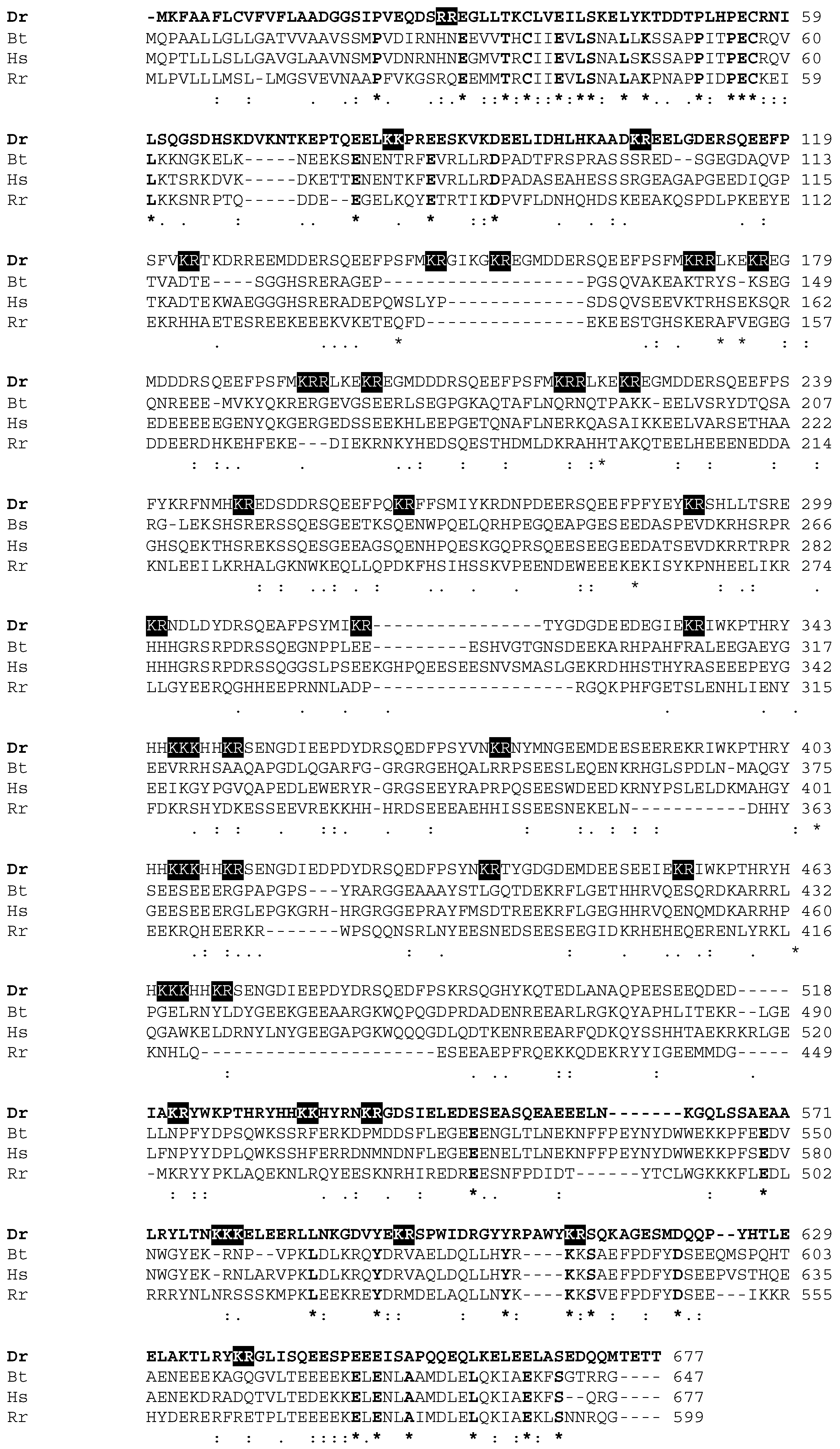
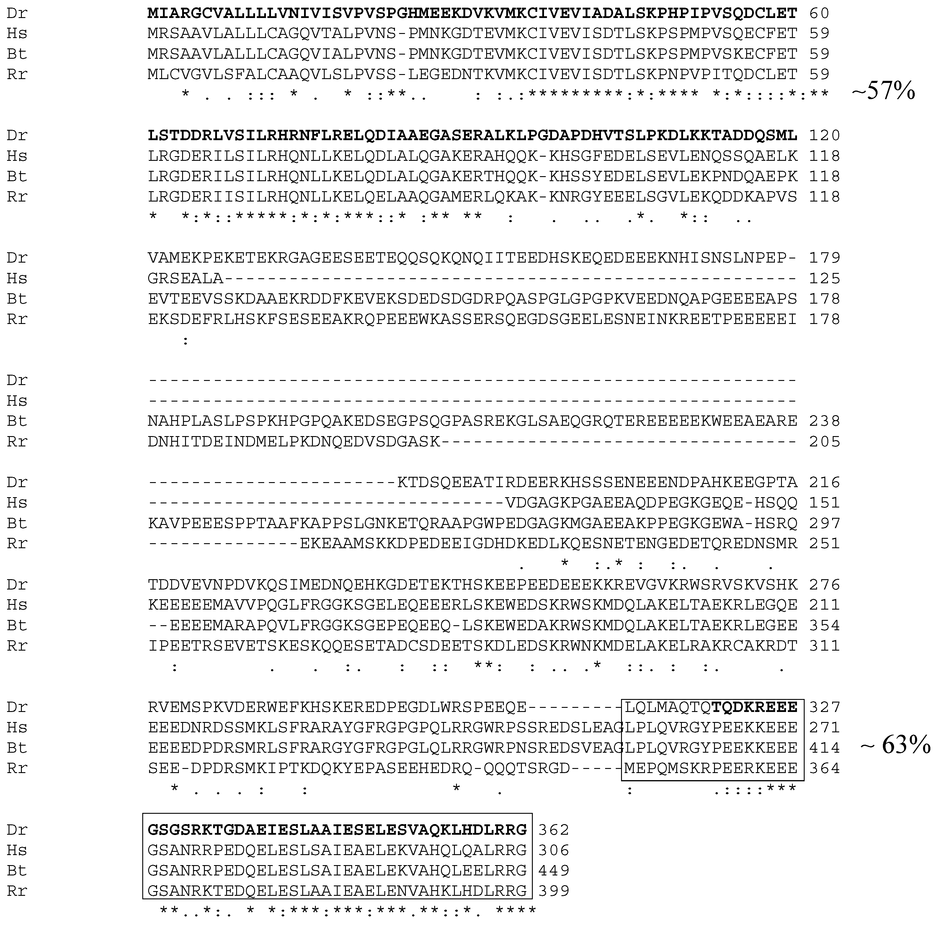
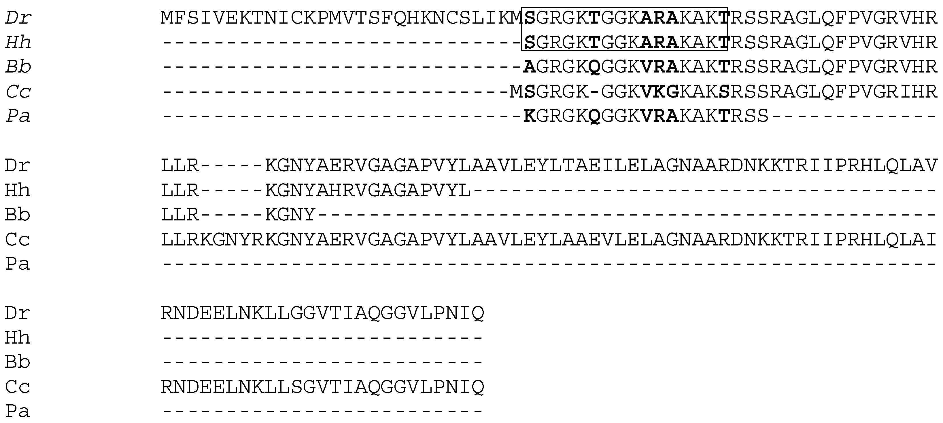
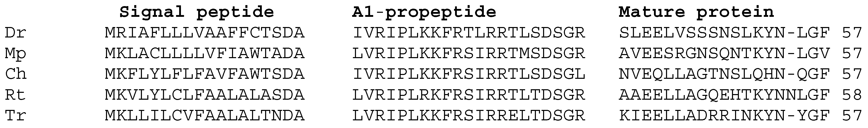
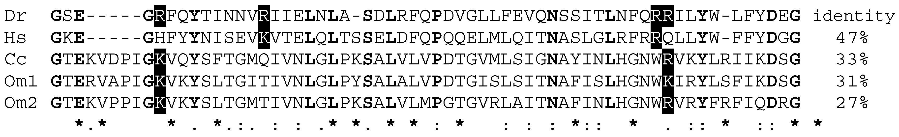
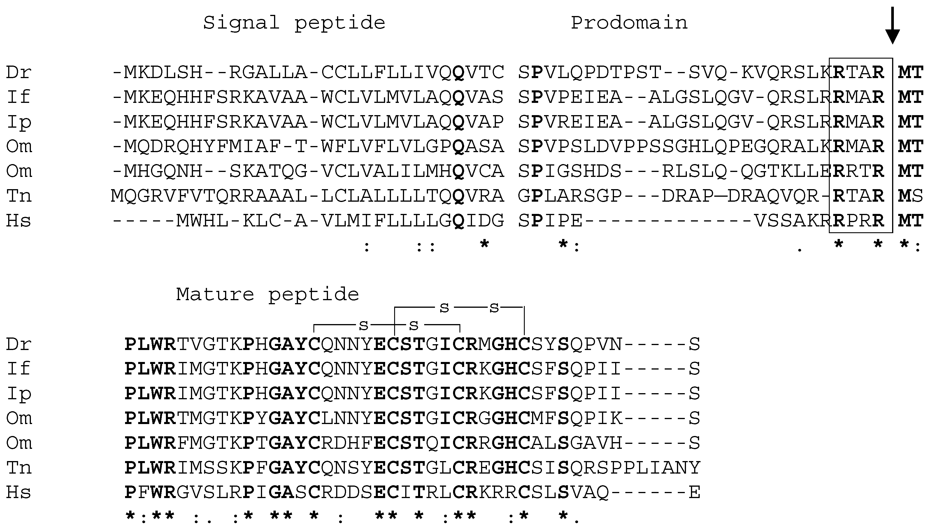
2.1. Cloning and Identification of Zebrafish Antimicrobial Peptidess

A preliminary analysis of the zebrafish genome enabled the identification of sequences homologous to those from AMPs already characterized in other vertebrate species, i.e.: chromogranin A and B, parasin I and its corresponding proteolytic enzyme, cathepsin D, BPI, LEAP 2 and β-defensin (DB1). The zebrafish homologous sequences were used to design pairs of primers for partial or complete amplification of each AMP gene of interest (Table 1). The absence of contamination with DNA was confirmed using RNA samples as template, and by RT-PCR assays, employing specific primers for the zebrafish β-actin.
The length of the seven sequences obtained was between 242 bp (DB1) and 504 bp (CgB). Each amplicon was cloned and sequenced, and the translated amino acid sequence was verified by alignment with those of known AMPs. Among peptides related to the neuroendocrine and immune system, the partial sequences of zebrafish chromogranins A and B (zfCgA and zfCgB) were amplified and characterized with specific primers (Table 1). The alignment of the zfCgA amino acid sequence with those of mammals and amphibians, exhibited between 31% and 34% identity (Table 2), showing a greater conservation in the N-terminal (57%) and C-terminal (63%) regions, while no similarity was found in the rest of the molecule (Figure 1). Alignment of the zfCgB sequence with that of amphibians and mammals showed an identity between 20% and 37% (Table 2) with a higher homology in the N-terminal region (Figure 1 and Figure 2).
The amino acid sequence of zebrafish parasin I (zfparasin I; 154 amino acid residues) shows significant homology with the N-terminal region of histone H2A (Table 2).
In Figure 3, the alignment of zfparasin I with buforin I, an antimicrobial peptide characterized in the Asian toad [26], and with hipposin, a polypeptide of 51 amino acids isolated from mucus of catfish and Atlantic halibut, is shown. High identity is evident from amino acid residues 1 to 33 [27] (Figure 3).
Cat-D, a protease that could be responsible for production of Parasin I from histone H2A, was also identified in zebrafish (zfCat-D). zfCat-D showed similarity to an eukaryotic lysosomal aspartic protease (Table 2) and BLAST analysis indicated that zfCat-D displays a conserved motif, approximately 39 amino acid residues, in which a cleavage site and a pre-domain, typical regions of many eukaryotic endo-peptidase, can be identified (Figure 4).
Analysis of the amino acid sequence of zfBPI indicated that this protein shows a significant identity with other members of BPI family (Table 2). In particular, the sequence of zfBPI shows a possible functional domain in the N-terminal region (residues 59–118) similar to the lipopolysaccharide(LPS)-binding domain found in human, carp and trout BPI proteins (Figure 5).
The analyses of encoded primary structure of the zfLEAP2, a member of the tissue-expressed AMP family, indicated that the protein contains92 amino acids, and includes a putative signal sequence (identified using the Signal P 3.0 program, (http://www.cbs.dtu.dk/services/SignalP-3.0/) suggesting that the peptide is secreted. A comparison of the sequences shows that the percentage identity between zfLEAP2 and equivalent proteins in other species is between 43% (H. sapiens) and 62% (I. punctatus) (Table 2), and that the mature functional region is more conserved than the signal peptide region and pre-domain (Figure 6). Interestingly, amino acid residues in the region between the pre-domain and the mature region are conserved from fish to mammals. Moreover, it is possible localize a motif (RXXR) in the cleavage site found in zfLEAP2 and all other LEAP2 sequences analyzed. In addition, the zfLEAP2mature protein contains four cysteines that have been found in the same position in other species [18]. The coding region for the β-defensin (another member of the tissue-expressed AMPs) of zebrafish (zfβ-defensin) corresponds to that described by [16].

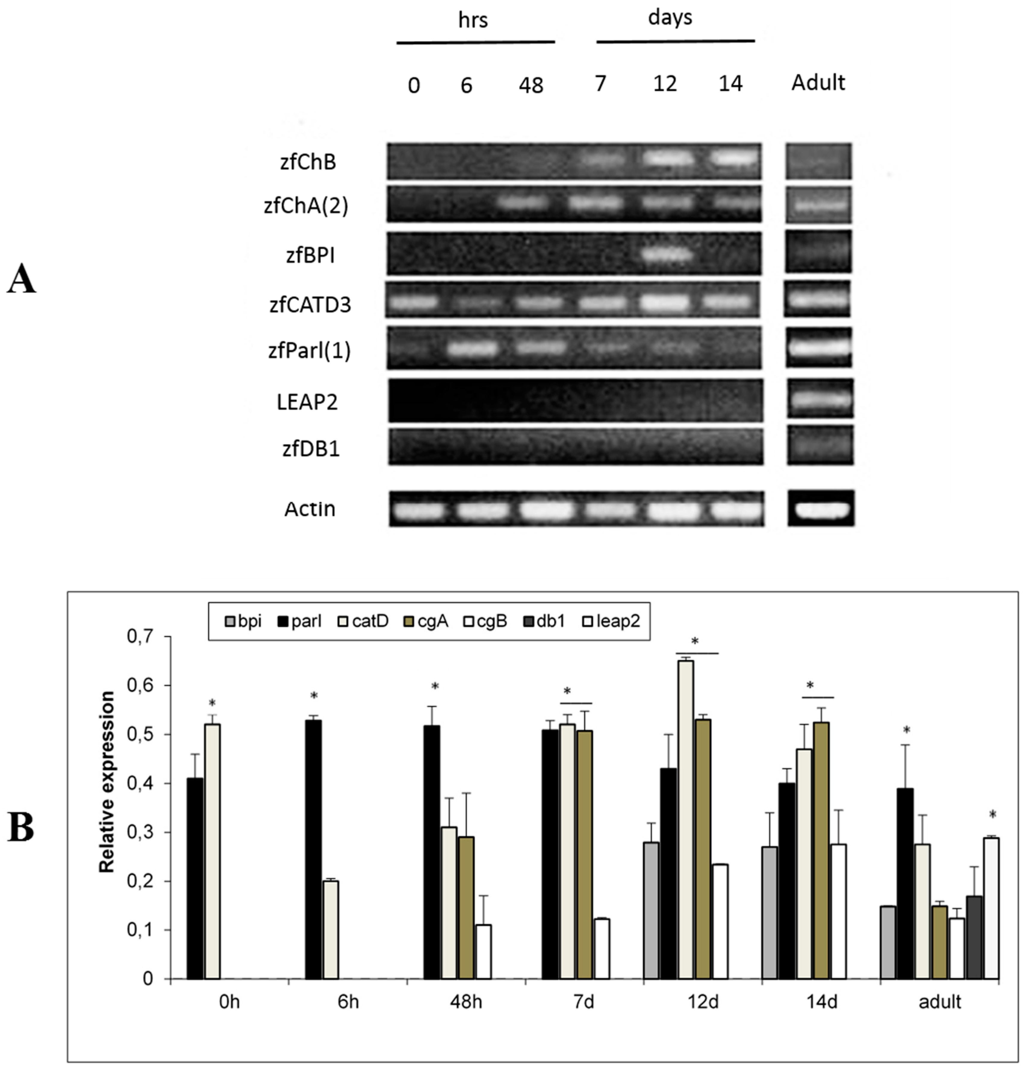
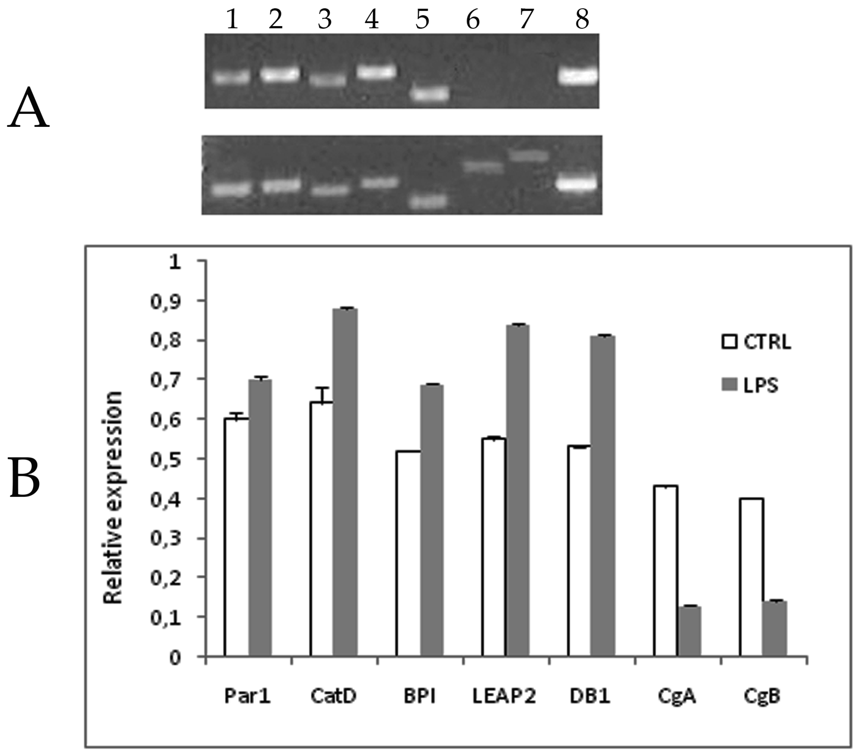
2.2. Ontogeny of AMP Gene Expression in Zebrafish and Response to Experimental Immune Challenge

The temporal expression of each gene for AMPs was analyzed in eggs, embryos and adult zebrafish by semi-quantitative RT-PCR analysis (Figure 7A,B). To check quality and concentration of RNA obtained a series of RT-PCR was carried out employing specific primers for the zebrafish β-actin as an internal control for the normalization of band intensity. Both transcripts of zfCgA and zfCgB are evident starting from 48 hours post fertilization (hpf), while zfParI and zfCat-D transcripts were already detectable in the fertilized zygote (0 hpf) (Figure 7A,B) and resulted in being higher expressed during all of the development period. The mature transcripts zfLEAP2 and zfDB1 were only detectable in the adult stage, while zfBPI transcripts are evident from the 12th day post fertilization (dpf) onwards (Figure 7A,B).
Experimental immune challenge with an LPS/LTA (lipoteichoic acid) cocktail in adult zebrafish showed that, except for zfCgA and zfCgB, which are under expressed, all other AMP mRNAs tested showed a stronger signal 48 h after the intraperitoneal injection as compared with untreated controls (Figure 8).

3. Discussion

AMPs are present in all metazoans (plants, fungi, invertebrates and vertebrates) and probably represent one of the earliest modalities of action of multicellular organisms against invading microorganisms [28]. AMPs have been described as inducible components with bacteriostatic or bacteriocidal activity [29], and their presence in fluids (blood, connective tissue and mucus) and tissues of vertebrates has been reported since the mid-20th century [30]. The model of the AMP action mechanism involves the electrostatic contact between the negative charge of bacterial membrane and the positive charge of peptide. As a consequence, a conformational change of the membrane occurs, bringing about the formation of a channel or pore, through which occurs the passage of the peptide until the blockage of internal metabolic processes, leading to the death of the bacterial cell [31]. An interesting relationship of some endogenous peptides with the specific immune system has been highlighted [11,32,33]. Several AMPs, such as pleurocidin, daxin, misgurin, piscidin, chrysophsin, morocidin, parasin, defensin, hepcidin and dicentracin have been identified in fish, and the results of these investigations have revealed the AMPs’ capability to protect fish from infections. Recently, a study on synthetic hepcidin injected in sea bass has confirmed the capability to potentiate the antimicrobial activity against bacteria by administration of AMPs as a new frontier of therapeutic application in farmed fish species [34]. The aim of this study was to evaluate, through the biomolecular approach, the dynamic of expression of a panel of various AMPs during development and in the adult zebrafish. Taking advantage of homologous genes identified in the zebrafish genome, pairs of specific primers were designed for AMPs of interest already identified in fish and other vertebrate species (chromogranin A and B; parasin I and cathepsin D; BPI, LEAP2, DBI).
Chromogranins A and B represent AMPs belonging to the endocrine and neuroendocrine systems and are expressed in many animal species [21,22,35]. Chromogranins are released in mammals during stress from the adrenal medulla, along with the adrenaline [36,37]. Our data indicate that D. rerio chromogranins are relatively different from those of tetrapods [38,39], as, although the identity with amphibians and mammals is high in the C-terminal region of the peptide (60–70%), in other regions, the identity is lower than 14–30%. The presence of chromogranins in 48 hpf embryos is noteworthy and is probably related to the protection of the nervous system during early development. Furthermore, our study showed that zebrafish chromogranins are not inducible by LPS; indeed, it seems depressed by this in the adult. Previous studies reported that chromogranins’ presence is dependent on the hypothalamus-hypophysis-adrenal axis, and a depression of glucocorticoits production could deplete their presence [40]. These results suggest the possibility, previously proposed in other vertebrates [35,40], that chromogranins can exert their function with tissue and organ specificity, conferring an early protection before the activation of the humoral and cellular adaptive immune system.
Parasin I has a molecular weight of approximately 2.4 kDa and consists of 19 amino acids, including three arginins and five lysines [41]. On the mucosal surfaces of injured catfish [14], parasin I showed a strong antimicrobial and extracellular activity, which seems to be about 12–100 times more potent than magainin 2 [41,42]. This evidence indicates that parasin I, coating the mucosal surface, could contribute to the effective local microbial control. Parasin I is produced by the cleavage of histone H2A by the cathepsin D [14,41]. The conservation of amino acid sequence of histone H2A and cathepsin D in fish suggests that the sequence of parasin I is a biologically important part of the molecule, and confers a benefit during fish evolution [42]. Since the biological roles of ubiquitous molecules, such as histones, for different functions (DNA condensation and antimicrobial action) have been characterized in trout (histone H1 and oncorincin II, [43]) and Atlantic halibut (H1 and H2A histones and hipposin), it is possible that similar functions could be attributed to the zebrafish homologues. The Atlantic halibut hipposin, a polypeptide of 51 amino acids isolated from mucus of skin, is similar to the amino acid sequence of parasin I. Like parasin I, hipposin is endowed with strong antimicrobial activity against both Gram-positive and Gram-negative bacteria [44], and probably highly conserved in teleost fish. The results of LPS/LTA injection experiments performed in the present study supports this hypothesis. After 48 h, LPS/LTA stimulated transcription of zfCat-D and histone H2A catalysis in zfParasin I. These results suggest possible alternative approaches for the stimulation of mucosal immunity. Moreover, as zfParasin I and zfCat-D appear to be constitutively expressed in the newly formed zygotes and during zebrafish ontogeny, their roles in immune defense are probably critical during early development.
BPI proteins belong to the family of lipid binding proteins, including LPS-binding proteins (LBP), cholesterol transferase proteins (CETP) and phospholipid transferase proteins (PLTP) [45]. Both LBP and BPI bind LPS, but with opposite biological modality [20]. The peptide has revealed in the amino terminal half of the molecule a high concentration of basic residues, mostly lysines, which represents an advantage in blocking the replicative activity of bacteria [46]. The primary role of the amino-terminal domain of BPI has been shown by a similar sequence of 21–25 residues in zebrafish and in other species [47]. At first, this peptide was isolated from the secondary granules of neutrophil granulocytes of rabbit [48]; subsequently, in human eosinophil granulocytes and epithelial cells, low expression levels have been highlighted [20]. After isolation of the cDNA of BPI gene from Atlantic cod (Gadus morhua L.), its expression has been observed primarily in the head kidney leukocytes and, only as a result of a bacterial stimulation, in the blood [49]. The study of expression of BPI in ontogenesis and in adulthood revealed higher expression starting from the 12th dpf. In cyprinid fish, phagocytes, which are responsible of BPI production, differentiate very early (in the carp at second day post-fertilization [50]), but no information are available about BPI production. It could be assumed that, in zebrafish, these cells are fully functional in secreting BPI at 12 dpf, but further studies are needed to localize and quantify the AMP presence because the analysis has done in total body, and it is possible that a very low amount of peptide was undetected. Our study revealed that 48 h after LPS/LTA injection, mRNA transcription of BPI is significantly enhanced, suggesting that it can be inducible by infectious challenge as reported in previous research in other vertebrates [35,40].
The LEAP-2 gene was only identified and characterized in a few teleost fish species, including trout [18] and catfish [19]. In both species, LEAP-2 is constitutively expressed in the liver. However, after experimental bacterial challenge, a stronger signal was shown in the intestine, skin (in trout, [18]) and spleen (in catfish, [19]). The zfLEAP-2 protein shows a signal peptide, as a pre-domain motif RXXR that is located before to the cutting site. The terminal part of the mature peptide is linked by the two disulfide bridges typical of LEAP-2 proteins, as reported in other species [18]. Our study showed that zfLEAP-2 transcripts are detectable only in the adult zebrafish and, consistently with observations in catfish and trout, that zfLEAP-2 is inducible by infectious challenge.
The vertebrate defensins can be classified into three sub-families α, β, and θ, on the base of the pairs of cysteines that form intramolecular disulphide bridges [51,52,53,54]. The β-defensins are expressed in a wide range of tissues and organs such as the intestine and epithelium [55]. They are expressed in pre-peptidic form by many cell types (keratinocytes, macrophages, neutrophils, lymphocytes), and stored in vesicles that are released upon infection [56]. Multiple β-defensin genes have been identified in puffer fish (Takifugu rubripes), spotted puffer fish (Tetraodon nigroviridis), zebrafish (Danio rerio), and trout (Oncorhynchus mykiss) [16,57]. The β-defensin gene is constitutively expressed in all below species during adulthood, and their expression and localization includes gills, head kidney, liver, intestine, gonads, and spleen. Our study revealed that the β-defensin gene is not expressed in zebrafish embryos, and not only confirmed the presence of β-defensin in the adult zebrafish, but also that it is inducible by infectious challenge.
Considering the resistance to antibiotics that has been observed in aquaculture settings, and in line with the practice of prophylaxis adopted on January 2006 and the new EU directives (COM (2005/297)), new preventive and therapeutic approaches based on AMPs could represent an innovative strategy for addressing infectious disease in farmed fish [34]. Even if this study is pivotal research and further studies are needed to localize the different AMPs in the adult and during development, our study supports the zebrafish as a suitable model system for rigorous investigations on the roles of AMPs in infectious disease. The aim is to suggest that AMPs employ as an alternative to antibiotics in therapeutic interventions in aquaculture of commercially relevant fish species. Moreover, the expression of some AMPs during early development suggests that new prophylactic strategies, actuated during larval stages, may represent a promising alternative, where parasine I could be a good candidate to use in combination with other AMPs.

4. Materials and Methods

4.1. Animals

Zebrafish (D. rerio, wild type) were reared in freshwater 80 L aquaria at 28 °C, placed in small tanks for egg deposition and fertilization, and the fertilized eggs collected and maintained in an incubator at 28 °C. Approximately 100 embryos were collected at each time point (0, 6, 24, 48 hpf and 5, 7, 12, 14 dpf), euthanized by immersion in water with 0.03 g/L Tricaine methansulphonate (Crescent Research Chemicals, Phoenix, AZ, USA), and processed for RNA extraction.

4.2. Extraction of Total RNA

For the extraction of total RNA from zebrafish embryos and adult tissues, samples were homogenized in TriReagent (Sigma-Aldrich, St. Louis, MO, USA) following the manufacturer’s instructions, using either disposable or electric homogenizers. Then, 0.2 mL of chloroform per ml of initial TriReagent were added to each sample, followed by vortexing for 15 s, incubated for 10 min at room temperature, and subsequently centrifuged at 12,000× g for 15 min at 4 °C. RNA precipitation was performed by adding 0.5 mL of isopropanol per mL of initial TriReagent for 10 min at room temperature, and then centrifuged at 12,000× g for 10 min at 4 °C. The supernatant was discarded and 1 mL of ethanol 75% (v/v) was added for each ml of initial TriReagent, and the mixtures centrifuged at 7500× g for 5 min at 4 °C. The supernatant was discarded and any remaining ethanol in the RNA pellet was removed by evaporation. The RNA was re-suspended in diethyl pyrocarbonate-treated water (DEPC) 0.1%. RNA quantification was carried out by measuring the absorbance at 260 nm (A260).

4.3. RT-PCR Analysis

cDNA was synthesized from 5 μg of total RNA, using the SuperScript III kit (Invitrogen, Carlsbad, CA, USA). In addition, 5 μg of total RNA were mixed with 1 mL of dNTP (10 mM), 1 μL of oligo (dT), and nuclease-free water added to a total volume of 10 μL. The mixtures were incubated for 5 min at 65 °C and cooled in ice; subsequently, 2 μL of reaction buffer 10×, 4 μL of MgCl2, 2 μL of DTT (0.1 M), 1 μL of RNase OUT, and 1 μL of Superscript III RT were added. The mix was incubated at 50 °C for 50 min, subsequently at 85 °C for 5 min, and then in ice. Then, 1 μL of RNase H was added, and samples were placed at 37 °C for 20 min. To check the quality and quantity of RNA, a PCR was performed, using primers specific for zebrafish β-actin (generally used in AMPs research, 7). For each gene of interest, one or more pairs of primers were designed (Table 1), using the program Primer 3 (Rozen and Skaletsky, 2000, http://www-genome.wi.mit.edu) available online). The PCR program was the following: 94 °C for 5 min, 35 cycles at 94 °C for 30 s, 50–55 °C for 1 min, 72 °C for 1 min, and 72 °C for 5 min.

4.4. cDNA Electrophoresis

The separation and identification of the cDNA fragments was performed by electrophoresis on agarose gels. For the preparation of a 1% (w/v) gel, 1 g of agarose was dissolved in 100 mL of 1× TAE (Tris-acetate-EDTA) buffer (50× TAE: 2M Tris-acetate, 50 mM EDTA). The same buffer was employed for the electrophoretic run at a constant voltage of 90 V, using a Power Pac Basic Bio-Rad apparatus (Bio-Rad, Hercules, CA, USA). cDNA bands were identified by adding ethidium bromide (10 ng/μL) and visualized under UV light.

4.5. cDNA Purification from Agarose Gel and Cloning of cDNA Fragments

cDNA from the specific bands of interest was purified from the gel using the Qiagen kit (QIAquick Gel Extraction, Quiagen, Carlsbad, CA, USA), following the manufacturer’s instructions. The bands were cut out from the gel, transferred to 1.5 mL Eppendorf tube and weighed. Buffer QG (5.5 M guanidine thiocyanate (GuSCN); 20 mM Tris HCl pH 6.6; three times the gel volume) was added, and samples were incubated at 50 °C for 10 min, inverting the tube every 2–3 min to facilitate the dissolution of the gel. A volume of isopropanol equal to the volume of the extracted gel bands was added. Then, the samples were transferred to chromatographic columns, and these were centrifuged at 13,000× rpm for 1 min. To remove any trace of agarose, 0.5 mL of buffer QG were added to each column and repeating the centrifugation. A final wash was carried out by adding 0.75 mL of PE buffer (10 mM Tris-HCl pH 7.5, 80% ethanol; Qiagen) and centrifuging at 13,000× rpm for 1 min. To eliminate all of the PE buffer, a “vacuum” centrifugation was performed, and to elute the DNA, columns were transferred to a new tube of 1.5 mL; in addition, 10 μL of nuclease-free water were added and the samples were centrifuged at 13,000× rpm for 1 min. The concentration of purified samples was assessed by gel electrophoresis. The cDNA fragments were cloned in pGEM-T Easy (Promega, Madison, WI, USA) (details in supplementary data).

4.6. Bacterial Cultures and Transformation

Bacteria were grown in Luria Bertani agar medium (LB, Sigma-Aldrich). The individual colonies, obtained from plates with recombinant bacteria, were incubated in the growth medium LB + ampicillin (100 μg/mL), leaving in stirring overnight at 37 °C. Furthermore, 3 μL of each ligation mixture were added to an aliquot of 50 μL of competent JM109 cells (High Efficiency Competent Cells, Promega). After 20 min in ice, the sample was exposed to heat-shock at 42 °C for 45 s and then placed on ice for 2 min. Then, 1 mL of bacterial growth medium LB was added and sample was incubated at 37 °C for 45 min, after which 50 μL of cell suspension were taken from the sample and plated on LB medium+ agar + ampicillin (100 μg/mL). The remaining sample was centrifuged at 6000× rpm for 5 min and approximately 800 μL of the supernatant were discarded; the pellet was resuspended in the remaining 200 μL, and 100 μL were plated in a second plate. The recombinant transformants were selected by means of the “blue-white screening” method (Sigma-Aldrich). The plates were incubated at 37 °C overnight.

4.7. Extraction of Plasmid DNA

The kit employed was the Miniprep DNA Purification System (Promega) following the manufacturer’s instruction (Supplementary data). The sequences obtained by using the ABI PRISM (BigDye Terminator Cycle Sequencing, Applied Biosystems, Foster City, CA, USA) were analysed for similarity to using the BLAST program [58].

4.8. Semi-Quantitative Analysis of AMP Expression Levels

Total RNA from zebrafish embryos at 0, 6, 48 hpf, at 7, 12, 14 dpf, and from adult animals, was amplified by RT-PCR using primers specific for the AMPs of interest. The bands were analyzed densito-metrically using TotalLab software, and the transcript levels were expressed as the relative ratio of each transcript to β-actin.

4.9. Experimental Immune Challenge of Adult Zebrafish

Adult wild type zebrafish were challenged with a cocktail of lipopolysaccharide (LPS, Sigma-Aldrich)) and lipoteichoic acid (LTA Sigma-Aldrich) 1:1. LPS from the producer is constituted from Klebsiella pneumoniae, Salmonella typhimurium, Salmonella minnesota, Vibrio cholerae, Shigella flexneri 1A, Escherichia coli 0111:B4, and Escherichia coli 055:B5; and LTA from Bacillus subtilis, Streptococcus aureus, S. pyrogenes, S. faecalis (liquid, the LPS/LTA cocktail components powder were reconstituted in saline buffer (PBS) at a final concentration of 0.5 mg/mL. The experimental zebrafish group (6 fish, males and females) was injected intra-peritoneally with 20 μL of the cocktail following [59]. A similarly composed control group received only PBS. Animals were sacrifice with an overdose of anesthetic (MS222, as described above) 48 h after the injection, RNA was extracted (Dynalkit, Carlsbad, CA, USA) from whole animal and cDNA synthesized (Life Technologies, Carlsbad, CA, USA) as described above. RT-PCR was performed in 25 μL of final volume with the following components: 10× NH4 buffer, 100 mM dNTP, 50 mM MgCl2, μL Taq(500 U), 100 μM primers and MQ water. The PCR program employed was the following: 94 °C for 5 min, 35 cycles at 94 °C for 30 s, 50–55 °C for 1 min, 72 °C for 1 min, and 72 °C for 5 min.

Supplementary Materials

The following are available online at www.mdpi.com/2410-3888/2/4/20/s1.

Acknowledgments

This study was supported by University of Tuscia Grant to International Universities Agreements, and Grant R01GM070589 from the National Institutes of Health and Grants IOS 1050518, IOB-0618409, and IOS-0822257 from the National Science Foundation (GRV, USA). An International Scholarship to Elisabetta Caccia was awarded by Tuscia University. This research is part of Italian CONISMA organization projects.

Author Contributions

E.C., G.R.V. and N.R. conceived and designed the experiments; E.C. and P.S.D. performed the experiments; M.A., M.C. and N.R. analyzed the data; G.R.V. contributed reagents/materials/analysis tools and supervised experiments in his lab; N.R. and G.R.V. wrote the paper.

Conflicts of Interest

The authors declare no conflict of interest.

References

  1. Park, C.H.; Valore, E.V.; Waring, A.J.; Ganz, T. Hepcidin, a urinary antimicrobial peptide synthesized in the liver. J. Biol. Chem. 2001, 276, 7806–7810. [Google Scholar] [CrossRef] [PubMed]
  2. Lauth, X.; Shike, H.; Burns, J.C.; Westerman, M.E.; Ostland, V.E.; Carlberg, J.M.; Van Olst, J.C.; Nizet, V.; Taylor, S.W.; Shimizu, C.; et al. Discovery and characterization of two isoforms of moronecidin, a novel antimicrobial peptide from hybrid striped bass. J. Biol. Chem. 2002, 277, 5030–5039. [Google Scholar] [CrossRef] [PubMed]
  3. Asmamaw, B. Hepcidin and its roles in fish. J. Zool. Stud. 2016, 3, 1–10. [Google Scholar]
  4. Katzenback, B.A. Antimicrobial peptides as mediators of innate immunity in teleosts. Biology (Basel) 2015, 4, 607–639. [Google Scholar] [CrossRef] [PubMed]
  5. Wang, G.; Li, X.; Wang, Z. APD3: The antimicrobial peptide database as a tool for research and education. Nucleic Acids Res. 2016, 44, D1087–D1093. [Google Scholar] [CrossRef] [PubMed]
  6. Park, C.B.; Lee, J.H.; Park, I.Y.; Kim, M.S.; Kim, S.C. A novel antimicrobial peptide from the loach, Misgurnusanguillicaudatus. FEBS Lett. 1997, 411, 173–178. [Google Scholar] [CrossRef]
  7. Douglas, S.E.; Patrzykat, A.; Pytyck, J.; Gallant, J.W. Identification, structure and differential expression of novel pleurocidins clustered on the genome of the winter flounder, Pseudopleuronectes americanus (Walbaum). Eur. J. Biochem. 2003, 270, 3720–3730. [Google Scholar] [CrossRef] [PubMed]
  8. Iijima, N.; Tanimoto, N.; Emoto, Y.; Morita, Y.; Uematsu, K.; Murakami, T.; Nakai, T. Purification and characterization of three isoforms of chrysophsin, a novel antimicrobial peptide in the gills of the red sea bream, Chrysophrys major. Eur. J. Biochem. FEBS 2003, 270, 675–686. [Google Scholar] [CrossRef]
  9. Shi, J.; Camus, A.C. Hepcidins in amphibians and fishes: Antimicrobial peptides or iron-regulatory hormones? Dev. Comp. Immunol. 2006, 30, 746–755. [Google Scholar] [CrossRef] [PubMed]
  10. Huang, P.H.; Chen, J.Y.; Kuo, C.M. Three different hepcidins from tilapia, Oreochromis mossambicus: Analysis of their expressions and biological functions. Mol. Immunol. 2007, 44, 1922–1934. [Google Scholar] [CrossRef] [PubMed]
  11. Yang, D.; Liu, Z.H.; Tewary, P.; Chen, Q.; de la Rosa, G.; Oppenheim, J.J. Defensin participation in innate and adaptive immunity. Curr. Pharm. Des. 2007, 13, 3131–3139. [Google Scholar] [CrossRef] [PubMed]
  12. Falco, A.; Mas, V.; Tafalla, C.; Perez, L.; Coll, J.M.; Estepa, A. Dual antiviral activity of human alpha-defensin-1 against viral haemorrhagic septicaemia rhabdovirus (VHSV): Inactivation of virus particles and induction of a type I interferon-related response. Antivir. Res. 2007, 76, 111–123. [Google Scholar] [CrossRef] [PubMed]
  13. Salerno, G.; Parrinello, N.; Roch, P.; Cammarata, M. cDNA sequence and tissue expression of an antimicrobial peptide, dicentracin; a new component of the moronecidin family isolated from head kidney leukocytes of sea bass, Dicentrarchus labrax. Comp. Biochem. Physiol. B Biochem. Mol. Biol. 2007, 146, 521–529. [Google Scholar] [CrossRef] [PubMed]
  14. Cho, J.H.; Park, I.Y.; Kim, H.S.; Lee, W.T.; Kim, M.S.; Kim, S.C. Cathepsin D produces antimicrobial peptide parasin I from histone H2A in the skin mucosa of fish. FASEB J. 2002, 16, 429–431. [Google Scholar] [CrossRef] [PubMed]
  15. Bulet, P.; Stöcklin, R. Insect antimicrobial peptides: Structures, properties and gene regulation. Protein Pept. Lett. 2005, 12, 3–11. [Google Scholar] [CrossRef] [PubMed]
  16. Zou, J.; Mercier, C.; Koussounadis, A.; Secombes, C. Discovery of multiple beta-defensin-like homologues in teleost fish. Mol. Immunol. 2007, 44, 638–647. [Google Scholar] [CrossRef] [PubMed]
  17. Krause, A.; Sillard, R.; Kleemeier, B.; Klüver, E.; Maronde, E.; Conejo-García, J.R.; Forssmann, W.G.; Schulz-Knappe, P.; Nehls, M.C.; Wattler, F.; et al. Isolation and biochemical characterization of LEAP-2, a novel blood peptide expressed in the liver. Protein Sci. 2003, 12, 143–152. [Google Scholar] [CrossRef] [PubMed]
  18. Zhang, Y.A.; Zou, J.; Chang, C.I.; Secombes, C.J. Discovery and characterization of two types of liver-expressed antimicrobial peptide 2 (LEAP-2) genes in rainbow trout. Vet. Immunol. Immunopathol. 2004, 101, 259–269. [Google Scholar] [CrossRef] [PubMed]
  19. Bao, B.; Peatman, E.; Xu, P.; Li, P.; Zeng, H.; He, C.; Liu, Z. The catfish liver-expressed antimicrobial peptide 2 (LEAP-2) gene is expressed in a wide range of tissues and developmentally regulated. Mol. Immunol. 2006, 43, 367–377. [Google Scholar] [CrossRef] [PubMed]
  20. Elsbach, P. The bactericidal/permeability-increasing protein (BPI) in antibacterial host defense. J. Leukoc. Biol. 1998, 64, 14–18. [Google Scholar] [PubMed]
  21. Hendy, G.N.; Bevan, S.; Mattei, M.G.; Mouland, A.J. Chromogranin A. Clin. Investig. Med. 1995, 18, 47–65. [Google Scholar]
  22. Briolat, J.; Wu, S.D.; Mahata, S.K.; Gonthier, B.; Bagnard, D.; Chasserot-Golaz, S.; Helle, K.B.; Aunis, D.; Metz-Boutigue, M.H. New antimicrobial activity for the catecholamine release-inhibitory peptide from chromogranin A. Cell. Mol. Life Sci. 2005, 62, 377–385. [Google Scholar] [CrossRef] [PubMed]
  23. Lin, W.; Liu, S.; Hu, L.; Zhang, S. Characterization and bioactivity of hepcidin-2 in zebrafish: Dependence of antibacterial activity upon disulfide bridges. Peptides 2014, 57, 36–42. [Google Scholar] [CrossRef] [PubMed]
  24. Feng, C.; Nita-Lazar, M.; González-Montalbán, N.; Wang, J.; Mancini, J.; Ravindran, C.; Ahmed, H.; Vasta, G.R. Manipulating galectin expression in zebrafish (Danio rerio). Methods Mol. Biol. 2015, 1207, 327–341. [Google Scholar] [PubMed]
  25. Nita-Lazar, M.; Mancini, J.; Feng, C.; González-Montalbán, N.; Ravindran, C.; Jackson, S.; Heras-Sánchez, A.L.; Giomarelli, B.; Ahmed, H.; Haslam, S.M.; et al. The zebrafish galectins Drgal1-L2 and Drgal3-L1 bind in vitro to the infectious hematopoietic necrosis virus (IHNV) glycoprotein and reduce viral adhesion to fish epithelial cells. Dev. Comp. Immunol. 2016, 55, 241–252. [Google Scholar] [CrossRef] [PubMed]
  26. Kim, H.S.; Yoon, H.; Minn, I.; Park, C.B.; Lee, W.T.; Zasloff, M.; Kim, S.C. Pepsin-mediated processing of the cytoplasmic histone H2A to strong antimicrobial peptide Buforin I. J. Immunol. 2000, 165, 3268–3274. [Google Scholar] [CrossRef] [PubMed]
  27. Birkemo, G.A.; Mantzilas, D.; Lüders, T.; Nes, I.F.; Nissen-Meyer, J. Identification and structural analysis of the antimicrobial domain in hipposin, a 51-mer antimicrobial peptide isolated from Atlantic halibut. Biochim. Biophys. Acta 2004, 1699, 221–227. [Google Scholar] [CrossRef]
  28. Zasloff, M. Antimicrobial peptides of multicellular organisms. Nature 2002, 415, 389–395. [Google Scholar] [CrossRef] [PubMed]
  29. Brogden, K.A.; Guthmiller, J.M.; Salzet, M.; Zasloff, M. The nervous system and innate immunity: The neuropeptide connection. Nat. Immunol. 2005, 6, 558–564. [Google Scholar] [PubMed]
  30. Skarnes, R.C.; Watson, D.W. Antimicrobial factors of normal tissues and fluids. Bacteriol. Rev. 1957, 21, 273–294. [Google Scholar] [PubMed]
  31. Sarmasik, A.; Warr, G.; Chen, T.T. Production of transgenic medaka with increased resistance to bacterial pathogens. Mar. Biotechnol. 2002, 4, 310–322. [Google Scholar] [CrossRef] [PubMed]
  32. Cuesta, A.; Meseguer, J.; Esteban, M.A. The antimicrobial peptide hepcidin exerts an important role in the innate immunity against bacteria in the bony fish gilthead seabream. Mol. Immunol. 2008, 45, 2333–2342. [Google Scholar] [CrossRef] [PubMed]
  33. Liu, A.; Claesson, H.E.; Mahshid, Y.; Klein, G.; Klein, E. Leukotriene B4 activates T cells that inhibit B-cell proliferation in EBV-infected cord blood-derived mononuclear cell cultures. Blood 2008, 111, 2693–2703. [Google Scholar] [CrossRef] [PubMed]
  34. Masso-Silva, J.A.; Diamond, G. Antimicrobial peptides from fish. Pharmaceuticals 2014, 7, 265–310. [Google Scholar] [CrossRef] [PubMed]
  35. Simon, J.P.; Aunis, D. Biochemistry of the chromogranin A protein family. Biochem. J. 1989, 262, 1–13. [Google Scholar] [CrossRef] [PubMed]
  36. Metz-Boutigue, M.H.; Kieffer, A.E.; Goumon, Y.; Aunis, D. Innate immunity: Involvement of new neuropeptides. Trends Microbiol. 2003, 11, 585–592. [Google Scholar] [CrossRef] [PubMed]
  37. Miyakawa, M.; Matsui, T.; Kishikawa, H.; Murayama, R.; Uchiyama, I.; Itoh, T.; Yoshida, T. Salivary chromogranin A as a measure of stress response to noise. Noise Health 2006, 8, 108–113. [Google Scholar] [CrossRef] [PubMed]
  38. Turquier, V.R.; Vaudry, H.; Je Gou, S.; Anouar, Y. Frog chromogranin A messenger ribonucleic acid encodes three highly conserved peptides. Coordinate regulation of proopiomelanocortin and chromogranin a gene expression in the pars intermedia of the pituitary during background colour adaptation. Endocrinology 1999, 140, 4104–4112. [Google Scholar] [CrossRef] [PubMed]
  39. Ait-Ali, D.; Turquier, V.; Alexandre, D.; Grumolato, L.; Jegou, S.; Vaudry, H.; Anouar, Y. Molecular characterization of frog chromogranin B reveals conservation of selective sequences encoding potential novel regulatory peptides. FEBS Lett. 2002, 511, 127–132. [Google Scholar] [CrossRef]
  40. Strub, J.-M.; Garcia-Stablone, P.; Lonning, K.; Taupenot, L.; Hubert, P.; Van Dorsselaers, A.; Aunis, D.; Metz-Boutigue, M.C. Processing of chromogranin B in bovine adrenal medulla-Identification of secretolytin, the endogenous C-terminal fragment of residues 614–626 with antibacterial activity. Eur. J. Biochem. (FEBS) 1995, 229, 356–368. [Google Scholar] [CrossRef]
  41. Park, I.Y.; Park, C.B.; Kim, M.S.; Kim, S.C. Parasin I, an antimicrobial peptide derived from histone H2A in the catfish, Parasilurus asotus. FEBS Lett. 1998, 437, 258–262. [Google Scholar] [CrossRef]
  42. Diamond, G.M.; Beckloff, N.; Weinberg, A.; Kisich, K.O. The Roles of Antimicrobial Peptides in Innate Host Defense. Curr. Pharm. Des. 2009, 15, 2377–2392. [Google Scholar] [CrossRef] [PubMed]
  43. Fernandes, J.M.; Molle, G.; Kemp, G.D.; Smith, V.J. Isolation and characterisation of oncorhyncin II, a histone H1-derived antimicrobial peptide from skin secretions of rainbow trout, Oncorhynchu smykiss. Dev. Comp. Immunol. 2004, 28, 127–138. [Google Scholar] [CrossRef]
  44. Birkemo, G.A.; Lüders, T.; Andersen, Ø.; Nes, I.F.; Nissen-Meyer, J. Hipposin, a histone-derived antimicrobial peptide in Atlantic halibut (Hippoglossus hippoglossus L.). Biochim. Biophys. Acta 2003, 1646, 207–215. [Google Scholar] [CrossRef]
  45. Tall, A. Plasma lipid transfer proteins. Annu. Rev. Biochem. 1995, 64, 235–257. [Google Scholar] [CrossRef] [PubMed]
  46. Ooi, C.E.; Weiss, J.; Doerfler, M.E.; Elsbach, P. Endotoxin-neutralizing properties of the 25 kD N-terminal fragment and a newly isolated 30 kD C-terminal fragment of the 55–60 kD bactericidal/permeability-increasing protein of human neutrophils. J. Exp. Med. 1991, 174, 649–655. [Google Scholar] [CrossRef] [PubMed]
  47. Horwitz, A.H.; Leigh, S.D.; Abrahamson, S.; Gazzano-Santoro, H.; Liu, P.S.; Williams, R.E.; Carroll, S.F.; Theofan, G. Expression and characterization of cysteine-modified variants of an amino-terminal fragment of bactericidal/permeability-increasing protein. Protein Expr. Purif. 1996, 8, 28–40. [Google Scholar] [CrossRef] [PubMed]
  48. Weiss, J.; Franson, C.; Schmeidler, K.; Elsbach, P. Reversible envelope effects during and after killing of Escherichia coli Wby a highly-purified rabbit polymorpho-nuclear leukocyte fraction. Biochim. Biophys. Acta 1976, 436, 154–169. [Google Scholar] [CrossRef]
  49. Solstad, T.; Stenvik, J.; Jørgensen, T.Ø. mRNA expression patterns of the BPI/LBP molecule in the Atlantic cod (Gadusmorhua L.). Fish Shellfish Immunol. 2007, 23, 260–271. [Google Scholar] [CrossRef] [PubMed]
  50. Romano, N.; Picchietti, S.; Taverne-Thiele, J.J.; Tavern, N.; Abelli, L.; Mastrolia, L.; Verburg-van Kemenade, B.M.L.; Rombout, J.H.W.M. Distribution of macrophages during fish development: An immunohistochemical study in carp (Cyprinuscarpio L.). Anat. Embryol. 1998, 198, 31–41. [Google Scholar] [CrossRef] [PubMed]
  51. Ganz, T. Defensins: Antimicrobial peptides of innate immunity. Nat. Rev. Immunol. 2003, 3, 710–720. [Google Scholar] [CrossRef] [PubMed]
  52. Lynn, D.J.; Lloyd, A.T.; Fares, M.A.; O’Farrelly, C. Evidence of positively selected sites in mammalian alpha-defensins. Mol. Biol. Evol. 2004, 21, 819–827. [Google Scholar] [CrossRef] [PubMed]
  53. Xiao, Y.; Hughes, A.L.; Ando, J.; Matsuda, Y.; Cheng, J.F.; Skinner-Noble, D.; Zhang, G. A genome-wide screen identifies a single beta-defensin gene cluster in the chicken: Implications for the origin and evolution of mammalian defensins. BMC Genom. 2004, 5, 56–61. [Google Scholar] [CrossRef] [PubMed] [Green Version]
  54. Selsted, M.E.; Ouellette, A.J. Mammalian defensins in the antimicrobial immune response. Nat. Immunol. 2005, 6, 551–557. [Google Scholar] [CrossRef] [PubMed]
  55. Diamond, G.; Kaiser, V.; Rhodes, J.; Russell, J.P.; Bevins, C.L. Transcriptional regulation of beta-defensin gene expression in tracheal epithelial cells. Infect. Immun. 2000, 68, 113–119. [Google Scholar] [CrossRef] [PubMed]
  56. Zhao, M.H.; Lockwood, C.M. A comprehensive method to purify three major ANCA antigens: Proteinase 3, myeloperoxidase and bactericidal/permeability-increasing protein from human neutrophil granule acid extract. J. Immunol. Methods 1996, 197, 121–130. [Google Scholar] [CrossRef]
  57. Falco, A.; Chico, V.; Marroquí, L.; Perez, L.; Coll, J.M.; Estepa, A. Expression and antiviral activity of a beta-defensin-like peptide identified in the rainbow trout (Oncorhynchus mykiss) EST sequences. Mol. Immunol. 2008, 45, 757–765. [Google Scholar] [CrossRef] [PubMed]
  58. Altschul, S.F.; Madden, T.L.; Schäffer, A.A.; Zhang, J.; Zhang, Z.; Miller, W.; Lipman, D.J. Gapped BLAST and PSI-BLAST: A new generation of protein database search programs. Nucleic Acids Res. 1997, 25, 3389–3402. [Google Scholar] [CrossRef] [PubMed]
  59. Neely, M.; Pfeifer, J.; Caparon, M.G. Streptococcus-zebrafish model of bacterial pathogenesis. Infect. Immun. 2002, 70, 3904–3914. [Google Scholar] [CrossRef] [PubMed]
Figure 1. Amino acid sequence alignment of Danio rerio ChromograninA (Dr, drCgA) with CgA from Homo sapiens (Hs) Bos taurus (Bt) and Rana ridibunda (Rr). The boxes indicate the N- and C-termini of drCgA protein, who share higher similarity with the reported sequences.
Fishes 02 00020 g001
Figure 2. Primary structure CgB of a 677-amino acid mature protein that encompasses 25% acidic residues. The zebrafish (Dr, Danio rerio) CgB amino acid sequence was aligned with the human, bovine and amphibian sequences (Bt, Bos taurus, Hs, Homo sapiens, Rr, Rana ridibunda). The amino acids of zebrafish CgB that are conserved in the other species are in bold. The positions of the di- or tribasic residues are indicated by highlight and represent amphiphilic part of the molecule active in antimicrobial interaction. The hyphens represent gaps that have been introduced for the alignment.
Fishes 02 00020 g002
Figure 3. The deduced amino acid sequence of zebrafish parasin I was aligned with those of hipposin, buforin, scallop (from H2A histome) and parasin. The different residues are bolded. The box indicates the high conservation of the amino acid sequence in zebrafish parasin and hipposin. Hh, Hippoglossus hyppoglossus; Bb, Bufo bufo; Cc Cyprinus carpio, Pa, Parasilurus asotus; Dr, Danio rerio.
Fishes 02 00020 g003
Figure 4. The first 57 N-terminus residues, deduced from CatDcDNA of zebrafish, is aligned with those of Mp (Macroscelide sproboscideus), Ch (Clupeaharengus), Rt (Rainbow trout, Oncorinchus mikissis) and Tarku (Takifugu rubripes, Tf). The typical motifs (signal peptide, A1 propetide, and mature protein) are indicated.
Fishes 02 00020 g004
Figure 5. Alignment of the predicted lipopolysaccharide(LPS)-binding domain (LPD) of zebrafish (Dr; Danio rerio) BPI/LDP with that of rainbow trout, LBP/BPI-1 and -2, human, and carp. Identical and similar amino acid residues are indicated by asterisks (*) and colon (:), respectively. Bars (–) indicate gaps that are introduced for optimal alignment. The number at the end of each sequence represents amino acid identities (%) to zebrafish BPI/LBD. The positive charged amino acid residues (K or R) conserved in BPI/LPD of carp (Cc; Cyprinus carpio), rainbow trout (Om1,2; Oncorinchus mikissis), human (Hs, Homo sapiens) are indicated by shaded region.
Fishes 02 00020 g005
Figure 6. Multiple alignment of zebrafish (Dr, Danio rerio) liver-expressed antimicrobial peptide 2(LEAP2) protein sequence with known LEAP-2 sequences. The multiple alignment was generated using the Clustal W program (version 1.8) using the LEAP-2 sequences from Ictalurus furcatus (AAX45792), Ictalurus punctatus (AAX45791), Oncorhynchus mykiss (AAS49157, AAR11766), Tetraodon nigroviridis (CAF87882) and Homo sapiens (NP_443203). The arrow indicates the known cleavage site in the human molecule and cleavage motif is boxed. The cysteine bridges are also indicated. Asterisks show identity whilst (.) or (:) indicate similarity.
Fishes 02 00020 g006
Figure 7. RT-PCR analysis of AMP expression during development (0, 6, 48 h and 7, 12 and 14 days) and in adult zebrafish. (A) the products separated on agarose gel by electrophoresis show expression of chromogranin A (cgA), chromogranin B (cgB), BPI (bpi), cathepsin D (catD), parasin I (ParI), db1 (β-defensin), and liver-expressed antimicrobial peptide (LEAP2); (B) densitometric analysis performed with Totallab software (CLIQS 1D pro; http://totallab.com/). In abscissa: stages of development; hours (h) and days (d). In ordinate: relative expression of AMPs. Results were obtained from a mean of triplicate experiments. Standard deviations are indicated on top of the bars. (*) From 0 h to 14 d: statistically higher vs. other AMPs, p < 0.001; in adult stage: parI, statistically higher vs. other AMPs; LEAP2, statistically higher vs. PBI, CgA, CgB, DB1 (Student t-test, two tails).
Fishes 02 00020 g007
Figure 8. RT-PCR analysis of AMP expression in adult zebrafish challenged by LPS/LTA injection: (1) parasin I (Par1), (2) Cathepsin D (CatD), (3) BPI, (4) LEAP-2, (5) DB1, (6) CgA, (7) CgB, and (8) β-Actin. (A) samples treated with LPS (above line), and untreated control (below line); (B) densitometric analysis performed by Totallab software; in abscissa: antimicrobial peptides, in ordinate: relative expression of AMPs. Results were obtained from a mean of triplicate experiments. Statistically different (Student t-test, two tails) not treated (CTRL) vs. LPS p < 0.001 for all AMPs tested. Data represented the mean ± standard deviation of three different experiments of LPS injection in adult zebrafish.
Fishes 02 00020 g008
Table 1. Sequences of the pairs of primers (Forward and Reverse) employed for the reverse-transcriptase-polymerase-chain-reaction (RT-PCR) analysis in zebrafish (zf). Parasin I (ParI), β-defensin 1 (DB1), liver-expressed antimicrobial peptide 2 (LEAP2), catepsin D (CATD), bactericidal permeability-increasing protein (BPI), Chromogranins A and B (CgA, CgB), β-actin (ACT); (*) Access number: NM_001081553.
TargetProduct Size (bp)Forward (5′–3′)Reverse (5′–3′)
zfParI350CGGCAAAGCTAGAGCTAAGGCAAGAAGACCGAGAAGGCTG
zfDB1 *242GAAAACTGGAGCTCCTGATCATGCTCTGGGGCTTCATGTT
zfLEAP2360CAGTGCCTACTGCCAGAACAGGATTGGCATGACAGTTTTG
zfCATD362GGAGGATGTGAAGCCATTGTAACAGAGTGGGCTTCGCTAA
zfBPI325CATCTCAGCAAAGACGACGATATGGATTTCAGGGGCATGT
zfChA463CATCTCCTGGGAGTTTCAGCTTTCGTCGGTTCCTCAGATC
zfChB504TTCCCTCACGTTTACCCTTGTCTCCTGTCTTTGTTATCCGG
zfACT380GACAACGGCTCCGGTATGTCTCTGTTGGTTTGGGATT
Table 2. Homology of amino acid sequences of zebrafish parasin I.
GeneGene Product (aa/kDa)Similar Polypeptide (aa)% IdentityOrganismAccession n.
CgA246/27,806 34Bos taurusAAB21297
31Rana ridibundaAAD38522
31Homo sapiensEDW81506
CgB677/81,950 37Homo sapiensNP001810
22Bos taurus18142994
20Rana ridibundaAAL76931
Par I154/16,741unamed protein product 100Tetraodon negroviridisCAF98588
histone H2A99Oncorhynchus mykissCAA25528
histone H2A96Xenopus tropicalisCAJ82985
similar to histone H2A93Canis familiarisXP545430
similar to histone H2A86Rattus norvegicusXP001062486
CatD398/43,187.51cathepsin D prepo 86Silarus asotusAAM62283
cathepsin D precursor 85Clupea harengusAAG27733
cathepsin D 83Oncorhynchus mykissAAC60301
cathepsin D1 80Takifugu rubripesNP001072052
unanamed protein prod. 77Tetraodon negroviridisCAF91576
cathepsin D precursor 75Chionodraco hamatusCAA07719
67Homo sapiensNP001900
BPI487/54,463unnamed protein product73Tetraodon negroviridisCAF90160
phospholipid transfer protein 70Xenopus tropicalisAAI36237
similar to phospholipid 64Gallus gallusXP425722
phospholipid60Homo sapiensEAW75784
pbi23Cyprinus carpioBAC56095
Om223Oncorhynchus mykiss
LEAP-2 92/10,212leap2 62Ictalurus punctatusAAX45791
leap2 62Ictalurus furcatusAAX45792
leap2 isoform A61Oncorhynchus mykissAAR11766
unnamed protein product59Tetraodon negroviridisCAF87882
peptide2B47Oncorhynchus mykissAAR11767
43Homo sapiensNP443203
DB167/7263unnamed protein product 85Tetraodon negroviridisCAG02912
beta defensin like peptides 1 78Oncorhynchus mykissCAK54951
CgA, chromogranin A; CgB, chromogranin B; LEAP-2, liver-expressed antimicrobial peptide 2; ParI, parasin I; CatD, cathepsin D; BPI, bactericidal permeability-increasing protein; DB1, Beta defensing.

