Establishment of Orthotopic Liver Tumors by Surgical Intrahepatic Tumor Injection in Mice with Underlying Non-Alcoholic Fatty Liver Disease


Abstract
:1. Introduction
2. Experimental Design
2.1. Materials
- Phosphate-buffered saline (PBS) without Ca/Mg (ThermoFisher, Grand Island, NY, USA; Cat. no.: 14190-144)
- Cell culture medium, RPMI (ThermoFisher; Cat. no.: 61870-036)
- Fetal bovine serum (FBS; Gemini Bio-Products, West Sacramento, CA, USA; Cat. no.: 100-106)
- Glutamate (ThermoFisher; Cat. no.: 25030081)
- Penicillin/streptomycin antibiotics (ThermoFisher; Cat. no.: 15140-122)
- Trypsin (Sigma-Aldrich, St. Louis, MO, USA; Cat. no.: 59427C)
- Matrigel (Corning, Corning, NY, USA; Cat. no.: 354230)
- 70% ethanol (Sigma-Aldrich; Cat. no.: 459836-2L)
- Povidone iodine 10% solution (Qualitest, Huntsville, AL, USA; Cat. no.: NDC 0603-1550-58)
- Methionine- and choline- deficient (MCD) diet (Research Diets, New Brunswick, NJ, USA; Cat. no.: A02082002B)
- Luciferase-expressing cell line: RIL-175 [16].
- Vicryl suture (Ethicon, Blue Ash, OH, USA; Cat. no.: J391H)
- G (0.33 mm) × 12.7 mm BD Insulin Syringe (BD Bioscience, Franklin Lakes, NJ, USA; Cat. no.: 324702)
- Serological pipettes and pipette tips
- TC Flask T75 (Sarstedt, Newton, NC, USA; Cat. no.: 83.3911)
- Serological pipettes and pipette tips (Sarstedt)
- Eppendorf and centrifuge tubes tubes (Sarstedt)
- Cotton-tipped applicators (Medline, Northfield, IL, USA; Cat. no.: MDS202000)
2.2. Equipment
- Incubator
- Centrifuge
- Counter top vortex
- Isoflurane (gas anesthesia system)
- Electric clipper
- Bead sterilizer
- Heating pad
- Heat lamp
- Straight forceps
- Straight scissor
- Needle driver
- Xenogen in vivo imaging system (IVIS Spectrum, Caliper Live Sciences, Hopkinton, MA, USA)
3. Procedure
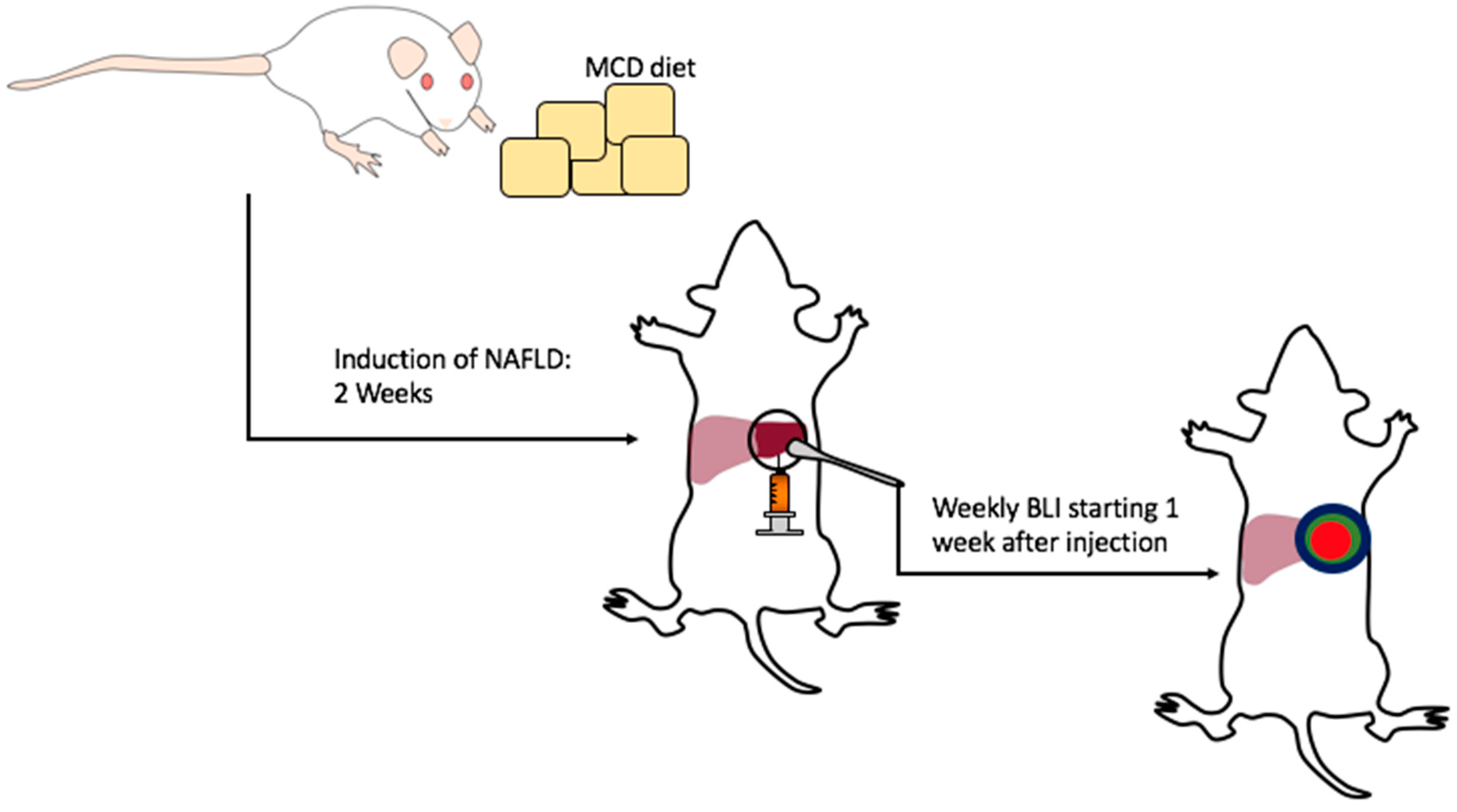
3.1. Induction of NAFLD/NASH. Time for Completion: 2 Weeks
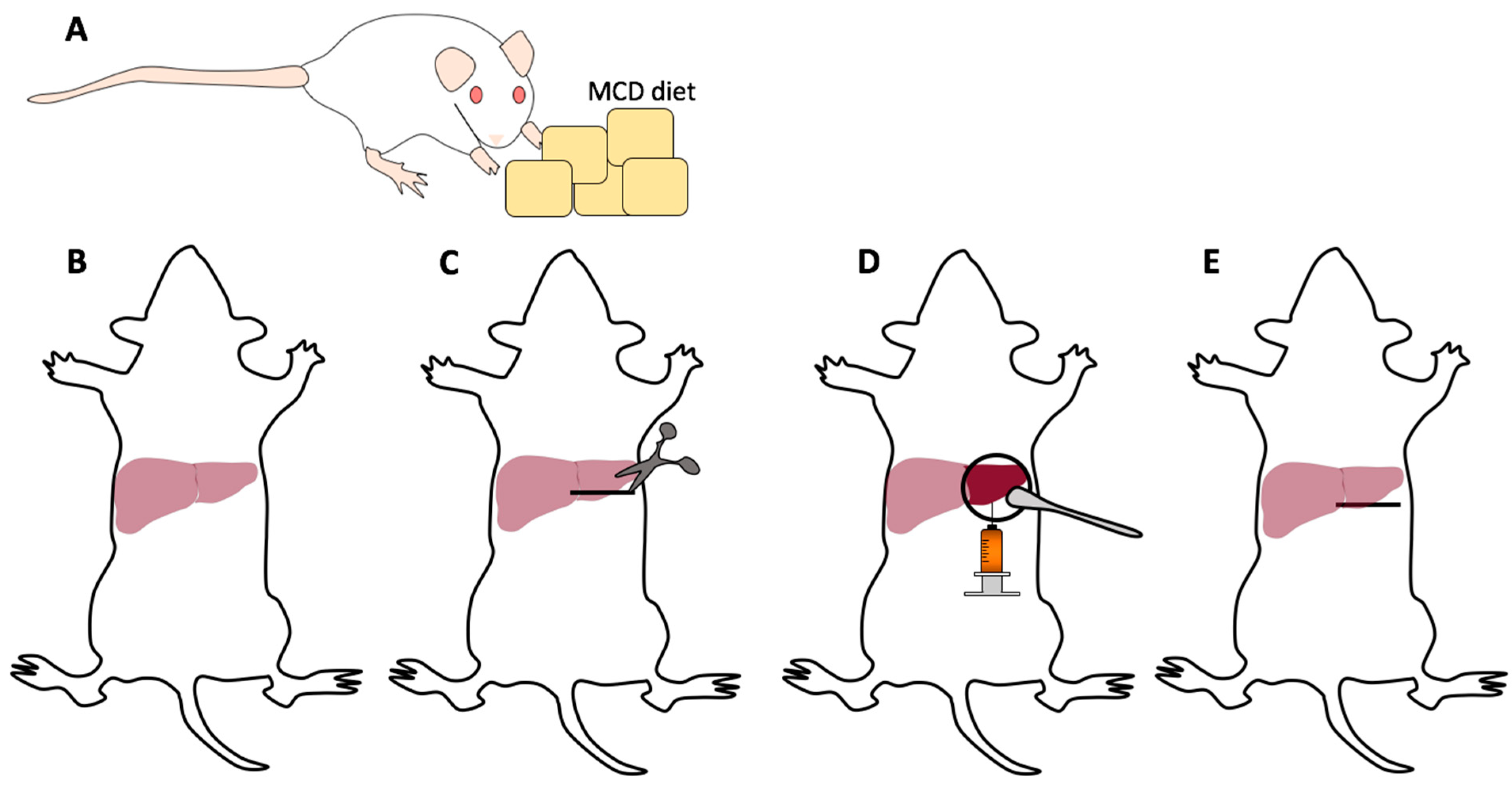
- Mice should be started on the MCD diet at approximately 6–8-weeks-old for two weeks for the induction of NAFLD/NASH (Figure 2A).NOTE: Mice on MCD may lose up to 30% of their body weight while on the MCD diet and as such weight should be monitored periodically during the course of the experiment. Any mouse with severe weight loss should be sacrificed in accordance with institutional protocol.
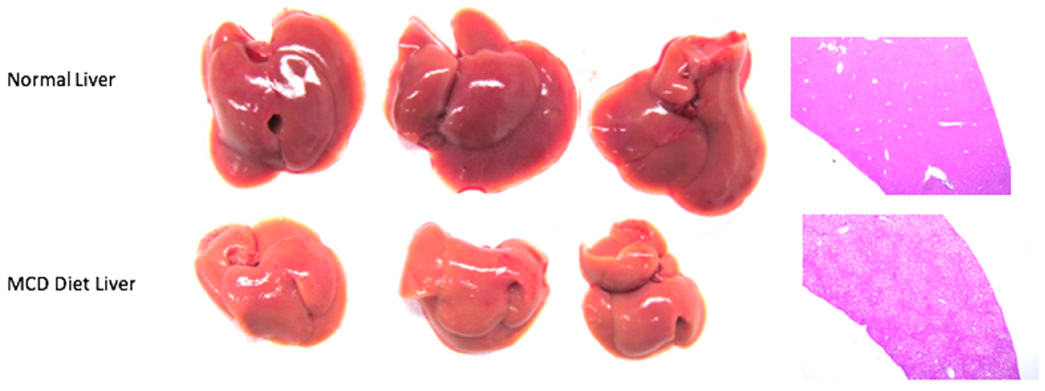
- Intrahepatic tumor injections should take place approximately two weeks after induction of the MCD diet when the mice are approximately 8–10-weeks-old. The investigator can visualize the liver disease as discolored often shrunken livers with steatosis on hematoxylin and eosin (H & E) staining (Figure 3).NOTE: Although the full extent of liver disease may not be realized after two weeks on diet, this allows for in parallel development of liver disease and HCC which mimics the human condition.NOTE: In general, older mice are easier to inject as the liver capsule is often more developed. We advise avoiding intrahepatic injection in mice <8 weeks old.
3.2. Cell Preparation. Time for Completion: 2 Weeks
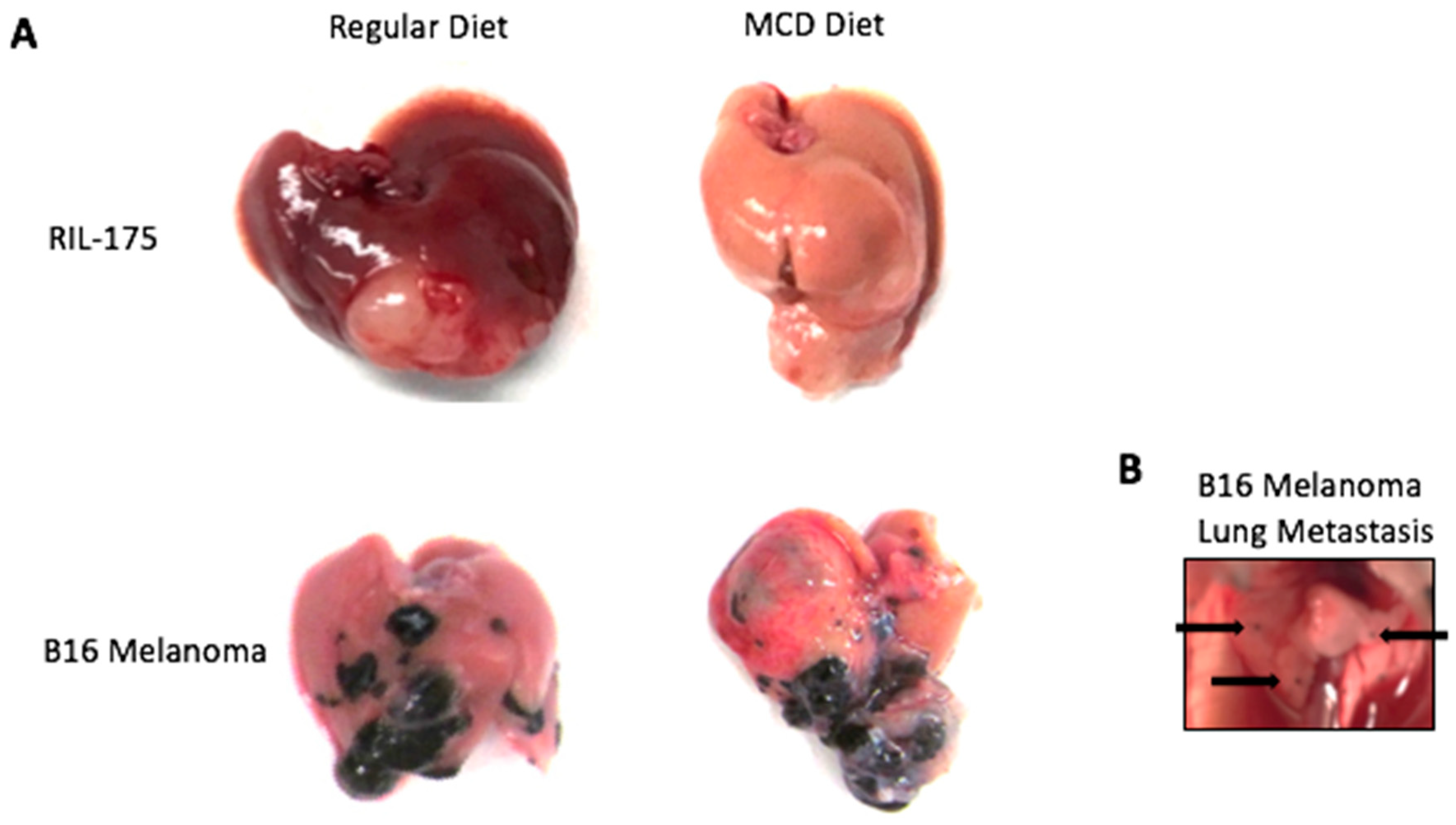
- Prepare a single cell suspension of a luciferase expressing cell line. Luciferase-expressing RIL-175 HCC tumor cells were cultured in complete RPMI medium supplemented with 10% FBS. Cells should be in culture for approximately four passages prior to injection.
- Prepare cell injection solution. For tumor establishment, 20 µL of tumor cell suspension should be injected per mouse. Cells should be counted and suspended 105–106 cells per 20 µL in a 50:50 solution of PBS and Matrigel. The concentration of the cell in the cell suspension may vary depending on growth kinetics of the cells being injected.
![Mps 01 00021 i001]() CRITICAL STEP Cell suspension and insulin syringes should be kept on ice to prevent Matrigel from solidifying. Stock of Matrigel should be aliquoted during first thaw and divided up into Eppendorf tubes of desired volume and stored at −20 °C.NOTE: Matrigel is used for tumor injection to provide a matrix plug allowing tumor cells to remain in liver after injection [15]. Injection of tumor cells in PBS has been performed but Matrigel allows for more consistent results.
CRITICAL STEP Cell suspension and insulin syringes should be kept on ice to prevent Matrigel from solidifying. Stock of Matrigel should be aliquoted during first thaw and divided up into Eppendorf tubes of desired volume and stored at −20 °C.NOTE: Matrigel is used for tumor injection to provide a matrix plug allowing tumor cells to remain in liver after injection [15]. Injection of tumor cells in PBS has been performed but Matrigel allows for more consistent results.

3.3. Surgical Procedure. Time for Completion: 15–30 Min per Mouse
- Anesthetize the mice using 2% isoflurane in an anesthesia chamber.
- Once mice are anesthetized, shave the abdomen clearing the surgical field of fur.
- Place and keep the mice on a heating pad with anesthesia nose cone to maintain sedation (Figure 2B).
- Use 10% povidone iodine solution followed by 70% ethanol to disinfect the skin, repeating this process three times.
- Sterilize the tips of surgical instruments in a bead sterilizer.
- Grab the skin with forceps and make an approximately 1 cm horizontal incision from the midline toward the left upper abdomen (Figure 2C).
![Mps 01 00021 i001]() CRITICAL STEP Proper placement of the incision makes the procedure much easier. Improper placement may lead to a struggle delivering the liver out of the wound for injection. We recommend making the incision just below the left costal margin but inferior enough not to cut into the thoracic cavity.NOTE: Mice on MCD diet have diseased liver and maybe smaller and retracted into the upper abdomen making the incision placement critical.
CRITICAL STEP Proper placement of the incision makes the procedure much easier. Improper placement may lead to a struggle delivering the liver out of the wound for injection. We recommend making the incision just below the left costal margin but inferior enough not to cut into the thoracic cavity.NOTE: Mice on MCD diet have diseased liver and maybe smaller and retracted into the upper abdomen making the incision placement critical. - Gently free the skin from the peritoneum. Grab the peritoneum lifting straight upward and make a small incision. Extend the peritoneal incision horizontally following the skin incision ensuring to see the tip of the scissors as to not injure underlying organs.NOTE: Bleeding may occur from superficial vessels of the abdominal wall. Use gentle pressure or the application of silver nitrate to help control the bleeding. Blood will distort the field of view and make following steps more difficult.
- Insert a cotton-tipped applicator into the left upper abdomen under the left lobe of the liver. Remove the cotton-tipped applicator slowly and gently rotate it toward yourself (Figure 2D).
![Mps 01 00021 i001]() CRITICAL STEP We aim to inject the left lobe of the murine liver as it is generally easily accessible and larger than the right or middle lobes. Additionally, the left lobe better accommodates the injected volume without tearing the liver capsule.NOTE: The tumor cell suspension should be loaded into the syringe prior to extraction of the liver and kept on ice.
CRITICAL STEP We aim to inject the left lobe of the murine liver as it is generally easily accessible and larger than the right or middle lobes. Additionally, the left lobe better accommodates the injected volume without tearing the liver capsule.NOTE: The tumor cell suspension should be loaded into the syringe prior to extraction of the liver and kept on ice. - With gentle traction stabilize the left lobe of the liver with the left hand against the underlying cotton-tipped applicator.
- Insert the 29 G needle into the liver parenchyma traversing several millimeters and slowly inject the tumor suspension.
![Mps 01 00021 i001]() CRITICAL STEP Proper needle location for injection is vital for consistent and reproducible results. The needle should traverse several millimeters of liver to prevent backflow of the tumor suspension. The needle should be superficial and visible through the liver and the tumor suspension should be injected just below the liver capsule, which would raise a wheel/bubble. Avoid excessive superior traction of the needle while in the liver parenchyma to prevent lacerating the liver. NOTE: This procedure takes surgical skill and practice to perfect in order to obtain reproducible results. It should be noted the amount of time it takes individual investigators to perform the procedure as you do not want the tumor cells to be on ice for an extended period of time. From our experience, after approximately 2–3 h, tumor cells lose viability while on ice.
CRITICAL STEP Proper needle location for injection is vital for consistent and reproducible results. The needle should traverse several millimeters of liver to prevent backflow of the tumor suspension. The needle should be superficial and visible through the liver and the tumor suspension should be injected just below the liver capsule, which would raise a wheel/bubble. Avoid excessive superior traction of the needle while in the liver parenchyma to prevent lacerating the liver. NOTE: This procedure takes surgical skill and practice to perfect in order to obtain reproducible results. It should be noted the amount of time it takes individual investigators to perform the procedure as you do not want the tumor cells to be on ice for an extended period of time. From our experience, after approximately 2–3 h, tumor cells lose viability while on ice. - After the injection, slowly retract the needle from the liver and place gentle pressure on the needle insertion site with a cotton-tipped applicator for several minutes to stop bleeding.
- Using a 5-0 Vicryl suture, close the peritoneal layer with a single figure-of-eight or two single interrupted sutures (Figure 2E).
- Using a 5-0 Vicryl suture, close the skin with two figure-of-eight or multiple single interrupted sutures.
![Mps 01 00021 i001]() CRITICAL STEP Do not use a running suture to close the skin. Mice will bite the sutures. If one suture is used and the mouse bites it, the entire wound may open.NOTE: Clips may be used if BLI imaging is not planned on being performed. If following tumor growth with BLI is planned, clips may cause interference with the imaging.
CRITICAL STEP Do not use a running suture to close the skin. Mice will bite the sutures. If one suture is used and the mouse bites it, the entire wound may open.NOTE: Clips may be used if BLI imaging is not planned on being performed. If following tumor growth with BLI is planned, clips may cause interference with the imaging. - Apply post-operative analgesia as per institutional protocol.
- Recover the mice from anesthesia in a cage with a heat lamp.
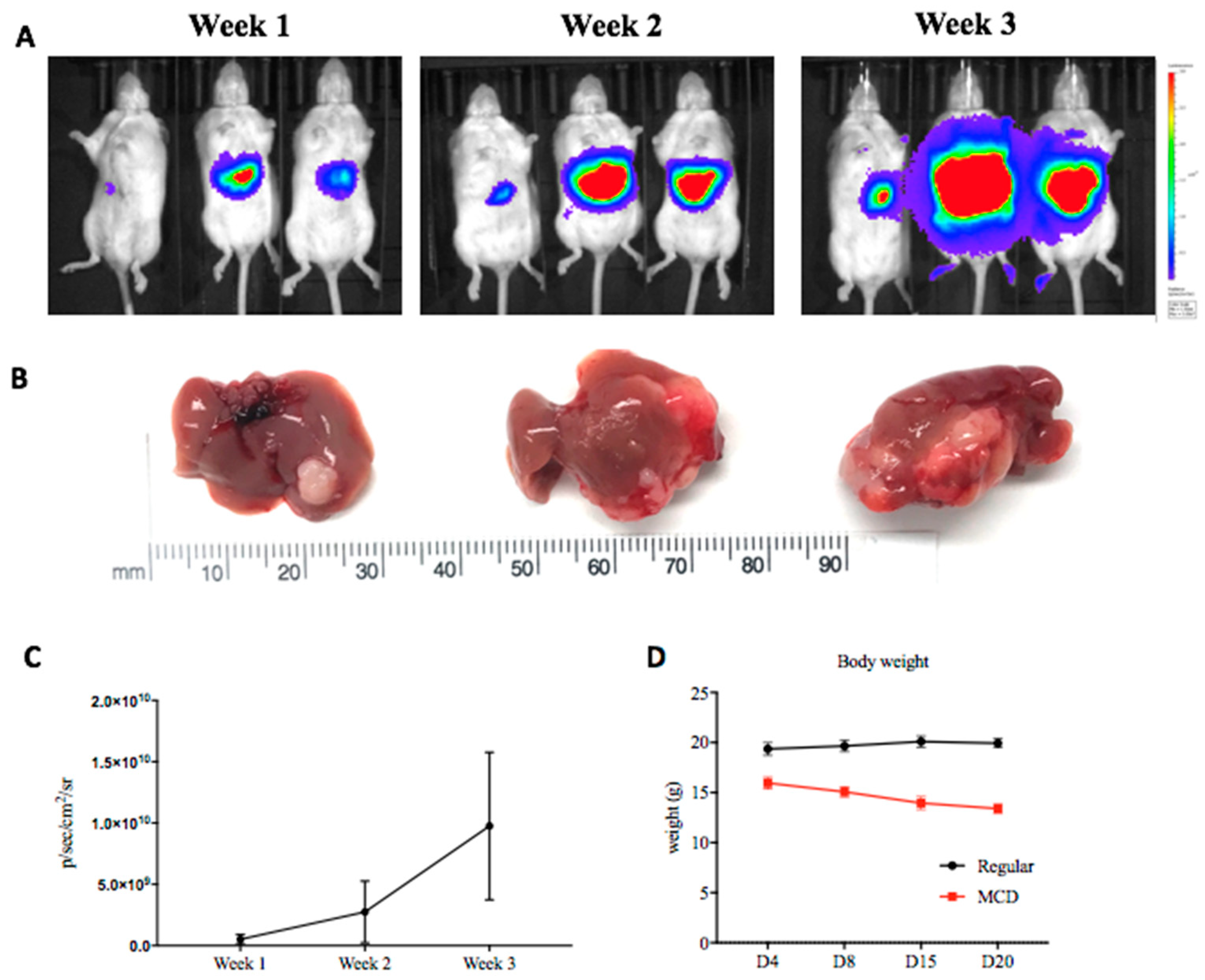
- Closely monitor the mice post-operatively for signs of distress. In combination with MCD diet, mice with intrahepatic tumors generally survive three weeks post-injection (five weeks after the initiation of MCD diet) after which the mice should be sacrificed for humane endpoints.NOTE: Upon sacrifice of mice, tumor growth throughout the abdomen is likely the results of a failed intrahepatic injection.
3.4. Tumor Growth Monitoring by Bioluminescent Imaging. Time for Completion: 15 Min per Group of Mice
- Tumor growth is monitored on a weekly basis with BLI.
- Anesthetize the mice using 2% Isoflurane in an anesthesia chamber.
- Shave the abdomen to decrease the interference with BLI.
![Mps 01 00021 i001]() CRITICAL STEP From our experience, monitoring intrahepatic injections with BLI in mice with black fur gives unreliable results as the black fur produces increased background signal. Therefore, we utilized B6(Cg)-Tyrc-2J/J mice to reduce the background signal. Shave the mice prior to every imaging session to ensure consistent results.
CRITICAL STEP From our experience, monitoring intrahepatic injections with BLI in mice with black fur gives unreliable results as the black fur produces increased background signal. Therefore, we utilized B6(Cg)-Tyrc-2J/J mice to reduce the background signal. Shave the mice prior to every imaging session to ensure consistent results. - Administer an intraperitoneal injection of 150 mg/kg of D-luciferin in PBS.
![Mps 01 00021 i001]() CRITICAL STEP Wait for the tumor saturation plateau to be reached. For our RIL-175 cells this was at approximately eight minutes after injection.
CRITICAL STEP Wait for the tumor saturation plateau to be reached. For our RIL-175 cells this was at approximately eight minutes after injection. - Perform imaging. We utilized the Xenogen in vivo imaging system The CCD camera was cooled to between −105 °C and −120 °C and the field of view set to 25 cm. Images were acquired with an exposure time of 30 s, medium binning, 1.2 f/stop, with an open filter.
![Mps 01 00021 i001]() CRITICAL STEP Prior to starting a large experiment, it is important to determine the proper exposure time required as to not saturate the image.
CRITICAL STEP Prior to starting a large experiment, it is important to determine the proper exposure time required as to not saturate the image. - Recover the mice from anesthesia in a cage with a heat lamp.
4. Expected Results
Author Contributions
Conflicts of Interest
References
- Fitzmaurice, C.; Allen, C.; Barber, R.M.; Barregard, L.; Bhutta, Z.A.; Brenner, H.; Dicker, D.J.; Chimed-Orchir, O.; Dandona, R.; Dandona, L.; et al. Global, Regional, and National Cancer Incidence, Mortality, Years of Life Lost, Years Lived With Disability, and Disability-Adjusted Life-years for 32 Cancer Groups, 1990 to 2015: A Systematic Analysis for the Global Burden of Disease Study. JAMA Oncol. 2017, 3, 524–548. [Google Scholar] [CrossRef] [PubMed]
- Duffy, A.G.; Greten, T.F. Liver cancer: Regorafenib as second-line therapy in hepatocellular carcinoma. Nat. Rev. Gastroenterol. Hepatol. 2017, 14, 141–142. [Google Scholar] [CrossRef] [PubMed]
- Younossi, Z.M.; Koenig, A.B.; Abdelatif, D.; Fazel, Y.; Henry, L.; Wymer, M. Global epidemiology of nonalcoholic fatty liver disease-Meta-analytic assessment of prevalence, incidence, and outcomes. Hepatology 2016, 64, 73–84. [Google Scholar] [CrossRef] [PubMed] [Green Version]
- Younossi, Z.; Anstee, Q.M.; Marietti, M.; Hardy, T.; Henry, L.; Eslam, M.; George, J.; Bugianesi, E. Global burden of NAFLD and NASH: Trends, predictions, risk factors and prevention. Nat. Rev. Gastroenterol. Hepatol. 2017, 15, 11. [Google Scholar] [CrossRef] [PubMed]
- Llovet, J.M.; Zucman-Rossi, J.; Pikarsky, E.; Sangro, B.; Schwartz, M.; Sherman, M.; Gores, G. Hepatocellular carcinoma. Nat. Rev. Dis. Primers 2016, 2, 16018. [Google Scholar] [CrossRef] [PubMed] [Green Version]
- Ma, C.; Kesarwala, A.H.; Eggert, T.; Medina-Echeverz, J.; Kleiner, D.E.; Jin, P.; Stroncek, D.F.; Terabe, M.; Kapoor, V.; ElGindi, M.; et al. NAFLD causes selective CD4+ T lymphocyte loss and promotes hepatocarcinogenesis. Nature 2016, 531, 253–257. [Google Scholar] [CrossRef] [PubMed]
- Brown, Z.J.; Fu, Q.; Ma, C.; Kruhlak, M.; Zhang, H.; Luo, J.; Heinrich, B.; Yu, S.J.; Zhang, Q.; Wilson, A.; et al. Carnitine palmitoyltransferase gene upregulation by linoleic acid induces CD4(+) T cell apoptosis promoting HCC development. Cell Death Dis. 2018, 9, 620. [Google Scholar] [CrossRef] [PubMed]
- Dela Pena, A.; Leclercq, I.; Field, J.; George, J.; Jones, B.; Farrell, G. NF-κB activation, rather than TNF, mediates hepatic inflammation in a murine dietary model of steatohepatitis. Gastroenterology 2005, 129, 1663–1674. [Google Scholar] [CrossRef] [PubMed]
- Ip, E.; Farrell, G.; Hall, P.; Robertson, G.; Leclercq, I. Administration of the potent PPARα agonist, Wy-14,643, reverses nutritional fibrosis and steatohepatitis in mice. Hepatology 2004, 39, 1286–1296. [Google Scholar] [CrossRef] [PubMed]
- Ibrahim, S.H.; Hirsova, P.; Malhi, H.; Gores, G.J. Animal Models of Nonalcoholic Steatohepatitis: Eat, Delete, and Inflame. Dig. Dis. Sci. 2016, 61, 1325–1336. [Google Scholar] [CrossRef] [PubMed]
- Rinella, M.E.; Green, R.M. The methionine-choline deficient dietary model of steatohepatitis does not exhibit insulin resistance. J. Hepatol. 2004, 40, 47–51. [Google Scholar] [CrossRef] [PubMed]
- Nakae, D.; Mizumoto, Y.; Andoh, N.; Tamura, K.; Horiguchi, K.; Endoh, T.; Kobayashi, E.; Tsujiuchi, T.; Denda, A.; Lombardi, B.; et al. Comparative changes in the liver of female Fischer-344 rats after short-term feeding of a semipurified or a semisynthetic l-amino acid-defined choline-deficient diet. Toxicol. Pathol. 1995, 23, 583–590. [Google Scholar] [CrossRef] [PubMed]
- Hebbard, L.; George, J. Animal models of nonalcoholic fatty liver disease. Nat. Rev. Gastroenterol. Hepatol. 2011, 8, 35–44. [Google Scholar] [CrossRef] [PubMed]
- Kodama, Y.; Kisseleva, T.; Iwaisako, K.; Miura, K.; Taura, K.; De Minicis, S.; Osterreicher, C.H.; Schnabl, B.; Seki, E.; Brenner, D.A. c-Jun N-terminal kinase-1 from hematopoietic cells mediates progression from hepatic steatosis to steatohepatitis and fibrosis in mice. Gastroenterology 2009, 137, 1467–1477. [Google Scholar] [CrossRef] [PubMed]
- Reiberger, T.; Chen, Y.; Ramjiawan, R.R.; Hato, T.; Fan, C.; Samuel, R.; Roberge, S.; Huang, P.; Lauwers, G.Y.; Zhu, A.X.; et al. An orthotopic mouse model of hepatocellular carcinoma with underlying liver cirrhosis. Nat. Protoc. 2015, 10, 1264–1274. [Google Scholar] [CrossRef] [PubMed] [Green Version]
- Eggert, T.; Wolter, K.; Ji, J.; Ma, C.; Yevsa, T.; Klotz, S.; Medina-Echeverz, J.; Longerich, T.; Forgues, M.; Reisinger, F.; et al. Distinct Functions of Senescence-Associated Immune Responses in Liver Tumor Surveillance and Tumor Progression. Cancer Cell 2016, 30, 533–547. [Google Scholar] [CrossRef] [PubMed]
- Lee, T.K.; Na, K.S.; Kim, J.; Jeong, H.J. Establishment of animal models with orthotopic hepatocellular carcinoma. Nucl. Med. Mol. Imaging 2014, 48, 173–179. [Google Scholar] [CrossRef] [PubMed]
- Sarraf-Yazdi, S.; Mi, J.; Dewhirst, M.W.; Clary, B.M. Use of in vivo bioluminescence imaging to predict hepatic tumor burden in mice. J. Surg. Res. 2004, 120, 249–255. [Google Scholar] [CrossRef] [PubMed]
- Fleten, K.G.; Bakke, K.M.; Maelandsmo, G.M.; Abildgaard, A.; Redalen, K.R.; Flatmark, K. Use of non-invasive imaging to monitor response to aflibercept treatment in murine models of colorectal cancer liver metastases. Clin. Exp. Metastasis 2017, 34, 51–62. [Google Scholar] [CrossRef] [PubMed]
- Zinn, K.R.; Chaudhuri, T.R.; Szafran, A.A.; O’Quinn, D.; Weaver, C.; Dugger, K.; Lamar, D.; Kesterson, R.A.; Wang, X.; Frank, S.J. Noninvasive bioluminescence imaging in small animals. ILAR J. 2008, 49, 103–115. [Google Scholar] [CrossRef] [PubMed]
- Makarova-Rusher, O.V.; Medina-Echeverz, J.; Duffy, A.G.; Greten, T.F. The yin and yang of evasion and immune activation in HCC. J. Hepatol. 2015, 62, 1420–1429. [Google Scholar] [CrossRef] [PubMed]


© 2018 by the authors. Licensee MDPI, Basel, Switzerland. This article is an open access article distributed under the terms and conditions of the Creative Commons Attribution (CC BY) license (http://creativecommons.org/licenses/by/4.0/).
Share and Cite
Brown, Z.J.; Heinrich, B.; Greten, T.F. Establishment of Orthotopic Liver Tumors by Surgical Intrahepatic Tumor Injection in Mice with Underlying Non-Alcoholic Fatty Liver Disease. Methods Protoc. 2018, 1, 21. https://doi.org/10.3390/mps1020021
Brown ZJ, Heinrich B, Greten TF. Establishment of Orthotopic Liver Tumors by Surgical Intrahepatic Tumor Injection in Mice with Underlying Non-Alcoholic Fatty Liver Disease. Methods and Protocols. 2018; 1(2):21. https://doi.org/10.3390/mps1020021
Chicago/Turabian StyleBrown, Zachary J., Bernd Heinrich, and Tim F. Greten. 2018. "Establishment of Orthotopic Liver Tumors by Surgical Intrahepatic Tumor Injection in Mice with Underlying Non-Alcoholic Fatty Liver Disease" Methods and Protocols 1, no. 2: 21. https://doi.org/10.3390/mps1020021