Application of NMR Screening Methods with 19F Detection to Fluorinated Compounds Bound to Proteins

1
Division of Agriculture and Agricultural Life Sciences, The University of Tokyo, Yayoi, Bunkyo-ku, Tokyo 113-8657, Japan
2
Department of Chemistry, College of Science and Technology, Meisei University, Hino, Tokyo 191-8506, Japan
*
Author to whom correspondence should be addressed.
Magnetochemistry 2018, 4(1), 3; https://doi.org/10.3390/magnetochemistry4010003
Submission received: 11 November 2017
/
Revised: 7 December 2017
/
Accepted: 21 December 2017
/
Published: 27 December 2017
(This article belongs to the Special Issue Nuclear Magnetic Resonance Spectroscopy)
Abstract
:The combinational use of one-dimensional (1D) NMR-based screening techniques with 1H and 19F detections were applied to a human serum albumin–diflunisal complex. Since most NMR screening methods observe 1H spectra, the overlapped 1H signals were unavailable in the binding epitope mapping. However, the NMR experiments with 19F detection can be used as an effective complementary method. For the purpose of identifying the 1H and 19F binding epitopes of diflunisal, this paper carries out a combinatorial analysis using 1H{1H} and 19F{1H} saturation transfer difference experiments. The differences of the 1H-inversion recovery rates with and without target irradiation are also analyzed for a comprehensive interpretation of binding epitope mapping.
1. Introduction
Protein–ligand interactions can provide useful insights for understanding the molecular recognition system. However, arriving at such understandings requires the developments of useful methods for selectively observing the ligand. Although X-ray analyses can determine such interactions of the complex at the atomic level, difficulties in crystallization often interfere with the process of X-ray studies. In some cases, NMR spectroscopy can be a useful alternative for analyzing macromolecular complexes and screening compounds with an affinity to target proteins. Various NMR-based screening methods to observe the ligand signals have been proposed. It has been shown that NOE-pumping [1], saturation transfer difference (STD) [2], water–ligand observed via gradient spectroscopy (WaterLOGSY) [3,4], and reverse NOE-pumping [5] experiments could directly detect 1H of the bound ligands. Recently, the NMR-based methods have been extended to fluorine detection [6,7,8]. Since the spectral elucidation in the aforementioned experiments [1,2,3,4,5] depends on the dispersion of 1H signals, its signal degeneracy leads to a lack of information for the target molecules. Considering these difficulties, the NMR-based screening methods with 19F-detection were applied to the human serum albumin (HSA)-diflunisal complex. HSA is an abundant plasma protein that binds to a wide range of drugs. Diflunisal contains two fluorine atoms in a molecule, and is a nonsteroidal anti-inflammatory drug that is effective in treating fever, pain, and inflammation. Since the X-ray crystal structure of a diflunisal-HSA complex has been determined (pdb: 2BXE), this complex could be a suitable model system for studying the molecular interactions of 1H and 19F using NMR spectroscopy. Information of the binding epitopes can be obtained for 19F as well as 1H of the fluorinated compound.
2. Results and Discussion
To investigate the 1H binding epitopes of ligands, two representative methods—the 1H{1H} STD method acquired with various saturation times [9], or the difference of inversion recovery rates with and without target irradiation (DIRECTION) [10] method—were generally used. In the present study, the binding epitopes of diflunisal (Figure 1) were investigated using both methods. In the 1H{1H} STD experiments, the STD build-up curves were obtained at various saturation times. The slope of the STD build-up curve at a saturation time of 0 s was obtained by fitting to the monoexponential equation: STD = STDmax(1 − e(−ksat × t)), where STD stands for the STD signal intensity at saturation time t, STDmax is the maximal STD intensity at long saturation times, and ksat stands for the observed saturation constant. The values of ksat × STDmax correspond to the slope of the curve at zero saturation time with an elimination of T1 bias. In the DIRECTION experiments, 1H-T1 were measured with and without the selective irradiation of protein, and its reciprocals, corresponding to the inversion recovery rates, were calculated for each separated 1H signal.
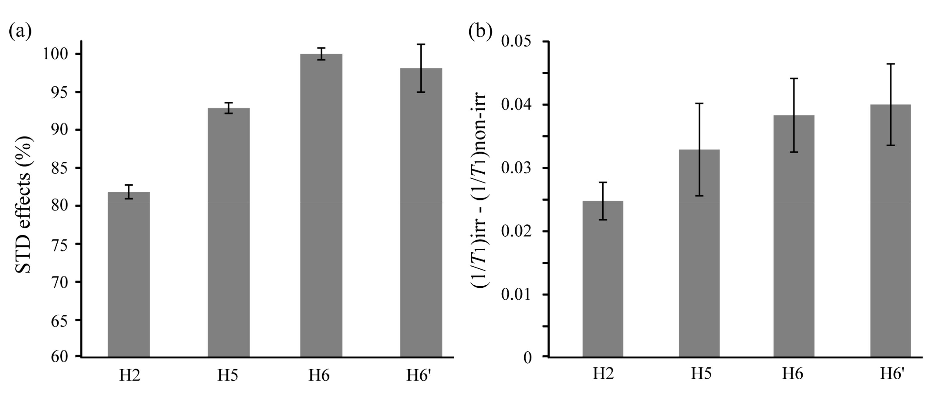
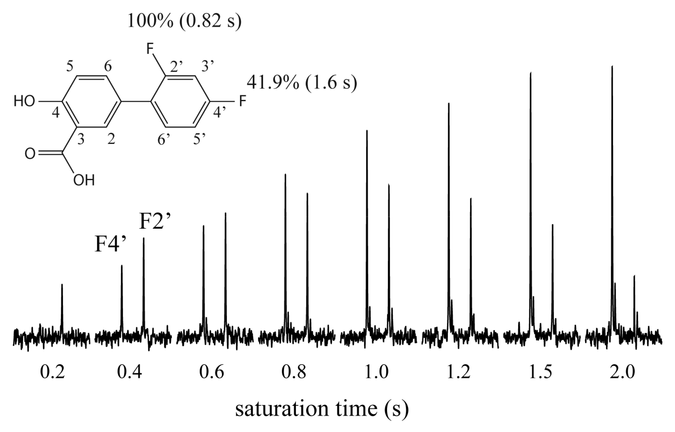
The values of the STD effect were normalized by referencing the signal of H6 with the largest STD effect. The relative values are shown in Figure 2a. The values of the STD effect were larger in H5 and H6, indicating that these protons contributed as the binding epitopes. The smallest value was obtained in H2, which made less contact with the protein. The binding epitopes were also investigated using the DIRECTION method, evaluating the difference between the 1H-inversion recovery rates with and without the irradiation of protein [10]. The large differences reflect the proximity to the protein surface. H6 and H6′ showed relatively large values (Figure 2b). Since the H3′ and H5′ signals overlapped, H6′ was the only signal available for analysis in a 2′,4′-difluoro ring (Figure 1), indicating that the incomplete information was obtained in the 1H-detection NMR methods. To obtain more detailed information of the binding epitopes for the 2′,4′-difluoro ring, the 19F{1H} STD spectra were acquired with the arrayed saturation times (Figure 3). The 19F{1H} STD experiment was more insensitive than the 1H{1H} STD experiment. It can be considered that the saturation transfer from 1H to 19F is much less effective than that from 1H to 1H. However, the 19F{1H} STD experiment provided the useful information regarding the 19F binding epitopes. The normalized values of the STD effect of F2′ and F4′ were 100% and 41.9%, respectively, and the values of 19F-T1 were 0.82 s and 1.6 s in the aforesaid order. This result indicated that F2′ made more close contact to HSA than F4′. It can be considered that a portion comprising H6, H6′, and F2′ could play a key role as the binding portion of diflunisal. The H2 made less close contact, which could be caused by an interruption of the carboxyl group at position 3. In the X-ray crystal structure of HSA complexed with diflunisal (pdb: 2BXE), three molecules of diflunisal were bound with one molecule of HSA, where various close contacts were made in each binding site between two fluorine atoms of diflunisal, and protons of HSA. Since information from an epitope mapping that was obtained by the NMR experiments revealed average contacts in three HSA binding sites, some differences in the close contacts need to be considered between crystal and solution states.
3. Materials and Methods
3.1. Instrumentation and Chemicals
All of the NMR spectra were recorded at 20 °C on a Varian 600 MHz NMR system (Vaian, Palo Alto, CA, USA) or JEOL ECA-500 MHz spectrometer (JEOL Ltd., Tokyo, Japan). Diflunisal and HSA were purchased from Sigma-Aldrich (Tokyo, Japan). A 600-µL of solution containing 0.05 mM HSA and 5.0 mM diflunisal was prepared in 100% 2H2O.
3.2. NMR Spectroscopy
The experimental parameters of the 1H{1H} STD experiment were as follows: data points = 16,384, spectral width of 1H = 8012 Hz, number of scans = 1024, recycle time = 1.0 s. The saturation times for the selective excitation of proteins were arrayed in the range of 0.2–3.5 s, and the arrayed spectra were acquired five times. The on and off resonance frequencies of 1H were 0.6 and −20 ppm, respectively. Those of the 19F{1H} STD experiment were as follows: data points = 8192, spectral width of 19F = 6012 Hz, number of scans = 10,240, recycle time = 1.0 s. The saturation times for the selective excitation of protein were arrayed in the range of 0.2–2.0 s. The on and off resonance frequencies of 1H were 0.6 and −20 ppm, respectively. The values of the initial slope in the STD build-up curves were obtained by the least-square fitting in both of the STD experiments [9]. The experimental parameters for measuring 19F-T1 were as follows: data points = 8192, spectral width of 19F = 6012 Hz, number of scans = 128, recycle time = 5.0 s. The inversion recovery pulse sequence was used. In measurements of 1H-T1 with and without the selective excitation of protein resonance (DIRECTION method) [10], the measurements were repeated five times, and the program in the JEOL Delta software (JEOL Ltd., Tokyo, Japan) was used for calculation of 1H-T1. The on and off resonance frequencies of 1H were 0.6 and −20 ppm, respectively. The exponential window function was used with zero-filling by a factor of 2. The 1H and 19F chemical shifts were relative to 3-(Trimethylsilyl)-1-propanesulfonic acid sodium salt (DSS) and trichlorofluoromethane, respectively, as external standards.
4. Conclusions
Although the sensitivity of the 19F{1H} STD experiment was lower than that of the 1H{1H} STD experiment, the obtained information was useful for the fluorinated compounds with the degenerated 1H signals. Comprehensive interpretations for the binding epitope mapping are essential, while considering some discrepancies in the results of various NMR experiments. The 19F{1H} STD experiment can be a complimentary method for the 1H detection methods.
Acknowledgments
This study was supported by two Grant-in-Aids for Scientific Research (15K05550 for M.T.) from the Ministry of Education, Culture, Sports, Science, and Technology.
Conflicts of Interest
The authors declare no conflict of interest.
References
- Chen, A.; Shapiro, M.J. NOE Pumping: A novel NMR technique for identification of compounds with binding affinity to macromolecules. J. Am. Chem. Soc. 1998, 120, 10258–10259. [Google Scholar] [CrossRef]
- Maye, M.; Meyer, B. Group epitope mapping by saturation transfer difference NMR to identify segments of a ligand in direct contact with a protein receptor. J. Am. Chem. Soc. 2001, 123, 6108–6117. [Google Scholar]
- Dalvit, C.; Pevarello, P.; Tatò, M.; Veronesi, M.; Vulpetti, A.; Sundström, M. Identification of compounds with binding affinity to proteins via magnetization transfer from bulk water. J. Biomol. NMR 2000, 18, 65–68. [Google Scholar] [CrossRef] [PubMed]
- Dalvit, C.; Fogliatto, G.P.; Stewart, A.; Veronesi, M.; Stockman, B.J. WaterLOGSY as a method for primary NMR screening: Practical aspects and range of applicability. J. Biomol. NMR 2001, 21, 349–359. [Google Scholar] [CrossRef] [PubMed]
- Chen, A.; Shapiro, M.J. NOE Pumping. 2. A high-throughput method to determine compounds with binding affinity to macromolecules by NMR. J. Am. Chem. Soc. 2000, 122, 414–415. [Google Scholar] [CrossRef]
- Dalvit, C.; Fagerness, P.E.; Hadden, S.T.A.; Sarver, R.W.; Stockman, B.J. Fluorine-NMR experiments for high-throughput screening: Theoretical aspects, practical considerations, and range of applicability. J. Am. Chem. Soc. 2003, 125, 7696–7703. [Google Scholar] [CrossRef] [PubMed]
- Dalvit, C.; Flocco, M.; Stockman, B.J.; Veronesi, M. Competition binding experiments for rapidly ranking lead molecules for their binding affinity to human serum albumin. Comb. Chem. High Throughput Screen. 2002, 5, 645–650. [Google Scholar] [CrossRef] [PubMed]
- Sakuma, C.; Kurita, J.; Furihata, K.; Tashiro, M. Achievement of 1H-19F heteronuclear experiments using the conventional spectrometer with a shared single high band amplifier. Magn. Reson. Chem. 2015, 53, 327–329. [Google Scholar] [CrossRef] [PubMed]
- Mayer, M.; James, T.L. NMR-based characterization of phenothiazines as a RNA binding scaffold. J. Am. Chem. Soc. 2004, 126, 4453–4460. [Google Scholar] [CrossRef] [PubMed]
- Mizukoshi, Y.; Abe, A.; Takizawa, T.; Hanzawa, H.; Fukunishi, Y.; Shimada, I.; Takahashi, H. An accurate pharmacophore mapping method by NMR spectroscopy. Angew. Chem. Int. Ed. 2012, 51, 1362–1365. [Google Scholar] [CrossRef] [PubMed]
Figure 1.
Structure and 1H NMR spectrum of diflunisal.
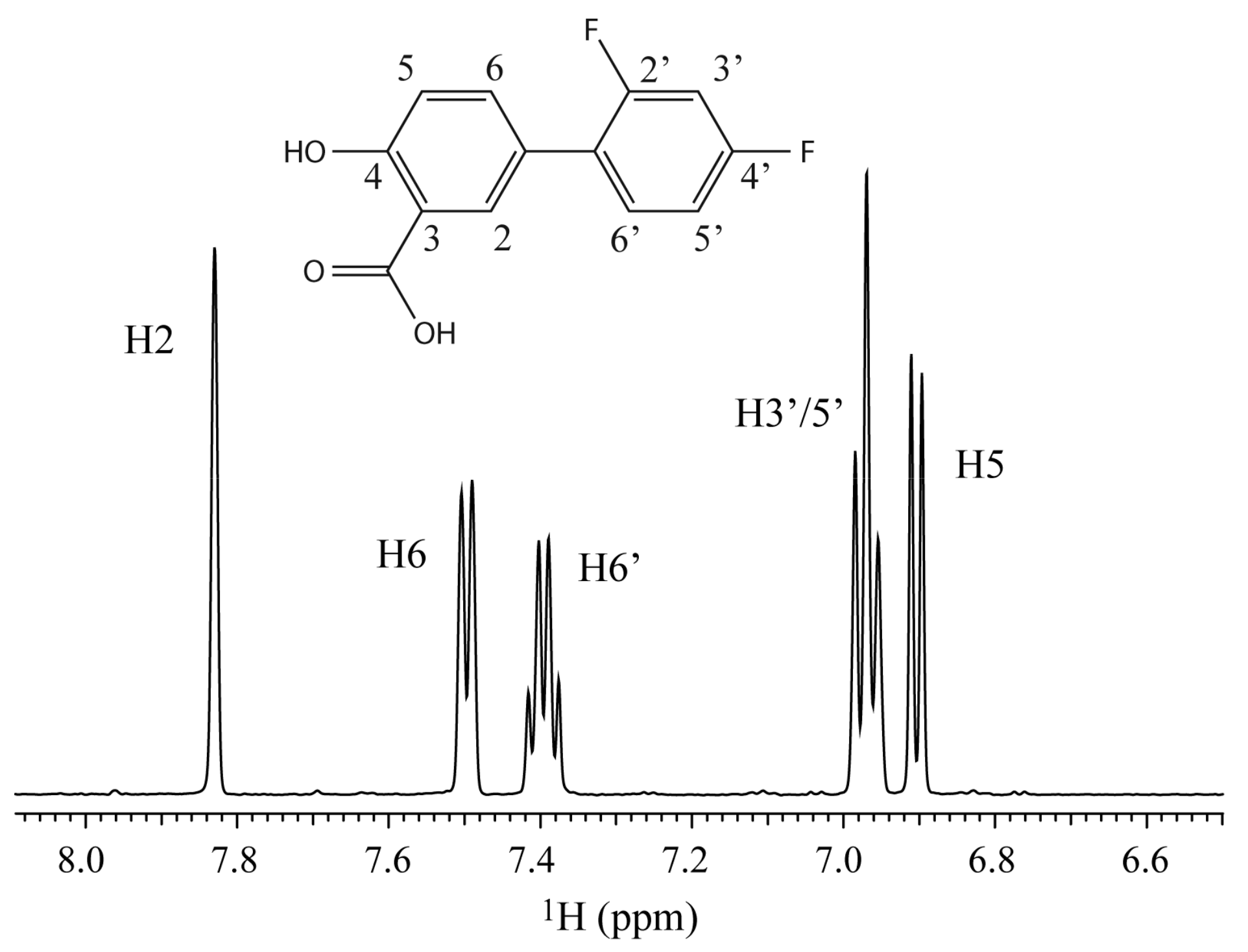
Figure 2.
(a) The values of the saturation transfer difference (STD) effect of diflunisal. The values were normalized by referencing the signal of H6 with the largest STD effect; (b) The difference of inversion recovery rate with and without target irradiation.
Figure 2.
(a) The values of the saturation transfer difference (STD) effect of diflunisal. The values were normalized by referencing the signal of H6 with the largest STD effect; (b) The difference of inversion recovery rate with and without target irradiation.

Figure 3.
The 19F{1H} STD spectra acquired with the arrayed saturation times. The normalized values of the STD effect (%) and the values of 19F-T1 (s) are shown.
Figure 3.
The 19F{1H} STD spectra acquired with the arrayed saturation times. The normalized values of the STD effect (%) and the values of 19F-T1 (s) are shown.
© 2017 by the authors. Licensee MDPI, Basel, Switzerland. This article is an open access article distributed under the terms and conditions of the Creative Commons Attribution (CC BY) license (http://creativecommons.org/licenses/by/4.0/).
Share and Cite
MDPI and ACS Style
Furihata, K.; Usui, M.; Tashiro, M. Application of NMR Screening Methods with 19F Detection to Fluorinated Compounds Bound to Proteins. Magnetochemistry 2018, 4, 3. https://doi.org/10.3390/magnetochemistry4010003
AMA Style
Furihata K, Usui M, Tashiro M. Application of NMR Screening Methods with 19F Detection to Fluorinated Compounds Bound to Proteins. Magnetochemistry. 2018; 4(1):3. https://doi.org/10.3390/magnetochemistry4010003
Chicago/Turabian StyleFurihata, Kazuo, Moe Usui, and Mitsuru Tashiro. 2018. "Application of NMR Screening Methods with 19F Detection to Fluorinated Compounds Bound to Proteins" Magnetochemistry 4, no. 1: 3. https://doi.org/10.3390/magnetochemistry4010003
Note that from the first issue of 2016, this journal uses article numbers instead of page numbers. See further details here.