Review

Cd, Pb and Hg Biomonitoring in Fish of the Mediterranean Region and Risk Estimations on Fish Consumption

by
Elisavet A. Renieri
1,
Athanasios K. Alegakis
1,
Michalis Kiriakakis
1,
Marco Vinceti
2,
Eren Ozcagli
3,
Martin F. Wilks
4 and
Aristidis M. Tsatsakis
1,*
1
Centre of Toxicology Sciences and Research, Division of Morphology-Medical School University of Crete, Vassilika Vouton, Heraklion 12258, Crete, Greece
2
Environmental, Genetic and Nutritional Epidemiology Research Center, University of Modena and Reggio Emilia, Reggio Emilia 42122, Italy
3
Department of Pharmaceutical Toxicology, Faculty of Pharmacy, Istanbul University, Beyazit-Istanbul 34116, Turkey
4
Swiss Centre for Applied Human Toxicology, Universität Basel, Basel CH-4056, Switzerland
*
Author to whom correspondence should be addressed.
Toxics 2014, 2(3), 417-442; https://doi.org/10.3390/toxics2030417
Submission received: 3 March 2014 / Revised: 21 July 2014 / Accepted: 6 August 2014 / Published: 18 August 2014
(This article belongs to the Collection Heavy Metals Toxicology)

Abstract

:
Cadmium (Cd), lead (Pb) and mercury (Hg) are toxic metals with increasing interest due to their tendency to bioaccumulate in fish tissue which may pose a threat to human health via fish consumption. This review of the recent literature on Cd, Pb, Hg levels summarizes data of fish biomonitoring studies in the Mediterranean Sea in order to determine potential risks due to dietary intake of metals. The analytical methods applied are described, with Atomic Absorption Spectroscopy and Inductively Coupled Plasma Mass Spectroscopy being the most popular. Most of the literature reviewed is focused on the Eastern Mediterranean. Results from the studies indicate that metals mostly accumulate in liver, followed by muscle. Although there are few studies reporting metal levels in fish exceeding the maximum residue levels (MRLs), the bulk of the studies cite levels below the MRLs. The hazard index (HI) of fish consumption, namely the ratio of estimated weekly intake to provisional tolerable weekly intake (EWI/PTWI) was estimated for adult consumers and no risk emerged. The EWI/PTWI ratios of lead and mercury for Italy (0.14 and 0.22 respectively) represent the highest HI levels estimated. In view of maximizing the benefits while minimizing the risks of fish consumption, a more detailed fish-specific database on intakes for consumers is required and extended bimonitoring in as many regions as possible.

1. Introduction

The Mediterranean Sea, a semi-closed basin surrounded by densely populated and industrialized countries, has a low capacity of water interchange with the Atlantic Ocean and other surrounding seas. Human development in the Mediterranean region has extensively influenced the coastal areas and has led to a constant rate of pollution with toxic compounds. The United Nations Environment Programme has estimated that 650 million tons of sewage, 129,000 tons of mineral oil, 60,000 tons of mercury, 3800 tons of lead and 36,000 tons of phosphates are dumped into the Mediterranean each year. Meanwhile, 70 percent of the wastewater dumped into the Mediterranean is untreated. The sea is also a major oil transportation route and up to one million tons of crude oil are discharged annually from accidental spills, illegal bunkering and tank cleaning practices, as well as inadequate harbor facilities. The Mediterranean Sea is additionally burdened by its major river systems: the Po, the Ebro, the Nile, and the Rhone which carry substantial amounts of agricultural and industrial wastes. As the Mediterranean is almost entirely landlocked, its waters have a very low renewal rate (80 to 90 years) making them excessively sensitive to pollution [1]. The escalating contaminant load in the Mediterranean Sea has created the need for more comprehensive studies and legislation establishment [2,3]. Metals are one of the most important groups of contaminants health-wise. Marine ecosystems receive metal loads through atmospheric and onshore waste origins, but the main source of metal pollution in the aquatic ecosystems is their discharge as industrial waste. The metals’ bioavailability and potential toxicity to organisms is determined by their chemical form [4]. Aside from biomonitoring studies, assessments on sediment quality as well as the water column are necessary.
As fish populations tend to be stable and easy to collect, they are widely used in biomonitoring of environmental pollution. In order to assess the body burden of toxic contaminants in fish, as means of estimating the contamination in several Mediterranean regions, monitoring programs have been used as a tool of investigation for many years [5]. Certain species have been widely used as sentinels of contamination in the aquatic environment due to the fact that they accumulate metals in their tissue at rates and concentrations well above the background levels. One of these species is Mullets (Mullus sp.) [6] which has been recommended as a monitoring species for metal pollution [7,8]. Other typical Mediterranean species used as sentinel fish are the Sea bass which is also the species mostly produced in aquaculture of Mediterranean areas [9] and the Sea bream which is widely distributed throughout the entire Mediterranean Sea and is selected for its native sandy costal habitat [10].
The metals considered most toxic and apparently most poisonous to marine life which are reviewed in this study, include mercury, cadmium, and lead [11]. As recently pointed out, in several areas of the Mediterranean Sea high concentrations of these elements are present in many types of commercially important fish [2]. In South Eastern (SE) Spain in particular, an area which has been polluted with metals as a result of mine tailings and industrial waste disposal, high concentrations of lead and cadmium in sediment and biota have been reported [12]. In the southern part of the Mediterranean, on the coast of Alexandria where the main fishing sources in Egypt are located, the levels of metal pollution have increased dramatically during the past few years due to domestic waste disposal in addition to waste refuse from shipping activities [13]. Moreover, in Iskenderun Bay, Turkey, as reported by Yilmaz, 2003 [14], lead levels detected in fish tissue exceeded the acceptable values for human consumption. With respect to mercury, the Mediterranean Sea is characterized by variations in mercury distribution, creating zones with extremely high mercury concentrations; more particularly Mediterranean deep-sea fishes, such as tuna and swordfish tend to exhibit higher levels of metal accumulation than those of populations inhabiting other areas such as the Atlantic [2].
On the other hand, areas with much traffic such as Izmir Bay, North Eastern (NE) Mediterranean, provide evidence of considerably lower lead concentrations than those found in other polluted areas of Turkey [15]. Similarly, study areas located on the NE Ionian coast of Italy which receive large quantities of untreated or partially treated industrial and domestic sewage along with spillages from vessel and harbor operations, show concentrations of the same proportion to those reported for non-polluted areas except for high mercury content in specimens of Capo Passero [16]. In the SE Aegean Sea (Turkey), Güllük Bay, the moderately high cadmium level in sediments can be explained by the adjacent Güllük Port and the presence of aquaculture farms nearby. The lead content of the Güllük Bay sediments is considered low and metal concentrations found in a study of this area were lower than those found in other Turkish polluted areas of Aegean Sea [17].
Metals bioaccumulated in aquatic inhabitants are subsequently transferred to humans through the food chain [18]. Fish constitutes a major part of the human diet mainly due to its high nutritional value; however it could carry serious health risks when contaminated. Long term consumption of foodstuff contaminated with metals may lead to the accumulation of toxic metals in several vital organs. This accumulation may result in perturbation of biochemical processes, which may cause liver, kidney, cardiovascular, nervous and bone disorders [19].
Table 1. Maximum Residue Levels (MRLs) in fish tissue and Intakes (TDI, PTWI) Cd, Pb, Hg and methylmercury (MeHg), set by JEFCA, EFSA and EC. (ww: wet weight).
MetalJECFAEFSAEC
TDI (µg/kg bw)PTWI (μg/kg bw)PTWI (μg/kg bw)MRLs (mg/g ww)MRLs (mg/g ww)
HgNA *NA *40.5 (1.0 **)0.5 (1.0 **)
MeHg0.231.61.61.3NA *
CdNA *7 (2003)2.50.05 (0.1–0.3 **)0.050–0.30 ***
PbNA *25250.30.30 ***
* NA: Not Available; ** certain fish; *** Maximum level of cadimium in the muscle meat of the following fish; bonito (Sarda sarda), common two-banded seabream (Diplodus vulgaris), eel (Anguilla anguilla), grey mullet (Mugil labrosus labrosus), horse mackerel or scad (Trachurus species), louvar or luvar (Luvarus imperialis), mackerel (Scomber species), sardine (Sardina pilchardus), sardinops (Sardinops species), tuna (Thunnus species, Euthynnus species, Katsuwonus pelamis), wedge sole (Dicologoglossa cuneata) is 0.10 mg/kg wet weight. Maximum level of cadimium in the muscle meat is 0.20 mg/kg wet weight for bullet tuna (Auxis species) and 0.30 mg/kg wet weight for anchovy (Engraulis species), swordfish (Xiphias gladius). As for those fishes outside of these exceptions, maximum level of cadimium in the muscle meat is 0.050 mg/kg wet weight.
The European Union (EU) has set maximum levels for certain contaminants (Table 1) with a view to reducing their presence in foodstuffs to the lowest levels reasonably achievable so as to secure high levels of public health protection, especially for sensitive population groups, such as children or people with allergies. In addition, levels of metals detected in fish need to be considered with respect to human intake and more specifically, the Provisional Tolerable Weekly Intake (PTWI) levels, the Tolerable Daily Intake (TDI) recommended by FAO/WHO experts (JECFA) EFSA and EC (Table 1) [20,21,22,23,24,25,26,27].
This review aims to summarise aspects of the biomonitoring studies on metal levels (Cd, Pb, Hg), such as the levels of the metals in fish tissue, the analytical methods applied and metal accumulation in fish. In addition, calculations for risks associated to fish consumption in the Mediterranean populations were made, using data for the countries and fish categories represented in the studies.
Figure 1. Sampling sites.
Toxics 02 00417 g001

2. Study Selection and Methods

2.1. Criteria for the Selection of the Studies

a)
The literature collection regarding biomonitoring studies was based on the following criteria: At least one of the three metals (Cd, Pb, Hg) was studied,
b)
The samples collected for each study included sea fish samples and only data on sea fish samples were recorded and listed for the review, excluding data on other species i.e., fresh water fish, crustaceans etc.
c)
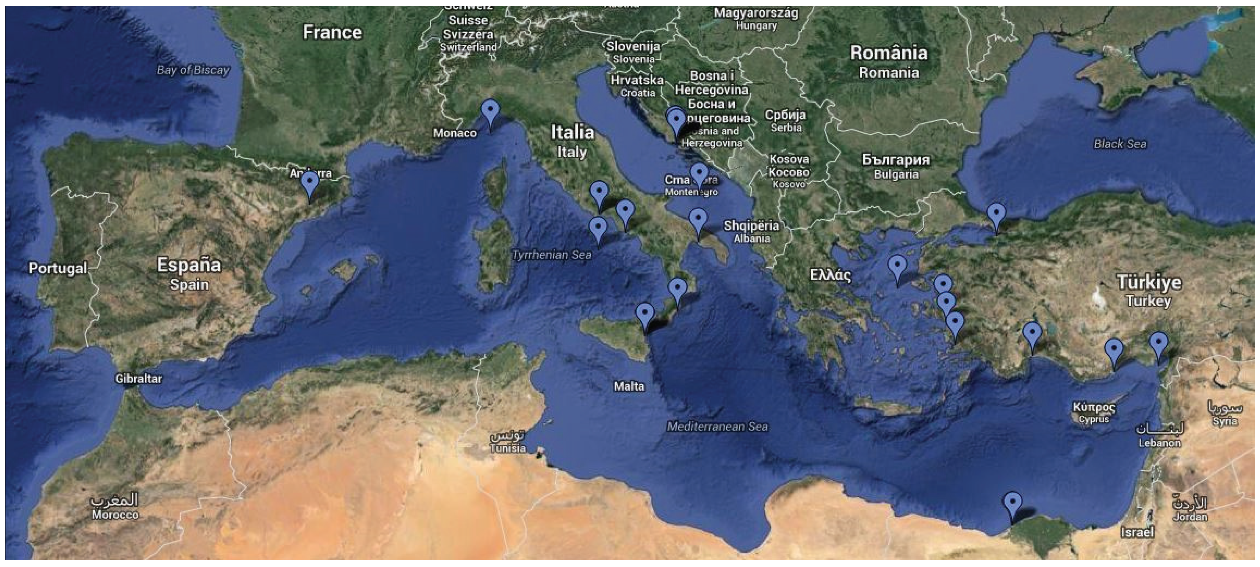
The sampling was conducted in the broader area of the Mediterranean Sea (Figure 1),
d)
The publication year of each study was between 2000 and 2012.
For listing of the analytical methods (Table 2, Chapter 3.1.) the biomonitoring studies of the initial dataset were considered.
For metal levels (Table 3, Chapter 3.3.) data reported in the studies from the initial dataset with the following exceptions were recorded:
(a)
Sampling was not conducted according to seasons or for a period longer than 2 years,
(b)
Analysis of the fish samples was not done by age or sex groups.
For Chapter 4.2. studies reporting data on human intakes of metals, in Mediterranean countries were collected, which were included under the following conditions:
(a)
Human intakes (EDI/EWI) of at least one of the metals (Cd, Pb and Hg) were estimated through the consumption of sea fish species and/or
(b)
Risk of human intake, expressed as a hazard index, was estimated by using human intake data (either obtained from Country’s Institutions or estimated experimentally, depending on the study) and metal levels determined in fish tissue, in the respective studies.
(c)
Studies performed on fish bought from supermarkets, were not considered.

2.2. Data Process on Metal Levels

Mean values of concentration of metals were selected from the published papers. When it was necessary transformations of Cd, Pb and Hg levels into μg/g tissue were made. All selected values follow the inclusion criteria described in Section 2.1. Bivariate associations among metal levels in fish tissues were examined using Spearman’s rho coefficients. Scatterplots of the above pairs (Cd-Pb, Cd-Hg and Pb-Hg) were made. Additionally, in order to investigate tissue distributions of mean metal values, error bars plots using quartiles (1st, median and 3rd) were used. IBM Statistics 20.0 was used for statistical analysis.

2.3. Risk Assessment Methods

Risk assessment calculations for each country (Chapter 4.3, Table 4.) were made, utilizing data on metal levels presented in Table 3 combined with consumption information for each country obtained from the FAO database [28] (accessed 2013). The consumption data for the time period 2000–2001 are derived from the FAOSTAT database based on the item “Food supply quantity g/capita/day” and categorized per type of fish “Pelagic Fish”, “Demersal Fish” “Marine fish” and “Total” when they are available. The worst case consumption scenario (the highest consumption rates) for each country and per type of fish was selected and the EWI/PTWI ratios for adult consumers were estimated.
The formula used for the aforementioned estimations are based on Copat C et al. 2012 [5].
Table 2. Analytical techniques applied in the reviewed literature.
Ref NO.MuscleLiverGonadsOtherAnalytical TechniqueLimit of Detection/(Min) (ppm)
CdPbHgCdPbHg
[29] a++--GF-AASGF-AASCV-AAS0.0020.040.02
[30] a-+-+GF-AAS--NR--
[31] b-+++GF-AAS--1--
[32] a++--GF-AASGF-AAS-0.00410.049-
[33] a++++ICP-OESICP-OES-11-
[5] a+---AASAAS-NRNR-
[34] a++--ICP-OESICP-OES-0.010.04-
[17] b+---GF-AASGF-AAS-(<0.01)(<0.02)-
[35] a+---GF-AASGF-AAS-0.00030.005-
[36] a++--ICP-OESICP-OES-NRNR-
[37] b+---FAASFAAS-0.080.19-
[2] a+---GF-AASGF-AASHG-AASNRNRNR
[38] a+---ICP-OESICP-OES-NRNR-
[7] a+---GF-AASGF-AASCV-AAS0.170.05ND
[13] b+---AAS--NR--
[39] a++--ICP-OESICP-OES-0.020.33-
[40] b+---ICP-OESICP-OES-14.2-
[41] b++-+FAASFAAS-2828-
[14] a+-++-ICP-OES--NR-
[42] a+---GF-AASGF-AASCV-AAS0.450.4
[43] a+++-GF-AASGF-AASCV-AAS0.10.10.5
[44] a++--GF-AASGF-AASCV-AAS0.050.030.03
[45] a+---GF-AASGF-AASGF-AASNRNRNR
[46] aNSNSNSNSDPSAVDPSAV-NRNR-
[47] a+---ET-AASET-AASCV-AAS0.10.93.3
[48] b+-----AMANR0.3240.2
[15] a+---GF-AASGF-AASCV-AAS0.00010.00010.0005
[49] b+-----ICP-OES--0.1
[50] a++--ICP-OESICP-OES-−0.01NR-
[51] a++-+ICP-OESICP-OES-NRNR-
[52] a+-----CV-AAS--NR
[53] a++++ICP-OESICP-OES-0.10.19-
[54] b+-----AAS--7
[55] a+-----CV-AAS--0.04
[56] b+---FF-AASFF-AASCV-HGUnit10010050
[57] b++-+FAASFAAS-NRNR0.04
[58] b+---GF-AASGF-AAS-0.450.33NR
[13] b+---AASAAS-0.160.88-
[59] a+---GF-AASGF-AASMHS10.0050.003
[60] a++++GF-AASGF-AASCV-AAS0.00010.00010.00005
[8] a+---GF-AASGF-AASCV-AAS0.00010.00010.00005
[61] b+---GF-AAS-AFSNR-NR
[62] b+-----AuCV-AAS--6
[63] a+-----CV-AAS--0.16
[64] b+---FAASFAAS-20100-
a wet weight; b dry weight; NR: Not reported; NS: not specified.
Table 3. Mean levels of Cd, Pb and Hg in fish tissue (μg/g).
Ref NoSampling SiteFishTissueMean (μg/g)
CdPbHg
[29]Ionian Sea, ITXiphias gladiusMuscle0.0050.050.07
Thunnus thynnusMuscle0.160.090.19
Xiphias gladiusLiver0.020.10.2
Thunnus thynnusLiver1.50.210.39
[32]E.Adriatic Sea, CRMerluccius merlucciusMuscleNENENE
Merluccius merlucciusLiverNENENE
Mullus barbatusMuscleNENENE
Mullus barbatusLiverNENENE
[33] Paradeniz Lagoon, TULiza saliensMuscle0.480.52NE
Liza saliensLiver0.630.82NE
Liza saliensGill0.480.73NE
Liza saliensGonad0.490.51NE
Mugil cephalusMuscle0.490.63NE
Mugil cephalusLiver0.620.88NE
Mugil cephalusGill0.520.54NE
Mugil cephalusGonad0.50.57NE
Dicentrarchus labraxMuscle0.670.67NE
Dicentrarchus labraxLiver0.740.74NE
Dicentrarchus labraxGill0.750.75NE
Dicentrarchus labraxGonad1.251.25NE
Sparus aurataMuscle0.20.2NE
Sparus aurataLiver0.270.27NE
Sparus aurataGill0.260.26NE
Sparus aurataGonadNENENE
[37]Iskenderun Bay, TUSaurida undosquamisMuscle1.313.474NE
Mullus barbatusMuscle0.8311.808NE
Sparus aurata Muscle1.3412.314NE
[17]Gullk Bay, TUDicentrarchus labraxMuscle<0.01<0.02NE
[13]Eastern Harbour and El-Mex Bay, EGSaurida undosquamisMuscle1.79NENE
Lithognathus mormyrusMuscle1.62NENE
Sphyraena sphyraenaMuscle1.83NENE
Siganus rivulatusMuscle2.82NENE
[40]Iskenderun Bay, TUMerlangius merlangusMuscle1.6850.426NE
Engraulis encrasicholusMuscle0.1830.055NE
Mullus barbatusMuscle0.4940.559NE
[42]Adriatic Sea, CREngraulis encrasicholusMuscle0.0020.010.04
Scomber japonicusMuscle0.0060.010.08
Mullus surmuletusMuscle0.0020.020.06
Spicara smarisMuscle0.0030.020.08
[44]Adriatic and Ionian seas, ITFishMuscle0.050.080.85
FishLiver0.280.470.57
[49]Central Adriatic Sea, ITMullus barbatus HomogenizedNENE0.48
Merluccius merlucciusHomogenizedNENE0.59
Micromesistius poutassouHomogenizedNENE0.38
Scomber scombrusHomogenizedNENE0.36
[51]Iskenderun Bay TUTriglia lucernaLiver0.242.48NE
Triglia lucernaSkin0.121.81NE
Triglia lucernaMuscle0.010.14NE
Lophius budegassaLiver0.261.77NE
Lophius budegassaSkin0.091.69NE
Lophius budegassaMuscle0.020.17NE
Solea lascarisLiver0.392.98NE
Solea lascarisSkin0.082.09NE
Solea lascarisMuscle0.040.39NE
[53]Yelkoma Lagoon, TUSparus aurataMuscle0.30.45NE
Dicentrarchus labraxMuscle0.10.19NE
Liza carinataMuscle0.470.47NE
Sparus aurataLiver0.560.59NE
Dicentrarchus labraxLiver0.160.52NE
Liza carinataLiver0.580.71NE
Sparus aurataGonad0.620.43NE
Dicentrarchus labraxGonad0.130.42NE
Liza carinataGonad0.490.68NE
Sparus aurataGill0.430.44NE
Dicentrarchus labraxGill0.140.54NE
Liza carinataGill0.620.41NE
[54]Gulf of Lions, FRMullus barbatusMuscleNENE1.11
Mullus surmuletusMuscleNENE0.92
[55]Ionian and Adriatic Seas, ITM. merluccius (Ionian sea)MuscleNENE0.09
M.merluccius (Adriatic sea)MuscleNENE0.18
Mullus barbatusMuscleNENE0.4
Mullus barbatusMuscleNENE0.49
[57]N.E.Mediterranean Sea, TUSparus auratusMuscle0.375.54NE
Atherina hepsetusMuscle0.376.12NE
Trigla cuculusMuscle0.665.32NE
Sardina pilchardusMuscle0.794.27NE
Scomberesox saurusMuscle0.555.57NE
Sparus aurataLiver0.968.87NE
Atherina hepsetusLiver1.1741.24NE
Trigla cuculusLiver1.6412.59NE
Sardina pilchardusLiver4.523.01NE
Scomberesox saurusLiver2.9939.43NE
Sparus auratusGill1.7913.31NE
Atherina hepsetusGill1.8512.37NE
Trigla cuculusGill2.088.95NE
Sardina pilchardusGill2.2512.81NE
Scomberesox saurusGill1.568.99NE
[58]Aegean Sea, TUSparus aurataNS0.50.62NE
Scorpaena porcusNS0.80.66NE
[62]Ligurian Sea, ITFarmed Sparus aurataMuscleNENE0.12
Wild Sparus aurataMuscleNENE0.54
[63]Adriatic and Ionian Sea, ITThunnus alalungaMuscleNENE1.17
Thunnus thynnusMuscleNENE1.18
NE: not estimated.
The estimated Weekly Intake (EWI) (μg/kg bw) was determined using the following equation:
Toxics 02 00417 i001
Where IRw is the weekly ingestion rate (seven times the daily ingestion rate IRd), BW: Body weight, Cm: the metal concentration in seafood (μg/g). Body weight was set to 70 kg.
The Provisional Tolerable Weekly Intake (PTWI) (μg/kg bw) for each metal was used from the EFSA values (Table 1.).

3. Biomonitoring

3.1. Analytical Methods Applied in the Studies

The laboratory techniques used differ in many cases although most of them follow a similar sampling process and sample preparation methods.
There are usually two major steps to trace element determination, i.e., sample digestion and detection method. There are four main methods of sample digestion commonly reported by laboratories: (1) dry ashing of the sample in a conventional oven; (2) microwave digestion of the sample in a strong acid; (3) acid digestion of the sample by heating in a pressure vessel and (4) dissolving the sample directly into acid. The analytical techniques that dominate the majority of the literature are Atomic Absorption Spectrometry (AAS) and Inductively Coupled Plasma (ICP) techniques, as can be seen in Table 2.
Table 4. EWI (μg/week/70 kg bw) and EWI/PTWI ratio of metals in demersal, pelagic and marine fish per country (TU: Turkey, IT: Italy, EG: Egypt, FR: France, CR: Croatia).
CountryDemersal
Mean (μg/g)EWI (μg/Week/70 kg bw)EWI/PTWI Ratio
CdPbHgCdPbHgCdPbHg
TU0.231.05NR0.120.52NR0.050.02NR
IT0.080.130.280.160.250.530.060.010.13
EGNR1.74NRNR2.09NRNR0.08NR
FRNRNRNRNRNRNRNRNRNR
CR0.010.030.200.000.020.140.000.000.03
CountryPelagic
Mean (μg/g)EWI (μg/week/70 kg bw)EWI/PTWI ratio
CdPbHgCdPbHgCdPbHg
TU0.142.03NR0.202.84NR0.080.11NR
IT0.160.490.640.230.680.900.090.030.22
EG0.191.70NR0.191.70NR0.080.07NR
FRNRNRNRNRNRNRNRNRNR
CR0.010.020.180.030.050.490.010.000.12
CountryMarine
Mean (μg/g)EWI (μg/week/70 kg bw)EWI/PTWI ratio
CdPbHgCdPbHgCdPbHg
TU0.225.660.500.133.400.300.050.140.08
ITNRNR1.05NRNR0.53NRNR0.13
EG0.751.46NR0.450.87NR0.180.03NR
FR0.04NR0.260.01NR0.080.00NR0.02
CR0.000.020.320.000.000.100.000.000.02
NR: Not recorded.
AAS includes Flame AAS (FAAS) and Graphite furnace (GF-AAS) which are usually used for the determination of Cd and Pb, in addition to Cold Vapour (CVAAS) and Hydride Generation (HG) which are applied for mercury determination. There is a single study [48] where AMA is used. The AMA (Advanced Mercury Analyser) is a unique Atomic Absorption Spectrometer that is specifically designed to determine total mercury content in various solids and liquids—without sample pre-treatment or sample preconcentration. This is the most recent study, which marks the introduction of a new method for the determination of mercury. There were some less popular techniques, used by individual laboratories, such as Differential Pulse Stripping Anodic Voltammetry (DPSAV) [46] and Atomic Ffluorescence Spectrometer (AFS) [61]. Methylmercury (MeHg) was most frequently determined using Gas Chromatograph, equipped with a splitless capillary injection system and Ni Electron Capture Detector (GC-ECD) [29,52,63].
There do not seem to be significant differences among the analytical techniques and it is quite evident that the preferred technique in the decade 2000–2010 is AAS, most likely due to its lower cost of practice and maintenance than ICP-MS. Laboratories analysing few metals for specific research purposes and not on a routine basis, seem to favour AAS techniques.

3.2. Issues of Metal Accumulation in Fish

The fish organism assimilates metals via two routes: digestion and absorption through the gill [65]. It has been demonstrated that metal accumulation in fish tissue is influenced by a number of abiotic factors such as biological habitat, chemical form, temperature, salinity, pH, dissolved oxygen concentration, water transparency etc. [7] together with biotic ones such as species, sex, body mass, age, physiologic conditions and nourishment sources [66]. Therefore, predicting a rate of metal bioaccumulation in fish tissue is difficult as it depends on the various factors described [67].
The distribution of cadmium in the aquatic environment is mostly affected by the salinity and it can be found even in traces in the water column and the organisms. It accumulates mainly via diet [10], and accumulation is affected both by salinity and temperature. The form in which it is mainly absorbed in fish is CdCl2 [66]. In aquatic organisms Cd interacts with Ca2+ uptake at the gill surface leading to acute hypocalcaemia and eventual death. Lead accumulation, on the other hand, does not originate from diet but most probably from contaminated water [10]. Acute exposure to waterborne lead causes disruption of Na+, Cl and Ca2+ regulation and development of black tails, while the principal toxic effects of chronic lead exposure to fish include haematological and neurological effects, renal impairment and spinal curvature. Methylmercury in particular, bioaccumulates and biomagnifies in fish depending on the factors mentioned above with the highest levels detected in top predators [66]. The Maximum Levels (MLs) set for Hg, MeHg, Cd and Pb are presented in Table 1. The higher MLs for Hg reflect the tendency of this element to build up in fish muscle, largely as MeHg, the chemical form of most concern from a toxicological point of view.

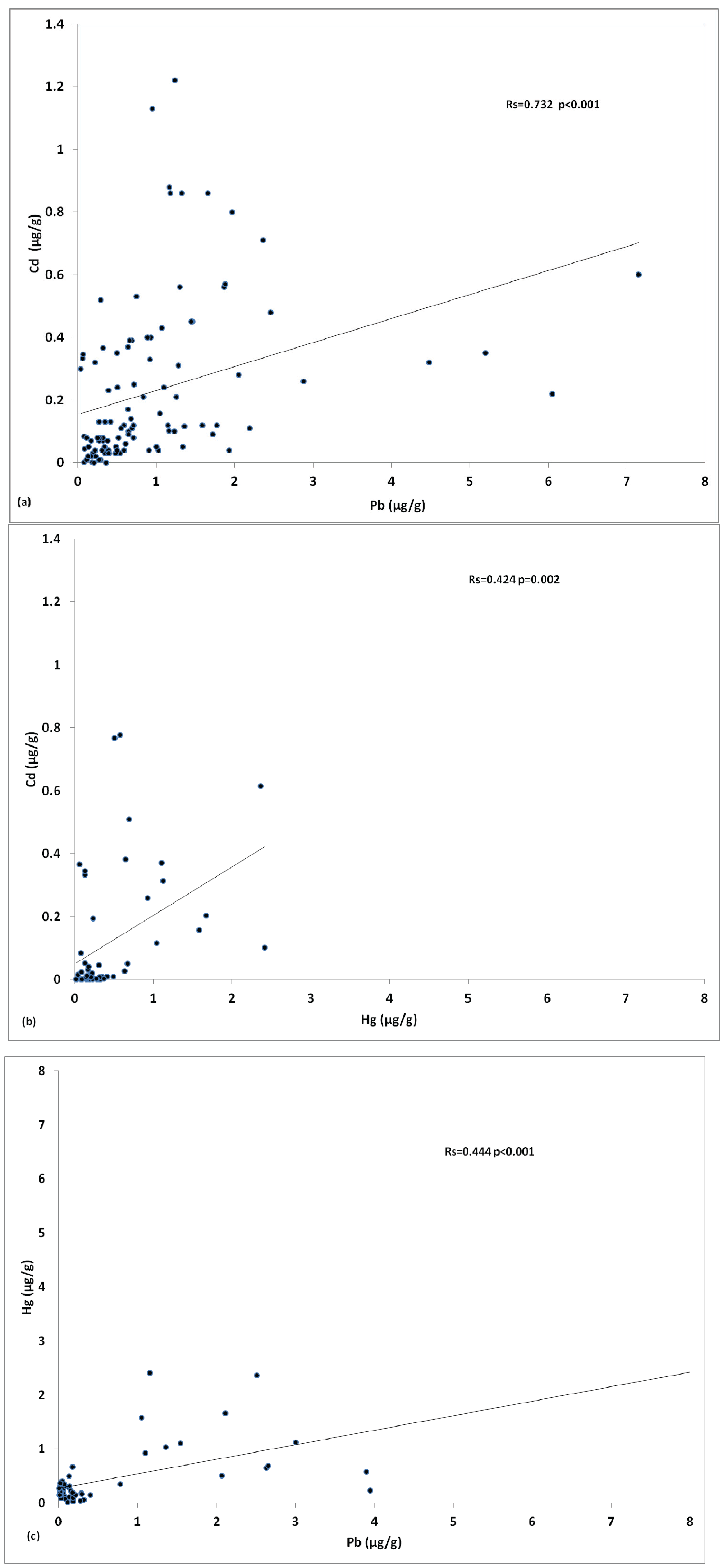
3.3. Cadmium, Lead and Mercury in Fish Tissues of the Mediterranean

The mean values of each metal reported in the selected studies, fish species and sampling sites can be viewed in Table 3. Sample size ranged from 5–161 samples with an average of 36 samples per study.
Significant correlations among metals were revealed upon investigation of the associations, with the use of Spearman’s rho coefficients. Correlations between metal levels are presented in Figure 2a–c.
There is a significant correlation (r = 0.732) between Cd and Pb levels (Figure 2a) This may be due to the fact that these metals have a similar distribution pattern in the medium of exposure (water), or that they share the same origin of dispersion, possibly waste disposals from human activity.
Cadmium also has a correlation to mercury (r = 0.424), though somewhat smaller than the one to Pb (Figure 2b). Finally, a similar correlation to Figure 2b appears between Hg and Pb Figure 2c. It must be also taken into consideration that the number of studies that examined all three metals concurrently was smaller than the initial dataset (15 studies).
Figure 2. (a) Correlation of Cd levels with Pb levels in fish tissue presented in the reviewed literature; (b) Correlation of Cd levels with Hg levels in fish tissue presented in the reviewed literature; (c) Correlation of levels Hg with Pb levels in fish tissue presented in the reviewed literature.
Toxics 02 00417 g002

3.4. Distribution of Cadmium, Lead and Mercury amongst Fish Species

Metal levels detected in fish tissue are affected by each species’ natural habitat. Certain species tend to accumulate metals at higher rates than others and it has been established in numerous studies that species with benthic behaviour or bottom dwellers to a point, such as Red mullets [8] or deep sea fauna such as angler fish, and frostfish [44] which have a close relationship with sediments, concentrate contaminants to a higher degree.
Apart from habitat, diet is an equally decisive factor for metal accumulation and therefore levels of metals in fish tissue. High trophic level predators such as tuna and swordfish are more likely to accumulate metals like mercury due to high mobility and metabolism [44]. Tuna’s ability to concentrate mercury, cadmium and lead at higher levels renders it a better bioindicator of pollution of the open-sea ecosystems than swordfish. It should also be taken into account that tuna shows a different growth rate with respect to swordfish [29].
When comparing the habitat of each species presented in the studies, to the results reported in the studies, a direct correlation of metal levels to the identification of fish as benthic, pelagic etc. is revealed (Table 3). Bottom dwellers (benthic) for instance, contain higher levels than pelagic. Other groups such as carnivorous and larger predators seem to bioaccumulate metals more intensively. Top predator species such as Xiphias gladius, due to highly active metabolism which requires high food consumption, tend to accumulate considerable amounts of metals in their tissues [2]. However, these relationships are not always clear-cut. Higher levels of metals detected in Mugil cephalus tissue can be attributed to seasonal changes concerning both intrinsic factors such as growth cycle, reproductive cycle and environmental, like changes in water temperature or local pollution [31].

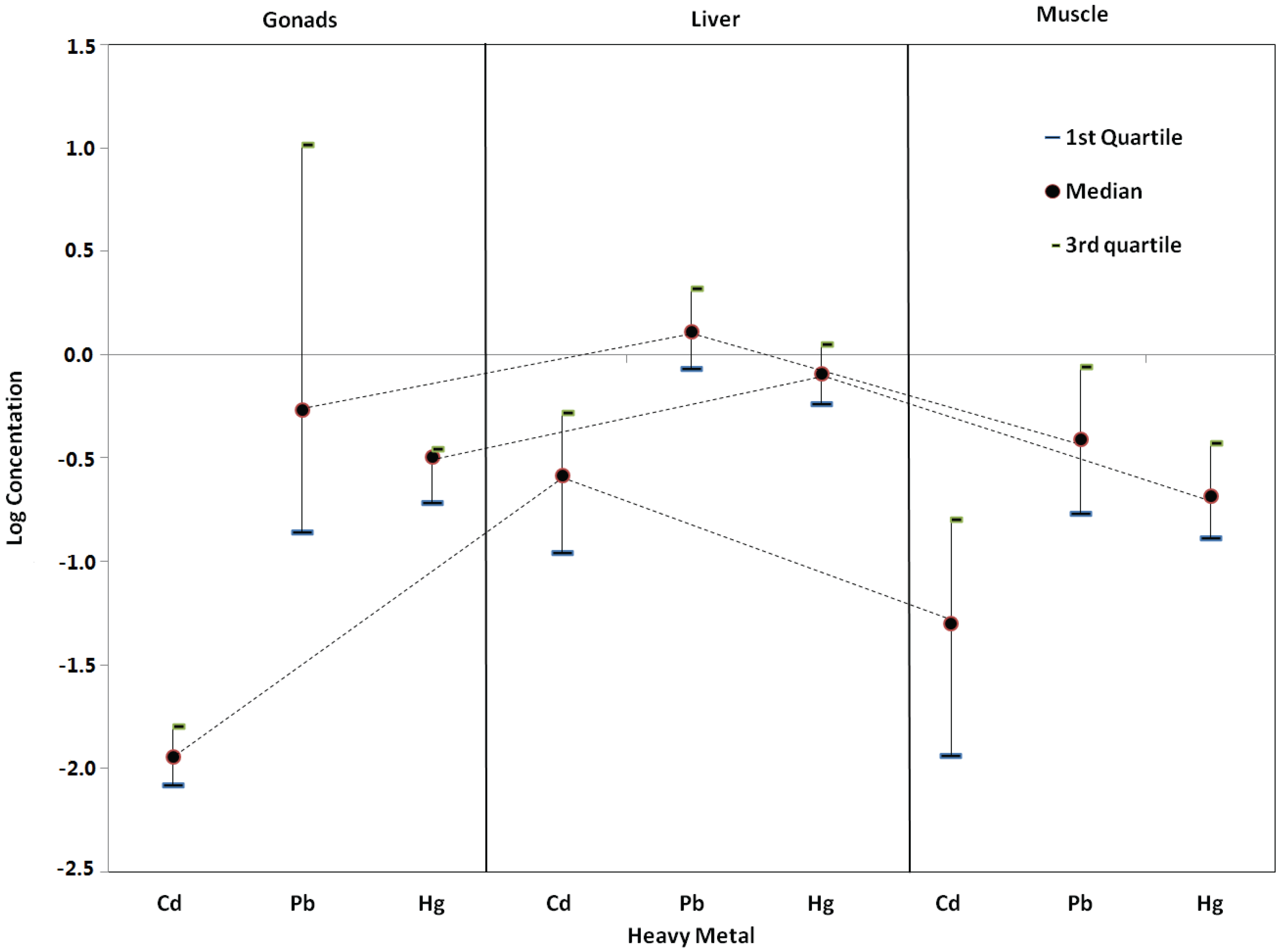
3.5. Distribution of Cadmium, Lead and Mercury amongst Fish Gonad, Liver and Muscle Tissues

Depending on various biomonitoring techniques, different fish tissues (muscle tissue, liver, gills, etc.) can be used as indicators of the extent of aquatic systems contamination [32]. Consequently, the use of biological indicator species can be determinant to the understanding of the fate and cycles of trace metals in the marine environment, especially for the areas where baseline data is sparse. Each metal has different accumulation properties and there is evidence of significant differences amongst species concerning metal levels, which represent differences of habitat, trophic levels and proclivity of metals to biomagnify in the food chain [66].
Figure 3. Median, 1st and 3rd quartiles of log-scale concentration of Cd, Pb and Hg per type of issue.
Toxics 02 00417 g003
The tissues most frequently examined in the studies reviewed in this work are muscle, liver, gill and gonads. In very few cases the skin was analysed as well. Metal concentration in tissues is related to various factors but mainly to their capacity to induce metal binding proteins such as metallothioneins. The reproduction period of each species also affects differentiations of metal levels in tissues, since metal accumulation and distribution it the fish body depends on organ size and activity.
Tissue distributions of mean metal values (quartiles 1st, median and 3rd) were investigated and results are presented in Figure 3. The highest levels of metals were detected in liver tissue, while gonads appeared to have the widest range. With liver being the main metabolically active tissue these results are justifiable. As previously mentioned, it is the most reliable indicator tissue of both water contamination and chronic exposure to metals due to the fact that is specialised in storage of metals whereas muscles are a primary site of metal uptake [65]. Hence, liver is usually the target tissue of biomonitoring studies in marine environments as it can give a picture of the contamination levels in a specific area at a specific time. As far as gonads are concerned, the broad range exhibited is most likely due to the seasonal changes which fish undergo during reproduction cycles.
On the other hand, muscle is mainly the edible part of the fish and the tissue most important for risk assessment concerning human intakes via fish consumption. Therefore, data collection on tissue basis, i.e., muscle, or liver, or both, is a matter of the research scope. Still, in order to form an integrated picture of the link between the environment (aquatic contamination) and human exposure assessment, monitoring studies encompassing both factors need to be performed.
The analysis also indicated similar patterns of Cd, Pb and Hg concentrations in each tissue. Medians of Pb were the highest in each tissue, whereas medians of Hg were approximately in the middle and of Cd the lowest.

4. Risk Assessment

4.1. Human Health Effects-Implications for Fish Consumption

The health benefits of fish consumption arise from their rich content in proteins, vitamins, valuable mineral compounds and mostly in essential ω-3 polyunsaturated fatty acids (PUFA). This kind of lipid composition, with high portions of PUFA and low portions of cholesterol, has been shown to offer effective protection against cardiovascular disease such as coronary heart disease, reducing arrhythmia and thrombosis, lowering plasma triglyceride levels and reducing blood clotting tendency [18,66,68]. However, the toxic metal load in fish can counteract the health benefits of the omega-3 fatty acids [69]. Research has shown evidence that high mercury levels in fish can diminish the cardio-protective effect of fish consumption. Dietary intake of metals such as methylmercury, cadmium and lead poses a potential risk since they have been associated with serious health complications in children and adults when found in high concentrations. Several authors have demonstrated that metal levels in different fish species may exceed the determined acceptable limits [38,70,71].
The adverse human health effects associated with exposure to metals, even at low concentrations, are diverse. The most common toxic effects of metals involve the brain and kidneys. Others include carcinogenic actions and may lead to chronic or acute diseases [44,66].
Mercury toxicity may cause permanent harm to the central nervous system, such as behavioral disorders and deficiencies in the immune system and development. All forms of mercury can pass through the placenta to the fetus during pregnancy, where it may affect the developing central nervous system (CNS) [72]. The fetal brain is more susceptible to damage due to mercury poisoning than the adult brain [73]. Unless actively removed, mercury has an extremely long half-life of somewhere between 15 and 30 years in the CNS. The most toxic form of mercury from environmental exposure which may affect living organisms is methylmercury. This is an organometallic compound which dissolves in the water column and is much more harmful than inorganic mercury [44] due to its ability to cross the blood-brain barrier. Furthermore, the main route of exposure for the general population is through fish consumption that may contain methylmercury [42,48].
Considering that almost 30,000 tons of cadmium is released into the environment annually, of which 4–13,000 come from human activities (UNEP, 2006) and that even in traces it is highly toxic, it is important to consider the effects on human health. Cadmium exposure in humans occurs through two main sources. The main route of exposure is diet, via contaminated water and food and more particularly products such as leafy vegetables, fruits, cereals, grains, organ meat and fish which often exhibit elevated cadmium levels. The second source of exposure is the environment. Cadmium is transported though the blood cells and distributed primarily to the liver and kidney organs [74]. It may also lead to skeletal damage and reproductive deficiencies, along with adverse effects on the brain and CNS. Acute high dose cadmium exposure can cause severe respiratory irritation, while long-term cadmium exposure has proven to be a risk factor for chronic lung disease and testicular degeneration [42]. Cadmium also affects the heart by increasing cholesterol and free fatty acid levels in addition to aortic and coronary atherosclerosis. It is also implicated in neurological disorders such as hyperactivity and learning disabilities in children since it affects their central nervous system [75]. The biological half-life of cadmium in humans is 17–30 years. The molecular mechanisms of cadmium toxicity are not fully understood yet.
Lead exposure can cause a wide spectrum of health problems, such as reduced cognitive development and intellectual performance in children aside from increased blood pressure, cardiovascular convulsions, coma and renal failure disease in adults [42]. The half-life of lead varies from about a month in blood, 1–1.5 months in soft tissue, and to about 25–30 years in bone.

4.2. Data from Human Dietary Intake Studies

There were 17 studies considered in this chapter, reporting human intake data via fish consumption over the years 2001–2012. Most of the studies present EWI and EDI values which were calculated for the fish samples examined in each study, below the recommended values (Table 1). Nevertheless, there were reports of intake values exceeding the established limits. Although toxicity and the resulting threat to human health due to metals are a consequence of concentration, it is well known that chronic exposure to Cd, Hg, and Pb even at relatively low levels can cause adverse effects [44,66].
On the one hand, the EWI and EDI in Türkmen et al. 2011 [33] study are in agreement with values reported for fish by Türkmen et al. 2010 [53] and far below the recommended values (Table 1). As a result, it is assumed that fish from Paradeniz lagoon are not considered a risk for human health. In the work of Copat et al. 2012 [5] although individual levels may exceed guidance values, there appears to be no overall concern due to overall dietary intake. Martí-Cid et al. 2008 [76] published some differentiated results on the dietary intakes of Cd, Hg, and Pb by the population of Catalonia, from an earlier study in 2000. The variation of metal levels in analysed foods has led to the increase of Pb daily intake, while intakes of Cd and total Hg decreased. However, intake levels remain below the respective PTWIs, indicating low risk for human health.
On the other hand, there are some cases where results from the studies raise certain concerns. As shown by Marcotrigiano and Storelli, 2003 [63], the concentrations of mercury in many fish species exceeded the maximum permitted limits. However, there do not seem to be significant risks associated with that particular seafood consumption. For lead and cadmium the estimated intake was considerably below the limits established by WHO, 1998.
In the work of Spada et al. 2012 [48], it is reported that for benthic fish Symphodus m. collected from the Taranto Gulf, the estimated total Hg (THg) per weight, calculated both for adults and children, exceeded the established PTWI, (2.5 and 3.2 μg/kg bw week for adults; 4.3 and 5.5 μg/kg bw week for children, respectively). In relation to average body weight, THg and MeHg intake for children surpassed that of the adults. Consequently, children consuming fish from this particular site are in danger of high THg and MeHg intakes. It must also be stated that the sampling sites for this study, Taranto Gulf, Mar Piccolo and Mar Grande basins are of great environmental concern due to industrial activity such as iron and steel factory, oil refinery, shipyard, and aquacultures. In an earlier study, Perugini et al. 2009 [49] investigated the presence of total mercury in fish from the Adriatic (European hake 0.59 ± 0.14 mg/kg, red mullet 0.48 ± 0.09 mg/kg, blue whiting 0.38 ± 0.09 mg/kg, Atlantic mackerel 0.36 ± 0.08 mg/kg) amongst other aquatic species and revealed that THg level was over the limits of EC 2006 (42.9%, 21.4%, 14.3% and 23.1% respectively). The TDI estimated 0.24 μg/kg-bw is greater than the established 0.23 μg/kg-bw (JECFA, 2003) and could be even higher when taking into account populations with high fish consumption.
Furthermore, Aksu et al. 2011 [56] state that even the lowest Pb levels in fish collected from the Marmara Sea presented in their study can lead to twice or three times the maximum TWI for consumers, since they were found to be higher than the critical limits set by both the Turkish Ministry of Environment for Aquatic Products (1 μg/g wet wt.) and by European countries (2.0 μg/g) (UNEP 1985). In addition, according to the Cd levels in fish reported in the same study, a person can consume safely only one fish meal per week originated from the Marmara Sea. It is also discussed that the high metal levels detected in fish, are related to the anthropogenic inputs in the Southern Marmara Shelf. In fish samples from the Aegean and Black Sea, Uluozlu et al. 2007 [58] have detected levels of lead and cadmium, higher than the recommended legal limits for human consumption (maximum level permitted for sea fishes is 0.4 mg/kg lead and 0.1 mg/kg for cadmium according to Turkish Food Codex).
In the Storelli et al. 2002 [63] study, levels of total mercury and methylmercury in the muscle tissue of albacore and bluefin tuna caught in the Mediterranean Sea were assessed and results revealed that for 78.6% of albacore samples and 61.1% of bluefin tuna samples, the total mercury concentrations exceeded the prescribed legal limit (1 mg/kg wet wt). With data obtained on seafood consumption per capita in Italy (Istituto Nazionale di Statistica 1998) (420 g) and the mean concentrations of THg and MeHg in albacore and bluefin tuna muscle, the EWI was determined and was found to be higher than the established PTWI for both species, namely for albacore: total mercury 8.19 μg/kg bodyweight (bw); methylmercury 7.42 μg/kg bw; bluefin tuna: total mercury 8.26 μg/kg bw, methylmercury 7.07 μg/kg bw. However, these findings are based on results of bluefin tuna consumption of weighing 30–40 kg. Considering the fact that it is improbable that one would consume 420 g per week of the same fish species, the estimated dietary exposure values may be misleading.
Falco et al. 2006 [71] estimated the daily intake of Cd, Hg, and Pb through the consumption of marine species by the general population of Catalonia for different age groups. Male seniors proved to be the group with the highest metal intake of Cd (1.34 μg/day) and Pb (2.48 μg/day) through fish. While comparing the daily intakes of each age group with PTWI, they were found to be lower in general although the estimated intake of methylmercury for boys, 1.96 μg/kg/week, was over the PTWI. Also, it was reported that even though Hg levels in swordfish exceeded the EU requirements, the consumption of this fish species by the population of Catalonia was very low and therefore its contribution to daily intake of Hg is irrelevant, supporting the notion that biomonitoring data are to be assessed invariably in association with consumption data.

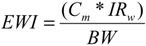
4.3. Estimation of Risk Assessment Scenario

Among the studies described in Chapter 4.2., there were some which showed data raising concern for human intakes in Italy [48,49], Turkey [56,58] and Spain [71]. Interestingly, both Italian and Turkish sampling sites were burdened by anthropogenic activities including mining and industrial activities. There were also some studies where even though the metal levels in fish tissue exceeded the established ones, low human intakes counterbalanced the risk of consumption [44,63,71]. Therefore, numerous factors need to be evaluated while conducting risk assessment, such as variation of consumption values per country and age groups along with proper assignment of metal levels detected, to consumption values.
A risk assessment based on the worst human intake scenario was attempted as described in Chapter 2.3. Results of the risk assessment that was conducted, based on the data collected from the studies, are presented in Table 4.
In most cases, there appears to be no risk for the consumers of the countries examined. Italy seems to have the highest EWI/PTWI for mercury (0.13, 0.22 and 0.13) in all cases (demersal, pelagic, and marine) as a result of having high levels of mean Hg values and EWI respectively. In addition, it is the country with somewhat high consumption values, which results in higher risk ratios, even in cases of fish consumption with relatively low metal levels.
The opposite seems to be taking place in Turkey, where the mean values for lead are high in pelagic (2.03 μg/g) and marine (5.66 μg/g), as are the consumption values EWI (2.84 and 3.40 respectively). However, the EWI/PTWI ratios are not of the same magnitude (0.11 and 0.14 respectively). There appears a similar situation in Egypt, where the mean values for lead in all cases, demersal (1.74 μg/g), pelagic (1.7 μg/g) and marine (1.46 μg/g) are high, as are the EWI values (2.09, 1.70 and 0.87 respectively), and the EWI/PTWI ratios are low (0.08, 0.07 and 0.03 respectively). This is most probably due to the low EWI values.
There are a number of unaddressed issues in the risk assessment process used in this review on account of the fact that there were no data on sensitive population groups such as children, pregnant women and seniors which we could analyse. Further studies are needed to obtain data on sensitive population groups. In addition, information on fish consumption obtained from FAO was inadequate for simultaneous processing of consumption data for all countries. Moreover, consumption data of other seafood such as crustaceans which contain high metal levels was not factored in. Finally, we did not include in our analysis selenium, a metalloid of considerable interest for human health not only for its own nutritional and toxicological properties [77,78], but also for its ability to modify and generally decrease the biological activity of several heavy metals such as cadmium, lead and mercury. However, the relation of these metals with selenium is very complex and still not well elucidated [79,80], and paradoxically in some cases concurrent administration of selenium along with heavy metals may not mitigate the toxicity of the single elements, but even exacerbate it [77,81,82,83], further hampering the risk assessment of mixed intake of these elements by seafood [84]. Overall, however, we consider it unlikely that lack of consideration of selenium in the present analysis may have substantially biased our results, though additional research is clearly required to adequately elucidate these potential inhibitory or additive interactions.

5. Conclusions

Comparative evaluation has proven to be challenging in view of the fact that studies vary greatly in terms of expression of results, presentation of findings and sensitivity of techniques (detection limits). Additionally, biomonitoring has focused mainly on the eastern part of the Mediterranean, leaving the greater part of the Sea uncharted regarding metal levels in fish. Greece, one of the countries with large fisheries in proportion to its size is, surprisingly, among the countries which are not represented in the studies. Such a low coverage of monitoring areas in published literature suggests a need for a complete observational network of heavy metals in fish in the Mediterranean Sea.
Assessing the biomonitoring results, the tissue which accumulates metals the most is liver, followed by muscle although gonads show the greatest levels, but this can lead to false conclusions due to seasonal and age variations. In addition, there seems to be a significant correlation between Cd and Pb levels accumulated in fish.
Regarding risk assessment, adult consumers of the countries represented in the studies do not seem to be at risk (HI varied from 0.00–0.22). Nevertheless, the fact that some studies reported that the established tolerance levels of heavy metals for fish consumption were exceeded supports the necessity for further investigation. For the benefit of safety evaluation, biomonitoring data must be linked to consumption data. The evaluation of potential health hazards for consumers should include the data of biomonitoring studies in as many regions as possible together with basic information about the specific dietary intake for comparison with safety levels such as the Acceptable Daily Intake (ADI). Definitely, more detailed approaches in the estimation of risk of heavy metals from fish consumption are needed. The application of scenarios including 95th and 80th percentiles of heavy metal levels, the estimation of risk in sensitive groups, the use of more sophisticated models and approaches could be a next step in those estimations.
In an attempt to maximize the health benefits from the high nutritional value of fish and minimize any contingent health risks, health institutions including public and private organizations should collaborate with a view to forming usable databases and revising the public policy under which fish consumption is monitored.

Abbreviations

JECFA
The Joint FAO/WHO Expert Committee on Food Additives
US-EPA
The US Environmental Protection Agency
EFSA
European Food Safety Authority
US FDA
The US Food and Drug Administration
EC
European Commission Regulation
EWI
Estimated Weekly Intake (μg/week/70 kg body weight)
EDI
Estimated Daily Intake in μg/week/70 kg body weight
PTWI
Provisional Tolerable Weekly Intake PTWI (μg/week/kg body weight)
PTWIa
PTWI for 70 kg adult person (μg/week/70 kg body weight)

Acknowledgments

Authors would like to thank Matthaios Kavvalakis for his contribution to the preparation of the manuscript.

Author Contributions

Elisavet A. Renieri collected the data and wrote the manuscript, Athanasios K. Alegakis conducted the statistical analysis, Michalis Kiriakakis contributed in the writing of the analytical methods’ section and Marco Vincenti in the selenium section. Finally, Eren Ozcagli, Martin F. Wilks and Aristidis M. Tsatsakis revised critically the manuscript.

Conflicts of Interest

The authors declare no conflict of interest.

References

  1. European Environment Agency (EEA). Priority Issues in the Mediterranean Environment; EEA: Copenhagen, Denmark, 2006. [Google Scholar]
  2. Damiano, S.; Papetti, P.; Menesatti, P. Accumulation of heavy metals to assess the health status of swordfish in a comparative analysis of mediterranean and atlantic areas. Mar. Pollut. Bull. 2011, 62, 1920–1925. [Google Scholar] [CrossRef]
  3. World Health Organization. Health Risks of Heavy Metals from Long-Range Transboundary Air Pollution; WHO Regional Office for Europe: Copenhagen, Denmark, 2007. [Google Scholar]
  4. Alonso Castillo, M.L.; Sanchez Trujillo, I.; Vereda Alonso, E.; Garcia de Torres, A.; Cano Pavon, J.M. Bioavailability of heavy metals in water and sediments from a typical mediterranean bay (Malaga Bay, region of Andalucia, Southern Spain). Mar. Pollut. Bull. 2013, 76, 427–434. [Google Scholar] [CrossRef]
  5. Copat, C.; Bella, F.; Castaing, M.; Fallico, R.; Sciacca, S.; Ferrante, M. Heavy metals concentrations in fish from sicily (Mediterranean sea) and evaluation of possible health risks to consumers. Bull. Environ. Contam. Toxicol. 2012, 88, 78–83. [Google Scholar] [CrossRef]
  6. UNEP/FAO/IAEA. Designing of Monitoring Programs and Management of Data Concerning Chemical Contaminants in Marine Organisms. In Map Technical Reports Series No. 77; UNEP: Athens, Greece, 1993. [Google Scholar]
  7. Storelli, M.M.; Marcotrigiano, G.O. Bioindicator organisms: Heavy metal pollution evaluation in the Ionian sea (Mediterranean sea–Italy). Environ. Monit. Assess. 2005, 102, 159–166. [Google Scholar] [CrossRef]
  8. Kucuksezgin, F.; Altay, O.; Uluturhan, E.; Kontas, A. Trace metal and organochlorine residue levels in red mullet (Mullus barbatus) from the Eastern Aegean, Turkey. Water Res. 2001, 35, 2327–2332. [Google Scholar]
  9. Ferreira, M.; Caetano, M.; Antunes, P.; Costa, J.; Gil, O.; Bandarra, N.; Pousao-Ferreira, P.; Vale, C.; Reis-Henriques, M.A. Assessment of contaminants and biomarkers of exposure in wild and farmed seabass. Ecotoxicol. Environ. Saf. 2010, 73, 579–588. [Google Scholar] [CrossRef]
  10. Creti, P.; Trinchella, F.; Scudiero, R. Heavy metal bioaccumulation and metallothionein content in tissues of the sea bream Sparus aurata from three different fish farming systems. Environ. Monit. Assess. 2010, 165, 321–329. [Google Scholar] [CrossRef]
  11. Shahidul Islam, M.; Tanaka, M. Impacts of pollution on coastal and marine ecosystems including coastal and marine fisheries and approach for management: A review and synthesis. Mar. Poll. Bull. 2004, 48, 624–649. [Google Scholar] [CrossRef]
  12. Cesar, A.; Marin, A.; Marin-Guirao, L.; Vita, R.; Lloret, J.; del Valls, T.A. Integrative ecotoxicological assessment of sediment in Portman bay (Southeast Spain). Ecotoxicol. Environ. Saf. 2009, 72, 1832–1841. [Google Scholar] [CrossRef]
  13. Abdallah, M.A.; Abdallah, A.M. Biomonitoring study of heavy metals in biota and sediments in the south eastern coast of Mediterranean Sea, Egypt. Environ. Monit. Assess. 2008, 146, 139–145. [Google Scholar] [CrossRef]
  14. Yilmaz, A.B. Levels of heavy metals (Fe, Cu, Ni, Cr, Pb, and Zn) in tissue of Mugil cephalus and Trachurus mediterraneus from Iskenderun Bay, Turkey. Environ. Res. 2003, 92, 277–281. [Google Scholar] [CrossRef]
  15. Kucuksezgin, F.; Kontas, A.; Uluturhan, E. Evaluations of heavy metal pollution in sediment and Mullus barbatus from the Izmir bay (Eastern Aegean) during 1997–2009. Mar. Pollut. Bull. 2011, 62, 1562–1571. [Google Scholar] [CrossRef]
  16. Houston, M.C. The role of mercury and cadmium heavy metals in vascular disease, hypertension, coronary heart disease, and myocardial infarction. Altern. Ther. Health Med. 2007, 13, S128–S133. [Google Scholar]
  17. Dalman, Ö.; Demirak, A.; Balcl, A. Determination of heavy metals (Cd, Pb) and trace elements (Cu, Zn) in sediments and fish of the Southeastern Aegean Sea (Turkey) by atomic absorption spectrometry. Food Chem. 2006, 95, 157–162. [Google Scholar]
  18. Fallah, A.A.; Saei-Dehkordi, S.S.; Nematollahi, A.; Jafari, T. Comparative study of heavy metal and trace element accumulation in edible tissues of farmed and wild rainbow trout (Oncorhynchus mykiss) using ICP-OES technique. Microchem. J. 2011, 98, 275–279. [Google Scholar] [CrossRef]
  19. Khan, M.U.; Malik, R.N.; Muhammad, S. Human health risk from heavy metal via food crops consumption with wastewater irrigation practices in Pakistan. Chemosphere 2013, 93, 2230–2238. [Google Scholar]
  20. European Union. Setting Maximum Levels for Certain Contaminants in Foodstuffs; Official Journal of the European Union, 2006; pp. 5–24. [Google Scholar]
  21. European Union. Setting Maximum Levels for Certain Contaminants in Foodstuffs; Official Journal of the European Union, 3 July 2008; pp. 6–9. [Google Scholar]
  22. European Food Safety Authority (EFSA). Scientific opinion of the panel on contaminants in the food chain on a request from the European commission on cadmium in food. EFSA J. 2009, 1–139. [Google Scholar]
  23. European Food Safety Authority (EFSA). Scientific opinion on lead in food. EFSA Panel on Contaminants in the Food Chain (CONTAM). EFSA J. 2010, 8, 147. [Google Scholar]
  24. European Food Safety Authority (EFSA). Scientific opinion on the risk for public health related to the presence of mercury and methylmercury in food. EFSA J. 2012, 10, 241. [Google Scholar]
  25. FAO/WHO. Summary and Conclusions of the Sixty-First Meeting of the Joint Fao/Who Expert Committee on Food Additives (Jecfa); World Health Organization: Rome, Italy, 2003. [Google Scholar]
  26. FAO/WHO. Summary and Conclusions of the Sixty-Seventh Meeting of the Joint Fao/Who Expert Committee on Food Additives (Jecfa); World Health Organization: Rome, Italy, 2006. [Google Scholar]
  27. FAO/WHO. Evaluation of Certain Food Additives and Contaminants: Seventy-Third Report of the Joint Fao/Who Expert Committee on Food Additives (Jecfa); World Health Organization: Geneva, Switzerland, 2011. [Google Scholar]
  28. FAO. Available online: http://faostat.Fao.Org/ (accessed on 9 October 2013).
  29. Storelli, M.M.; Giacominelli-Stuffler, R.; Storelli, A.; Marcotrigiano, G.O. Accumulation of mercury, cadmium, lead and arsenic in swordfish and bluefin tuna from the Mediterranean Sea: A comparative study. Mar. Pollut. Bull. 2005, 50, 1004–1007. [Google Scholar] [CrossRef]
  30. Marijic, V.F.; Raspor, B. Age- and tissue-dependent metallothionein and cytosolic metal distribution in a native Mediterranean fish, mullus barbatus, from the Eastern Adriatic Sea. Comp. Biochem. Physiol. C 2006, 143, 382–387. [Google Scholar] [CrossRef]
  31. Dural, M.; Goksu, M.Z.; Ozak, A.A.; Derici, B. Bioaccumulation of some heavy metals in different tissues of Dicentrarchus labrax l, 1758, Sparus aurata l, 1758 and Mugil cephalus l, 1758 from the camlik lagoon of the eastern coast of Mediterranean (Turkey). Environ. Monit. Assess. 2006, 118, 65–74. [Google Scholar] [CrossRef]
  32. Gaspic, Z.K.; Zvonaric, T.; Vrgoc, N.; Odzak, N.; Baric, A. Cadmium and lead in selected tissues of two commercially important fish species from the Adriatic Sea. Water Res. 2002, 36, 5023–5028. [Google Scholar]
  33. Turkmen, M.; Turkmen, A.; Tepe, Y. Comparison of metals in tissues of fish from paradeniz lagoon in the coastal area of Northern East Mediterranean. Bull. Environ. Contam. Toxicol. 2011, 87, 381–385. [Google Scholar] [CrossRef]
  34. Tepe, Y. Metal concentrations in eight fish species from aegean and mediterranean seas. Environ. Monit. Assess. 2009, 159, 501–509. [Google Scholar] [CrossRef]
  35. Sepe, A.; Ciaralli, L.; Ciprotti, M.; Giordano, R.; Funari, E.; Costantini, S. Determination of cadmium, chromium, lead and vanadium in six fish species from the Adriatic Sea. Food Addit. Contam. 2003, 20, 543–552. [Google Scholar] [CrossRef]
  36. Türkmen, M.; Türkmen, A.; Tepe, Y.; Töre, Y.; Ateş, A. Determination of metals in fish species from Aegean and Mediterranean seas. Food Chem. 2009, 113, 233–237. [Google Scholar]
  37. Türkmen, A.; Türkmen, M.; Tepe, Y.; Akyurt, İ. Heavy metals in three commercially valuable fish species from İskenderun bay, Northern East Mediterranean Sea, Turkey. Food Chem. 2005, 91, 167–172. [Google Scholar] [CrossRef]
  38. Abernathy, C.O.; Thomas, D.J.; Calderon, R.L. Health effects and risk assessment of Arsenic. J. Nutr. 2003, 133, 1536s–1538s. [Google Scholar]
  39. Türkmen, M.; Türkmen, A.; Tepe, Y.; Ateş, A.; Gökkuş, K. Determination of metal contaminations in sea foods from Marmara, Aegean and Mediterranean seas: Twelve fish species. Food Chem. 2008, 108, 794–800. [Google Scholar]
  40. Turan, C.; Dural, M.; Oksuz, A.; Ozturk, B. Levels of heavy metals in some commercial fish species captured from the black sea and Mediterranean Coast of Turkey. Bull. Environ. Contam. Toxicol. 2009, 82, 601–604. [Google Scholar] [CrossRef]
  41. Cogun, H.Y.; Yuzereroglu, T.A.; Firat, O.; Gok, G.; Kargin, F. Metal concentrations in fish species from the northeast Mediterranean sea. Environ. Monit. Assess. 2006, 121, 431–438. [Google Scholar] [CrossRef]
  42. Bilandžić, N.; Đokić, M.; Sedak, M. Metal content determination in four fish species from the Adriatic sea. Food Chem. 2011, 124, 1005–1010. [Google Scholar] [CrossRef]
  43. Uluturhan, E.; Kucuksezgin, F. Heavy metal contaminants in red pandora (Pagellus erythrinus) tissues from the eastern Aegean sea, Turkey. Water Res. 2007, 41, 1185–1192. [Google Scholar]
  44. Marcotrigiano, G.O.; Storelli, M.M. Heavy metal, polychlorinated biphenyl and organochlorine pesticide residues in marine organisms: Risk evaluation for consumers. Vet. Res. Commun. 2003, 27, 183–195. [Google Scholar] [CrossRef]
  45. Papetti, P.; Rossi, G. Heavy metals in the fishery products of low lazio and the use of metallothionein as a biomarker of contamination. Environ. Monit. Assess. 2009, 159, 589–598. [Google Scholar] [CrossRef]
  46. Çelik, U.; Oehlenschläger, J. High contents of cadmium, lead, zinc and copper in popular fishery products sold in Turkish supermarket. Food Control. 2007, 18, 258–261. [Google Scholar]
  47. Juresa, D.; Blanusa, M. Mercury, arsenic, lead and cadmium in fish and shellfish from the Adriatic Sea. Food Addit. Contam. 2003, 20, 241–246. [Google Scholar] [CrossRef]
  48. Spada, L.; Annicchiarico, C.; Cardellicchio, N.; Giandomenico, S.; di Leo, A. Mercury and methylmercury concentrations in mediterranean seafood and surface sediments, intake evaluation and risk for consumers. Int. J. Hyg. Environ. Health 2012, 215, 418–426. [Google Scholar] [CrossRef]
  49. Perugini, M.; Visciano, P.; Manera, M.; Zaccaroni, A.; Olivieri, V.; Amorena, M. Levels of total mercury in marine organisms from Adriatic Sea, Italy. Bull. Environ. Contam. Toxicol. 2009, 83, 244–248. [Google Scholar] [CrossRef]
  50. Turkmen, A.; Tepe, Y.; Turkmen, M. Metal levels in tissues of the European anchovy, Engraulis encrasicolus l., 1758, and picarel, spicara smaris l., 1758, from black, Marmara and Aegean Seas. Bull. Environ. Contam. Toxicol. 2008, 80, 521–525. [Google Scholar] [CrossRef]
  51. Yılmaz, A.B.; Sangün, M.K.; Yağlıoğlu, D.; Turan, C. Metals (major, essential to non-essential) composition of the different tissues of three demersal fish species from İskenderun Bay, Turkey. Food Chem. 2010, 123, 410–415. [Google Scholar]
  52. Gonul, L.T.; Kucuksezgin, F. Mercury accumulation and speciation in the muscle of red mullet (Mullus barbatus) and annular sea bream (Diplodus annularis) from Izmir bay (Eastern Aegean). Mar. Pollut. Bull. 2007, 54, 1962–1968. [Google Scholar] [CrossRef]
  53. Turkmen, A.; Turkmen, M.; Tepe, Y.; Cekic, M. Metals in tissues of fish from yelkoma lagoon, Northeastern Mediterranean. Environ. Monit Assess. 2010, 168, 223–230. [Google Scholar] [CrossRef]
  54. Harmelin-Vivien, M.; Cossa, D.; Crochet, S.; Bănaru, D.; Letourneur, Y.; Mellon-Duval, C. Difference of mercury bioaccumulation in red mullets from the North-Western Mediterranean and Black Seas. Mar. Poll. Bull. 2009, 58, 679–685. [Google Scholar] [CrossRef]
  55. Storelli, M.M.; Storelli, A.; Giacominelli-Stuffler, R.; Marcotrigiano, G.O. Mercury speciation in the muscle of two commercially important fish, hake (Merluccius merluccius) and striped mullet (Mullus barbatus) from the mediterranean sea: Estimated weekly intake. Food Chem. 2005, 89, 295–300. [Google Scholar]
  56. Aksu, A.; Balkis, N.; Taskin, O.S.; Ersan, M.S. Toxic metal (Pb, Cd, As and Hg) and organochlorine residue levels in hake (Merluccius merluccius) from the Marmara Sea, Turkey. Environ. Monit. Assess. 2011, 182, 509–521. [Google Scholar] [CrossRef]
  57. Canli, M.; Atli, G. The relationships between heavy metal (Cd, Cr, Cu, Fe, Pb, Zn) levels and the size of six Mediterranean fish species. Environ. Pollut. 2003, 121, 129–136. [Google Scholar] [CrossRef]
  58. Uluozlu, O.D.; Tuzen, M.; Mendil, D.; Soylak, M. Trace metal content in nine species of fish from the Black and Aegean Seas, Turkey. Food Chem. 2007, 104, 835–840. [Google Scholar]
  59. Amodio-Cocchieri, R.; Amoroso, S.; Arnese, A.; Cirillo, T.; Montuori, P.; Triassi, M. Pollution by mercury, arsenic, lead, chromium, cadmium, and polycyclic aromatic hydrocarbons of fish and mussels from the Gulf of Naples, Italy. Bull. Environ. Contam. Toxicol. 2003, 71, 551–560. [Google Scholar] [CrossRef]
  60. Kucuksezgin, F.; Uluturhan, E.; Kontas, A.; Altay, O. Trace metal concentrations in edible fishes from Izmir Bay, Eastern Aegean. Mar. Pollut. Bull. 2002, 44, 827–832. [Google Scholar] [CrossRef]
  61. Dierking, J.; Wafo, E.; Schembri, T.; Lagadec, V.; Nicolas, C.; Letourneur, Y.; Harmelin-Vivien, M. Spatial patterns in PCBs, pesticides, mercury and cadmium in the common sole in the nw mediterranean sea, and a novel use of contaminants as biomarkers. Mar. Pollut. Bull. 2009, 58, 1605–1614. [Google Scholar] [CrossRef]
  62. Minganti, V.; Drava, G.; De Pellegrini, R.; Siccardi, C. Trace elements in farmed and wild Gilthead seabream, Sparus aurata. Mar. Pollut. Bull. 2010, 60, 2022–2025. [Google Scholar] [CrossRef]
  63. Storelli, M.M.; Stuffler, R.G.; Marcotrigiano, G.O. Total and methylmercury residues in tuna-fish from the Mediterranean Sea. Food Addit. Contam. 2002, 19, 715–720. [Google Scholar] [CrossRef]
  64. Turkmen, A.; Turkmen, M.; Tepe, Y.; Mazlum, Y.; Oymael, S. Metal concentrations in blue crab (Callinectes sapidus) and mullet (Mugil cephalus) in Iskenderun Bay, Northern East Mediterranean, Turkey. Bull. Environ. Contam. Toxicol. 2006, 77, 186–193. [Google Scholar] [CrossRef]
  65. Tepe, Y.; Turkmen, M.; Turkmen, A. Assessment of heavy metals in two commercial fish species of four Turkish Seas. Environ. Monit. Assess. 2008, 146, 277–284. [Google Scholar] [CrossRef]
  66. Castro-González, M.I.; Méndez-Armenta, M. Heavy metals: Implications associated to fish consumption. Environ. Toxicol. Pharmacol. 2008, 26, 263–271. [Google Scholar] [CrossRef]
  67. Conti, G.O.; Copat, C.; Ledda, C.; Fiore, M.; Fallico, R.; Sciacca, S.; Ferrante, M. Evaluation of heavy metals and polycyclic aromatic hydrocarbons (pahs) in mullus barbatus from sicily channel and risk-based consumption limits. Bull. Environ. Contam. Toxicol. 2012, 88, 946–950. [Google Scholar] [CrossRef]
  68. Storelli, M.M. Potential human health risks from metals (Hg, Cd, and Pb) and polychlorinated biphenyls (PCBs) via seafood consumption: Estimation of target hazard quotients (THQs) and toxic equivalents (TEQs). Food Chem. Toxicol. 2008, 46, 2782–2788. [Google Scholar] [CrossRef]
  69. Chan, H.M.; Egeland, G.M. Fish consumption, mercury exposure, and heart diseases. Nutr. Rev. 2004, 62, 68–72. [Google Scholar] [CrossRef]
  70. Gochfeld, M.; Burger, J. Good fish/bad fish: A composite benefit-risk by dose curve. Neurotoxicology 2005, 26, 511–520. [Google Scholar] [CrossRef]
  71. Falco, G.; Llobet, J.M.; Bocio, A.; Domingo, J.L. Daily intake of arsenic, cadmium, mercury, and lead by consumption of edible marine species. J. Agric. Food Chem. 2006, 54, 6106–6112. [Google Scholar] [CrossRef]
  72. Gochfeld, M. Cases of mercury exposure, bioavailability, and absorption. Ecotoxicol. Environ. Saf. 2003, 56, 174–179. [Google Scholar] [CrossRef]
  73. Clarkson, T.W.; Magos, L.; Myers, G.J. The toxicology of mercury–current exposures and clinical manifestations. N. Engl. J. Med. 2003, 349, 1731–1737. [Google Scholar] [CrossRef]
  74. Goyer, R.A. Toxic and essential metal interactions. Annu. Rev. Nutr. 1997, 17, 37–50. [Google Scholar] [CrossRef]
  75. Wang, B.; Du, Y. Cadmium and its neurotoxic effects. Oxid Med. Cell. Longev. 2013, 2013, 898034. [Google Scholar]
  76. Marti-Cid, R.; Llobet, J.M.; Castell, V.; Domingo, J.L. Dietary intake of arsenic, cadmium, mercury, and lead by the population of Catalonia, Spain. Biol. Trace Elem. Res. 2008, 125, 120–132. [Google Scholar] [CrossRef]
  77. Vinceti, M.; Crespi, C.M.; Bonvicini, F.; Malagoli, C.; Ferrante, M.; Marmiroli, S.; Stranges, S. The need for a reassessment of the safe upper limit of selenium in drinking water. Sci. Total Environ. 2013, 443, 633–642. [Google Scholar] [CrossRef]
  78. Vinceti, M.; Dennert, G.; Crespi, C.M.; Zwahlen, M.; Brinkman, M.; Zeegers, M.P.; Horneber, M.; D’Amico, R.; del Giovane, C. Selenium for preventing cancer. Cochr. Database Syst. Rev. 2014, 3, CD005195. [Google Scholar]
  79. Zwolak, I.; Zaporowska, H. Selenium interactions and toxicity: A review. Selenium interactions and toxicity. Cell. Biol. Toxicol. 2012, 28, 31–46. [Google Scholar] [CrossRef]
  80. Zhang, H.; Feng, X.; Chan, H.M.; Larssen, T. New insights into traditional health risk assessments of mercury exposure: Implications of selenium. Environ. Sci. Technol. 2014, 48, 1206–1212. [Google Scholar] [CrossRef]
  81. Glaser, V.; Nazari, E.M.; Muller, Y.M.; Feksa, L.; Wannmacher, C.M.; Rocha, J.B.; de Bem, A.F.; Farina, M.; Latini, A. Effects of inorganic selenium administration in methylmercury-induced neurotoxicity in mouse cerebral cortex. Int. J. Dev. Neurosci. 2010, 28, 631–637. [Google Scholar] [CrossRef]
  82. Heinz, G.H.; Hoffman, D.J.; Klimstra, J.D.; Stebbins, K.R. A comparison of the teratogenicity of methylmercury and selenomethionine injected into bird eggs. Arch. Environ. Contam. Toxicol. 2012, 62, 519–528. [Google Scholar] [CrossRef]
  83. Penglase, S.; Hamre, K.; Ellingsen, S. Selenium and mercury have a synergistic negative effect on fish reproduction. Aquat. Toxicol. 2014, 149, 16–24. [Google Scholar] [CrossRef]
  84. Copat, C.; Vinceti, M.; D’Agati, M.G.; Arena, G.; Mauceri, V.; Grasso, A.; Fallico, R.; Sciacca, S.; Ferrante, M. Mercury and selenium intake by seafood from the Ionian Sea: A risk evaluation. Ecotoxicol. Environ. Saf. 2014, 100, 87–92. [Google Scholar] [CrossRef]

