
An Ingested Orthodontic Wire Fragment: A Case Report


Abstract
:1. Introduction
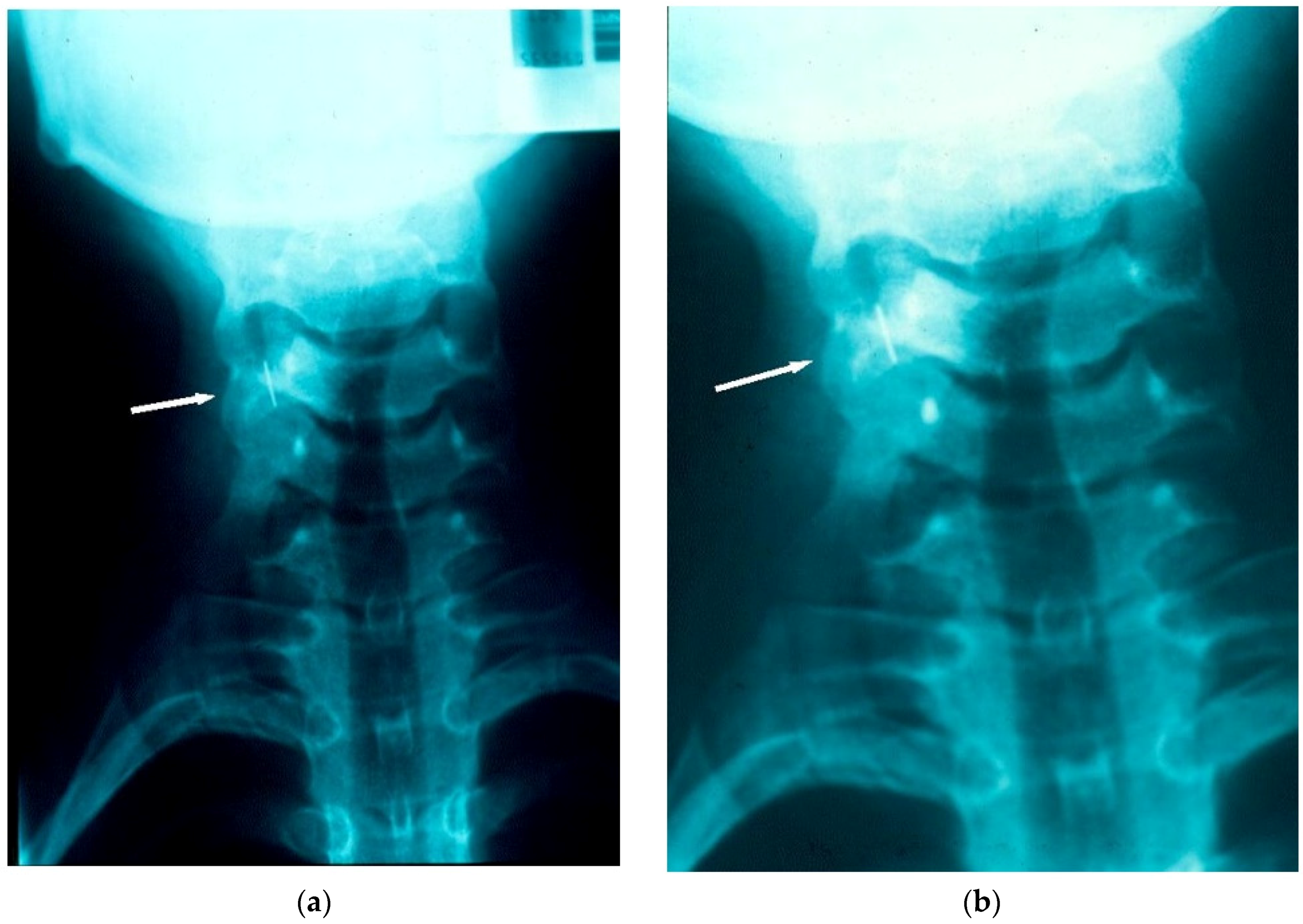
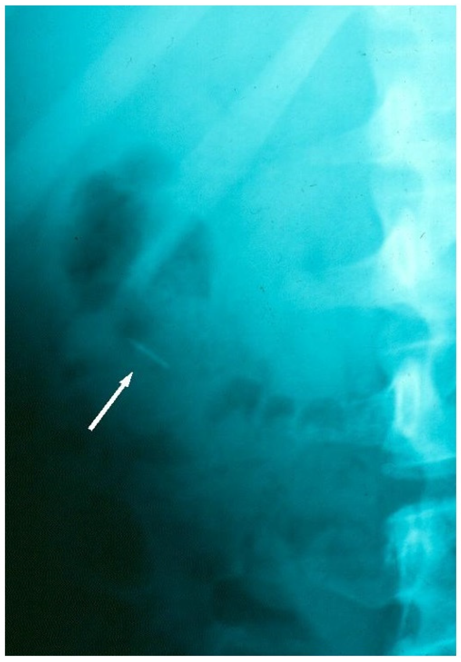
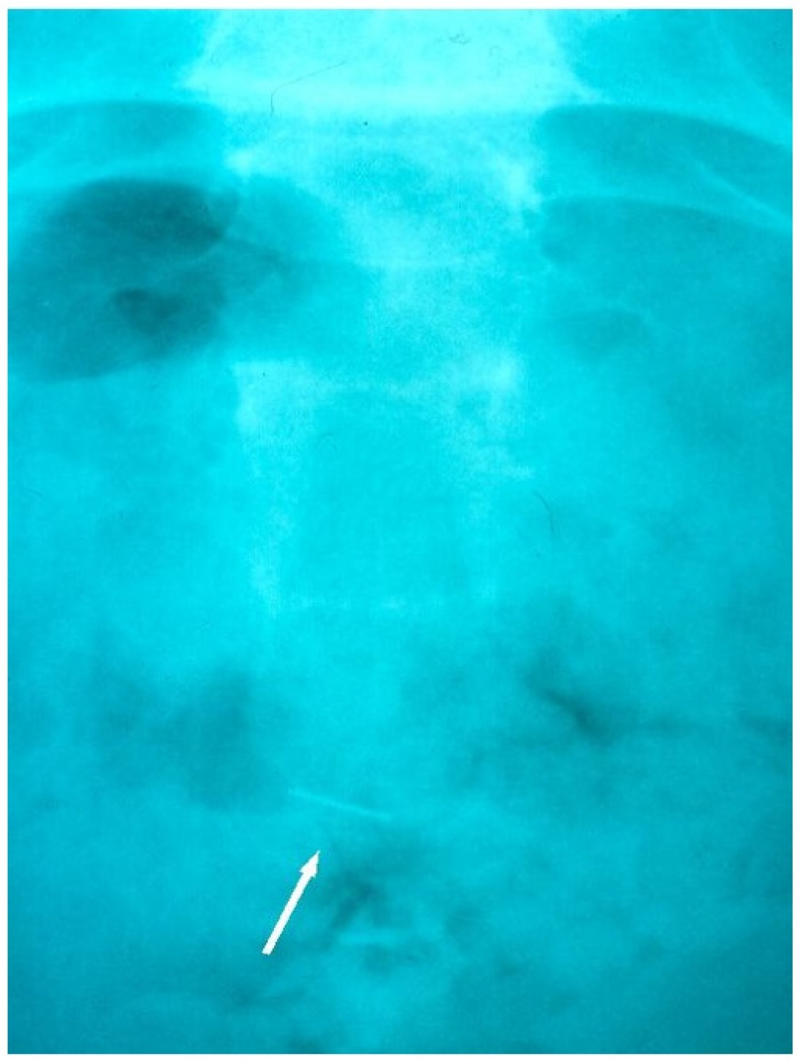
2. Case Report
3. Discussion
4. Conclusions
Author Contributions
Conflicts of Interest
References
- Al-Wahadni, A.; Al-Hamad, K.Q.; Al-Tarawneh, A. Foreign body ingestion and aspiration in dentistry: A review of the literature and reports of three cases. Dent. Update 2006, 33, 561–570. [Google Scholar] [PubMed]
- Klein, A.M.; Schoem, S.R. Unrecognised aspiration of a dental retainer—A case report. Otolaryngol. Head Neck Surg. 2002, 126, 438–439. [Google Scholar] [CrossRef] [PubMed]
- Dibiase, A.T.; Samuels, R.H.A.; Ozdiler, E.; Akcam, M.O.; Turkkahraman, H. Hazards of orthodontic appliances and the oropharynx. J. Orthod. 2000, 27, 295–302. [Google Scholar] [CrossRef] [PubMed]
- Allwork, E.G.; Edwards, I.R.; Welch, I.M. Ingestion of a quadhelix appliance requiring surgical removal: A case report. J. Orthod. 2007, 34, 154–157. [Google Scholar] [CrossRef] [PubMed]
- Tiwana, K.K.; Morton, T.; Tiwana, P.S. Aspiration and ingestion in dental practice: A ten year institutional review. J. Am. Dent. Assoc. 2004, 135, 1287–1291. [Google Scholar] [CrossRef] [PubMed]
- Nelson, J.F. Ingesting an onlay: A case report. J. Am. Dent. Assoc. 1992, 123, 73–74. [Google Scholar] [CrossRef] [PubMed]
- Nakajima, M.; Sato, Y. A method for preventing aspiration or ingestion of fixed restorations. J. Prosthet. Dent. 2004, 92. [Google Scholar] [CrossRef]
- Milton, T.M.; Hearing, S.D.; Ireland, A.J. Ingested foreign bodies associated with orthodontic treatment: Report of three cases and review of ingestion/aspiration incident management. Br. Dent. J. 2001, 190, 592–596. [Google Scholar] [PubMed]
- Nicolas, R.; Eggers, G.; Komposch, G. Orthodontic archwire in the nasal cavity: A case report. J. Orofac. Orthop. 2009, 70, 92–96. [Google Scholar] [CrossRef] [PubMed]
- Lee, B.W. Case report. Swallowed piece of archwire. Aust. Orthod. 1992, 12, 169–170. [Google Scholar]
- Quick, A.N.; Harris, A.M. Accidental ingestion of a component of a fixed orthodontic appliance—A case report. SADJ 2002, 57, 101–104. [Google Scholar] [PubMed]
- Sheridan, A. Orthodontic Bracket lost in airway. Am. J. Orthod. Dentofac. Orthop. 2009, 135. [Google Scholar] [CrossRef] [PubMed]
- Fiho, L.; Godoy, F.; O’Ryan, F. Orthodontic bracket lost in airway during orthognathic surgery. Am. J. Orthod. Dentofac. Orthop. 2008, 134, 288–290. [Google Scholar]
- Absi, E.G.; Buckley, J.G. The location and tracking of swallowed dental appliances; the role of radiology. Dentomaxillofac. Orthod. 1995, 24, 139–142. [Google Scholar] [CrossRef] [PubMed]
- Varho, R.; Oksala, H.; Tolvanen, M.; Svedstrom-Oristo, A.L. Inhalation or ingestion of orthodontic objects in Finland. Acta Odontol. Scand. 2015, 73, 408–413. [Google Scholar] [CrossRef] [PubMed]
- Hoseini, M.; Mostafavi, S.M.; Rezaei, N.; Boluri, E.J. Orthodontic wire ingestion during treatment: Reporting a case and review the management of foreign body ingestion or aspiration (Emergencies). Case Rep. Dent. 2013, 2013. [Google Scholar] [CrossRef] [PubMed]
- Abdel-Kader, H.M. Broken orthodontic trans-palatal archwire stuck to the throat of orthodontic patient: Is it strange? J. Orthod. 2003, 30. [Google Scholar] [CrossRef] [PubMed]
- Sfondrini, M.F.; Cacciafesta, V.; Lena, A. Accidental ingestion of a rapid palatal expander. J. Clin. Orthod. 2003, 37, 201–202. [Google Scholar] [PubMed]
- Hinkle, F.G. Ingested retainer: A case report. Am. J. Orthod. Dentofac. Orthop. 1987, 92, 46–49. [Google Scholar] [CrossRef]
- Park, J.H.; Owtad, P.; Milde, B. Incident Management guidelines for an Ingested Orthodontic Object. IJO 2013, 24, 43–47. [Google Scholar]
- Tiller, M.; Schepp, W.; Gundling, F.; Tuerck, J. Chronic pancreatitis caused by a swallowed orthodontic device. Endoscopy 2014, 46, E667–E668. [Google Scholar] [CrossRef] [PubMed]
- Umesan, U.K.; Ahmad, W.; Balakrishnan, P. Laryngeal impaction of an archwire segment after accidental ingestion during orthodontic adjustment. Am. J. Orthod. Dentofac. Orthop. 2012, 142, 264–268. [Google Scholar] [CrossRef] [PubMed]
- Monini Ada, C.; Maia, L.G.; Gandini, L.G. Accidental swallowing of an orthodontic expansion appliance key. Am. J. Orthod. Dentofac. Orthop. 2011, 140, 266–268. [Google Scholar] [CrossRef] [PubMed]
- Tipathi, T.; Rai, P.; Singh, H. Foreign body ingestion of orthodontic origin. Am. J. Orthod. Dentofac. Orthop. 2011, 139, 279–283. [Google Scholar] [CrossRef] [PubMed]
- Parkhouse, R.C. Medical complication in orthodontics. Br. J. Orthod. 1991, 18, 51–57. [Google Scholar] [CrossRef] [PubMed]
- Nazif, M.M.; Ready, M.A. Accidental swallowing of orthodontic expansion appliance keys: Report of two cases. ASDC J. Dent. Child. 1983, 50, 126–127. [Google Scholar] [PubMed]
- Wilmott, S.E.; Ikeaqwuani, O.; McLeod, N.M. An orthodontic bracket embedded in the medial pterygoid surface: A case report. J. Orthod. 2016, 43, 65–67. [Google Scholar] [CrossRef] [PubMed]
- Naragond, A.; Kengenal, S.; Rajasigamani, K.; Kumar, N.S. Accidental ingestion of molar band and its management: Maintenance is better than management. Case Rep. Dent. 2013. [Google Scholar] [CrossRef] [PubMed]
- Rohida, N.S.; Bhad, W.A. Accidental ingestion of a fractured Twin-block appliance. Am. J. Orthod. Dentofac. Orthop. 2011, 139, 123–125. [Google Scholar] [CrossRef] [PubMed]
- British Orthodontic Society. Advice Sheet—Guidelines for the Management of Inhaled or Ingested Foreign Bodies; British Orthodontic Society: London, UK, 2011. [Google Scholar]
- The Royal College of Radiologists. Making the Best Use of a Department of Clinical Radiology: Guidelines for Doctors, 6th ed.; The Royal College of Radiologists: London, UK, 2007. [Google Scholar]
- Cameron, S.M.; Whitlock, W.L.; Tabor, M.S. Foreign body aspiration in dentistry: A review. JADA 1996, 127, 1224–1229. [Google Scholar] [CrossRef] [PubMed]


| Authors | Year | Foreign Body | Inhaled or Ingested |
|---|---|---|---|
| Wilmott et al. [27] | 2016 | Fixed bracket | Ingested |
| Tiller et al. [21] | 2014 | Ligature wire | Ingested |
| Hoseini [16] | 2013 | Orthodontic wire | Ingested |
| Park et al. [20] | 2013 | Archwire fragment | Ingested |
| Naragon et al. [28] | 2013 | Orthodontic band | Ingested |
| Umesan et al. [22] | 2012 | Archwire fragment | Inhaled |
| Monini Ada et al. [23] | 2011 | Expansion key | Ingested |
| Tripathi T et al. [24] | 2011 | Expansion key | Ingested |
| Rohida et al. [29] | 2011 | Twin block appliance | Ingested |
| Nicolas et al. [9] | 2009 | Archwire fragment | Inhaled |
| Sheridan [12] | 2009 | Fixed bracket | Inhaled |
| Fiho et al. [13] | 2008 | Fixed bracket | Inhaled |
| Allwork et al. [4] | 2007 | Quadhelix | Ingested |
| Al-Wahadni et al. [1] | 2006 | Orthodontic band | Ingested |
| Abdel-Kader [17] | 2003 | Transpalatal archwire | Inhaled |
| Sfondrini et al. [18] | 2003 | Rapid palatal expander | Ingested |
| Klein et al. [2] | 2002 | Retainer | Inhaled |
| Quick et al. [11] | 2002 | Wire & coil Spring | Ingested |
| Milton et al. [8] | 2001 | Fixed bracket | Ingested |
| Archwire fragment | Ingested | ||
| Sectional wire | Ingested | ||
| Dibiase et al. [3] | 2000 | Removable appliance | Ingested |
| Absi et al. [14] | 1995 | Archwire | Ingested |
| Lee [10] | 1992 | Archwire fragment | Ingested |
| Parkhouse [25] | 1991 | Appliance segment | Ingested |
| Hinkle [19] | 1987 | Retainer | Ingested |
| Nazif et al. [26] | 1983 | Expansion key | Ingested |
| Expansion key | Ingested |
© 2016 by the authors; licensee MDPI, Basel, Switzerland. This article is an open access article distributed under the terms and conditions of the Creative Commons Attribution (CC-BY) license (http://creativecommons.org/licenses/by/4.0/).
Share and Cite
Puryer, J.; McNamara, C.; Sandy, J.; Ireland, T. An Ingested Orthodontic Wire Fragment: A Case Report. Dent. J. 2016, 4, 24. https://doi.org/10.3390/dj4030024
Puryer J, McNamara C, Sandy J, Ireland T. An Ingested Orthodontic Wire Fragment: A Case Report. Dentistry Journal. 2016; 4(3):24. https://doi.org/10.3390/dj4030024
Chicago/Turabian StylePuryer, James, Catherine McNamara, Jonathan Sandy, and Tony Ireland. 2016. "An Ingested Orthodontic Wire Fragment: A Case Report" Dentistry Journal 4, no. 3: 24. https://doi.org/10.3390/dj4030024