Review

Chirality in Optical Trapping and Optical Binding

School of Chemistry, University of East Anglia, Norwich, NR4 7TJ, UK
*
Author to whom correspondence should be addressed.
Photonics 2015, 2(2), 483-497; https://doi.org/10.3390/photonics2020483
Submission received: 26 March 2015 / Revised: 5 May 2015 / Accepted: 6 May 2015 / Published: 11 May 2015
(This article belongs to the Special Issue Optical Trapping in Biology and Nanotechnology)

Abstract

:
Optical trapping is a well-established technique that is increasingly used on biological substances and nanostructures. Chirality, the property of objects that differ from their mirror image, is also of significance in such fields, and a subject of much current interest. This review offers insight into the intertwining of these topics with a focus on the latest theory. Optical trapping of nanoscale objects involves forward Rayleigh scattering of light involving transition dipole moments; usually these dipoles are assumed to be electric although, in chiral studies, magnetic dipoles must also be considered. It is shown that a system combining optical trapping and chirality could be used to separate enantiomers. Attention is also given to optical binding, which involves light induced interactions between trapped particles. Interesting effects also arise when binding is combined with chirality.

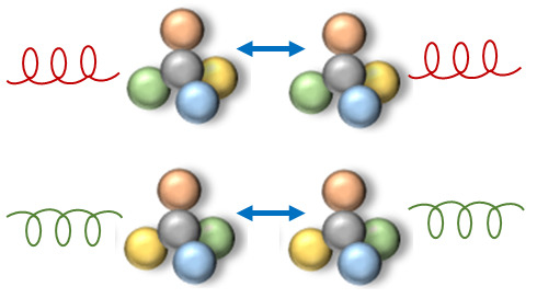
Graphical Abstract

1. Introduction

When a laser beam is scattered by a dielectric microparticle, resulting in light refraction on entering and leaving the particle, a small amount of momentum is transferred from the photons to the matter. This change in momentum, known as the gradient force, results in the attraction of the particle to the high intensity part of the beam (usually the centre). Optical trapping of microscale particles via this mechanism was first reported in the 1970s [1] and duly led to the initial observation of a single beam optical trap in 1986 [2]. These preliminary experiments, and many of the methodologies that developed from them, utilized the gradient force exerted by a single, tightly focused Gaussian laser beam to trap particles in solution through what has become known as the “optical tweezer” effect. Since these initial findings, optical technology has evolved significantly, and traps that facilitate three dimensional manipulation of particles are now readily available. While originally limited to the controlled manipulation of individual particles, multitrap setups involving either splitting [3,4] or time sharing [5,6] with a single laser beam are now also commonly utilized. As a more advanced form of the former, holographic optical tweezers that employ diffractive optical elements such as spatial light modulators now allow computer controlled, independent manipulation of multiple particles [7,8,9]. A number of multitrap devices have also been developed based on the application of laser beams with more complex phase and intensity profiles, as for example Bessel or higher order Laguerre Gaussian beams [10,11,12].
Optical tweezers have been successfully applied to confine an extensive range of materials, and they are utilized for a wide variety of purposes. They have been successfully employed in the fabrication—through manipulation and immobilization of single particles—of micro- and nano-scale structures [13,14,15]. In a manner analogous to the function of tipped cantilevers in atomic force microscopy, trapped particles are utilized in scanning microscopy as handles for probes that can map sample surfaces [16,17]. Through the selective trapping of particles it is also possible to sort individual molecules within a mixed solution [6,18,19]. Beyond the capacity to isolate and spatially arrange microparticles, modern optical traps can also be utilized to quantitatively measure displacement and applied force with nanometre and piconewton resolutions, respectively. This level of precision, along with the inherent non-contact and non-invasive characteristic of the technology, make optical tweezers particularly appealing for the study of biological systems [20]. For example, force and displacement measurements have been used with nucleic acids and proteins [21,22,23], cells [24,25], viruses [26,27] and biological molecular motors [28].
The role of chirality in optical trapping and optical binding is a subject which has received very little attention until recently. Physical entities—whether material entities such as molecules, or non-material in the case of circularly polarized light beams—are said to be chiral if they are non-superimposable on their mirror image; chiral effects in optical trapping may occur if either the matter or light is chiral. Chirality is not only important in connection with biology [29,30,31,32,33,34], but it is also increasingly significant in relation to nanoscale systems such as metamaterials [35,36,37,38,39]. However, it is noteworthy that different electrodynamical mechanisms operate in the manipulation of objects such as molecules or nanoparticles. This review summarises recent investigations of the associated issues, based on a description of the physical mechanisms rather than in-depth mathematical theory. Section 2 will address the capacity of chiral molecules to exhibit a differential optical response when irradiated with a non-absorbing laser beam. Such a trapping effect has the potential to optomechanically separate distinct enantiomers within a racemic mixture. Experimental trapping procedures typically involve near-micrometre scale particles, but similar levels of optical control are possible within the nanoscale [40,41,42]. Section 3 tackles the role of chirality in optical binding; a phenomenon, predicted initially by Thirunamachandran [43], which is distinct from optical trapping. In optical binding, applied electromagnetic radiation not only traps but also induces a force between particles. These laser-induced forces can be either attractive or repulsive (despite the term “binding”) and, crucially, they may override the intrinsic dispersion force that acts between particles. Here, too, experimental efforts that initially concentrated on dielectric microparticles [44,45,46,47,48,49] now increasingly focus on binding between nanoparticles [50,51]. Within the latter size regime, a host of theoretical developments have appeared in the literature—see [52] and references therein, including molecular studies based on quantum theory [53,54,55]. Our focus is on the novel opportunities afforded by the optical binding of chiral molecules and nanoparticles.

2. Chirality in Optical Trapping

Some of the most innovative recent research involving the optical interactions of chiral material focuses on trapping specific types of solid microparticle; for example, in connection with observation of the internal helical structure of trapped particles [56], the production of an omnidirectional chiral mirror enabling optomechanical effects [57], or the selective manipulation of chiral objects dependent on photon helicity [58]. Moreover, outside of trapping techniques, it has recently been shown that motion can be applied to chiral macroscopic material through use of an optical “tractor beam” [59]. Optical trapping at the nanoscale, where the wavelength of light exceeds the particle dimensions, is necessarily treated differently to the ray optics approach of microparticle trapping. Treatment of the former usually involves the interaction of transition electric dipole moments (E1) with the applied electromagnetic field, in a forward Rayleigh scattering mechanism [52,60,61,62]. Scattering of this type implies concerted single photon absorption and emission events, in other word a coupling of two photon interactions, with the direction and energy of the emergent light being identical to the input. When studying Rayleigh scattering (and most other optical effects) it is usually legitimate to assume that all the light−matter interactions are mediated by the transition electric dipole moments of the molecule; henceforth this will be termed an E12 interaction, denoting two electric−dipole interactions. However, scattering may still occur—though to a much lesser extent—when one of the interactions involves a transition magnetic dipole (i.e., concerted E1M1 couplings). In chiral discrimination studies, in which slightly different optical effects are observed using left-handed circularly polarized light compared to right-handed light, it is the E1M1 feature that offers the most important contribution—a feature that arises since electric and magnetic effects have fundamentally different symmetry properties with regard to spatial inversion (and also time reversal) [63]. As will be shown, the resulting dissimilarity may be exploited with respect to chiral molecules in an optical trap.
In the case of forward Rayleigh scattering the fundamental physical observable (whether or not chiral molecules are involved) is the optically induced potential energy [64], from which an optical trapping force is determined as the spatial gradient. This contrasts, for example, with the process of circular dichroism—the differential absorption of left- and right-handed circularly polarized light—whose observable is signified by a rate (as determined from the Fermi Rule) and is thus completely distinct from optical trapping. The electric dipole approximation is almost universally employed in optical trapping calculations, although the effects of electric quadrupole moments have been examined [65]. In contrast, as stated earlier, E1M1 optical trapping interactions are crucial in the study of chiral discrimination since differential scattering cannot occur via E12 interactions, i.e., identical optical effects arise for left- and right-handed input light in such cases. As shown in Figure 1, while discrimination is usually defined in terms of a difference in the optical response of a specific enantiomer (either a left- or right-handed molecule) irradiated with left-handed polarized light compared to right-handed light—illustrated in (a) and (c), or (b) and (d)—equally discriminatory results are attained for left-handed circularly polarized light, say, applied to a left-handed enantiomer relative to a right-handed one. The principle is depicted by the pairings (a) and (b), or (c) and (d) in Figure 1. The latter cases, involving a single beam of defined circularity, leads to the interesting prospect of enantiomer separation by optical means—a subject of much current interest, which is further outlined below.
Figure 1. Illustration of the equivalences (ab, cd) between the discriminatory optical trapping forces for chiral molecules of different handedness (depicted by red and green spheres) irradiated by circularly polarized light of either handedness (right- and left-handed forms shown with opposite twist) propagating in the direction shown by the black arrows. All other pairs are non-equivalent.
Photonics 02 00483 g001
Before pursuing this further, it is interesting to reflect on the range of other methods that are available for achieving enantiomer separation. While chromatographic or electromigration techniques, involving the detection of material chirality using a substance known as a “chiral selector”, are well-established industrial-scale separation procedures [66,67,68,69], other experiments independent of such selectors are offered in the literature. For microscale particles, most investigations rely on hydrodynamical forces associated with the shape of chiral objects, resulting in separate migratory pathways (driven by a steady fluid flow) [70,71,72,73,74]. Another system is based on a “propeller effect”, in which an applied radio frequency electric field of rotating polarization induces opposite enantiomers to rotate in opposing directions [75]; this effect has recently been experimentally verified using chiral helical colloids in a rotating magnetic field [76]. A promising optical method to separate mirror-imaged chiral microparticles, in a fluidic environment, has also recently been reported [77,78]. This optofluidic sorting scheme depends on chiral (circularly polarized) light to produce optical forces that deflects left-handed cholesteric droplets in an opposite direction to right-handed droplets. Despite these successes, downscaling into nanoscale dimensions remains elusive in practical terms since, for example, approaches based on circular Bragg reflection [79] suffer extensive complications due to thermal fluctuations; although there has been recent progress on enantiomer detection using microwave spectroscopy [80,81,82,83] it has not afforded a means of separation. Theoretical interest in optical forces on chiral objects is increasing continually [84,85,86,87], especially in connection with molecular enantiomers [88,89,90,91,92,93,94,95,96,97,98,99,100,101,102,103,104,105]; as a result, a workable optical system for separation of chiral molecules should, hopefully, be achievable in the not too distant future.
Against this context, although numerous forms of optical force are under consideration in the literature, it is of considerable interest to conceive an optomechanical method for the specific purpose of chiral separation, based on optical trapping. To this end, the physical mechanism—based on the concepts introduced in Figure 1—needs to be understood. A complete mathematical description, whose details are provided elsewhere [101], is attained by first determining an expression for the light-induced potential energy ΔE acting on a chiral molecule, where both E12 and E1M1 interactions arise;
Δ E ( L | R ) ( r ) = ( I ( r ) 2 ε 0 c 2 ) Re { c e ¯ i ( L | R ) e j ( L | R ) α i j + e ¯ i ( L | R ) b j ( L | R ) G i j + b ¯ i ( L | R ) e j ( L | R ) G ¯ j i }
where αij are components of the polarizability matrix—amongst which there are non-zero values for every molecule or nanoparticle—with i and j denoting Cartesian coordinates; Gij is a counterpart electric-magnetic scattering matrix and I(r) is the intensity of the trapping beam at position r. Moreover, e i ( L | R ) and b j ( L | R ) are Cartesian components of the circular polarization vectors for the electric and magnetic fields, respectively (either left- or right-handed chirality as denoted by the superscript); a summation over repeated indices i, j is implicit. Throughout, overbars signify complex conjugation (which effectively inverts chirality). In Equation (1), the first term corresponds to the E12 interaction and the other terms to the E1M1 couplings. Chiral discrimination is found from Δ E ( L R ) Δ E ( L )  Δ E ( R ) so that, since the first term is identical for left- and right-handed polarized light (and thus cancel out), the following is derived;
Δ E ( L R ) ( r ) = i ( I ( r ) 2 ε 0 c 2 ) ( e ¯ i ( L ) e j ( L ) + e ¯ i ( R ) e j ( R ) ) ( G i j G ¯ j i )
in which only the terms that correspond to E1M1 couplings arise. On trapping two opposite enantiomers (i.e., a right-handed and a left-handed chiral molecule), the differential force is found from ΔF = F(L) – F(R), where the optical trapping force for each enantiomer is determined from the potential energy through the expression F ( L|R ) ( r ) = ( Δ E ( L|R ) ( r ) ) . In cases where the molecules undergo free rotational motion, the middle term in Equation (2) simply delivers the value 2/3. It is discovered that the differential forces, due to the E1M1 interactions, are equal and opposite when comparing the right-handed enantiomer with the left-handed molecule—this is the origin of enantiomer separation due to optical trapping.
A simple calculation can illustrate the principle. For chiral molecules with modest polarizability volumes of 5 × 10⁻²⁸ m3, within a pulsed laser beam of intensity 4 × 10¹¹ W cm−2 and waist 10 µm, estimates for the typical magnitude of such a differential force is 10−15–10−14 N [104], considered to be well within experimental reach. To achieve the necessary levels of irradiance, we anticipate that the input beam would take the form of ultrashort (femtosecond) pulses, such that the molecular response would take the form of a series of impulses typically valued in the 2 × 10⁻²⁹ N s range. In the relatively long (picosecond) intervals between successive pulses, there would be very little drift, so that the net effect would be equivalent to that of continuous irradiation. One possible implementation for enantiomer separation, based on optical trapping, may involve enantiomers in a solution. In such a scenario, while all the enantiomers are attracted to the high-intensity part of a circularly polarized trapping beam (for example, the centre of a Gaussian beam), the left-handed molecule may be more inclined, in relation to its mirror image, to reside in the high intensity region of the beam [105]. This prospect does not account for the intermolecular interactions known as optical binding, which is the subject of the following section.

3. Chirality in Optical Binding

Optical binding is a laser-induced force that acts between trapped molecules—although it is fundamentally distinct from optical trapping. This phenomena is again described by forward Rayleigh scattering although, in this case, a photon is absorbed at one molecule and emitted at another, and electromagnetic couplings between the molecules occur; this mechanism, therefore, involves four photon interactions with the emergent beam unchanged. The first study [106] of optical binding between chiral molecules revealed that the binding force is independent of the handedness of the incident light, with the discrimination arising due to the electric-magnetic dipole polarizabilities of each molecule (the E1M1−E1M1 couplings); the magnitude of the corresponding forces is typically 10−4 times smaller than conventional optical binding, i.e., the situation when all four photon interactions relate to electric dipole transition moments. However, although E12 couplings are always non-discriminatory, it is possible for one chiral molecule to interact in this manner whilst the other engages E1M1 coupling; under such circumstances the optical binding force between the molecular pair, involving one α and one G matrix, is discriminatory. This system, denoted by E12−E1M1 and until recently [107] overlooked in the literature, can be shown to have a binding force that is discriminatory with respect to the handedness of the molecules and the incident light (although no discriminatory force arises when the laser light is linearly polarized). Typically, this force is two orders of magnitude larger than the E1M1−E1M1 case.
The optically induced potential energy between a pair of chiral molecules (A, B) in the arrangement k R , where k is the wave-vector of the incident circularly polarized beam and R is the inter-particle separation vector, is expressible as [107];
Δ E ( L|R ) ( r , R ) = I g ( 12 ) ( r A , r B ) 8 π ε 0 2 c 2 R 3 { ( G ( A ) α ( B ) α ( A ) G ( B ) ) × [ ± ( cos k R + k R sin k R ) ± k 2 R 2 cos k R ] }
where I is the mean input laser irradiance, g ( 12 ) ( r A , r B ) is a degree of second order coherence [108], and α and G are the scalar equivalents of the molecular polarizability scattering matrices defined earlier. In Equation (3) the upper sign corresponds to left-handed, and the lower to right-handed, optical input. If the laser beam is propagating parallel to the inter-particle separation vector, kR, then the potential energy becomes:
Δ E ( L|R ) ( r , R ) = I g ( 12 ) ( r A , r B ) 4 π ε 0 2 c 2 R 3 { ( G ( A ) α ( B ) α ( A ) G ( B ) ) × [ ( cos k R + k R sin k R ) ± k 2 R 2 cos k R ] } cos ( k R )
The result for the potential energy when the pair is orientationally averaged, which pertains to the situation where the molecular pair is allowed to tumble freely, takes the following form (suppressing the labelled position dependence):
Δ E ( L|R ) ( R ) = ± I g ( 12 ) ( α G G α ) 4 π ε 0 2 c 2 R 3 ( cos 2 k R k R sin 2 k R 2 + 3 sin 2 k R 2 k R + 3 cos 2 k R k 2 R 2 3 sin 2 k R 2 k 3 R 3 )
where G = G . From Equations (3–5), the differential forces of attraction or repulsion between the participant molecules can be derived from F ( L|R ) ( R ) = Δ E ( L|R ) ( R ) / R . It can thus be deduced that for a pair of chemically identical enantiomers the differential force will be zero, irrespective of the handedness of incident light. If, however, the pair of chiral particles have identical handedness then there exists a non-zero discriminatory binding force, whose sign depends on both the particle and of the radiation handedness. In this case, a pair of right-handed particles irradiated by right-handed light has the same discriminatory binding force as the corresponding pair of left-handed particles irradiated by left-handed light. This equivalence is illustrated in Figure 2, i.e., (a) ≡ (b). A pair of right-handed particles irradiated by left-handed light, however, differs from a pair of right-handed particles irradiated by right-handed light (or vice versa), so that in Figure 2, {(a), (b)} ≠ {(c), (d)}, whilst it is clearly the case that (c) ≡ (d).
Figure 2. Illustration of the equivalences (ab, cd), and non-equivalences (a, bc, d), between the discriminatory optical binding forces between chiral particles of different handedness (depicted by red and green spheres) irradiated by circularly polarized light of either handedness (right- and left-handed forms shown with opposite twist). For cases e, f, where the two species are enantiomers, the discriminatory force vanishes: the pair effectively represents a system that is achiral, and the optical binding force is accordingly independent of the handedness of light.
Photonics 02 00483 g002
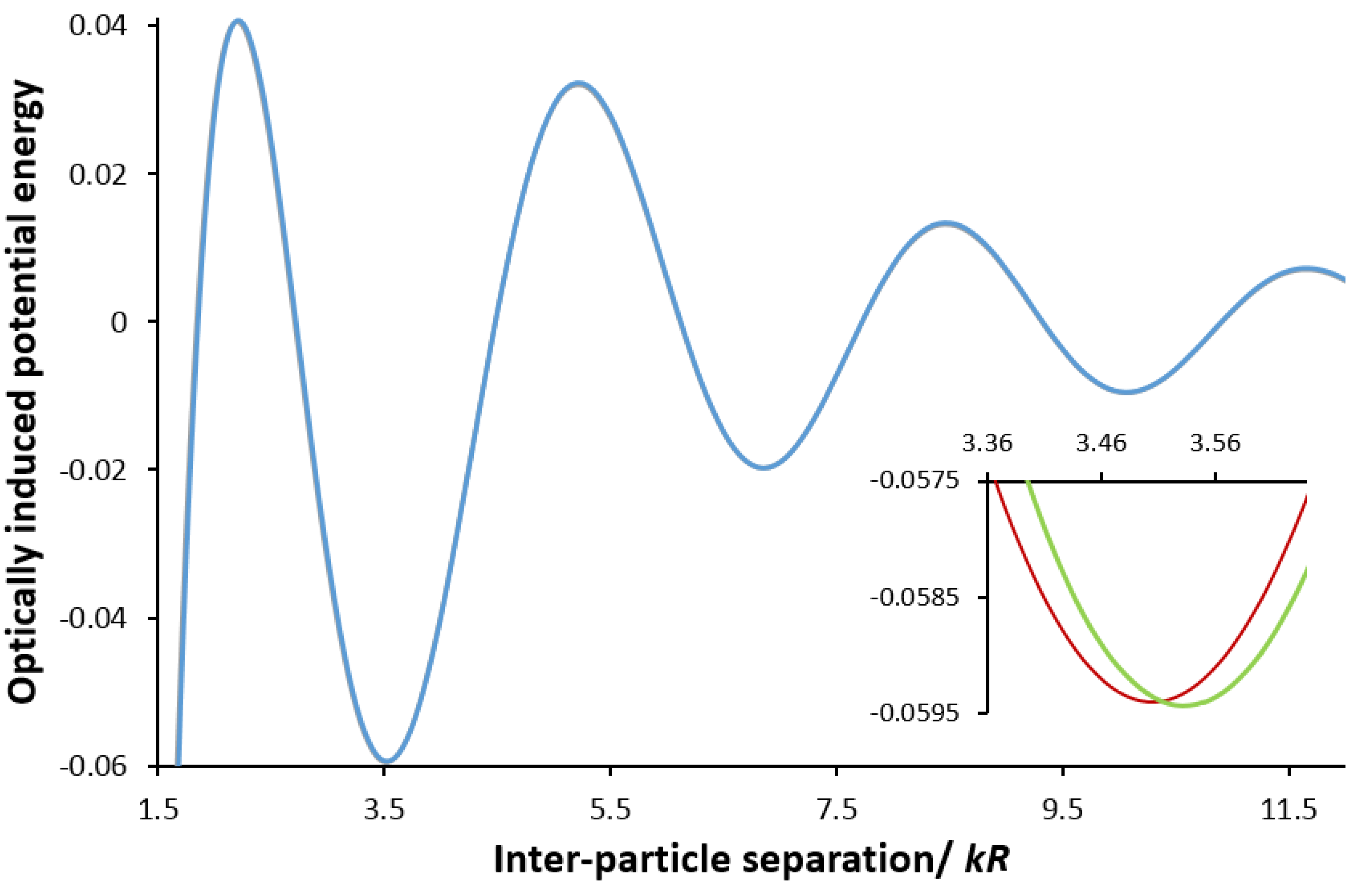
Possible routes to exploit these discriminatory optical binding forces may be found in the helicity-dependent optomechanical manipulation of chiral particles [57,58], or chiral sorting strategies based on optical forces [71,73,78,109,110]. Calculations based on the application of Equation (5) as a correction to the leading (E12−E12) result [43] produce graphs of the form shown in Figure 3, where the magnitude of the correction is taken to be of the order of the fine structure constant, ~1/137, (i.e., the fundamental physical constant that characterizes the relative strength of electromagnetic interactions) a factor that is typical for such cases. Here, the inset to Figure 3 shows the different positioning of the principal optical binding energy minima, for particles whose handedness is either both the same, or both opposite to, that of the radiation. It emerges that, for a laser wavelength of 628 nm, there is a displacement of 5 nm between these minima: in consequence, any modulation of the optical input between right and left circular polarizations will be a corresponding oscillation in their equilibrium positions. One potentially realizable application is therefore a means to identify chirality in optically bound systems. These and other possibilities for process implementation are now the subject of further active investigation.
Figure 3. Plot of the optical binding potential energy (in arbitrary units) for two chiral particles in a circularly polarized beam. The abscissa scale measures the inter-particle distance R in dimensionless units of kR, the wave-number k is defined as 2 π/λ with λ as the laser wavelength. On the scale of the main graph, there is an imperceptible difference between the results for particles whose handedness is either the same, or opposite to, that of the radiation. The inset exhibits the difference between the two cases, around the position of the first potential energy minimum.
Photonics 02 00483 g003

Acknowledgments

The authors are grateful to the Leverhulme Trust, the Engineering and Physical Sciences Research Council, and UEA for funding our research. We also thank Jack S. Ford for helping produce Figure 3.

Conflict of Interest

The authors declare no conflict of interest.

References

  1. Ashkin, A. Acceleration and trapping of particles by radiation pressure. Phys. Rev. Lett. 1970, 24, 156–159. [Google Scholar] [CrossRef]
  2. Ashkin, A.; Dziedzic, J.; Bjorkholm, J.; Chu, S. Observation of a single-beam gradient force optical trap for dielectric particles. Opt. Lett. 1986, 11, 288–290. [Google Scholar] [CrossRef]
  3. Liu, Y.; Yu, M. Multiple traps created with an inclined dual-fiber system. Opt. Express 2009, 17, 21680–21690. [Google Scholar] [CrossRef]
  4. Kotsifaki, D.G.; Makropoulou, M.; Serafetinides, A.A. Efficient and low cost multiple optical trap, based on interference. Optik 2013, 124, 617–621. [Google Scholar] [CrossRef]
  5. Ren, Y.-X.; Wu, J.-G.; Chen, M.; Li, H.; Li, Y.-M. Stability of novel time-sharing dual optical tweezers using a rotating tilt glass plate. Chinese. Phys. Lett. 2010, 27, 028703. [Google Scholar] [CrossRef]
  6. Dasgupta, R.; Verma, R.S.; Gupta, P.K. Microfluidic sorting with blinking optical traps. Opt. Lett. 2012, 37, 1739–1741. [Google Scholar] [CrossRef] [PubMed]
  7. Gibson, G.M.; Bowman, R.W.; Linnenberger, A.; Dienerowitz, M.; Phillips, D.B.; Carberry, D.M.; Miles, M.J.; Padgett, M.J. A compact holographic optical tweezers instrument. Rev. Sci. Instrum. 2012, 83, 113107. [Google Scholar] [CrossRef]
  8. Yevnin, M.; Kasimov, D.; Gluckman, Y.; Ebenstein, Y.; Roichman, Y. Independent and simultaneous three-dimensional optical trapping and imaging. Biomed. Opt. Express 2013, 4, 2087–2094. [Google Scholar] [CrossRef]
  9. Shaw, L.; Preece, D.; Rubinsztein-Dunlop, H. Kinect the dots: 3D control of optical tweezers. J. Opt. 2013, 15, 075303. [Google Scholar] [CrossRef]
  10. Dholakia, K.; Čižmár, T. Shaping the future of manipulation. Nat. Photonics 2011, 5, 335–342. [Google Scholar] [CrossRef]
  11. Yan, Z.; Jureller, J.E.; Sweet, J.; Guffey, M.J.; Pelton, M.; Scherer, N.F. Three-dimensional optical trapping and manipulation of single silver nanowires. Nano Lett. 2012, 12, 5155–5161. [Google Scholar] [CrossRef]
  12. Decombe, J.-B.; Mondal, S.K.; Kumbhakar, D.; Pal, S.S.; Fick, J. Single and multiple microparticle trapping using non-Gaussian beams from optical fiber nanoantennas. IEEE J. Sel. Top. Quant. 2015, 21, 1–6. [Google Scholar] [CrossRef]
  13. Dawood, F.; Qin, S.; Li, L.; Lin, E.Y.; Fourkas, J.T. Simultaneous microscale optical manipulation, fabrication and immobilisation in aqueous media. Chem. Sci. 2012, 3, 2449–2456. [Google Scholar] [CrossRef]
  14. Ghadiri, R.; Weigel, T.; Esen, C.; Ostendorf, A. Microassembly of complex and three-dimensional microstructures using holographic optical tweezers. J. Micromech. Microeng. 2012, 22, 065016. [Google Scholar] [CrossRef]
  15. Palima, D.; Glückstad, J. Gearing up for optical microrobotics: micromanipulation and actuation of synthetic microstructures by optical forces. Laser Photon. Rev. 2013, 7, 478–494. [Google Scholar] [CrossRef]
  16. Phillips, D.B.; Grieve, J.A.; Olof, S.N.; Kocher, S.J.; Bowman, R.; Padgett, M.J.; Miles, M.J.; Carberry, D.M. Surface imaging using holographic optical tweezers. Nanotechnology 2011, 22, 285503. [Google Scholar] [CrossRef] [PubMed]
  17. Phillips, D.B.; Gibson, G.M.; Bowman, R.; Padgett, M.J.; Hanna, S.; Carberry, D.M.; Miles, M.J.; Simpson, S.H. An optically actuated surface scanning probe. Opt. Express 2012, 20, 29679–29693. [Google Scholar] [CrossRef]
  18. Ma, B.; Yao, B.; Peng, F.; Yan, S.; Lei, M.; Rupp, R. Optical sorting of particles by dual-channel line optical tweezers. J. Opt. 2012, 14, 105702. [Google Scholar] [CrossRef]
  19. Li, Y.; Guo, Z.; Qu, S. Living cell manipulation in a microfluidic device by femtosecond optical tweezers. Opt. Laser Eng. 2014, 55, 150–154. [Google Scholar] [CrossRef]
  20. Capitanio, M.; Pavone, F.S. Interrogating biology with force: Single molecule high-resolution measurements with optical tweezers. Biophys. J. 2013, 105, 1293–1303. [Google Scholar] [CrossRef]
  21. Li, M.; Wang, M.D. Unzipping single DNA molecules to study nucleosome structure and dynamics. Methods Enzymol. 2012, 513, 29–58. [Google Scholar] [CrossRef]
  22. Monico, C.; Capitanio, M.; Belcastro, G.; Vanzi, F.; Pavone, F. Optical methods to study protein-DNA interactions in vitro and in living cells at the single-molecule level. Int. J. Mol. Sci. 2013, 14, 3961–3992. [Google Scholar] [CrossRef] [PubMed]
  23. Heller, I.; Hoekstra, T.P.; King, G.A.; Peterman, E.J.G.; Wuite, G.J.L. Optical tweezers analysis of DNA–protein complexes. Chem. Rev. 2014, 114, 3087–3119. [Google Scholar] [CrossRef]
  24. Chowdhury, S.; Thakur, A.; Svec, P.; Wang, C.; Losert, W.; Gupta, S. Automated manipulation of biological cells using gripper formations controlled by optical tweezers. IEEE T. Autom. Sci. Eng. 2014, 11, 338–347. [Google Scholar] [CrossRef]
  25. Jeorrett, A.H.; Neale, S.L.; Massoubre, D.; Gu, E.; Henderson, R.K.; Millington, O.; Mathieson, K.; Dawson, M.D. Optoelectronic tweezers system for single cell manipulation and fluorescence imaging of live immune cells. Opt. Express 2014, 22, 1372–1380. [Google Scholar] [CrossRef]
  26. Smith, D.E. Single-molecule studies of viral DNA packaging. Curr. Opin. Virol. 2011, 1, 134–141. [Google Scholar] [CrossRef]
  27. Pang, Y.; Song, H.; Kim, J.H.; Hou, X.; Cheng, W. Optical trapping of individual human immunodeficiency viruses in culture fluid reveals heterogeneity with single-molecule resolution. Nat. Nanotechnol. 2014, 9, 624–630. [Google Scholar] [CrossRef]
  28. Veigel, C.; Schmidt, C.F. Moving into the cell: single-molecule studies of molecular motors in complex environments. Nat. Rev. Mol. Cell Biol. 2011, 12, 163–176. [Google Scholar] [CrossRef]
  29. Bentley, R. Chirality in Biology. In Encyclopedia of Molecular Cell Biology and Molecular Medicine; Meyers, R.A., Ed.; Wiley-VCH: Weinheim, Germany, 2006; pp. 579–618. [Google Scholar]
  30. Barron, L.D. Chirality and life. Space Sci. Rev. 2008, 135, 187–201. [Google Scholar] [CrossRef]
  31. Mori, K. Bioactive natural products and chirality. Chirality 2011, 23, 449–462. [Google Scholar] [CrossRef]
  32. Cintas, P.; Amabilino, D.B. Biochirality: Origins, Evolution and Molecular Recognition; Springer: Heidelberg, Germany, 2013. [Google Scholar]
  33. Timsit, Y. DNA self-assembly: From chirality to evolution. Int. J. Mol. Sci. 2013, 14, 8252–8270. [Google Scholar] [CrossRef] [PubMed]
  34. Senge, M.; Ryan, A.; Letchford, K.; MacGowan, S.; Mielke, T. Chlorophylls, symmetry, chirality, and photosynthesis. Symmetry 2014, 6, 781–843. [Google Scholar] [CrossRef]
  35. Wang, B.; Zhou, J.; Koschny, T.; Kafesaki, M.; Soukoulis, C.M. Chiral metamaterials: simulations and experiments. J. Opt. A: Pure Appl. Opt. 2009, 11, 114003. [Google Scholar] [CrossRef]
  36. Valev, V.K.; Baumberg, J.J.; Sibilia, C.; Verbiest, T. Chirality and chiroptical effects in plasmonic nanostructures: fundamentals, recent progress, and outlook. Adv. Mater. 2013, 25, 2517–2534. [Google Scholar] [CrossRef] [PubMed]
  37. Li, Z.; Mutlu, M.; Ozbay, E. Chiral metamaterials: from optical activity and negative refractive index to asymmetric transmission. J. Opt. 2013, 15, 023001. [Google Scholar] [CrossRef]
  38. Wang, Y.; Xu, J.; Wang, Y.; Chen, H. Emerging chirality in nanoscience. Chem. Soc. Rev. 2013, 42, 2930–2962. [Google Scholar] [CrossRef]
  39. Bradshaw, D.S.; Leeder, J.M.; Coles, M.M.; Andrews, D.L. Signatures of material and optical chirality: Origins and measures. Chem. Phys. Lett. 2015, 626, 106–110. [Google Scholar] [CrossRef]
  40. Bendix, P.M.; Jauffred, L.; Norregaard, K.; Oddershede, L.B. Optical trapping of nanoparticles and quantum dots. IEEE J. Sel. Top. Quant. 2014, 20, 15–26. [Google Scholar] [CrossRef]
  41. Tinguely, J.-C.; Ding, M.; Brambilla, G.; Hohenau, A.; Krenn, J.R.; Hellesø, O.G. Nanostructured fibre tip for trapping of nanoparticles. Proc. SPIE 2014, 8999, 89991D. [Google Scholar] [CrossRef]
  42. Gelfand, R.M.; Wheaton, S.; Gordon, R. Cleaved fiber optic double nanohole optical tweezers for trapping nanoparticles. Opt. Lett. 2014, 39, 6415–6417. [Google Scholar] [CrossRef]
  43. Thirunamachandran, T. Intermolecular interactions in the presence of an intense radiation field. Mol. Phys. 1980, 40, 393–399. [Google Scholar] [CrossRef]
  44. Burns, M.M.; Fournier, J.-M.; Golovchenko, J.A. Optical binding. Phys. Rev. Lett. 1989, 63, 1233–1236. [Google Scholar] [CrossRef] [PubMed]
  45. Tatarkova, S.A.; Carruthers, A.E.; Dholakia, K. One-dimensional optically bound arrays of microscopic particles. Phys. Rev. Lett. 2002, 89, 283901. [Google Scholar] [CrossRef] [PubMed]
  46. Singer, W.; Frick, M.; Bernet, S.; Ritsch-Marte, M. Self-organized array of regularly spaced microbeads in a fiber-optical trap. J. Opt. Soc. Am. B 2003, 20, 1568–1574. [Google Scholar] [CrossRef]
  47. Karásek, V.; Čižmár, T.; Brzobohatý, O.; Zemánek, P.; Garcés-Chávez, V.; Dholakia, K. Long-range one-dimensional longitudinal optical binding. Phys. Rev. Lett. 2008, 101, 143601. [Google Scholar] [CrossRef] [PubMed]
  48. Gherardi, D.M.; Carruthers, A.E.; Čižmár, T.; Wright, E.M.; Dholakia, K. A dual beam photonic crystal fiber trap for microscopic particles. Appl. Phys. Lett. 2008, 93, 041110. [Google Scholar] [CrossRef]
  49. Brzobohatý, O.; Čižmár, T.; Karásek, V.; Šiler, M.; Dholakia, K.; Zemánek, P. Experimental and theoretical determination of optical binding forces. Opt. Express 2010, 18, 25389–25402. [Google Scholar] [CrossRef] [PubMed]
  50. Demergis, V.; Florin, E.-L. Ultrastrong optical binding of metallic nanoparticles. Nano Lett. 2012, 12, 5756–5760. [Google Scholar] [CrossRef]
  51. Yan, Z.; Gray, S.K.; Scherer, N.F. Potential energy surfaces and reaction pathways for light-mediated self-organization of metal nanoparticle clusters. Nat. Commun. 2014, 5, 3751. [Google Scholar] [CrossRef] [PubMed]
  52. Čižmár, T.; Dávila Romero, L.C.; Dholakia, K.; Andrews, D.L. Multiple optical trapping and binding: New routes to self-assembly. J. Phys. B: At. Mol. Opt. Phys. 2010, 43, 102001. [Google Scholar] [CrossRef]
  53. Bradshaw, D.S.; Andrews, D.L. Optically induced forces and torques: Interactions between nanoparticles in a laser beam. Phys. Rev. A 2005, 72, 033816. [Google Scholar] [CrossRef]
  54. Rodríguez, J.; Dávila Romero, L.C.; Andrews, D.L. Optical binding in nanoparticle assembly: Potential energy landscapes. Phys. Rev. A 2008, 78, 043805. [Google Scholar] [CrossRef]
  55. Dávila Romero, L.C.; Andrews, D.L. A retarded coupling approach to intermolecular interactions. J. Phys. B: At. Mol. Opt. Phys. 2009, 42, 085403. [Google Scholar] [CrossRef]
  56. Hernandez, R.J.; Mazzulla, A.; Pane, A.; Volke-Sepulveda, K.; Cipparrone, G. Attractive-repulsive dynamics on light-responsive chiral microparticles induced by polarized tweezers. Lab Chip 2013, 13, 459–467. [Google Scholar] [CrossRef] [PubMed]
  57. Donato, M.G.; Hernandez, J.; Mazzulla, A.; Provenzano, C.; Saija, R.; Sayed, R.; Vasi, S.; Magazzù, A.; Pagliusi, P.; Bartolino, R.; et al. Polarization-dependent optomechanics mediated by chiral microresonators. Nat. Commun. 2014, 5, 3656. [Google Scholar] [CrossRef] [PubMed]
  58. Tkachenko, G.; Brasselet, E. Helicity-dependent three-dimensional optical trapping of chiral microparticles. Nat. Commun. 2014, 5, 4491. [Google Scholar] [CrossRef] [PubMed]
  59. Fernandes, D.E.; Silveirinha, M.G. Optical tractor beam with chiral light. 2015. [Google Scholar]
  60. Dienerowitz, M.; Mazilu, M.; Dholakia, K. Optical manipulation of nanoparticles: A review. J. Nanophoton. 2008, 2, 021875. [Google Scholar] [CrossRef]
  61. Marago, O.M.; Jones, P.H.; Gucciardi, P.G.; Volpe, G.; Ferrari, A.C. Optical trapping and manipulation of nanostructures. Nat. Nanotechnol. 2013, 8, 807–819. [Google Scholar] [CrossRef] [Green Version]
  62. Bowman, R.W.; Padgett, M.J. Optical trapping and binding. Rep. Prog. Phys. 2013, 76, 026401. [Google Scholar] [CrossRef]
  63. Barron, L.D. Molecular Light Scattering and Optical Activity, 1st ed.; Cambridge University Press: Cambridge, UK, 1982. [Google Scholar]
  64. Bradshaw, D.S.; Andrews, D.L. Interparticle interactions: Energy potentials, energy transfer, and nanoscale mechanical motion in response to optical radiation. J. Phys. Chem. A 2013, 117, 75–82. [Google Scholar] [CrossRef] [PubMed]
  65. Klimov, V.V.; Letokhov, V.S. Gradient optical force on atoms: beyond dipole approximation. Opt. Commun. 1996, 126, 45–48. [Google Scholar] [CrossRef]
  66. Gübitz, G.; Schmid, M.G. Chiral separation principles in chromatographic and electromigration techniques. Mol. Biotechnol. 2006, 32, 159–179. [Google Scholar] [CrossRef]
  67. Subramanian, G. Chiral Separation Techniques: A Practical Approach; Wiley-VCH: Weinheim, Germany, 2008. [Google Scholar]
  68. Ward, T.J.; Baker, B.A. Chiral separations. Anal. Chem. 2008, 80, 4363–4372. [Google Scholar] [CrossRef] [PubMed]
  69. Ahuja, S. Chiral Separation Methods for Pharmaceutical and Biotechnological Products; Wiley: Hoboken, NJ, USA, 2011. [Google Scholar]
  70. Marcos, F.H.C.; Powers, T.R.; Stocker, R. Separation of microscale chiral objects by shear flow. Phys. Rev. Lett. 2009, 102, 158103. [Google Scholar] [CrossRef] [PubMed]
  71. Eichhorn, R. Microfluidic sorting of stereoisomers. Phys. Rev. Lett. 2010, 105, 034502. [Google Scholar] [CrossRef] [PubMed]
  72. Bogunovic, L.; Fliedner, M.; Eichhorn, R.; Wegener, S.; Regtmeier, J.; Anselmetti, D.; Reimann, P. Chiral particle separation by a nonchiral microlattice. Phys. Rev. Lett. 2012, 109, 100603. [Google Scholar] [CrossRef] [PubMed]
  73. Meinhardt, S.; Smiatek, J.; Eichhorn, R.; Schmid, F. Separation of chiral particles in micro- or nanofluidic channels. Phys. Rev. Lett. 2012, 108, 214504. [Google Scholar] [CrossRef] [PubMed]
  74. Aristov, M.; Eichhorn, R.; Bechinger, C. Separation of chiral colloidal particles in a helical flow field. Soft Matter 2013, 9, 2525–2530. [Google Scholar] [CrossRef] [PubMed]
  75. Baranova, N.; Zel'Dovich, B.Y. Separation of mirror isomeric molecules by radio-frequency electric field of rotating polarization. Chem. Phys. Lett. 1978, 57, 435–437. [Google Scholar] [CrossRef]
  76. Schamel, D.; Pfeifer, M.; Gibbs, J.G.; Miksch, B.; Mark, A.G.; Fischer, P. Chiral colloidal molecules and observation of the propeller effect. J. Am. Chem. Soc. 2013, 135, 12353–12359. [Google Scholar] [CrossRef] [PubMed]
  77. Tkachenko, G.; Brasselet, E. Spin controlled optical radiation pressure. Phys. Rev. Lett. 2013, 111, 033605. [Google Scholar] [CrossRef] [PubMed]
  78. Tkachenko, G.; Brasselet, E. Optofluidic sorting of material chirality by chiral light. Nat. Commun. 2014, 5, 3577. [Google Scholar] [CrossRef] [PubMed]
  79. Faryad, M.; Lakhtakia, A. The circular Bragg phenomenon. Adv. Opt. Photon. 2014, 6, 225–292. [Google Scholar] [CrossRef]
  80. Patterson, D.; Doyle, J.M. Sensitive chiral analysis via microwave three-wave mixing. Phys. Rev. Lett. 2013, 111, 023008. [Google Scholar] [CrossRef] [PubMed]
  81. Patterson, D.; Schnell, M.; Doyle, J.M. Enantiomer-specific detection of chiral molecules via microwave spectroscopy. Nature 2013, 497, 475–477. [Google Scholar] [CrossRef]
  82. Patterson, D.; Schnell, M. New studies on molecular chirality in the gas phase: enantiomer differentiation and determination of enantiomeric excess. Phys. Chem. Chem. Phys. 2014, 16, 11114–11123. [Google Scholar] [CrossRef]
  83. Shubert, V.A.; Schmitz, D.; Patterson, D.; Doyle, J.M.; Schnell, M. Identifying enantiomers in mixtures of chiral molecules with broadband microwave spectroscopy. Angew. Chem. Int. Ed. 2014, 53, 1152–1155. [Google Scholar] [CrossRef]
  84. Butcher, D.T.; Buhmann, S.Y.; Scheel, S. Casimir–Polder forces between chiral objects. New J. Phys. 2012, 14, 113013. [Google Scholar] [CrossRef]
  85. Shang, Q.-C.; Wu, Z.-S.; Qu, T.; Li, Z.-J.; Bai, L.; Gong, L. Analysis of the radiation force and torque exerted on a chiral sphere by a Gaussian beam. Opt. Express 2013, 21, 8677–8688. [Google Scholar] [CrossRef] [PubMed]
  86. Ding, K.; Ng, J.; Zhou, L.; Chan, C.T. Realization of optical pulling forces using chirality. Phys. Rev. A 2014, 89, 063825. [Google Scholar] [CrossRef]
  87. Wang, S.B.; Chan, C.T. Lateral optical force on chiral particles near a surface. Nat. Commun. 2014, 5, 3307. [Google Scholar] [CrossRef] [PubMed]
  88. Salam, A.; Meath, W.J. On the control of excited state relative populations of enantiomers using circularly polarized pulses of varying durations. J. Chem. Phys. 1997, 106, 7865–7868. [Google Scholar] [CrossRef]
  89. Salam, A.; Meath, W.J. On enantiomeric excesses obtained from racemic mixtures by using circularly polarized pulsed lasers of varying durations. Chem. Phys. 1998, 228, 115–129. [Google Scholar] [CrossRef]
  90. González, L.; Hoki, K.; Kröner, D.; Leal, A.S.; Manz, J.; Ohtsuki, Y. Selective preparation of enantiomers by laser pulses: From optimal control to specific pump and dump transitions. J. Chem. Phys. 2000, 113, 11134–11142. [Google Scholar] [CrossRef]
  91. González, L.; Kröner, D.; Solá, I.R. Separation of enantiomers by ultraviolet laser pulses in H2POSH: π pulses versus adiabatic transitions. J. Chem. Phys. 2001, 115, 2519–2529. [Google Scholar] [CrossRef]
  92. Hoki, K.; González, L.; Fujimura, Y. Control of molecular handedness using pump-dump laser pulses. J. Chem. Phys. 2002, 116, 2433–2438. [Google Scholar] [CrossRef]
  93. Král, P.; Thanopulos, I.; Shapiro, M.; Cohen, D. Two-step enantio-selective optical switch. Phys. Rev. Lett. 2003, 90, 033001. [Google Scholar] [CrossRef] [PubMed]
  94. Ma, Y.; Salam, A. On chiral selectivity of enantiomers using a circularly polarized pulsed laser under resonant and off-resonant conditions. Chem. Phys. 2006, 324, 367–375. [Google Scholar] [CrossRef]
  95. Li, Y.; Bruder, C.; Sun, C.P. Generalized Stern-Gerlach effect for chiral molecules. Phys. Rev. Lett. 2007, 99, 130403. [Google Scholar] [CrossRef] [PubMed]
  96. Jia, W.Z.; Wei, L.F. Distinguishing left- and right-handed molecules using two-step coherent pulses. J. Phys. B: At. Mol. Opt. Phys. 2010, 43, 185402. [Google Scholar] [CrossRef]
  97. Li, X.; Shapiro, M. Spatial separation of enantiomers by coherent optical means. J. Chem. Phys. 2010, 132, 041101. [Google Scholar] [CrossRef]
  98. Li, X.; Shapiro, M. Theory of the optical spatial separation of racemic mixtures of chiral molecules. J. Chem. Phys. 2010, 132, 194315. [Google Scholar] [CrossRef]
  99. Eilam, A.; Shapiro, M. Spatial separation of dimers of chiral molecules. Phys. Rev. Lett. 2013, 110, 213004. [Google Scholar] [CrossRef] [PubMed]
  100. Canaguier-Durand, A.; Hutchison, J.A.; Genet, C.; Ebbesen, T.W. Mechanical separation of chiral dipoles by chiral light. New J. Phys. 2013, 15, 123037. [Google Scholar] [CrossRef]
  101. Bradshaw, D.S.; Andrews, D.L. Chiral discrimination in optical trapping and manipulation. New J. Phys. 2014, 16, 103021. [Google Scholar] [CrossRef]
  102. Cameron, R.P.; Barnett, S.M.; Yao, A.M. Discriminatory optical force for chiral molecules. New J. Phys. 2014, 16, 013020. [Google Scholar] [CrossRef]
  103. Cameron, R.P.; Yao, A.M.; Barnett, S.M. Diffraction gratings for chiral molecules and their applications. J. Phys. Chem. A 2014, 118, 3472–3478. [Google Scholar] [CrossRef]
  104. Bradshaw, D.S.; Andrews, D.L. Laser optical separation of chiral molecules. Opt. Lett. 2015, 40, 677–680. [Google Scholar] [CrossRef]
  105. Bradshaw, D.S.; Andrews, D.L. Electromagnetic trapping of chiral molecules: Orientational effects of the irradiating beam. J. Opt. Soc. Am. B 2015, 32, B25–B31. [Google Scholar] [CrossRef]
  106. Salam, A. On the effect of a radiation field in modifying the intermolecular interaction between two chiral molecules. J. Chem. Phys. 2006, 124, 014302. [Google Scholar] [CrossRef]
  107. Forbes, K.A.; Andrews, D.L. Chiral discrimination in optical binding. Phys. Rev. A 2015, in press. [Google Scholar] [CrossRef]
  108. Loudon, R. The Quantum Theory of Light, 3rd ed.; Oxford University Press: Oxford, UK, 2000. [Google Scholar]
  109. Kostur, M.; Schindler, M.; Talkner, P.; Hänggi, P. Chiral separation in microflows. Phys. Rev. Lett. 2006, 96, 014502. [Google Scholar] [CrossRef]
  110. Eichhorn, R. Enantioseparation in microfluidic channels. Chem. Phys. 2010, 375, 568–577. [Google Scholar] [CrossRef]