Proposal of a Novel Natural Biomaterial, the Scleral Ossicle, for the Development of Vascularized Bone Tissue In Vitro

 , , ,
, , ,  and
and 
Abstract
: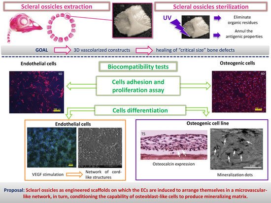
1. Introduction
2. Materials and Methods
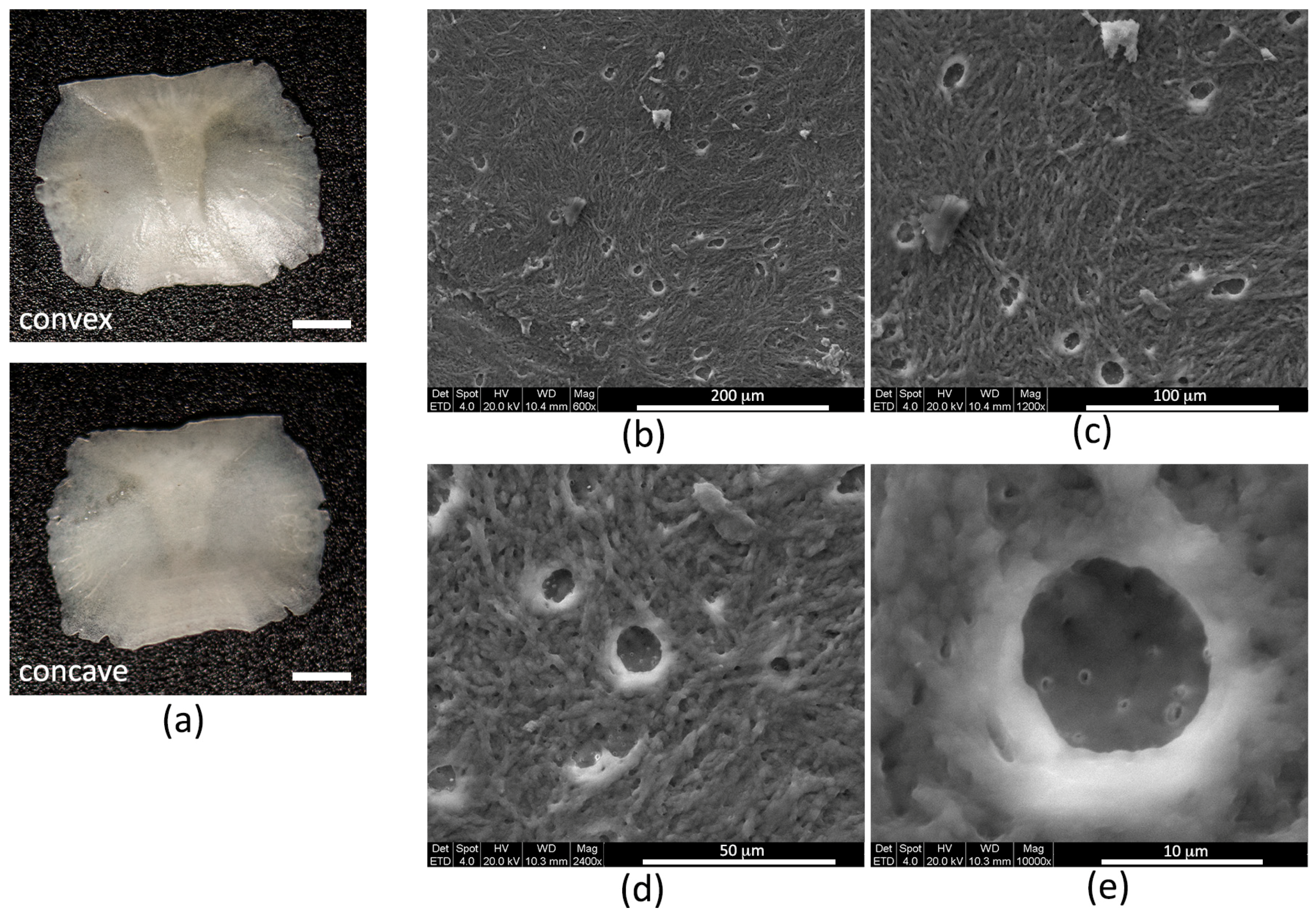
2.1. Scleral Ossicles (SOs) Preparation and Characterization
2.1.1. Extraction and Preparation of SOs
2.1.2. Size Calculation
2.1.3. Profilometry
2.1.4. Micro Computed Tomography (µCT) Analysis
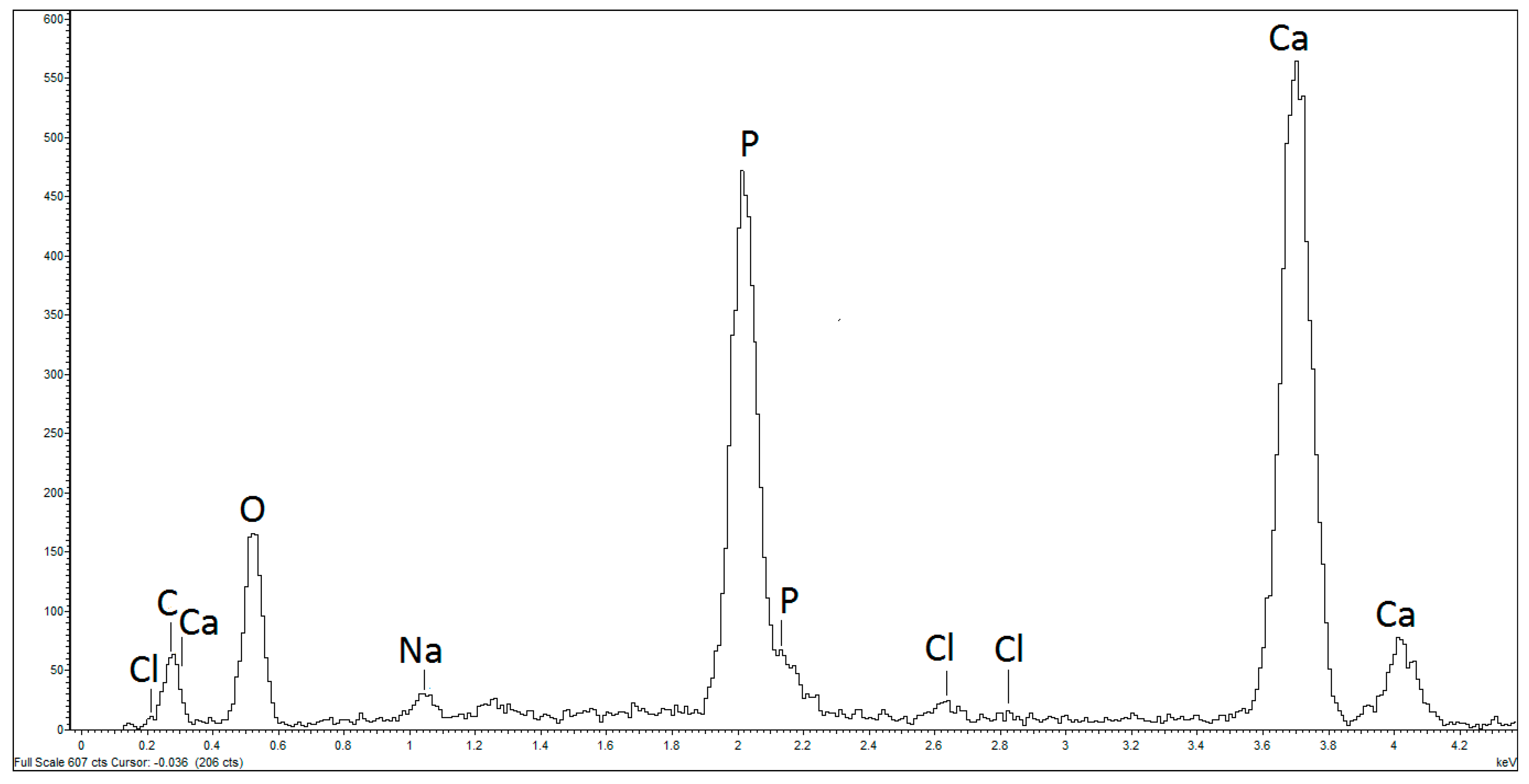
2.2. Scanning Electron Microscopy and X-ray Microanalysis
2.3. Cell Culture
2.4. Cell Differentiation
2.5. Immunofluorescence Analysis
2.6. Trypan Blue Viability Assay
2.7. MTT (3-(4,5-Dimethylthiazol-2-yl)-2,5-Diphenyltetrazolium Bromide) Viability Assay
2.8. Ultrastructural Observations
2.9. Paraffin Inclusion and Immunohistochemistry (IHC)
2.10. Statistical Analysis
3. Results
3.1. Morphological Characterization
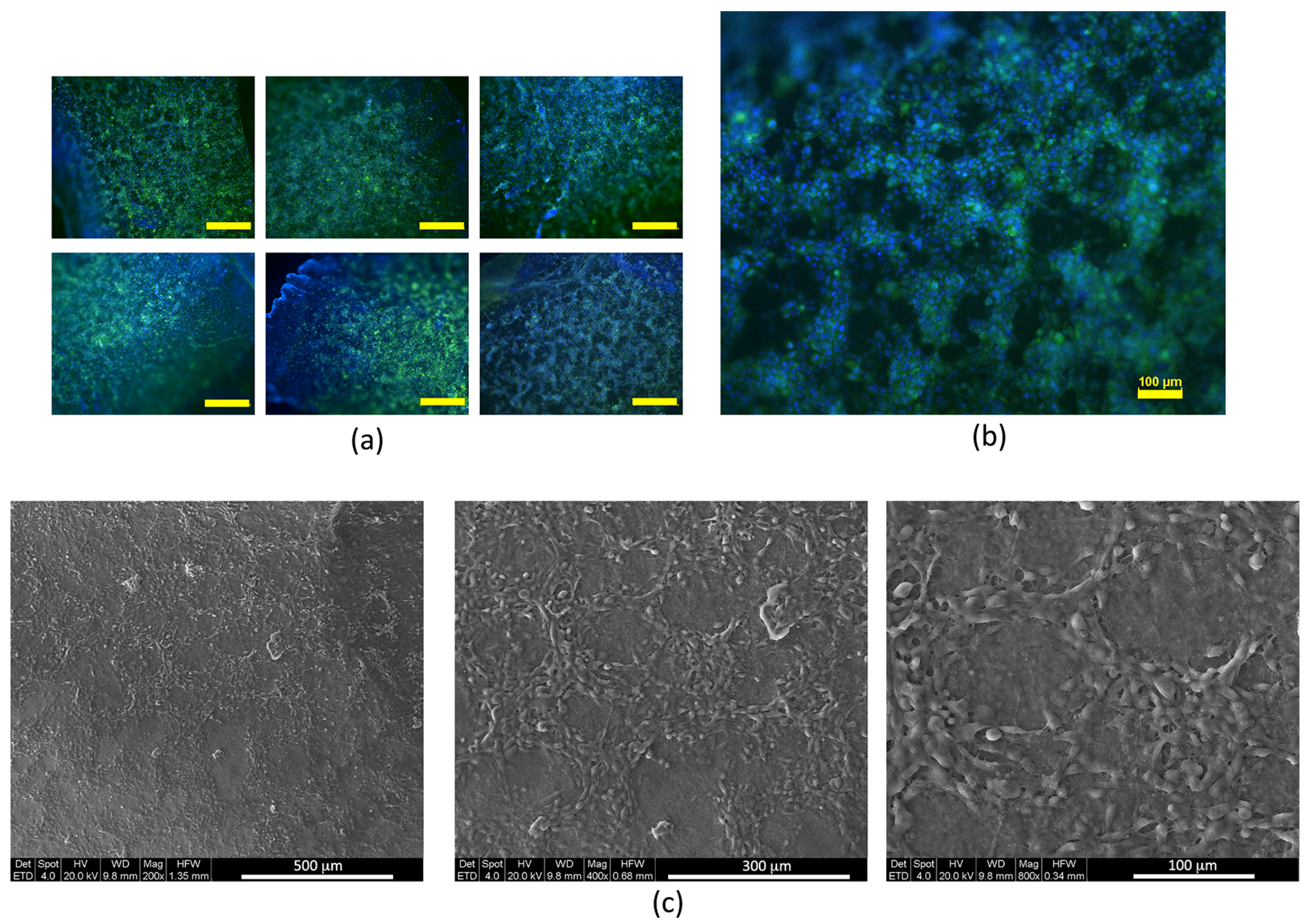
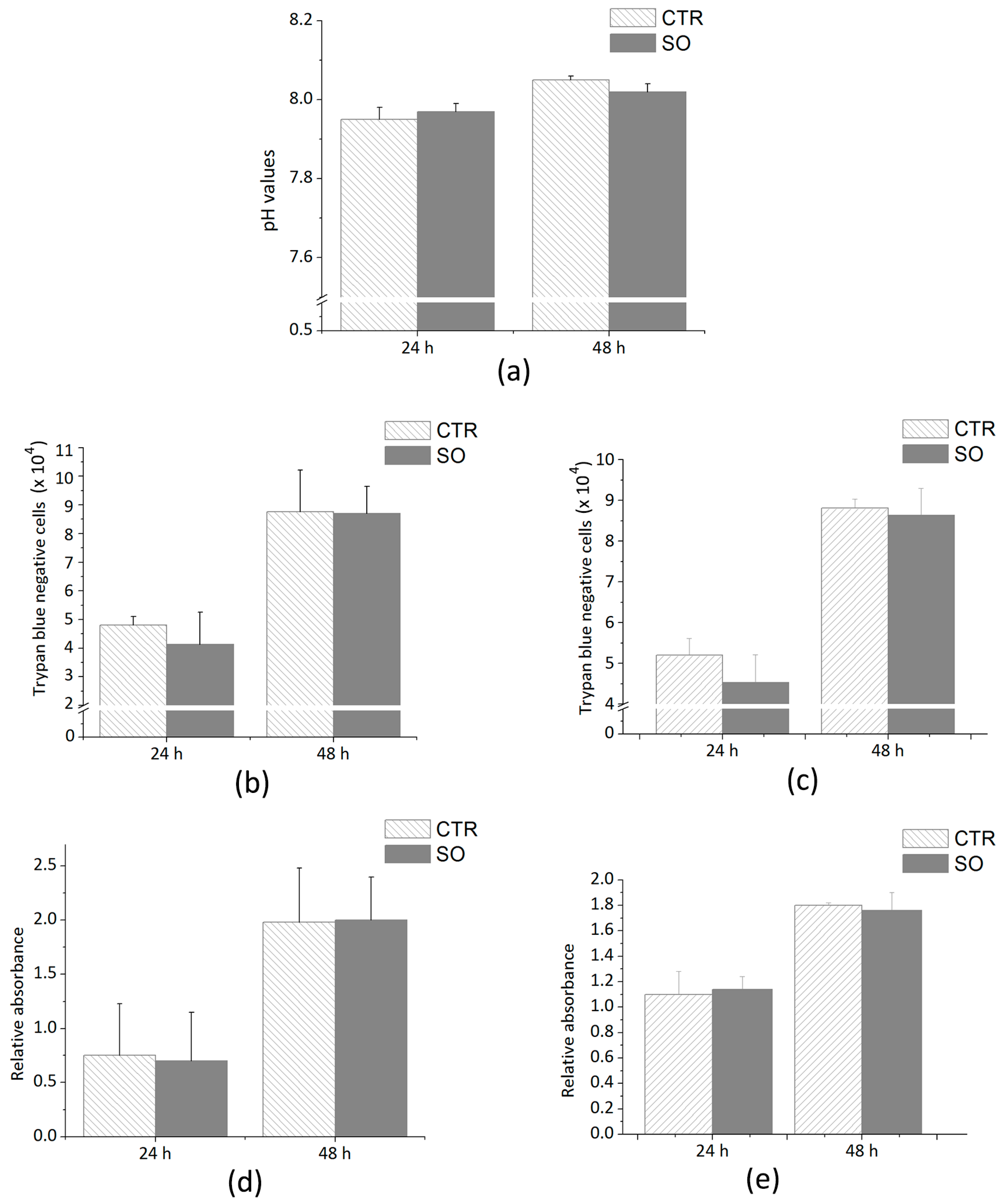
3.2. Biocompatibility Tests
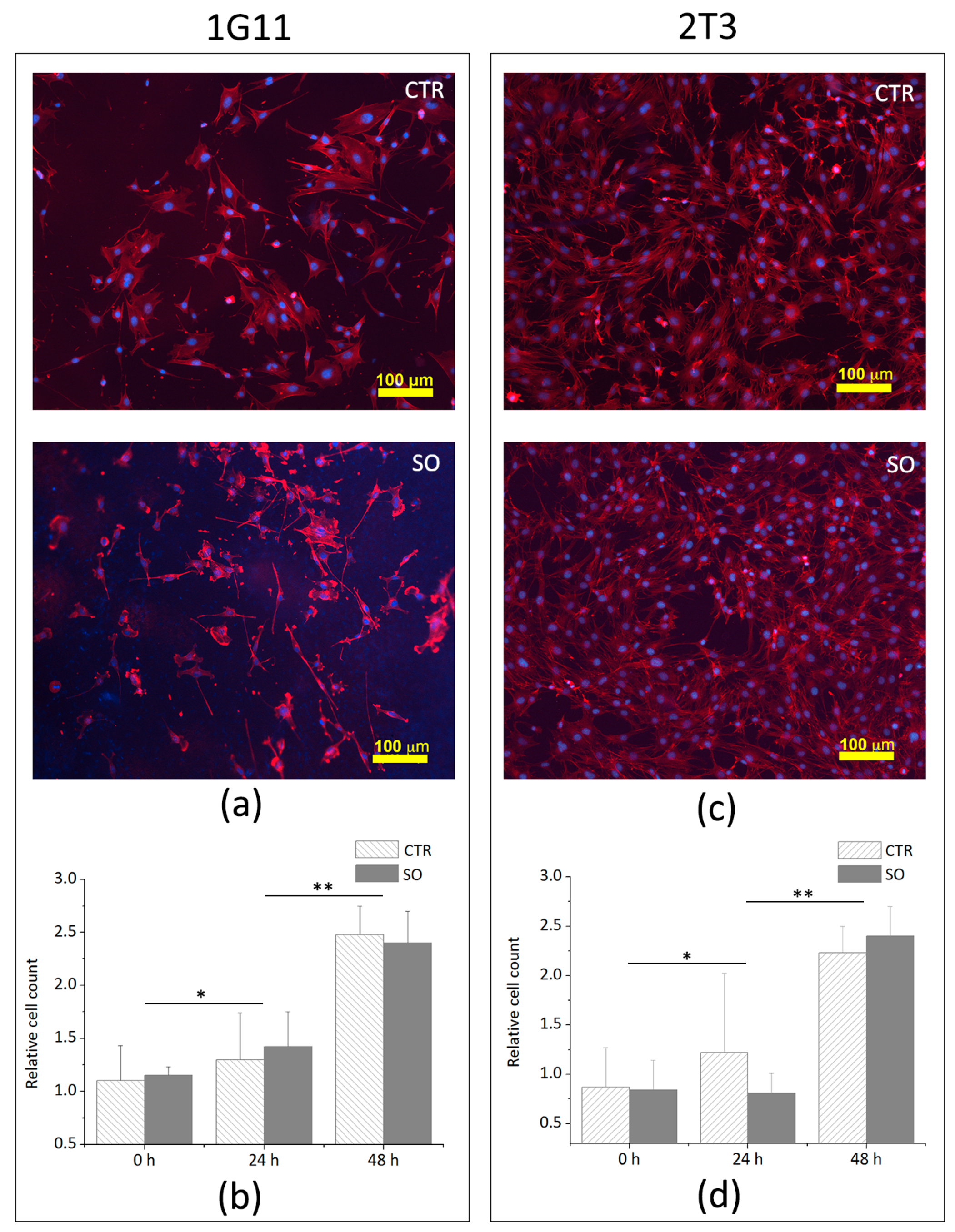
3.3. Analysis of Cells Adhesion and Proliferation
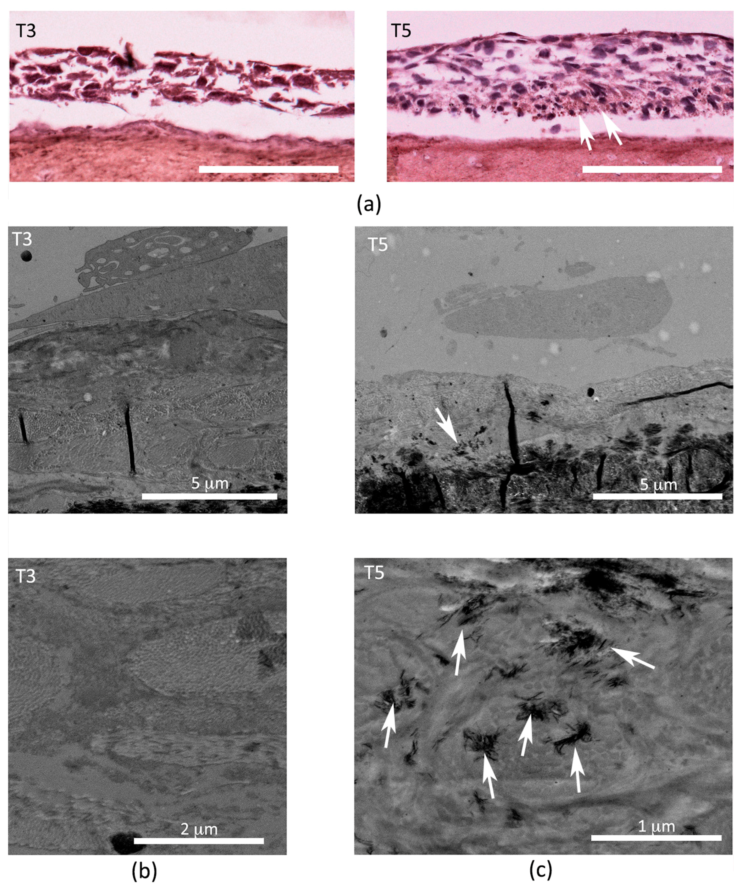
3.4. Analysis of Endothelial and Osteoblast Differentiation on SO Surfaces
3.4.1. Endothelial Differentiation
3.4.2. Osteogenic Differentiation
4. Discussion
5. Conclusions
Acknowledgments
Author Contributions
Conflicts of Interest
References
- Harris, J.S.; Bemenderfer, T.B.; Wessel, A.R.; Kacena, M.A. A review of mouse critical size defect models in weight bearing bones. Bone 2013, 55, 241–247. [Google Scholar] [CrossRef] [PubMed]
- Schroeder, J.E.; Mosheiff, R. Tissue engineering approaches for bone repair: Concepts and evidence. Injury 2011, 42, 609–613. [Google Scholar] [CrossRef] [PubMed]
- Polo-Corrales, L.; Latorre-Esteves, M.; Ramirez-Vick, J.E. Scaffold design for bone regeneration. J. Nanosci. Nanotechnol. 2014, 14, 15–56. [Google Scholar] [CrossRef] [PubMed]
- Laurencin, C.; Khan, Y.; El-Amin, S.F. Bone graft substitutes. Expert Rev. Med. Devices 2006, 3, 49–57. [Google Scholar] [CrossRef] [PubMed]
- Kusumbe, A.P.; Ramasamy, S.K.; Adams, R.H. Coupling of angiogenesis and osteogenesis by a specific vessel subtype in bone. Nature 2014, 507, 323–328. [Google Scholar] [CrossRef] [PubMed]
- Coulombre, A.J.; Coulombre, J.L. The skeleton of the eye. I. Conjunctival papillae and scleral ossicles. Dev. Biol. 1962, 5, 382–401. [Google Scholar] [CrossRef]
- Lima, F.C.; Vieira, L.G.; Santos, A.L.Q.; De Simone, S.B.S.; Hirano, L.Q.R.; Silva, J.M.M.; Romao, M.F. Anatomy of the scleral ossicles in Brazilian birds. Braz. J. Morphol. Sci. 2009, 26, 165–169. [Google Scholar]
- Warheit, K.I.; Good, D.A.; De Queiroz, K. Variation numbers of scleral ossicles and their phylogenetic transformations within the Pelecaniformes. Auk 1989, 106, 383–388. [Google Scholar]
- Palumbo, C.; Presutti, L.; Genovese, E.; Cavani, F.; Sena, P.; Benincasa, M.; Ferretti, M. Role of osteocyte apoptosis in peculiar ossicles of the hearing sense organ: Preliminary observations on hearing loss and osteoporosis. Ital. J. Anat. Embryol. 2012, 117, 143. [Google Scholar] [CrossRef]
- Palumbo, C.; Cavani, F.; Sena, P.; Benincasa, M.; Ferretti, M. Osteocyte apoptosis and absence of bone remodeling in human auditory ossicles and scleral ossicles of lower vertebrates: A mere coincidence or linked processes? Calcif. Tissue Int. 2012, 90, 211–218. [Google Scholar] [CrossRef] [PubMed]
- Schmitz, J.P.; Hollinger, J.O. The critical size defect as an experimental model for craniomandibulofacial nonunions. Clin. Orthop. Relat. Res. 1986, 299–308. [Google Scholar] [CrossRef]
- Schmitz, J.P.; Schwartz, Z.; Hollinger, J.O.; Boyan, B.D. Characterization of rat calvarial nonunion defects. Acta Anat. (Basel) 1990, 138, 185–192. [Google Scholar] [CrossRef] [PubMed]
- Cowan, C.M.; Shi, Y.-Y.; Aalami, O.O.; Chou, Y.-F.; Mari, C.; Thomas, R.; Quarto, N.; Contag, C.H.; Wu, B.; Longaker, M.T. Adipose-derived adult stromal cells heal critical-size mouse calvarial defects. Nat. Biotechnol. 2004, 22, 560–567. [Google Scholar] [CrossRef] [PubMed]
- Dong, Q.G.; Bernasconi, S.; Lostaglio, S.; De Calmanovici, R.W.; Martin-Padura, I.; Breviario, F.; Garlanda, C.; Ramponi, S.; Mantovani, A.; Vecchi, A. A General Strategy for Isolation of Endothelial Cells from Murine Tissues: Characterization of Two Endothelial Cell Lines from the Murine Lung and Subcutaneous Sponge Implants. Arterioscler. Thromb. Vasc. Biol. 1997, 17, 1599–1604. [Google Scholar] [CrossRef] [PubMed]
- Ghosh-Choudhury, N.; Windle, J.J.; Koop, B.A.; Harris, M.A.; Guerrero, D.L.; Wozney, J.M.; Mundy, G.R.; Harris, S.E. Immortalized murine osteoblasts derived from BMP 2-T-antigen expressing transgenic mice. Endocrinology 1996, 137, 331–339. [Google Scholar] [CrossRef] [PubMed]
- Guo, B.; Lei, B.; Li, P.; Ma, P.X. Functionalized scaffolds to enhance tissue regeneration. Regen. Biomater. 2015, 2, 47–57. [Google Scholar] [CrossRef] [PubMed]
- Mota, C.; Puppi, D.; Chiellini, F.; Chiellini, E. Additive manufacturing techniques for the production of tissue engineering constructs Carlos. J. Tissue Eng. Regen. Med. 2015, 9, 174–190. [Google Scholar] [CrossRef] [PubMed]
- Barabaschi, G.D.G.; Manoharan, V.; Li, Q.; Bertassoni, L.E. Engineering Pre-vascularized Scaffolds for Bone Regeneration. Adv. Exp. Med. Biol. 2015, 881, 79–94. [Google Scholar] [PubMed]
- Spicer, P.P.; Kretlow, J.D.; Young, S.; Jansen, J.A.; Kasper, F.K.; Mikos, A.G. Evaluation of bone regeneration using the rat critical size calvarial defect. Nat. Protoc. 2012, 7, 1918–1929. [Google Scholar] [CrossRef] [PubMed]
- Franz-Odendaal, T.A.; Hall, B.K.; Witten, P.E. Buried alive: How osteoblasts become osteocytes. Dev. Dyn. 2006, 235, 176–190. [Google Scholar] [CrossRef] [PubMed]
- Duench, K.; Franz-Odendaal, T.A. BMP and Hedgehog signaling during the development of scleral ossicles. Dev. Biol. 2012, 365, 251–258. [Google Scholar] [CrossRef] [PubMed]
- Jourdeuil, K.; Franz-Odendaal, T.A. Vasculogenesis and the Induction of Skeletogenic Condensations in the Avian Eye. Anat. Rec. 2012, 295, 691–698. [Google Scholar] [CrossRef] [PubMed]
- Jabalee, J.; Hillier, S.; Franz-Odendaal, T.A. An investigation of cellular dynamics during the development of intramembranous bones: The scleral ossicles. J. Anat. 2013, 223, 311–320. [Google Scholar] [CrossRef] [PubMed]
- Jabalee, J.; Franz-Odendaal, T.A. Vascular endothelial growth factor signaling affects both angiogenesis and osteogenesis during the development of scleral ossicles. Dev. Biol. 2015, 406, 52–62. [Google Scholar] [CrossRef] [PubMed]
- Franz-Odendaal, T.A. Toward understanding the development of scleral ossicles in the chicken, Gallus gallus. Dev. Dyn. 2008, 237, 3240–3251. [Google Scholar] [CrossRef] [PubMed]
- Edinger, T. Uber knocherne Scleralringe. Zool. Jahrb. Abt. Anat. 1929, 51, 163–226. [Google Scholar]
- Martin, G.R. Form and Function in the Optical Structure of Bird Eyes. In Perception and Motor Control in Birds; Academic Press: London, UK, 1985; pp. 311–373. [Google Scholar]
- Romer, A.S. Osteology of the Reptiles; University of Chicago Press: Chicago, IL, USA, 1956. [Google Scholar]
- Palumbo, C.; Ferretti, M.; Ardizzoni, A.; Zaffe, D.; Marotti, G. Osteocyte-osteoclast morphological relationships and the putative role of osteocytes in bone remodeling. J. Musculoskelet. Neuronal Interact. 2001, 1, 327–332. [Google Scholar] [PubMed]
- Dallas, S.L.; Prideaux, M.; Bonewald, L.F. The osteocyte: An endocrine cell … and more. Endocr. Rev. 2013, 34, 658–690. [Google Scholar] [CrossRef] [PubMed]
- Marotti, G.; Palumbo, C. The mechanism of transduction of mechanical strains into biological signals at the bone cellular level. Eur. J. Histochem. 2007, 51, 15–20. [Google Scholar] [PubMed]
- Viñals, F.; Pouysségur, J. Transforming Growth Factor β1 (TGF-β1) Promotes Endothelial Cell Survival during In Vitro Angiogenesis via an Autocrine Mechanism Implicating TGF-α Signaling. Mol. Cell. Biol. 2001, 21, 7218–7230. [Google Scholar] [CrossRef] [PubMed]
- Gonzalvo-Feo, S.; Del Prete, A.; Pruenster, M.; Salvi, V.; Wang, L.; Sironi, M.; Bierschenk, S.; Sperandio, M.; Vecchi, A.; Sozzani, S. Endothelial Cell-Derived Chemerin Promotes Dendritic Cell Transmigration. J. Immunol. 2014, 192, 2366–2373. [Google Scholar] [CrossRef] [PubMed]
- Cellier, E.; Midaoui, A.E.; Robitaille, G.A.; Richard, D.E.; Lariviere, R.; Lebel, M. Effects of TGF-beta1 on endothelial factors. Arch. Physiol. Biochem. 2010, 116, 50–55. [Google Scholar] [CrossRef] [PubMed]
- Zhang, R.; Oyajobi, B.O.; Harris, S.E.; Chen, D.; Tsao, C.; Deng, H.W.; Zhao, M. Wnt/β-catenin signaling activates bone morphogenetic protein 2 expression in osteoblasts. Bone 2013, 52, 145–156. [Google Scholar] [CrossRef] [PubMed]
- Liu, H.; Zhang, R.; Chen, D.; Oyajobi, B.O.; Zhao, M. Functional redundancy of type II BMP receptor and type IIB activin receptor in BMP2-induced osteoblast differentiation. J. Cell. Physiol. 2012, 227, 952–963. [Google Scholar] [CrossRef] [PubMed]
- Mandal, C.C.; Ganapathy, S.; Gorin, Y.; Mahadev, K.; Block, K.; Abboud, H.E.; Harris, S.E.; Ghosh-Choudhury, G.; Ghosh-Choudhury, N. Reactive oxygen species derived from Nox4 mediate BMP2 gene transcription and osteoblast differentiation Chandi. Biochem. J. 2011, 433, 393–402. [Google Scholar] [CrossRef] [PubMed]
- Lazzeri, L.; Cascone, M.G.; Danti, S.; Serino, L.P.; Moscato, S.; Bernardini, N. Gelatine/PLLA sponge-like scaffolds: Morphological and biological characterization. J. Mater. Sci. Mater. Med. 2006, 17, 1211–1217. [Google Scholar] [CrossRef] [PubMed]
- Mattii, L.; Battolla, B.; D’Alessandro, D.; Trombi, L.; Pacini, S.; Cascone, M.G.; Lazzeri, L.; Bernardini, N.; Dolfi, A.; Galimberti, S.; et al. Gelatin/PLLA sponge-like scaffolds allow proliferation and osteogenic differentiation of human mesenchymal stromal cells. Macromol. Biosci. 2008, 8, 819–826. [Google Scholar] [CrossRef] [PubMed]
- Trombi, L.; D’Alessandro, D.; Pacini, S.; Fiorentino, B.; Scarpellini, M.; Fazzi, R.; Galimberti, S.; Guazzini, S.; Petrini, M. Good manufacturing practice-grade fibrin gel is useful as a scaffold for human mesenchymal stromal cells and supports in vitro osteogenic differentiation. Transfusion 2008, 48, 2246–2251. [Google Scholar] [CrossRef] [PubMed]
- Trombi, L.; Mattii, L.; Pacini, S.; D’alessandro, D.; Battolla, B.; Orciuolo, E.; Buda, G.; Fazzi, R.; Galimberti, S.; Petrini, M. Human Autologous Plasma-Derived Clot as a Biological Scaffold for Mesenchymal Stem Cells in Treatment of Orthopedic Healing. J. Orthop. Res. 2007, 26, 176–183. [Google Scholar] [CrossRef] [PubMed]
- Danti, S.; Stefanini, C.; D’Alessandro, D.; Moscato, S.; Pietrabissa, A.; Petrini, M.; Berrettini, S. Novel biological/biohybrid prostheses for the ossicular chain: Fabrication feasibility and preliminary functional characterization. Biomed. Microdevices 2009, 11, 783–793. [Google Scholar] [CrossRef] [PubMed]
- Mercado-Pagàn, A.E.; Stahl, A.M.; Shanjani, Y.; Yang, Y. Vascularization in bone tissue engineering constructs. Ann. Biomed. Eng. 2016, 36, 1011–1014. [Google Scholar] [CrossRef] [PubMed]
- Checchi, M.; Smargiassi, A.; Ferretti, M.; Sena, P.; Benincasa, M.; Cavani, F.; Ranieri, A.; Mitola, S.; Palumbo, C. Preliminary observations on scleral ossicles in performing functionalized 3D vascularized scaffolds for “critical-size” bone defect healing. Ital. J. Anat. Embryol. 2016, 121, 168. [Google Scholar] [CrossRef]
- Checchi, M.; Grisendi, G.; Bertacchini, J.; Magarò, M.S.; Ferretti, M.; Benincasa, M.; Sena, P.; Cavani, F.; Palumbo, C. Scleral ossicles as natural biomaterials on which vascular-like network is promoted from Mouse Aortic Endothelial cells ( MAECs ): Preliminary results. Ital. J. Anat. Embryol. 2017, 122, 60. [Google Scholar] [CrossRef]
- Kuss, M.A.; Wu, S.; Wang, Y.; Untrauer, J.B.; Li, W.; Lim, J.Y.; Duan, B. Prevascularization of 3D printed bone scaffolds by bioactive hydrogels and cell co-culture. J. Biomed. Mater. Res. Part B Appl. Biomater. 2017. [Google Scholar] [CrossRef] [PubMed]
- Zhang, S.; Zhou, M.; Ye, Z.; Zhou, Y.; Tan, W.S. Fabrication of viable and functional pre-vascularized modular bone tissues by coculturing MSCs and HUVECs on microcarriers in spinner flasks. Biotechnol. J. 2017, 12. [Google Scholar] [CrossRef] [PubMed]


| Size of SOs (mm) | Lacuno-Canalicular Size (µm) | ||
| Length | 3.886 ± 0.326 | Lacunae | 12.790 ± 3.720 |
| Width | 3.011 ± 0.334 | Canaliculi | 0.503 ± 0.222 |
| Thickness | 0.16 ± 0.04 | ||
| Roughness Ra (µm) | µCT Analysis | ||
| 1.05 ± 0.50 | V (mm3) | 1.15 ± 0.31 | |
| S (mm2) | 26.95 ± 3.65 | ||
| St.Th (mm) | 0.13 ± 0.02 | ||
| C | O | Na | Cl | Ca | P |
|---|---|---|---|---|---|
| 40.05 ± 3.33 | 36.28 ± 1.60 | 0.44 ± 0.07 | 0.06 ± 0.03 | 15.48 ± 2.13 | 7.67 ± 0.68 |
© 2017 by the authors. Licensee MDPI, Basel, Switzerland. This article is an open access article distributed under the terms and conditions of the Creative Commons Attribution (CC BY) license (http://creativecommons.org/licenses/by/4.0/).
Share and Cite
Checchi, M.; Bertacchini, J.; Grisendi, G.; Smargiassi, A.; Sola, A.; Messori, M.; Palumbo, C. Proposal of a Novel Natural Biomaterial, the Scleral Ossicle, for the Development of Vascularized Bone Tissue In Vitro. Biomedicines 2018, 6, 3. https://doi.org/10.3390/biomedicines6010003
Checchi M, Bertacchini J, Grisendi G, Smargiassi A, Sola A, Messori M, Palumbo C. Proposal of a Novel Natural Biomaterial, the Scleral Ossicle, for the Development of Vascularized Bone Tissue In Vitro. Biomedicines. 2018; 6(1):3. https://doi.org/10.3390/biomedicines6010003
Chicago/Turabian StyleChecchi, Marta, Jessika Bertacchini, Giulia Grisendi, Alberto Smargiassi, Antonella Sola, Massimo Messori, and Carla Palumbo. 2018. "Proposal of a Novel Natural Biomaterial, the Scleral Ossicle, for the Development of Vascularized Bone Tissue In Vitro" Biomedicines 6, no. 1: 3. https://doi.org/10.3390/biomedicines6010003