Technical Note

A Low-Cost Label-Free AFB1 Impedimetric Immunosensor Based on Functionalized CD-Trodes

by
Marcos Vinicius Foguel
1,*,
Gabriela Furlan Giordano
1,
Célia Maria De Sylos
2,
Iracilda Zeppone Carlos
3,
Antonio Aparecido Pupim Ferreira
1,
Assis Vicente Benedetti
1 and
Hideko Yamanaka
1,*
1
Department of Analytical Chemistry, Institute of Chemistry, UNESP-Univ Estadual Paulista, Rua Francisco Degni, 55, Quitandinha, 14800-060 Araraquara, SP, Brazil
2
Department of Food and Nutrition, School of Pharmaceutical Sciences, UNESP-Univ Estadual Paulista, Rodovia Araraquara Jaú, Km 01, s/n, Campos Ville, 14800-903 Araraquara, SP, Brazil
3
Department of Clinical Analysis, School of Pharmaceutical Sciences, UNESP-Univ Estadual Paulista, Rodovia Araraquara Jaú, Km 01, s/n, Campos Ville, 14800-903 Araraquara, SP, Brazil
*
Authors to whom correspondence should be addressed.
Chemosensors 2016, 4(3), 17; https://doi.org/10.3390/chemosensors4030017
Submission received: 29 April 2016 / Revised: 22 June 2016 / Accepted: 19 August 2016 / Published: 1 September 2016
(This article belongs to the Special Issue Electrochemical Immunosensors and Aptasensors)

Abstract

:
This work describes the investigation of a label-free immunosensor for the detection of aflatoxin B1 (AFB1). CD-trodes (electrodes obtained from recordable compact disks) were used as low-cost and disposable transducers after modification with a self-assembled monolayer (SAM) of lipoic acid. The anti-aflatoxin B1 antibody was immobilized via EDC/NHS activation, followed by blocking with bovine serum albumin and immunoassays with AFB1. The optimization of analytical parameters and the detection were carried out using electrochemical impedance measurements. Using chemometric tools, the best conditions for the immunosensor development were defined as: anti-AFB1 antibody at 1:2000 dilution and surface blocking with 0.5% bovine serum albumin, both incubated for 1 h, and antibody–antigen immunoreaction for 30 min. The impedimetric immunosensor showed a linear range from 5 × 10⁻⁹ to 1 × 10⁻⁷ mol·L−1 (1.56–31.2 ng·mL−1), limit of detection and limit of quantification, respectively, 3.6 × 10⁻¹⁰ and 1.1 × 10⁻⁹mol·L−1 (0.11 and 0.34 ng·mL−1). The proposed immunosensor was applied to analyze peanut samples.

Graphical Abstract

1. Introduction

Aflatoxin B1 (AFB1) is a toxic metabolite produced by fungi Aspergillus flavus and A. parasiticus that is characterized by acute toxicity, teratogenicity, mutagenicity, and carcinogenicity [1]. The acute effects are observed mainly in the liver; they can result in necrosis, hemorrhage, injury, fibrosis, cirrhosis, and cancer. This toxin infects a wide range of agricultural products [2], especially peanuts, corn, wheat, rice, nuts, dried fruits, among others.
The detection of aflatoxin in food and feed is usually performed by instrumental methodologies based on synchronous fluorescence spectrometry [3], high-performance liquid chromatography with amperometric detection [4], fluorescence detection [5], thin layer chromatography [6], or immunochromatographic assay [7]. An alternative is offered by the use of immunosensors, due to their sensibility, stability, and ease of handling. In this case, the antibodies are immobilized on an electrode and must maintain their biological activity on the transducer [8]. One of the procedures is based on the formation of a self-assembled monolayer (SAM) on the electrode surface [9], which consists of the interaction of a highly-organized organic molecule layer on the surface and one of the functional groups of the selected organic molecule having the function of providing the biological material’s immobilization (e.g., enzymes, proteins, nucleic acid, antibody, etc.) via the free functional group of SAM [10]. Organic monolayers with a sulfur group on the electrode surface are of great interest, because the sulfur binds strongly to the gold surface and the reactive functional group is maintained free for the immobilization of biological molecules [11]. Surface Plasmon resonance devices [12], conductometric [13], fluorometric [14], and amperometric [15,16] biosensors indicated good performance. Related to the impedimetric immunosensor for AFB1, the literature registered the immobilization of antibody on Pt, glassy carbon, and gold electrode, as displayed in Table 1.
This work reports the development of an impedimetric immunosensor for the determination of aflatoxin B1, through gold CD-trode (electrode obtained from recordable compact disks) surface modification with a self-assembled monolayer (SAM) of lipoic acid activated via EDC (1-ethyl-3-(3-(dimethylamino)-propyl)carbodiimide)/NHS (N-hydroxy succinimide) for the immobilization of anti-aflatoxin B1 antibody. CD-trodes can be easily obtained by simple treatment of wasted gold CDs to obtain cheap but efficient electrochemical sensors [26,27]. The optimization of the experimental parameters involved in the development of the CD-trode sensor was performed chemometrically, by means of full factorial design. The proposed biosensor was applied to determine the antigen in peanut samples.

2. Materials and Methods

2.1. Reagents

69%–70% HNO3, 95%–98% H2SO4, methanol, and chloroform were purchased from J. T. Baker (Phillipsburg, NJ, USA). 1-ethyl-3-(3-(dimethylamino)-propyl)carbodiimide (EDC) was obtained from Fluka (Buchs, Switzerland). Lipoic acid (C8H14O2S2), N-hydroxy succinimide (NHS), aflatoxin B1 (AFB1) from Aspergillus flavus, anti-AFB1 antibody from rabbit, bovine serum albumin (BSA), NaH2PO4·H2O, Na2HPO4, K3Fe(CN)6, K4Fe(CN)6, CuSO4·5H2O, and KCl were purchased from Sigma (St. Louis, MO, USA). Ultra-pure water (resistivity 18.2 MΩ·cm) was used to prepare the solutions.

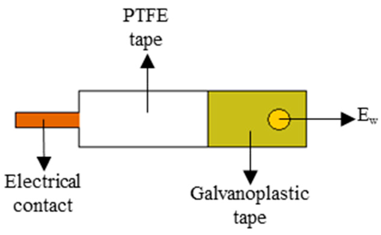
2.2. Apparatus and Electrochemical Cell

The electrochemical measurements were carried out using a potentiostat/galvanostat AUTOLAB PGSTAT 302 with impedance module FRA 2 (frequency response analyzer) and software version 4.9.005. The experiments were carried out using a one-compartment electrochemical cell with a volume of 5 mL, and a three-electrode system: gold CD-trode (Ageom = 0.071 cm2 and active area of 0.091 cm2, estimated from by Randles–Sevik equation [28]), Ag|AgCl|KCl(sat) and platinum wire (Ageom = 4 cm2) as work, reference, and auxiliary electrodes, respectively.

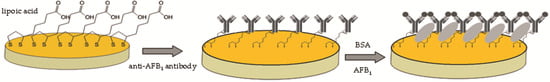
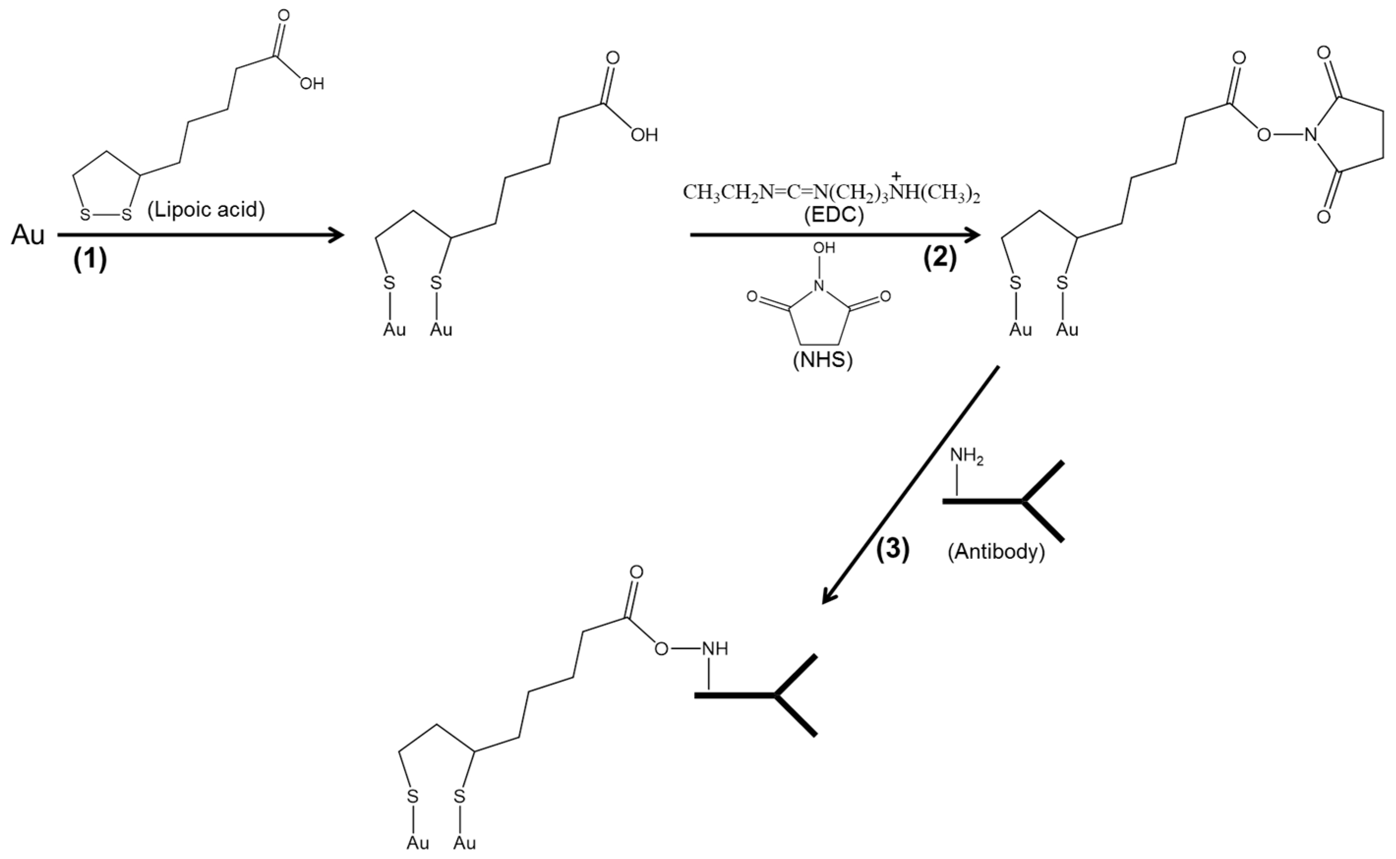
2.3. Construction of Gold Electrode (AuCD-Trode)

The working electrode was constructed from a recordable compact disc (Mitsui Archive Gold CD-R 100) containing a gold film with thickness between 50 and 100 nm, using a previously reported procedure [26]. Briefly, to access the metal layer of the CD-R, concentrated HNO3 was added to the surface; after 5–10 min, the protective layers were totally removed and the gold surface was washed thoroughly with distilled water [26]. The CD-R was cut and the working area of the electrode (Ew) was pre-set with perforated galvanoplastic tape. The electrical contact of the CD-trode was a laminated copper wire fixed and insulated with polytetrafluoroethylene (PTFE) tape. The characterization of the AuCD-trode as an electrochemical transducer was done previously [27]. CD-trodes are disposable, so they were used only once. A scheme of CD-trode is shown in Figure 1.

2.4. Immunosensor Development

Scheme 1 shows the schemes of the different steps of modification of CD-trode to obtain the impedimetric immunosensor [29,30].
The first step in the development of an impedimetric immunosensor (Scheme 1-1) is the addition of 10 µL of 1 × 10⁻³ mol·L−1 lipoic acid prepared in ethanol/water solution (1:10) to the gold CD-trode surface and incubation for 2 h to form a SAM [31]. According to literature, both thiol and disulfide groups interact with gold [11]. For the immobilization of biological material on the SAM, it was necessary to activate the carboxyl group of the lipoic acid with 10 µL of 0.4/0.1 mmol·L−1 EDC/NHS prepared in deionized water that was added on the surface of the modified electrode, and the incubation time was 60 min (Scheme 1-2) [32]. The immobilization of the anti-AFB1 antibody (Scheme 1-3) was performed by adding 10 µL of antibody solution prepared in 0.01 mol·L−1 phosphate buffer solution (pH 7.4) with different concentrations and incubation times on the modified electrode. In order to avoid unspecific interaction, the electrode surface was blocked with 10 µL of 0.5% bovine serum albumin (BSA) prepared in 0.01 mol·L−1 phosphate buffer solution (pH 7.4) containing tween 20 (PBS-T) for 60 min. Finally, 10 µL of aflatoxin B1, prepared in 0.5% BSA solution, was added on the immunosensor surface and incubated for 30 min. The incubation steps were performed at 25 °C. After each incubation time at different steps of modification, the electrode was washed by immersion in ultra-pure water three times for 10 s under stirring.
The analysis of factorial designs results was conducted with statistical and graphical analysis software MINITAB® Release 15, developed by Minitab Inc., State College, PA, USA. All experiments were carried out in triplicate and the reproducibility was evaluated statistically by the MINITAB® software.
After the construction of the immunosensor, a standard solution of AFB1—prepared in 0.01 mol·L−1 phosphate buffer solution at pH 7.4 containing 0.5% BSA—was added to the electrode, and the affinity reaction was monitored.

2.5. Electrochemical Measurements

In order to clean and homogenize the surface, the CD-trodes were submitted to pretreatments with 10 voltammetric cycles in 0.5 mol·L−1 sulfuric acid in the potential range between +0.2 and +1.5 V at 100 mV·s−1.
The impedance spectra were obtained by applying a sine wave of 10 mV (rms) on Eocp from 100 kHz to 100 mHz and recording 10 points/frequency decade. Measurements were performed in 0.1 mol·L−1 phosphate buffer solution pH 7.0 containing 1.0 × 10⁻³ mol·L−1 Fe(CN)63−/4− redox pair. All electrochemical measurements were done in triplicate at 25 ± 2 °C in a Faraday cage. The value of charge transfer resistance (Rct), calculated from the Nyquist plot, was used as parameter related to the response of the immunosensor. These values were obtained for each experiment. The real impedance (Zre) of the frequency in the maximum of the semicircle was taken; it is the solution resistance (RΩ) plus half of the charge transfer resistance. Therefore, the Rct can be defined by Equation (1) [33].
Rct = 2 Zre − 2 RΩ

2.6. Extraction of AFB1 from Peanut Samples

The immunosensor was applied to AFB1 analysis in peanut samples. The toxin extraction from the sample was carried out as follows: 50 g of ground raw peanuts were mixed with 270 mL of methanol and 30 mL of 4% KCl in a blender for 5 min, followed by filtration through a qualitative filter paper. Then, the filtrate was mixed with 150 mL of 10% CuSO4·5H2O and celite for 5 min, and then filtered again on filter paper. The filtrate was mixed with water (1:1), and the aflatoxin extracted with 10 mL of chloroform. Another aliquot of chloroform was added to the aqueous solution. The organic phases were mixed and dried in a water bath at 40 °C [34]. The dried aliquots were re-suspended with 1.0 mL methanol and diluted in 0.01 mol·L−1 phosphate buffer solution (pH 7.4, containing 0.5% BSA) and analyzed.

3. Results and Discussion

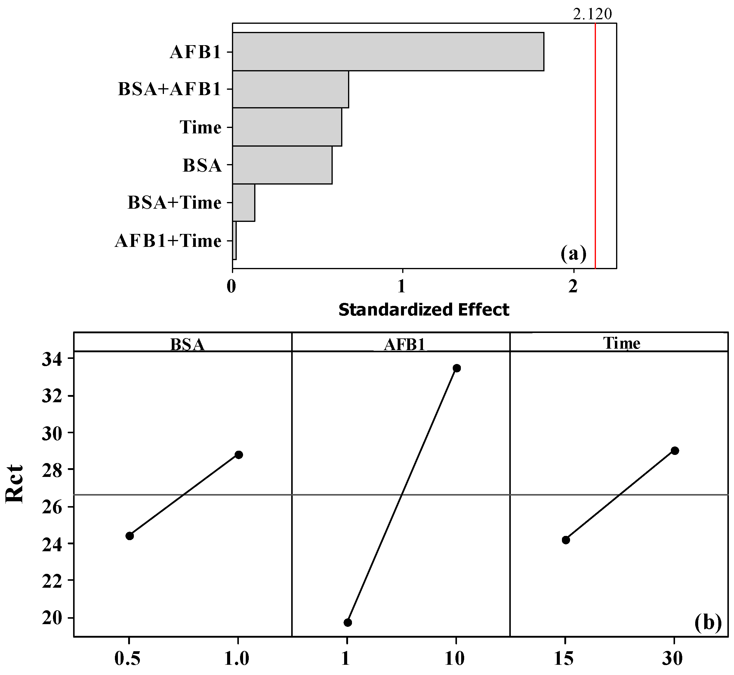
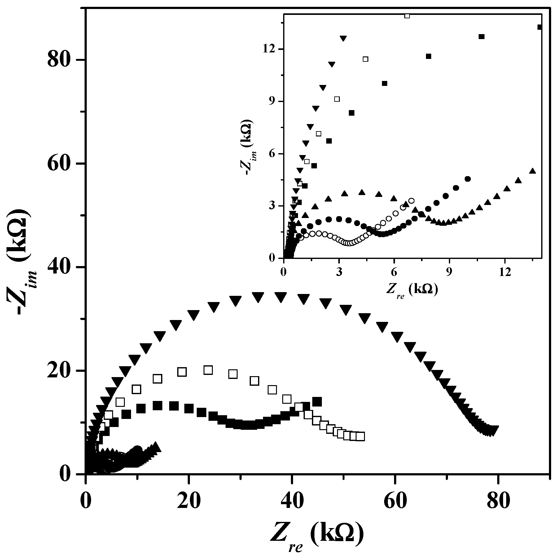
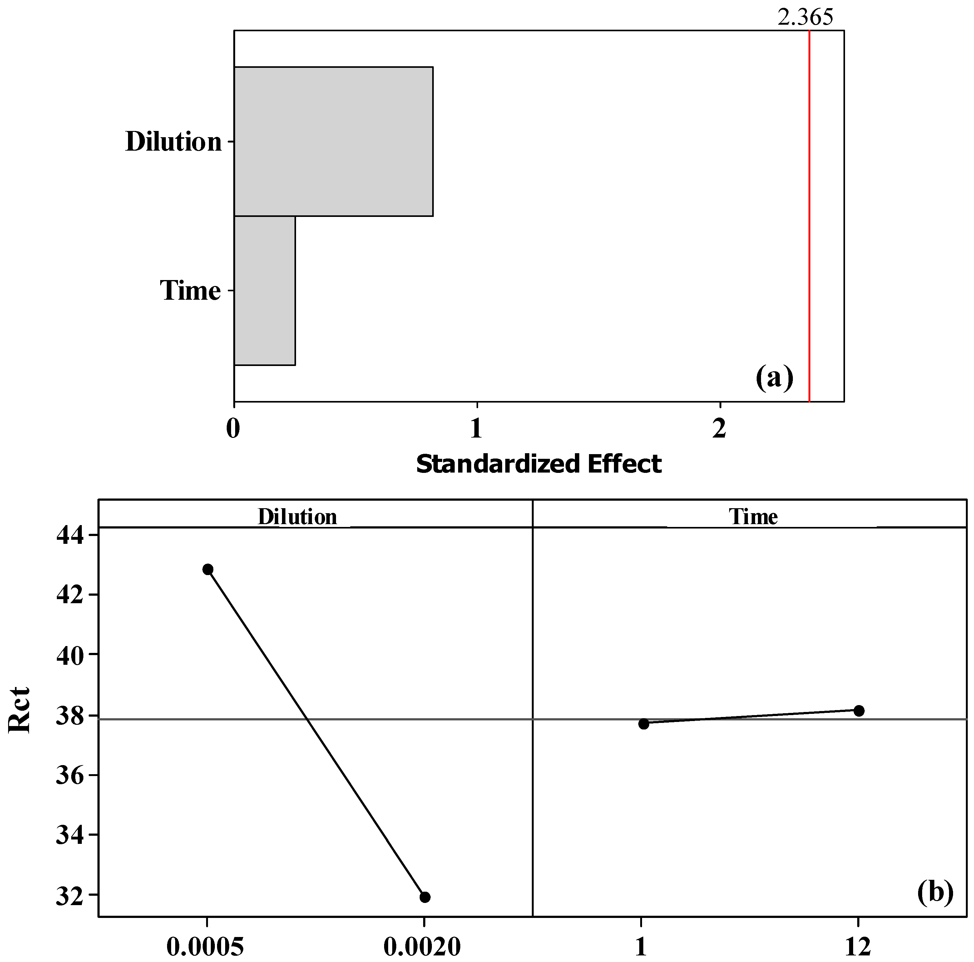
3.1. Chemometric Study of Antibody, BSA, and AFB1 Concentrations and Incubation Time

The first step in the development of the immunosensor concerned the optimization of the concentration of anti-AFB1 (1:2000 and 1:500, or 0.0017 and 0.0067 µg·µL−1) and its incubation time (1 and 12 h). When the antibodies were incubated for 1 or 12 h, the results indicated almost the same value of Rct, and the capacitive circle presented no inductive loop [35]. This means that one hour of incubation is enough.
The concentration of the anti-AFB1 antibody and incubation time optimization were performed by full factorial design of type 22. The Pareto plot in Figure 2a shows that the most important parameter is the antibody dilution (DAb), while the incubation time of the solution (tinc) and interactions between factors (DAb and tinc) on the modified CD-trode do not have great influence on the impedance measurements. The influences of the high and low levels of each variable were also determined. The lines shown in Figure 2b express the trend of the Rct values when changing the parameter from low to high level. The line with higher slope indicates the most influential parameter of the system; thus, the dilution presents the greater influence to the system. The 1:2000 dilution of the antibody with 1 h of incubation tended to result in higher impedance value.
The optimization of bovine serum albumin and aflatoxin B1 concentration and incubation time of antigen were investigated by means of full factorial design of type 23. In Figure 3a, the Pareto plot shows that the most important parameter is the AFB1 concentration, followed by incubation time of the antigen. The BSA concentration and interactions between the factors have little influence on the response of the system. The plot in Figure 3b indicates that at high AFB1 concentration, Rct is higher when the incubation time is 30 min, because these were the parameters that tended to higher values. As the variation of the BSA concentration does not present influence, the 0.5% concentration was adopted. To evaluate the incubation time of BSA, different times were studied: 30, 40, and 60 min (data not shown). The Rct values for 30 and 40 min of incubation were quite similar to the Rct of the previous modification step (Au-SAM-Ab). However, with 60 min of incubation, the Rct value was higher than the previous step, and the reproducibility of the measurements was better. Thereby, the incubation time of 60 min was used for the blocking step.
Figure 4 presents the Nyquist diagram for the various steps of the construction of the optimized immunosensor: SAM layer, active SAM layer, antibody layer, blocking with BSA, and interaction of antigen with the immunosensor. The figures indicate that each layer deposition on the immunosensor surface increases the impedance of the system. This increase is due to the changes of the electrical characteristics of the gold/electrolyte interface.

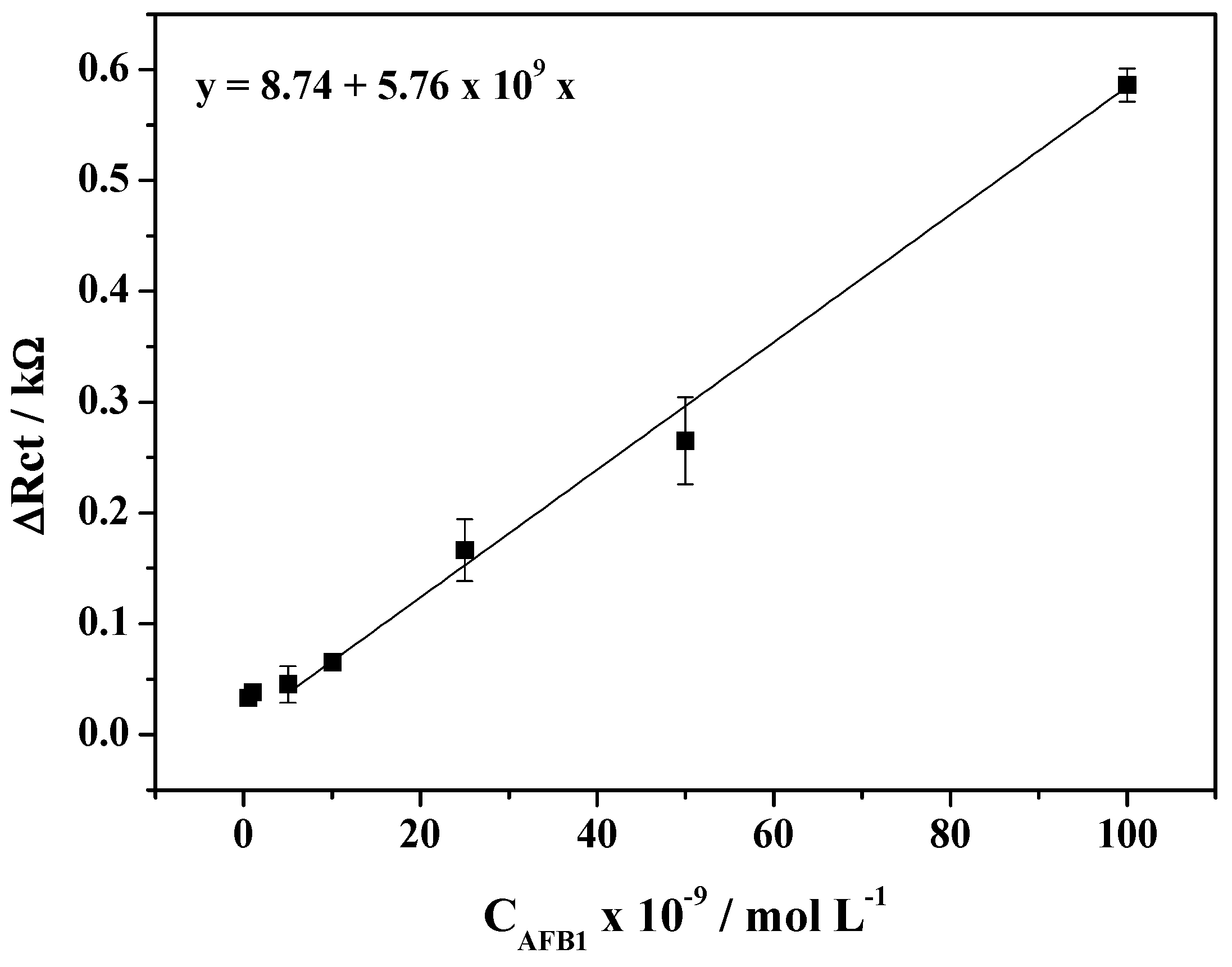
3.2. Analytical Curve

After optimization of all steps for the development of the impedimetric immunosensor, an analytical curve was constructed from 5.0 × 10⁻⁹ to 1.0 × 10⁻⁷ mol·L−1 (1.56–31.2 ng·mL−1) as shown in Figure 5. The curve presented a linear range with a correlation coefficient of 0.99858, and the limits of detection and quantification of 3.6 × 10⁻¹⁰ and 1.1 × 10⁻⁹ mol·L−1 (0.11 and 0.34 ng·mL−1), respectively.

3.3. Application in Peanut Samples

Impedimetric immunosensors were applied to three different samples of raw peanuts, provided by a food industry that performs quality analysis to verify the eventual contamination of the products with aflatoxin. According to the supplier industry, two of these samples were contaminated with AFB1, and in the third one the concentration level was unknown, but below the limit established by the Brazilian National Agency of Sanitary Surveillance (20 µg of total aflatoxin in 1 kg of peanut).
Table 2 shows the AFB1 concentration for each sample, analyzed by interpolating in the analytical curve. Samples A and B presented levels around three times above the limit set by Brazilian legislation. In sample C, a concentration of 25 µg·kg−1 was detected by the sensor, which is slightly above the legal limit. This contrasts the datum determined previously by the producer, using classical analytical procedures. This could indicate a better sensitivity of the sensor with respect to standard methods, however we cannot exclude a possible growth of Aspergillus sp. and subsequent production of aflatoxin during storage, because of the long time interval elapsed between the two analyses (around 7 months).

4. Conclusions

On the proposed methodology, the dynamic range is 0.16 to 3 ng·mL−1 and LOD 0.35 ng·mL−1. In comparison with data reported in previous literature, displayed in Table 1, the performances of the CD-trode sensor are comparable with those obtained with some of the immunosensors previously described in the literature [20,22,25], but with the advantage of being based on the use of disposable and low-cost CD-trodes. These preliminary results are encouraging in order to progress with an in-depth validation of the sensor, in particular concerning matrix effects and recovery tests.

Acknowledgments

The authors thank FAPESP (Proc. 2008/07729-8), FP7–PEOPLE–IRSES (Proc. 2008-230849) and CNPq (Proc. 309275/2012-1) for financial support.

Conflicts of Interest

The authors declare no conflict of interest.

Abbreviations

The following abbreviations were used in this manuscript:
AFB1Aflatoxin B1
CD-trodeElectrode obtained from recordable compact disk
EDC1-Ethyl-3-(3-(dimethylamino)-propyl)carbodiimide
NHSN-hydroxy succinimide
SAMSelf-assembled monolayer

References

  1. Nayak, S.; Sashidhar, R.B. Metabolic intervention of aflatoxin B1 toxicity by curcumin. J. Ethnopharmacol. 2010, 127, 641–644. [Google Scholar] [CrossRef] [PubMed]
  2. Ye, Y.; Zhou, Y.; Mo, Z.; Cheng, W.; Yang, S.; Wang, X.; Chen, F. Rapid detection of aflatoxin B1 on membrane by dot-immunogold filtration assay. Talanta 2010, 81, 792–798. [Google Scholar] [CrossRef] [PubMed]
  3. Aghamohammadi, M.; Hashemi, J.; Kram, G.A.; Alizadeh, N. Enhanced synchronous spectrofluorimetric determination of aflatoxin B1 in pistachio samples using multivariate analysis. Anal. Chim. Acta 2007, 582, 288–294. [Google Scholar] [CrossRef] [PubMed]
  4. Elizalde-González, M.P.; Mattusch, J.; Wennrich, R. Stability and determination of aflatoxins by high-performance liquid chromatography with amperometric detection. J. Chromatogr. A 1998, 828, 439–444. [Google Scholar] [CrossRef]
  5. Khayoon, W.S.; Saad, B.; Yan, C.B.; Hashim, N.H.; Ali, A.S.M.; Salleh, M.I.; Salleh, B. Determination of aflatoxins in animal feeds by HPLC with multifunctional column clean-up. Food Chem. 2010, 118, 882–886. [Google Scholar] [CrossRef]
  6. Gimeno, A.; Martins, M.L. Rapid thin layer chromatographic determination of patulin, citrinin, and aflatoxin in apples and pears, and their juices and jams. J. Assoc. Off. Anal. Chem. 1983, 66, 85–91. [Google Scholar] [PubMed]
  7. Xiulan, S.; Xiaolian, Z.; Jian, T.; Xiaohong, G.; Jun, Z.; Chu, F.S. Development of an immunochromatographic assay for detection of aflatoxin B1 in foods. Food Control 2006, 17, 256–262. [Google Scholar] [CrossRef]
  8. Ansari, A.A.; Kaushik, A.; Solanki, P.R.; Malhotra, B.D. Nanostructured zinc oxide platform for mycotoxin detection. Bioelectrochemistry 2010, 77, 75–81. [Google Scholar] [CrossRef] [PubMed]
  9. Moccelini, S.K.; Fernandes, S.C.; Vieira, I.C. Bean sprout peroxidase biosensor based on l-cysteine self-assembled monolayer for the determination of dopamine. Sens. Actuators B 2008, 133, 364–369. [Google Scholar] [CrossRef]
  10. Asav, E.; Akyilmaz, E. Preparation and optimization of a bienzymic biosensor based on self-assembled monolayer modified gold electrode for alcohol and glucose detection. Biosens. Bioelectron. 2010, 25, 1014–1018. [Google Scholar] [CrossRef] [PubMed]
  11. Biebuyck, H.A.; Bain, C.D.; Whitesides, G.M. Comparison of organic monolayers on polycrystalline gold spontaneously assembled from solutions containing dialkyl disulfides or alkanethiols. Langmuir 1994, 10, 1825–1831. [Google Scholar] [CrossRef]
  12. Van der Gaag, B.; Spath, S.; Dietrich, H.; Stigter, E.; Boonzaaijer, G.; van Osenbruggen, T.; Koopal, K. Biosensors and multiple mycotoxin analysis. Food Control 2003, 14, 251–254. [Google Scholar] [CrossRef]
  13. Liu, Y.; Qin, Z.; Wu, X.; Jiang, H. Immune-biosensor for aflatoxin B1 based bio-electrocatalytic reaction on micro-comb electrode. Biochem. Eng. J. 2006, 32, 211–217. [Google Scholar] [CrossRef]
  14. Carlson, M.A.; Bargeron, C.B.; Benson, R.C.; Fraser, A.B.; Phillips, T.E.; Velky, J.T.; Groopman, J.D.; Strickland, P.T.; Ko, H.W. An automated, handheld biosensor for aflatoxin. Biosens. Bioelectron. 2000, 14, 841–848. [Google Scholar] [CrossRef]
  15. Piermarini, S.; Micheli, L.; Ammida, N.H.S.; Palleschi, G.; Moscone, D. Electrochemical immunosensor array using a 96-well screen-printed microplate for aflatoxin B1 detection. Biosens. Bioelectron. 2007, 22, 1434–1440. [Google Scholar] [CrossRef] [PubMed] [Green Version]
  16. Sun, A.L.; Qi, Q.A.; Dong, Z.L.; Liang, K.Z. An electrochemical enzyme immunoassay for aflatoxin B1 based on bio-electrocatalytic reaction with room-temperature ionic liquid and nanoparticle-modified electrodes. Sens. Instrum. Food Qual. Saf. 2008, 2, 43–50. [Google Scholar] [CrossRef]
  17. Owino, J.H.O.; Ignaszak, A.; Al-Ahmed, A.; Baker, P.G.L.; Alemu, H.; Ngila, J.C.; Iwuoha, E.I. Modelling of the impedimetric responses of an aflatoxin B1 immunosensor prepared on an electrosynthetic polyaniline platform. Anal. Bioanal. Chem. 2007, 388, 1069–1074. [Google Scholar] [CrossRef] [PubMed]
  18. Zaijun, L.; Zhongyun, W.; Xiulan, S.; Yinjun, F.; Peipei, C. A sensitive and highly stable electrochemical impedance immunosensor based on the formation of silica gel-ionic liquid biocompatible film on the glassy carbon electrode for the determination of aflatoxin B1 in bee pollen. Talanta 2010, 80, 1632–1637. [Google Scholar] [CrossRef] [PubMed]
  19. Chen, L.; Jiang, J.; Shen, G.; Yu, R. A label-free electrochemical impedance immunosensor for the sensitive detection of aflatoxin B1. Anal. Methods 2015, 7, 2354–2359. [Google Scholar] [CrossRef]
  20. Srivastava, S.; Ali, M.A.; Umrao, S.; Parashar, U.K.; Srivastava, A.; Sumana, G.; Malhotra, B.D.; Pandey, S.S.; Hayase, S. Graphene oxide-based biosensor for food toxin detection. Appl. Biochem. Biotechnol. 2014, 174, 960–970. [Google Scholar] [CrossRef] [PubMed]
  21. Wang, D.; Hu, W.; Xiong, Y.; Xu, Y.; Li, C.M. Multifunctionalized reduced graphene oxide-doped polypyrrole/pyrrolepropylic acid nanocomposite impedimetric immunosensor to ultra-sensitively detect small molecular aflatoxin B1. Biosens. Bioelectron. 2015, 63, 185–189. [Google Scholar] [CrossRef] [PubMed]
  22. Castillo, G.; Spinella, K.; Poturnayová, A.; Šnejdárková, M.; Mosiello, L.; Hianik, T. Detection of aflatoxin B1 by aptamer-based biosensor using PAMAM dendrimers as immobilization platform. Food Control 2015, 52, 9–18. [Google Scholar] [CrossRef]
  23. Yu, L.; Zhang, Y.; Hu, C.; Wu, H.; Yang, Y.; Huang, C.; Jia, N. Highly sensitive electrochemical impedance spectroscopy immunosensor for the detection of AFB1 in olive oil. Food Chem. 2015, 176, 22–26. [Google Scholar] [CrossRef] [PubMed]
  24. Hu, H.; Cao, L.; Li, Q.; Ma, K.; Yan, P.; Kirk, D.W. Fabrication and modeling of an ultrasensitive label free impedimetric immunosensor for Aflatoxin B1 based on poly(o-phenylenediamine) modified gold 3D nano electrode ensembles. RSC Adv. 2015, 5, 55209–55217. [Google Scholar] [CrossRef]
  25. Li, Z.; Ye, Z.; Fu, Y.; Xiong, Y.; Li, Y. A portable electrochemical immunosensor for rapid detection of trace aflatoxin B 1 in rice. Anal. Methods 2016, 8, 548–553. [Google Scholar] [CrossRef]
  26. Angnes, L.; Richter, E.M.; Augelli, M.A.; Kume, G.H. Gold electrodes from recordable CDs. Anal. Chem. 2000, 72, 5503–5506. [Google Scholar] [CrossRef] [PubMed]
  27. Foguel, M.V.; Santos, G.P.; Ferreira, A.A.P.; Magnani, M.; Mascini, M.; Skladal, P.; Benedetti, A.V.; Yamanaka, H. Comparison of gold CD-R types as electrochemical device and as platform for biosensors. J. Braz. Chem. Soc. 2016, 27, 650–662. [Google Scholar] [CrossRef]
  28. Zoski, C.G. Preface. In Handbook of Electrochemistry; Elsevier: Oxford, UK, 2007. [Google Scholar]
  29. Su, X.L.; Li, Y. A self-assembled monolayer-based piezoelectric immunosensor for rapid detection of Escherichia coli O157:H7. Biosens. Bioelectron. 2004, 19, 563–574. [Google Scholar] [CrossRef]
  30. Hermanson, G.T. Bioconjugate Techniques; Academic Press: San Diego, CA, USA, 2013. [Google Scholar]
  31. Uliana, C.V.; Tognolli, J.O.; Yamanaka, H. Application of factorial design experiments to the development of a disposable amperometric DNA biosensor. Electroanalysis 2011, 23, 2607–2615. [Google Scholar] [CrossRef]
  32. Hnaien, M.; Diouani, M.F.; Helali, S.; Hafaid, I.; Hassen, W.M.; Renault, N.J.; Ghram, A.; Abdelghani, A. Immobilization of specific antibody on SAM functionalized gold electrode for rabies virus detection by electrochemical impedance spectroscopy. Biochem. Eng. J. 2008, 39, 443–449. [Google Scholar] [CrossRef]
  33. Brett, C.; Brett, A. Electrochemistry: Principles, Methods, and Applications; Oxford University Press: Oxford, UK, 1993. [Google Scholar]
  34. Ensminger, L.G. The association of official analytical chemists. Clin. Toxicol. 1976, 9, 471. [Google Scholar] [CrossRef]
  35. Pupim Ferreira, A.A.; Alves, M.J.M.; Barrozo, S.; Yamanaka, H.; Benedetti, A.V. Optimization of incubation time of protein Tc85 in the construction of biosensor: Is the EIS a good tool? J. Electroanal. Chem. 2010, 643, 1–8. [Google Scholar] [CrossRef]
Figure 1. Scheme of modification of the gold CD-trode surface. PTFE: polytetrafluoroethylene.
Chemosensors 04 00017 g001
Scheme 1. Scheme of the gold CD-trode surface modification.
Chemosensors 04 00017 sch001
Figure 2. (a) Pareto plot: the influence of parameters on immobilization of Ab-AFB1 on modified CD-trode with a self-assembled monolayer (SAM). DAb = dilution of anti-AFB1 antibody and tincub. = incubation time; (b) Trend of Rct to high and low levels of each variable.
Chemosensors 04 00017 g002
Figure 3. (a) Pareto plot: the influence of parameters on immobilization of AFB1 antigen on the immobilized antibody on the SAM; (b) Trend of Rct to high and low levels of each variable. CBSA = bovine serum albumin concentration, CAFB1 = aflatoxin B1 concentration, and tincub. = incubation time of aflatoxin B1.
Chemosensors 04 00017 g003
Figure 4. Nyquist plot in 0.1 mol·L−1 phosphate buffer solution pH 7.0 containing 1.0 × 10⁻³ mol·L−1 Fe(CN)63/-4− for unmodified CD-trode (○), modified CD-trode with 1.0 × 10⁻³ mol·L−1 lipoic acid (●), CD-trode with carboxyl group of SAM activated with EDC/NHS (▲), CD-trode with anti-AFB1 antibody immobilized on active SAM (■), Ab immobilized and blocked with 0.5% BSA and 60 min of incubation (□), and CD-trode with 1.0 × 10⁻⁷ mol·L−1 AFB1 on the Ab with 30 min of incubation (▼). Eocp vs. Ag|AgCl|KCl(sat). Inset: Zoom of high frequency data.
Chemosensors 04 00017 g004
Figure 5. Analytical curve of AFB1 using impedimetric immunosensor (n = 3).
Chemosensors 04 00017 g005
Table 1. Comparison of the performances of different impedimetric immunosensors for the determination of aflatoxin B1 (AFB1).
MatrixDynamic RangeLOD 1Ref.
Pt electrodes modified with polyaniline and polystyrene sulphonic acid0–6 mg·L−10.1 mg·L−1[17]
Silica gel–ionic liquid biocompatible film on the glassy carbon electrode0.1–10 ng·mL−10.01 ng·mL−1[18]
1,6-hexanedithiol, colloidal Au, and aflatoxin B1—bovine serum albumin conjugate on a gold electrode0.08–100 ng·mL−10.05 ng·mL−1[19]
Graphene oxide on Au electrode0.5–5 ng·mL−10.23 ng·mL−1[20]
Graphene/polypyrrole/pyrrolepropylic acid composite film on glassy carbon electrode10 fg·mL−1–10 pg·mL−110 fg·mL−1[21]
Poly(amidoamine) dendrimers of fourth generation immobilized on gold electrode covered by cystamine0.03–3.1 ng·mL−10.12 ng·mL−1[22] *
MWCNT 2/ionic liquid composite films on glassy carbon electrode0.1–10 ng·mL−10.03 ng·mL−1[23]
Poly(ophenylenediamine) electropolymerized film modified gold three-dimensional nanoelectrode ensembles0.04–8.0 ng·mL−10.019 ng·mL−1[24]
Screen-printed interdigitated microelectrodes modified with 3-Dithiobis-(sulfosuccinimidyl-propionate) and Protein G5–20 ng·mL−15 ng·mL−1[25]
1 LOD = Limit of detection, 2 MWCNT: multi-walled carbon nanotubes. * Original value of dynamic range: 0.1–10 nmol·L−1 and LOD: 0.4 nmol·L−1.
Table 2. AFB1 concentration values found in peanuts samples (n = 3).
SampleConcentration × 10−9/mol·L−1Concentration/µg·kg−1
A13 ± 165 ± 6
B13.3 ± 0.667 ± 3
C5.0 ± 0.325 ± 1

