Article

Simultaneous Determination of the Main Peanut Allergens in Foods Using Disposable Amperometric Magnetic Beads-Based Immunosensing Platforms

by
Víctor Ruiz-Valdepeñas Montiel
1,
Rebeca Magnolia Torrente-Rodríguez
1,
Susana Campuzano
1,*,
Alessandro Pellicanò
2,
Ángel Julio Reviejo
1,
Maria Stella Cosio
2 and
José Manuel Pingarrón
1,*
1
Departamento de Química Analítica, Facultad de CC. Químicas, Universidad Complutense de Madrid, E-28040 Madrid, Spain
2
Department of Food, Environmental and Nutritional Sciences (DEFENS), University of Milan, Via Celoria 2, 20133 Milan, Italy
*
Authors to whom correspondence should be addressed.
Chemosensors 2016, 4(3), 11; https://doi.org/10.3390/chemosensors4030011
Submission received: 26 March 2016 / Revised: 5 June 2016 / Accepted: 24 June 2016 / Published: 28 June 2016
(This article belongs to the Special Issue Electrochemical Immunosensors and Aptasensors)

Abstract

:
In this work, a novel magnetic beads (MBs)-based immunosensing approach for the rapid and simultaneous determination of the main peanut allergenic proteins (Ara h 1 and Ara h 2) is reported. It involves the use of sandwich-type immunoassays using selective capture and detector antibodies and carboxylic acid-modified magnetic beads (HOOC-MBs). Amperometric detection at −0.20 V was performed using dual screen-printed carbon electrodes (SPdCEs) and the H2O2/hydroquinone (HQ) system. This methodology exhibits high sensitivity and selectivity for the target proteins providing detection limits of 18.0 and 0.07 ng/mL for Ara h 1 and Ara h 2, respectively, with an assay time of only 2 h. The usefulness of the approach was evaluated by detecting the endogenous content of both allergenic proteins in different food extracts as well as trace amounts of peanut allergen (0.0001% or 1.0 mg/kg) in wheat flour spiked samples. The developed platform provides better Low detection limits (LODs) in shorter assay times than those claimed for the allergen specific commercial ELISA kits using the same immunoreagents and quantitative information on individual food allergen levels. Moreover, the flexibility of the methodology makes it readily translate to the detection of other food-allergens.

Graphical Abstract

1. Introduction

Food allergies, i.e., adverse immunologic (IgE and non-IgE mediated) reactions to food, have resulted in considerable morbidity and reached high proportions in the industrialized world, affecting up to 10% of young children and 2%–3% of adults [1]. Analysis for food allergens is required both for consumer protection and food fraud identification. The eight food major allergens are peanuts, wheat, eggs, milk, soy, tree nuts, fish, and shellfish [2]. Peanut allergy deserves particular attention because very small amounts of peanut proteins can induce severe allergic reactions. It persists throughout life and accounts for most of food-induced anaphylactic reactions with a prevalence that has doubled in a five-year time span [3,4]. Consequently, there is an increasing concern and need to protect food allergic consumers from acute and potentially life-threatening allergic reactions through detection of peanut trace contamination and accurate food labeling [5]. Although Regulation No. 1169/2011 established food allergen labelling and information requirements under the EU Food Information for Consumers [6], food allergic patients are still at high risk of consuming unintentional trace amounts of allergens that may have contaminated the food product at some point along the production line.
The detection of peanut allergens in food products is sometimes challenging since they are often present unintentionally and in trace amounts, or can be masked by compounds of the constituting food matrix. Moreover, since there are no established thresholds below which an allergen poses only a small risk of causing harm to an allergic consumer so far, there is general agreement in the analytical community and especially standardization bodies to look for validated methods that can detect food allergens in the low ppm range (1–10 mg allergenic ingredient kg−1 food product) [7,8].
Analytical techniques used to detect peanut allergens can be divided into protein-or DNA-based assays. The former detect specific peanut protein allergens, using enzyme-linked immunosorbent assays (ELISAs), or total soluble peanut proteins. Commercially available ELISA kits constitute the most widely used analytical tool by food industries and official food control agencies for monitoring adventitious contamination of food products by allergenic ingredients [4]. However, these methods are limited to providing only qualitative or semi-quantitative information and can suffer from unexpected cross-reactivity in complex food matrices [3]. On the other hand, DNA-based techniques allow the presence of allergens to be detected by PCR amplification of a specific DNA fragment of a peanut allergen gene. False positive results due to cross-reactivity with other nuts [9], significant differences regarding quantification with respect to ELISA kits [1,4] and the high number of replicates for samples required by the PCR methods are important limitations hindering their applicability to processed foods or complex food matrices [8,10]. Most importantly, these methods require different assays to detect each of the different food allergens [11].
Recently, liquid chromatography-mass spectrometry (LC-MS)/MS has emerged as an interesting alternative for food allergen analysis because it provides wide linear dynamic ranges and absolute identification and quantification of allergens. However, apart from a high level of expertise and costly equipment, multiple extraction and cleanup steps are necessary making this method laborious and time consuming [1]. Therefore, the development of accurate and simpler methods for performing highly sensitive and specific simultaneous detection of multiple food-product allergens is highly demanded.
In this context, electrochemical immunosensors constitute clear alternatives to the above-mentioned techniques due to their simplicity, low cost and easy use. However, their applications for the detection and quantification of allergens are still scarce [12]. Although some electrochemical immunosensors have been reported recently for the determination of peanut allergenic proteins [12,13,14,15], to the best of our knowledge, no electrochemical immunosensor has been so far reported for the multiplexed determination of food allergens. This paper describes the first electrochemical immunosensor for the simultaneous determination of the two major peanut allergenic proteins, Ara h 1 and Ara h 2, in one single experiment. More than 65% of peanut allergic individuals have specific IgE to Ara h 1 and more than 71% to Ara h 2 [16]. The implemented methodology involved the use of functionalized magnetic beads (MBs), a specific pair set of antibodies for sandwiching each target protein and amperometric detection at dual screen-printed carbon electrodes (SPdCEs) using the hydroquinone (HQ)/horseradish peroxidase (HRP)/H2O2 system. The dual immunosensor was successfully applied to the detection of both endogenous target proteins in food extracts and, in addition, to the detection of peanut traces (0.0001% or 1.0 mg·kg−1) in wheat flour spiked samples.

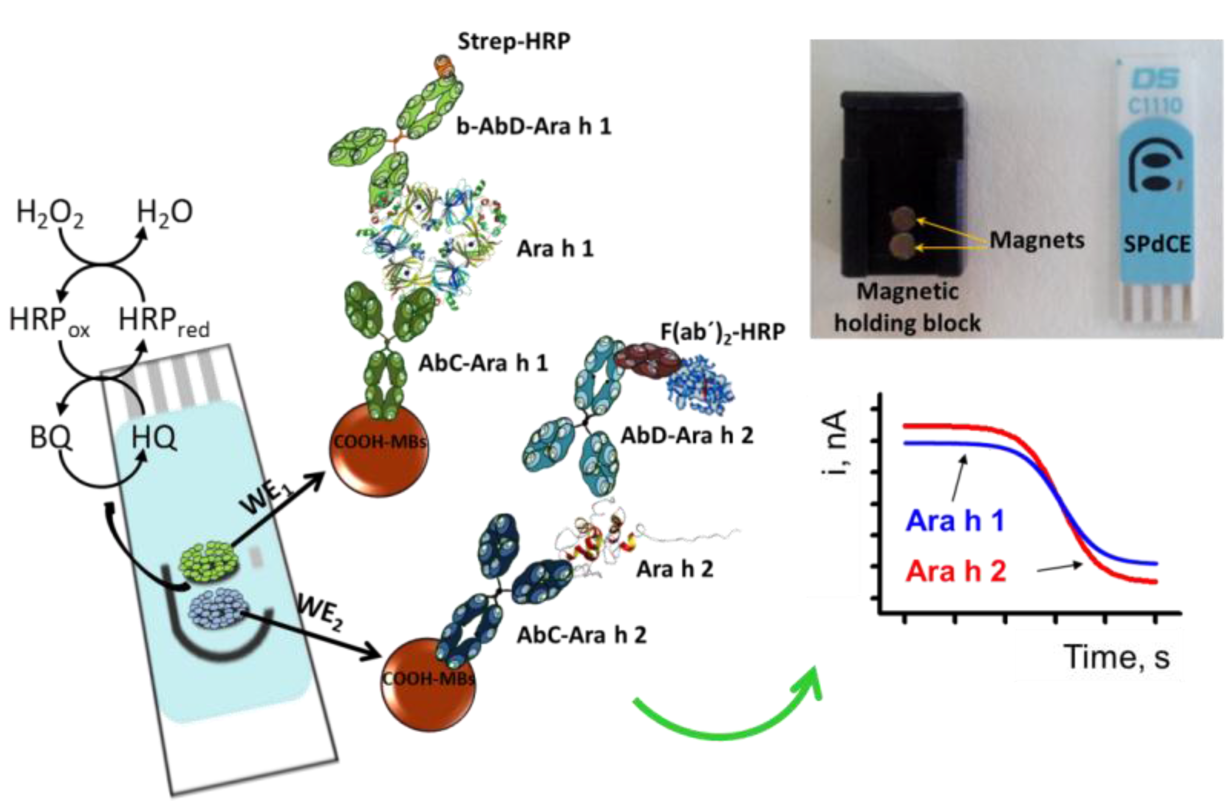
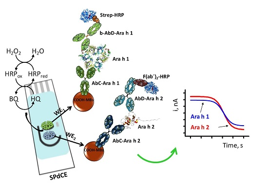
2. Materials and Methods

2.1. Materials

Amperometric measurements were performed with a CHI812B potentiostat (CH Instruments, Austin, TX, USA) controlled by software CHI812B. Dual screen-printed carbon electrodes (SPdCEs) (DRP-C1110, Dropsens, Oviedo, Spain) consisting of two elliptic carbon working electrodes (6.3 mm2 each), a carbon counter electrode and an Ag pseudo-reference electrode were employed as transducers. A specific cable connector (ref. DRP-BICAC also from DropSens, S.L.) acted as interface between the SPdCEs and the potentiostat. Single screen-printed carbon electrodes (SPCEs) and their specific connector (DRP-C110 and DRP-CAST, respectively, Dropsens) were also used. All measurements were carried out at room temperature.
A Bunsen AGT-9 Vortex (Lab Merchant Limited, London, UK) was used for the homogenization of the solutions. A Thermomixer MT100 constant temperature incubator shaker (Universal Labortechnik GmbH & Co. KG, Leipzig, Alemania) and a magnetic separator Dynal MPC-S (Thermo Fisher Scientific Inc., Madrid, Spain) were also employed. Capture of the modified-MBs onto the SPCE surface was controlled by a neodymium magnet (AIMAN GZ S.L., Madrid, Spain) embedded in a homemade casing of Teflon. Centrifuges Cencom (J.P. Selecta S.A., Barcelona, Spain) and MPW-65R (Biogen Científica, Madrid, Spain) were used in the separation steps.
All reagents were of the highest available grade. Sodium di-hydrogen phosphate, di-sodium hydrogen phosphate, Tris-HCl, NaCl and KCl were purchased from Scharlab (Barcelona, Spain). Tween®20, N-(3-dimethylaminopropyl)-N′-ethylcarbodiimide (EDC), N-hydroxysulfosuccinimide (sulfo-NHS), ethanolamine, hydroquinone (HQ), hydrogen peroxide (30%, w/v), lysozyme (from chicken egg white) and albumin from chicken egg white (OVA) were purchased from Sigma-Aldrich (Madrid, Spain). 2-(N-morpholino)ethanesulfonic acid (MES) and bovine serum albumin (BSA Type VH) were purchased from Gerbu Biotechnik GmbH (Heidelberg, Alemania) and commercial blocker casein solution (a ready-to-use, phosphate buffered saline (PBS), solution of 1% w/v purified casein) was purchased from Thermo Fisher Scientific (Madrid, Spain). Carboxylic acid-modified MBs (HOOC-MBs, 2.7 μm Ø, 10 mg/mL, Dynabeads® M-270 Carboxylic Acid) were purchased from Invitrogen (San Diego, CA, USA). Peanut allergen Ara h 1 Enzyme-linked immunosorbent assay (ELISA) kit (EL-AH1, containing mouse monoclonal IgG1 (2C12) antiAra h 1 capture antibody, AbC-Ara h 1, purified Ara h 1 standard, and biotinylated mouse monoclonal IgG1 (2F7) antiAra h 1 detection antibody, b-AbD-Ara h 1) and Ara h 2 ELISA kit (EL-AH2, containing mouse monoclonal IgG1 (1C4) antiAra h 2 capture antibody, AbC-Ara h 2, purified peanut allergen Ara h 2 standard, and Polyclonal rabbit antiserum raised against natural purified Ara h 2 as detection antibody, AbD-Ara h 2) were purchased from Indoor Biotechnologies, Inc. (Charlottesville, VA, USA). Peroxidase-conjugated AffiniPure F(ab’)2 Fragment Goat anti-Rabbit IgG (F(ab’)2-HRP), Fc Fragment Specific was purchased from Jackson ImmunoResearch Laboratories, Inc. (West Grove, PA, USA). A high sensitivity Strep-HRP conjugate from Sigma Aldrich (Ref: 000000011089153001, 500 U/mL) (Madrid, Spain) was also used.
All buffer solutions were prepared with water from Milli-Q Merck Millipore purification system (18.2 MΩ cm) (Darmstadt, Germany). Phosphate-buffered saline (PBS) consisting of 0.01 M phosphate buffer solution containing 137 mM NaCl and 2.7 mM KCl; 0.01 M sodium phosphate buffer solution consisting of PBS with 0.05% Tween®20 (pH 7.5, PBST); 0.05 M phosphate buffer, pH 6.0; 0.1 M phosphate buffer, pH 8.0; 0.025 M MES buffer and 0.1 M Tris-HCl buffer, pH 7.2. Activation of the HOOC-MBs was carried out with an EDC/sulfo-NHS mixture solution (50 mg/mL each in MES buffer, pH 5.0). The blocking step was accomplished with a 1 M ethanolamine solution prepared in a 0.1 M phosphate buffer solution of pH 8.0.

2.2. Modification of MBs

Dual Ara h 1 and Ara h 2 determinations at SPdCEs were accomplished by simultaneously preparing two different batches of MBs each of them suitable for the determination of each protein receptor following slightly changed protocols (in order to rearrange the assay times) with respect to those described previously for the individual determination of each protein [14,15]. In brief, 3-µL aliquot of the HOOC-MBs commercial suspension was transferred into a 1.5 mL Eppendorf tube for each batch. MBs were washed twice with 50 µL MES buffer solution for 10 min under continuous stirring (950 rpm, 25 °C). Between washings, the particles were captured using a magnet and, after 4 min, the supernatant was discarded. The MBs-surface confined carboxylic groups were activated by incubation during 35 min at 25 °C under continuous stirring (950 rpm) in 25 µL of the EDC/sulfo-NHS mixture solution. The activated MBs were washed twice with 50 µL of MES buffer and re-suspended in 25 µL of the corresponding capture antibody solution (25 µg/mL AbC-Ara h 1 and 50 µg/mL AbC-Ara h 2, prepared in MES buffer) during 30 min at 25 °C under continuous stirring (950 rpm). Subsequently, the AbC-modified MBs were washed twice with 50 µL of MES buffer solution. Thereafter, the unreacted activated groups on the MBs were blocked by adding 25 μL of a 1 M ethanolamine solution in 0.1 M phosphate buffer, pH 8.0, and incubating the suspension under continuous stirring (950 rpm) for 60 min at 25 °C. After one washing step with 50 µL of 0.1 M Tris-HCl buffer solution (pH 7.2) and two more with 50 µL of the commercial blocker casein solution, the magnetic beads modified with the capture antibody (AbC-MBs) were re-suspended in 25 µL of the target analyte standard solution or the sample (prepared in blocker casein solution) and incubated during 45 min (950 rpm, 25 °C). Then, the modified MBs were washed twice with 50 µL of the blocker casein solution and immersed into the corresponding AbD solution (b-AbD-Ara h 1 and AbD-Ara h 2 1/10,000 and 1/1000 diluted, respectively, in blocker casein solution) during 45 min (950 rpm, 25 °C). After two washing steps with 50 µL of PBST buffer solution (pH 7.5), the resulting beads were incubated during 30 min (950 rpm, 25 °C) in the corresponding labeling reagent solution: Strep-HRP (1/1000) for Ara h 1 and F(ab’)2-HRP (1/10,000) for Ara h 2, both prepared in PBST, pH 7.5. Finally, the modified-MBs were washed twice with 50 µL of PBST buffer solution (pH 7.5) and re-suspended in 5 µL of 0.05 M sodium phosphate buffer solution (pH 6.0).
Total determination of Ara h 1 and Ara h 2 was performed at SPCEs. In this case, 3 µL of AbC-Ara h 1-MBs and 3 µL of AbC-Ara h 2-MBs (after the blocking step with ethanolamine) were commingled together into a 1.5 mL Eppendorf tube and incubated 45 min (950 rpm, 25 °C) in a 25 µL of the standard/sample solution (prepared in blocker casein solution). This MB mixture was washed twice with 50 µL of the blocker casein solution and immersed into a mixture solution containing both AbDs 1/10,000 (b-AbD-Ara h 1) and 1/1000 (AbD-Ara h 2) diluted in commercial blocker casein solution during 45 min (950 rpm, 25 °C). After two washing steps with 50 µL of PBST buffer solution (pH 7.5), the resulting beads were incubated during 30 min in a mixture solution containing the two labeling reagents: Strep-HRP (1/1000) and F(ab’)2-HRP (1/10,000), prepared in PBST, pH 7.5. Finally, the modified-MBs were re-suspended in 45 µL of 0.05 M sodium phosphate buffer solution (pH 6.0) to perform the amperometric detection.

2.3. Amperometric Measurements

The amperometric measurements at the SPdCEs were performed as follows: the 5 µL of the resuspended MBs modified for Ara h 1 determination were magnetically captured onto one of the working electrodes of the dual SPCE. Similarly, the 5 µL suspension of the modified MBs for the Ara h 2 determination were captured on the second working electrode by keeping the dual SPCE in a horizontal position after placing it in the corresponding homemade magnet holding block. Then, the magnet holding block was immersed into an electrochemical cell containing 10 mL of 0.05 M phosphate buffer of pH 6.0 and 1.0 mM HQ (prepared just before performing the electrochemical measurement). Amperometric measurements in stirred solutions were made by applying a detection potential of −0.20 V vs. Ag pseudo-reference electrode upon addition of 50 μL of a 0.1 M H2O2 solution until the steady-state current was reached at both working electrodes (approx. 100 s). The amperometric signals given through the manuscript corresponded to the difference between the steady-state and the background currents.
To perform the detection at SPCEs, the 45 µL of the MBs mixture solution were magnetically captured on the working electrode of the SPCE. The same protocol described before for the detection at SPdCEs was followed.

2.4. Analysis of Real Samples

The dual Ara h 1 and Ara h 2 amperometric magnetoimmunosensor was applied to the analysis of different food samples containing unknown endogenous amounts of both proteins and also samples free of peanuts (wheat flour) spiked at trace levels.
Different types of foodstuffs, purchased in local supermarkets, were analyzed: wheat flour, hazelnuts; peanuts (peanut flour, raw and fried); chocolate bars with roasted peanuts and peanut cream.
Regarding the analysis of spiked samples, peanut-free wheat flour (verified using the commercial Ara h 1 and Ara h 2 ELISA spectrophotometric kits) was spiked with different amounts of peanut flour that consisted of 100% raw peanut (unknown variety) from a commercial retailer (Frinuts). Accordingly, a series of mixtures containing 1.0%, 0.5%, 0.1%, 0.05%, 0.025%, 0.01%, 0.0075%, 0.005%, 0.001%, 0.0005% and 0.0001% (w/w) of peanut were prepared.
The following protocol was used for the extraction of proteins present in peanuts in all the food samples analyzed: 0.5 g of accurately weighted ground sample (previously blended) were introduced in plastic tubes and incubated in 5.0 mL of Tris-HCl (pH 8.2) overnight at 60 °C under continuous stirring (950 rpm). Regarding the chocolate sample, it was frozen at −20 °C before blending, and 0.5 g of skimmed milk powder (Central Lechera Asturiana®, Asturias, Spain) were added during the extraction in order to avoid masking of the target protein by tannins [7]. Subsequently, the aqueous phase was isolated by centrifugation involving a first step at 3600 rpm during 10 min and a second step at 10,000 rpm during 3 min (4 °C) for a 1 mL aliquot of the first supernatant [14,17,18]. The resulting supernatant appropriately diluted was used to perform the determinations with the MBs-based immunosensor. No significant differences between the Ara h 1 and Ara h 2 content determined was observed after one month storage of these food extracts at 4 °C.
In order to make comparison, the same food extracts were also analyzed by applying both ELISA methods involving the use of the same immunoreagents.

3. Results

Figure 1 shows schematically the principles on which the dual electrochemical magnetoimmunosensor is based. Similar to that previously reported for the individual determination of each allergen protein [14,15], sandwich immunoassays were performed onto HOOC-MBs. Target proteins were sandwiched between respective specific capture antibodies and a biotinylated detector antibody for Ara h 1 (b-AbD-Ara h 1) and a non-biotinylated detector antibody for Ara h 2 (AbD-Ara h 2). These detector antibodies were labeled in a latter step with a streptavidin-HRP (Strep-HRP) polymer in the case of Ara h 1 or an HRP-conjugated secondary antibody in the case of Ara h 2. The MBs bearing the sandwich immunocomplexes for each target protein were magnetically captured on the corresponding working electrode (WE 1 and WE 2) of the SPdCE and amperometric detection at −0.20 V of the catalytic currents generated upon H2O2 addition and using HQ as redox mediator in solution at each working electrode was employed to determine each target protein concentration. It is important to note that this methodology implied that the SPdCEs acted only as the electrochemical transducer while all the affinity reactions occurred on the surface of the MBs, thus minimizing unspecific adsorptions of the bioreagents on the electrode surfaces.
The working variables used in the assays are summarized in Table S1 (in the Supporting Information) and were the same as those optimized for the single determination of each target protein with the exception of the incubation time in the AbC-Ara h 2 solution, which has been extended from 15 to 30 min in order to finish the preparation of both MBs batches at the same time. The detection potential value was also previously optimized for the HQ/HRP/H2O2 system [19]. Moreover, the working conditions used in the HOOC-MBs activation procedure, the successive washings and the unreacted carboxylic groups blocking step were established according to the protocols provided by the MBs supplier.
Cross-talking between the adjacent working electrodes is considered a potential major drawback to be avoided in the design of electrochemical multisensory platforms [20]. In addition, cross-reactivity amongst antibody pairs selected should be evaluated to demonstrate the feasibility of the bioplatform to perform the simultaneous determination of Ara h 1 and Ara h 2. Figure 2 shows the amperometric measurements obtained with the dual MBs-based immunosensor in solutions containing different Ara h 1 and Ara h 2 mixtures. As it can be deduced, no significant cross-talking between electrodes was apparent and the selected antibody pairs gave rise to significant responses only for the target protein despite the similar structural motifs described in both proteins [21]. These results endorsed the viability of the dual MBs-based immunosensing platform for the simultaneous specific detection of both allergenic proteins. Furthermore, the currents measured in the absence of the target protein can be considered as the negative control to account for any nonspecific binding of the AbDs or the enzymatic labels on the functionalized MBs. As it is shown in Figure 2, the immunosensor responses were mostly due to the selective sandwich immunocomplexes attached to the MBs surface.

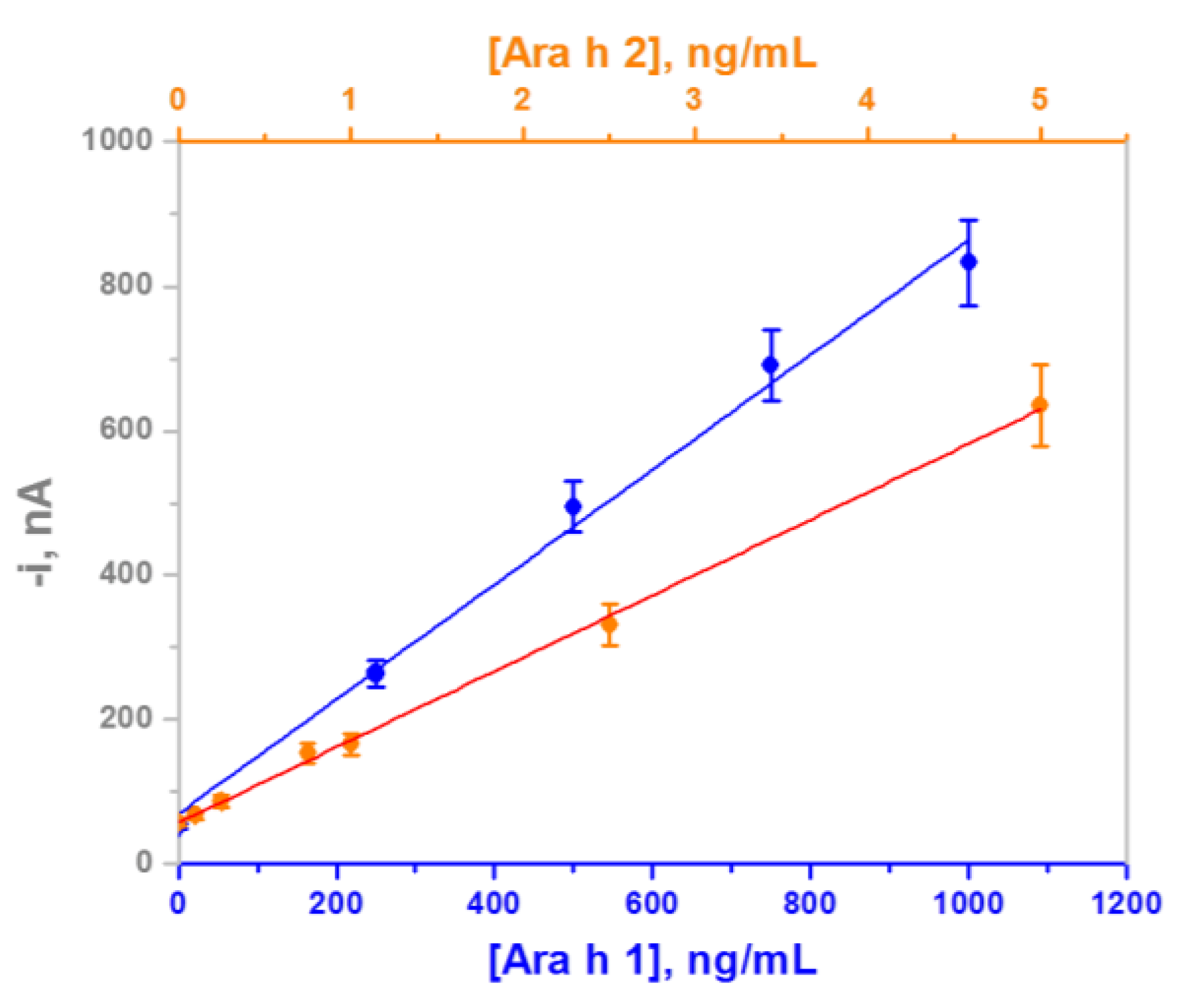
3.1. Analytical Characteristics

The reproducibility of the simultaneous amperometric responses for 500 ng/mL Ara h 1 and 1.0 ng/mL Ara h 2 was checked using eight different dual MBs-based immunosensors. Relative standard deviation (RSD) values of 7.3% and 8.9% were calculated for Ara h 1 and Ara h 2, respectively, confirming that the whole dual immunosensor preparation process, including MBs modification, MBs magnetic capture on the surface of each working electrode and amperometric measurements, was reliable and that reproducible amperometric responses can be obtained with different immunosensors constructed in the same manner.
Figure 3 displays the calibration plots for both target protein standards with the dual immunosensor. The corresponding analytical characteristics are summarized in Table 1. It is worth to note the remarkably higher sensitivity obtained for the determination of Ara h 2, which is in agreement with that reported by other authors using the same immunoreagents [5], and attributed to a better affinity of the antibody pair used for this target protein. Low detection limits (LODs) of 18 and 0.07 ng/mL (450 and 1.75 pg in 25 μL) were calculated according to the 3 × sb/m criterion, where sb was estimated as the standard deviation for 10 blank signal measurements and m is the slope value of the calibration plot. These low LODs are relevant from a clinical point of view since some patients exhibit strong allergic reactions against allergen levels as low as in the ng/mL range [5]. These LODs are slightly higher than those reported with the immunosensors developed for the individual determination of each proteins (6.3 and 0.026 ng/mL for Ara h 1 and Ara h 2, respectively), which is most likely due to the remarkably smaller active surface area of the dual SPCEs working electrodes when compared with the single SPCEs (6.3 vs. 12.6 mm2). Nevertheless, the LOD values achieved with the dual immunosensor were shown to be sufficient to allow detecting both target proteins in food extracts as well as peanut traces, as it will be demonstrated below.
It is also important to note that the achieved LODs are better than those claimed with commercial ELISA kits for the individual detection of Ara h 1 and Ara h 2 (31.5 and 2 ng/mL, respectively) using the same immunoreagents employed in the dual immunosensor.
The storage stability of the AbC-MBs was tested by keeping them at 4 °C in Eppendorf tubes containing 50 μL of filtered PBS. Two replicates of the stored AbC-Ara h 1-MBs and AbC-Ara h 2-MBs conjugates were incubated each working day in solutions containing no target protein, 250 ng/mL Ara h 1 and 2.5 ng/mL Ara h 2. Control charts were constructed by setting the average current value calculated from 10 measurements made the first day of the study (when the AbC-Ara h 1-MBs and AbC-Ara h 2-MBs were prepared) as the central values, while the upper and lower limits of control were set at ±3 × SD of these central values. The obtained results (not shown) showed that the immunosensors prepared with the stored AbC-MBs provided measurements within the control limits during 25 and 50 days, for Ara h 1 and Ara h 2, respectively. This good storage stability suggests the possibility of preparing sets of AbC-Ara h 1-MBs and AbC-Ara h 2-MBs conjugates and storing them under the above-mentioned conditions until the dual bioplatform needs to be prepared.

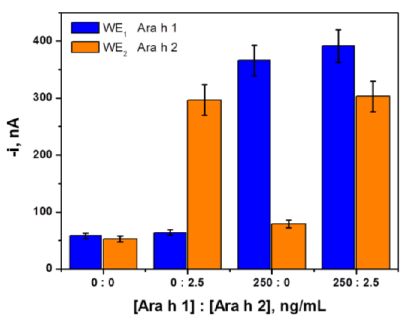
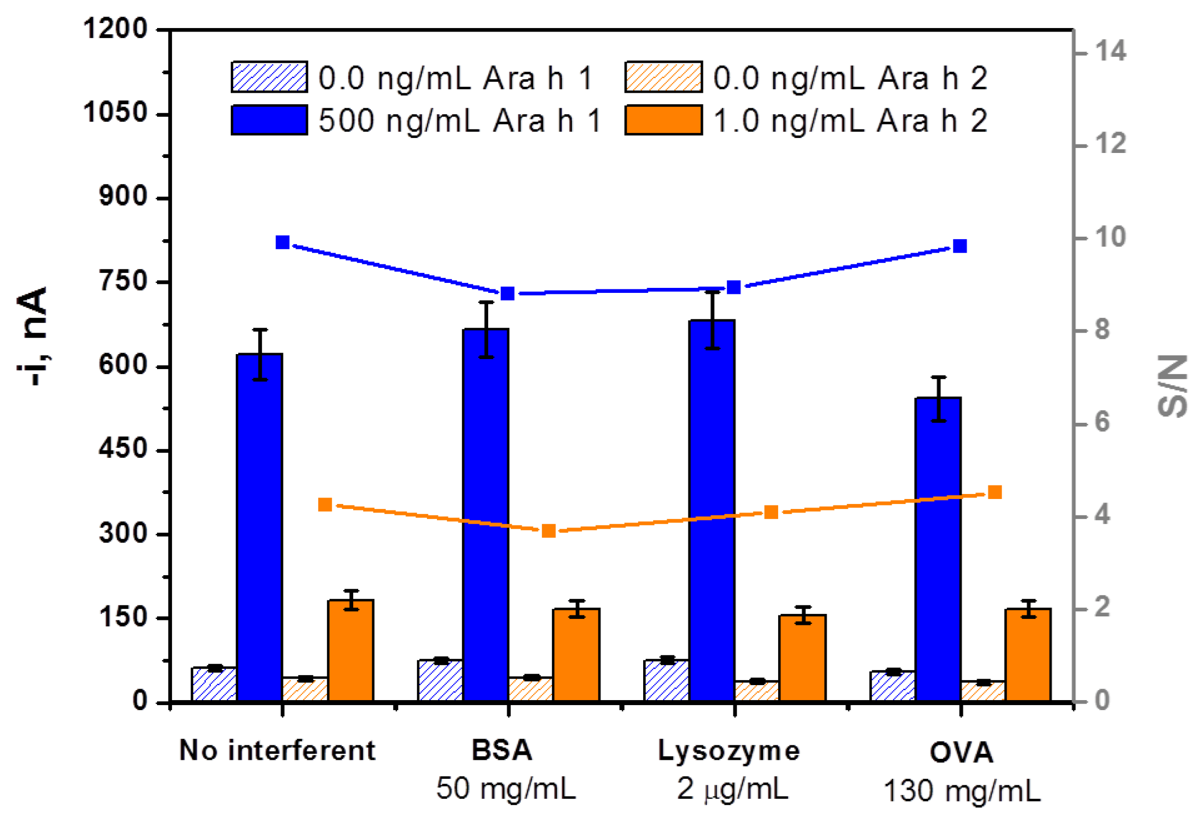
3.2. Selectivity of the Dual Magnetoimmunosensor

The selectivity of the dual magnetoimmunosensor was evaluated towards non-target proteins such as BSA, lysozyme and OVA, which can coexist with the target proteins in food extracts. A comparison of the current values measured with the dual immunosensing platform for 0 and 500 ng/mL Ara h 1 and 0 and 1.0 ng/mL Ara h 2 in the absence and in the presence of these potential interfering compounds is shown in Figure 4. No significant effect in the measurements for Ara h 1 and Ara h 2 was apparent as a result of the presence of the three non-target proteins even at the large concentrations tested. Moreover, no noticeable cross-reactivity was observed between Ara h 1 and Ara h 2 despite these proteins showing similar structural motifs [21], and even although Ara h 1 was tested at a 500 times larger concentration than Ara h 2. The high selectivity of the developed platform against other Ara h, legumes and nuts proteins will be also evidenced in the analysis of different complex food extracts where other non-targeted proteins are present in a large extent.

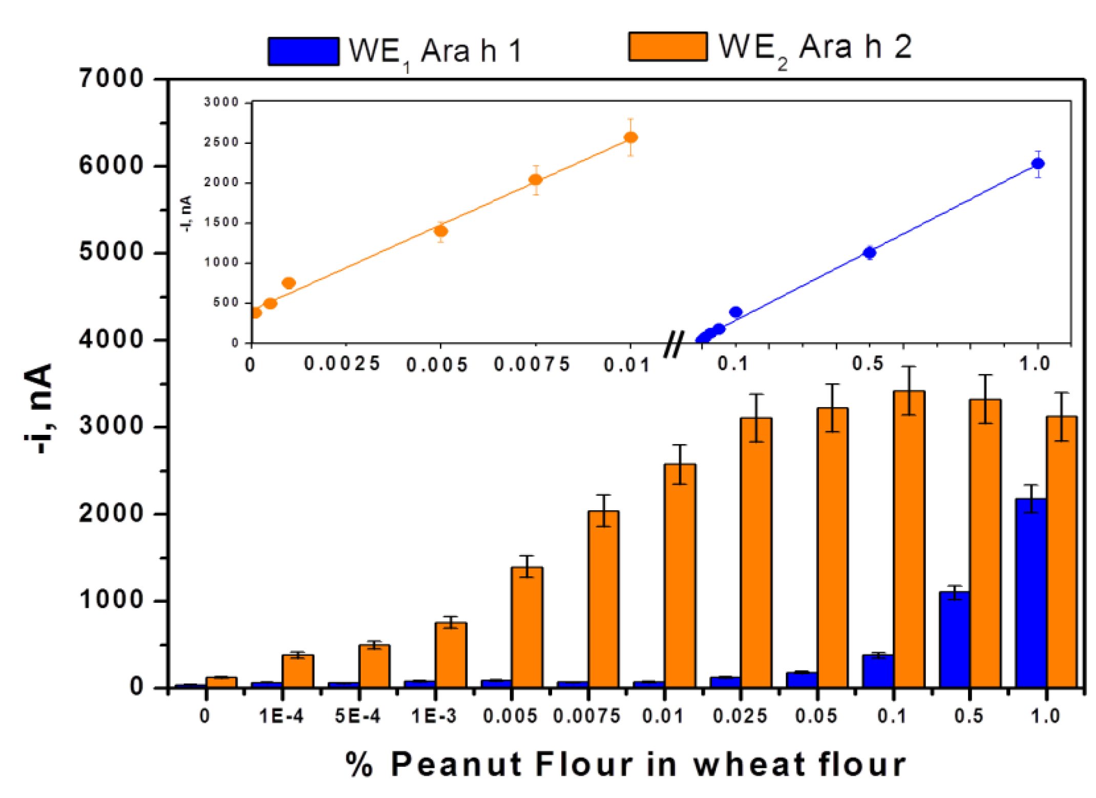
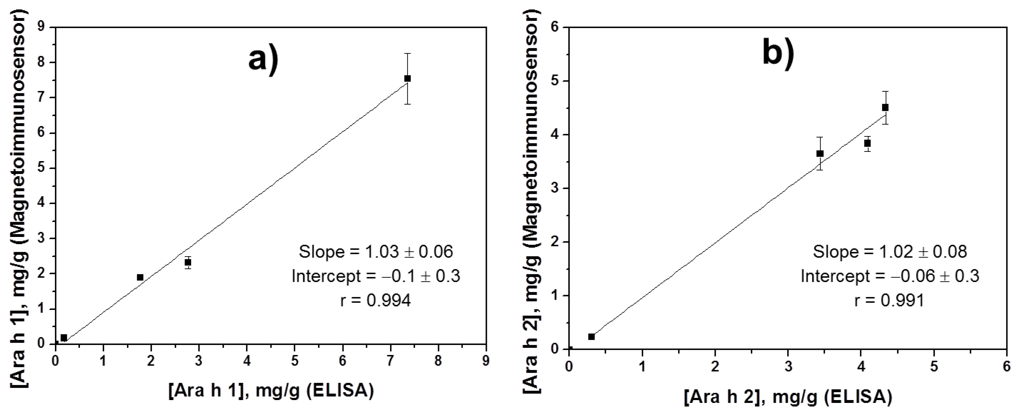
3.3. Simultaneous Determination of Ara h 1 and Ara h 2 in Food Samples

The usefulness of the dual immunosensor for the analysis of real samples was verified by determining both target allergen proteins in different food extracts containing variable and unknown amount of endogenous Ara h 1 and Ara h 2 as well as in target-spiked protein-free samples. Most interestingly, no significant matrix effects were found once the sample extracts were appropriately diluted with blocker casein solution. Using the dilution factors summarized in Table 2, no statistically significant differences were observed between the slope value of the calibration plots constructed with Ara h 1 and Ara h 2 standards (Figure 3) and the slope values of the calibration graphs constructed from all the extracts by spiking with growing amounts of standards solution (up to 750 and 2.5 ng/mL for Ara h 1 and Ara h 2, respectively). It is worth remarking that the dilution factors mentioned in Table 2 correspond to those required to fit the target analyte concentration into the linear range of the calibration graphs. Therefore, Ara h 1 and Ara h 2 quantification could be accomplished by simple interpolation of the measured currents in the properly diluted samples at each working electrode of the SPdCE into the calibration plot constructed with standard solutions.
The results obtained for all the analyzed food extracts are given in Table 2. In addition, these results were compared with those obtained by using the ELISA kits containing the same immunoreagents. As it can be deduced in Figure 5, excellent correlations were found for both proteins’ concentrations determined both with the amperometric multiplexed platform and the single-plexed ELISA kits. RSD values obtained (n = 3) are between 2.3%–9.6% and 3.4%–10.5% using the developed dual platform and the conventional ELISA methodology, respectively. For both target proteins, the confidence intervals (at a significance level of α = 0.05) for the slope and intercept included the unit and the zero values, respectively. These results demonstrated the great selectivity of the developed platforms against other legumes and nuts proteins since no detectable amperometric responses were obtained for undiluted raw halzenuts and wheat flour extracts.
Furthermore, wheat flour containing no detectable content of the target allergen was spiked with different increasing amounts of peanut flour and the corresponding extracts were obtained and analyzed as described in Section 2.4. Figure 6 shows as the dual immunosensor was able to discriminate samples contaminated with 0.0001%–0.01% peanut through Ara h 2 detection whereas samples contaminated with 0.01%–1.0% peanut could be identified by means of Ara h 1 detection. The ability to detect clearly 0.0001% (1.0 mg·kg−1) peanut improves in a factor of 500–1000 the lowest detectable trace peanuts concentration reported previously by other authors based on Ara h 1 determination, 0.05 [14] and 0.1% [12,13]. This enhanced sensitivity means a major comparative advantage taking into account the serious public health problem that the contamination degree with peanuts of commercial food samples, whether fraudulent or accidental, may cause in sensitive individuals. Moreover, it is important to highlight that although a level of 10 mg·kg−1 is considered relevant for the detection of potentially hazardous residues of undeclared allergens in foods, the achievement of a 10 times lower detection limit, as it is the case of this work, may be highly helpful since minimal amounts of the target allergen can be critical [9]. In addition, it is also important to note that the detection level of about 1.0 mg/kg peanut is achieved without any amplification step and, even so, is lower than those reported so far with PCR-based approaches, which are in the 2 to 10 mg/kg range [9,22,23,24]. Furthermore, the developed immunosensor is suitable to allow allergen determination in a simple way without requirements of a high number of sample replicates and the use of a high precision thermocycler. These features make this methodology possess inherent advantages with respect to PCR-based assays for an easy implementation in analytical food quality and safety laboratories performing routine or decentralized analyses.

4. Discussion

Apart from the better sensitivity achieved, it is important to note that the multiplexing capability of the developed platform provides higher levels of information from samples that are unavailable with current commercial ELISA detection kits. Some ELISA kits and detection methods are only capable of providing information on total allergen amounts, therefore lacking information and breakdown of the actual individual allergens in the sample, assuming that patients exhibit the same levels of allergic reaction against all peanut allergens, which is not the case [25,26]. Moreover, data about the individual content of food allergens could be important also in processed foods since some treatments have demonstrated having major roles in changing the allergenic characteristics of particular allergenic proteins (i.e., enhancing allergenic properties of Ara h 2 with roasting) [27,28].
The ability to perform multiplexed allergen detection using the developed platform would therefore provide a means to quantitatively detect specific peanut allergens like Ara h 1 and Ara h 2. This could potentially open up opportunities for patients who, provided with information from clinicians, understand their specific threshold levels for particular allergens to ingest foods that are labeled in detail.
Furthermore, the possibility of shortening the assay time and simplifying as much as possible the sample treatment was considered. With this purpose, measurements in spiked extracts performed according with the whole protocol described in Section 2.4, were compared with those carried out by omitting the centrifugation steps or replacing them by a 30 min natural decantation process. Figure S1 (in the Supporting Information) shows that although, as expected, the resulting S/N ratios were smaller when the extracts were not centrifuged, the amperometric responses were still sufficiently large to discriminate clearly between spiked and non-spiked samples. This relevant result outlined the potentiality of the developed methodology to be employed as a rapid method for alarm or screening purposes able to discriminate samples containing only 0.005% peanut through detection of Ara h 2 protein in a cloudy extract (see orange bars 3 in Figure S1a and tube 3) in Figure S1b).

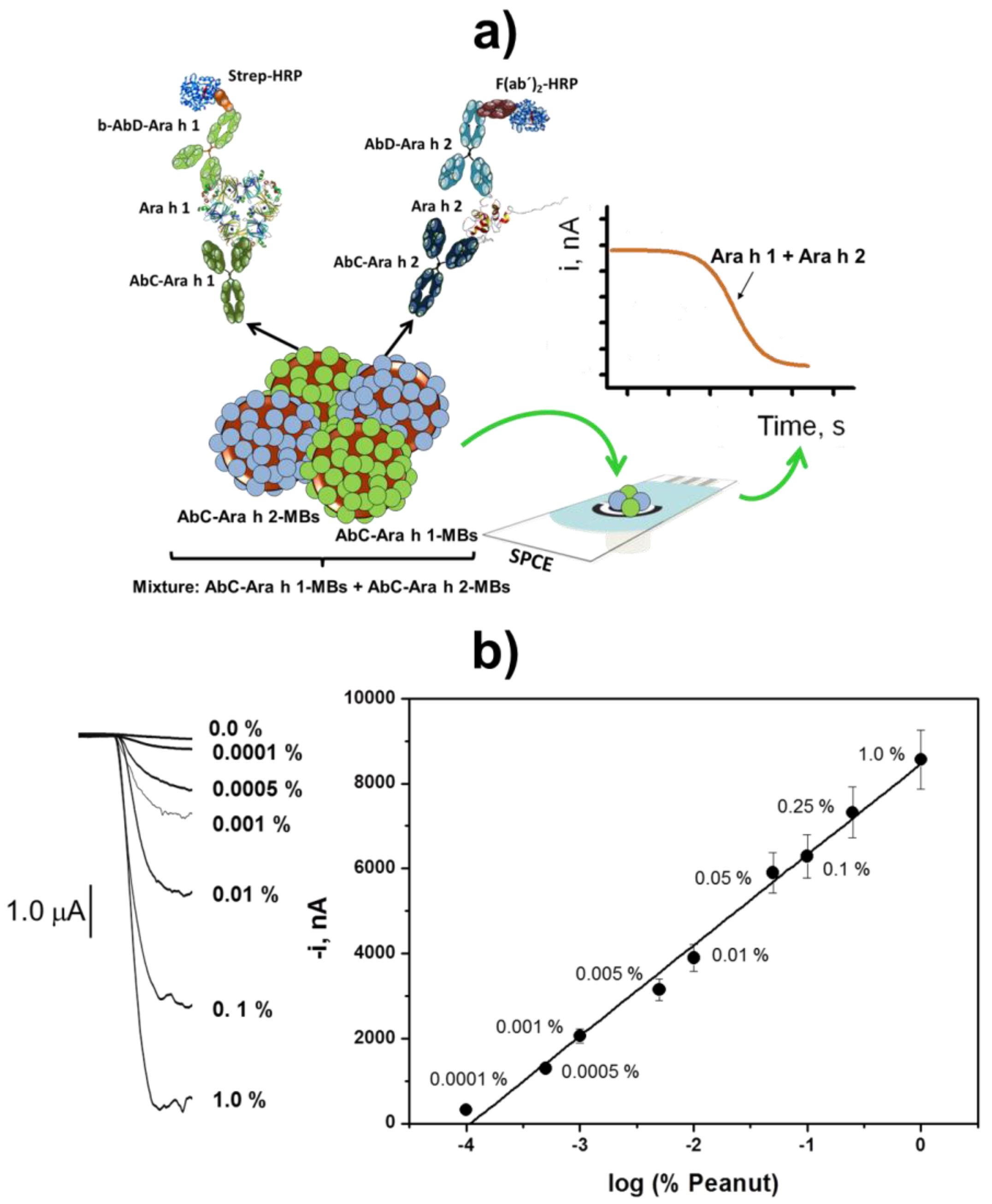
Total Detection of Both Major Peanut Allergenic Proteins in Food Samples

In view of the results presented in Figure 6 and with the aim of developing a simple methodology for screening the presence of peanuts in foods in a wider range of concentrations, the total detection of both target proteins was evaluated by using a mixture of target specific modified magnetic beads (MBs) sets and amperometric detection at a single SPCE.
Figure 7a shows a schematic display of the approach. The modified MBs are commingled together and incubated sequentially in the sample and mixture of the two AbD solutions as well as in the labeling reagents’ solutions and then captured on the surface of a SPCE. The results presented in Figure 7b demonstrate that this methodology allows a clear discrimination of samples contaminated with peanut over five orders of magnitude concentrations, between 0.0001% and 1.0%. Therefore, this approach, although not designed to identify the type of peanut allergenic protein detected, proves to be a user-friendly, attractive, effective and rapid tool (2 h) for detecting the presence in a wide concentration range or verifying the absence of peanuts in foods.

5. Conclusions

In this work, a dual electrochemical immunoplatform for the simultaneous detection of peanut allergenic proteins Ara h 1 and Ara h 2 in one single experiment has been described for the first time. This platform provides quantitative information on individual food allergen levels, and the achieved LODs for both target proteins are lower than those claimed for standard well-based ELISAs using the same immunoreagents. Apart from its successful applicability for trace allergen contaminant detection and quantification, the flexibility of this MBs-based electrochemical design allows for further expansion to any allergen that patients are potentially allergic to and to produce a comprehensive array for determining the most important food allergens with a single assay on a single chip. Furthermore, the sensors can be mass produced, making them low cost and disposable. Given its demonstrated important advantages, this novel bioplatform provides food manufacturers and allergic patients or caretakers with an attractive solution to the need for highly sensitive and specific detection of any multiple trace allergen contaminants in food samples.

Supplementary Materials

The following are available online at https://www.mdpi.com/2227-9040/4/3/11/s1, Table S1: Optimized experimental variables affecting the performance of the electrochemical dual MBs-based immunosensing platform for the simultaneous determination of Ara h 1 and Ara h 2, Figure S1: (a) Current values measured with the dual immunosensing platform from extracts prepared with unspiked and spiked with 0.5 (Ara h 1) and 0.005 (Ara h 2) % (w/w) wheat flour using different sample treatments: protocol described in Section 2.4 (1); centrifugation steps were substituted by a 30-min natural decantation process (2); centrifugation steps were omitted (3). (b) Real picture of the extracts obtained after applying each sample treatment. Supporting electrolyte, 0.05 M sodium phosphate solution, pH 6.0; Eapp = −0.20 V vs. Ag pseudo-reference electrode. Error bars estimated as triple of the standard deviation (n = 3).

Acknowledgments

The financial support of the Spanish Ministerio de Economía y Competitividad Research Projects, CTQ2015-64402-C2-1-R, and the NANOAVANSENS Program from the Comunidad de Madrid (S2013/MT-3029) are gratefully acknowledged. Financial support by COFIN 2010–2011 (Programme di Ricerca Scientifica di Relevante Interesse Nazionale, MIUR, 2010AXENJ8_002) is also acknowledged. R.M. Torrente-Rodríguez and V. Ruiz-Valdepeñas Montiel acknowledge predoctoral contracts from the Spanish Ministerio de Economía y Competitividad and Universidad Complutense de Madrid, respectively.

Author Contributions

V.R-V.M. and R.M.T-R. contributed equally to this work. V.R-V.M., R.M.T-R., S.C. and J.M.P. conceived and designed the experiments; V.R-V.M., R.M.T-R. and A.P. performed the experiments; V.R-V.M., R.M.T-R., S.C., M.S.C. and A.J.R. analyzed the data; S.C. and J.M.P. wrote the paper.

Conflicts of Interest

The authors declare no conflict of interest.

Abbreviations

The following abbreviations are used in this manuscript:
ELISAsEnzyme-linked immunosorbent assays
HQHydroquinone
LODLimit of detection
MBsMagnetic beads
PCRPolymerase chain reaction
SPdCEsScreen-printed dual carbon electrodes

References

  1. Walker, M.J.; Burns, D.T.; Elliott, C.T.; Gowland, M.H.; Clare Mills, E.N. Is food allergen analysis flawed? Health and supply chain risks and a proposed framework to address urgent analytical needs. Analyst 2016, 141, 24–35. [Google Scholar] [CrossRef] [PubMed]
  2. U.S. Food and Drug Administration. Consumers-Food Allergies: What You Need to Know. 2015. Available online: http://www.fda.gov/Food/ResourcesForYou/Consumers/ucm079311.htm (accessed on 25 August 2015). [Google Scholar]
  3. Van Hengel, A.J. Food allergen detection methods and the challenge to protect food-allergic consumers. Anal. Bioanal. Chem. 2007, 389, 111–118. [Google Scholar] [CrossRef] [PubMed]
  4. Montserrat, M.; Sanz, D.; Juan, T.; Herrero, A.; Sánchez, L.; Calvo, M.; Pérez, M.D. Detection of peanut (Arachis hypogaea) allergens in processed foods by immunoassay: Influence of selected target protein and ELISA format applied. Food Control 2015, 54, 300–307. [Google Scholar] [CrossRef]
  5. Ng, E.; Nadeau, K.; Wang, S.X. Giant magneto resistive sensor array for sensitive and specific multiplexed food allergen detection. Biosens. Bioelectron. 2016, 80, 359–365. [Google Scholar] [CrossRef] [PubMed]
  6. Food Allergen Labelling and Information Requirements under the EU Food Information for Consumers, Regulation No. 1169/2011: Technical Guidance. Available online: http://www.food.gov.uk/sites/default/files/food-allergen-labelling-technical-guidance.pdf (accessed on 27 June 2016).
  7. Poms, R.E.; Agazzi, M.E.; Bau, A.; Brohee, M.; Capelletti, C.; Nørgaard, J.V.; Anklam, E. Inter-laboratory validation study of five commercial ELISA test kits for the determination of peanut proteins in biscuits and dark chocolate. Food Addit. Contam. 2005, 22, 104–112. [Google Scholar] [CrossRef] [PubMed]
  8. Pedreschi, R.; Nørgaard, J.; Maquet, A. Current Challenges in Detecting Food Allergens by Shotgun and Targeted Proteomic Approaches: A Case Study on Traces of Peanut Allergens in Baked Cookies. Nutrients 2012, 4, 132–150. [Google Scholar] [CrossRef] [PubMed] [Green Version]
  9. López-Calleja, I.M.; de la Cruz, S.; Pegels, N.; González, I.; García, T.; Martín, R. Development of a real time PCR assay for detection of allergenic trace amounts of peanut (Arachis hypogaea) in processed foods. Food Control 2013, 30, 480–490. [Google Scholar] [CrossRef]
  10. Holzhauser, T.; Kleiner, K.; Janise, A.; Röder, M. Matrix-normalised quantification of species by threshold-calibrated competitive real-time PCR: Allergenic peanut in food as one example. Food Chem. 2014, 163, 68–76. [Google Scholar] [CrossRef] [PubMed]
  11. Cho, C.Y.; Nowatzke, W.; Oliver, K.; Garber, E.A.E. Multiplex detection of food allergens and gluten. Anal. Bioanal. Chem. 2015, 407, 4195–4206. [Google Scholar] [CrossRef] [PubMed]
  12. Alves, R.C.; Pimentel, F.B.; Nouws, H.P.A.; Marques, R.C.B.; González-García, M.B.; Oliveira, M.B.P.P.; Delerue-Matos, C. Detection of Ara h 1 (a major peanut allergen) in food using an electrochemical gold nanoparticle-coated screen-printed immunosensor. Biosens. Bioelectron. 2015, 64, 19–24. [Google Scholar] [CrossRef] [PubMed]
  13. Alves, R.C.; Pimentel, F.B.; Nouws, H.P.A.; Correr, W.; González-García, M.B.; Oliveira, M.B.P.P.; Delerue-Matos, C. Detection of the peanut allergen Ara h 6 in foodstuffs using a voltammetric biosensing approach. Anal. Bioanal. Chem. 2015, 407, 7157–7163. [Google Scholar] [CrossRef] [PubMed]
  14. Ruiz-Valdepeñas Montiel, V.; Campuzano, S.; Pellicanò, A.; Torrente-Rodríguez, R.M.; Reviejo, A.J.; Cosio, M.S.; Pingarrón, J.M. Sensitive and selective magnetoimmunosensing platform for determination of the food allergen Ara h 1. Anal. Chim. Acta 2015, 880, 52–59. [Google Scholar] [CrossRef] [PubMed]
  15. Ruiz-Valdepeñas Montiel, V.; Campuzano, S.; Pellicanò, A.; Torrente-Rodríguez, R.M.; Reviejo, A.J.; Cosio, M.S.; Pingarrón, J.M. Electrochemical detection of peanuts at trace levels in foods using a magnetoimmunosensor for the allergenic protein Ara h 2. Sens. Actuator B Chem. 2016. [Google Scholar] [CrossRef]
  16. Koppelman, S.J.; Vlooswijk, R.A.; Knippels, L.M.; Hessing, M.; Knol, E.F.; Van Reijsen, F.C.; Bruijnzeel-Koomen, C.A. Quantification of major peanut allergens Ara h 1 and Ara h 2 in the peanut varieties Runner, Spanish, Virginia, and Valencia, bred in different parts of the world. Allergy 2001, 56, 132–137. [Google Scholar] [CrossRef] [PubMed]
  17. Pele, M.; Brohée, M.; Anklam, E.; Van Hengel, A. Presence of peanut and hazelnut in cookies and chocolates: The relationship between analytical results and the declaration of food allergens on product labels. Food Addit. Contam. 2007, 24, 1334–1344. [Google Scholar] [CrossRef] [PubMed]
  18. Jollet, P.; Delport, F.; Janssen, K.P.F.; Tran, D.T.; Wouters, J.; Verbiest, T.; Lammertyn, J. Fast and accurate peanut allergen detection with nanobead enhanced optical fiber SPR biosensor. Talanta 2011, 83, 1436–1441. [Google Scholar]
  19. Gamella, M.; Campuzano, S.; Conzuelo, F.; Reviejo, A.J.; Pingarrón, J.M. Amperometric magnetoimmunosensor for direct determination of D-dimer in human serum. Electroanal. 2012, 24, 2235–2243. [Google Scholar] [CrossRef]
  20. Escamilla-Gómez, V.; Hernández-Santos, D.; González-García, M.B.; Pingarrón-Carrazón, J.M.; Costa-García, A. Simultaneous detection of free and total prostate specific antigen on a screen-printed electrochemical dual sensor. Biosens. Bioelectron. 2009, 24, 2678–2683. [Google Scholar] [CrossRef] [PubMed]
  21. Bublin, M.; Breiteneder, H. Cross-reactivity of peanut allergens. Curr. Allergy Asthma Rep. 2014, 14. [Google Scholar] [CrossRef] [PubMed]
  22. Watanabe, T.; Akiyama, H.; Maleki, S.; Yamakawa, H.; Iijima, K.; Yamazaki, F.; Matsumoto, T.; Futo, S.; Arakawa, F.; Watai, M.; et al. A specific qualitative detection method for peanut (Arachis hypogaea) in foods using polymerase chain reaction. J. Food Biochem. 2006, 30, 215–233. [Google Scholar] [CrossRef]
  23. Scaravelli, E.; Brohee, M.; Marchelli, R.; Van Hengel, A. Development of three real-time PCR assays to detect peanut allergen residue in processed food products. Eur. Food Res. Technol. 2008, 227, 857–869. [Google Scholar] [CrossRef]
  24. Köppel, R.; Dvorak, V.; Zimmerli, F.; Breitenmoser, A.; Eugster, A.; Waiblinger, H.U. Two tetraplex real-time PCR for the detection and quantification of DNA from eight allergens in food. Eur. Food Res. Technol. 2010, 230, 367–374. [Google Scholar] [CrossRef]
  25. Koppelman, S.J.; Wensing, M.; Ertmann, M.; Knulst, A.C.; Knol, E.F. Relevance of Ara h 1, Ara h 2 and Ara h 3 in peanut-allergic patients, as determined by immunoglobulin E Western blotting, basophil-histamine release and intracutaneous testing: Ara h 2 is the most important peanut allergen. Clin. Exp. Allergy 2004, 34, 583–590. [Google Scholar] [CrossRef] [PubMed]
  26. Mitchell, D.C. Peanut allergy diagnosis: As simple as Ara h 1, 2, and 3. Consultant for pediatricians. Consult. Pediatr. 2013, 12, 347–350. [Google Scholar]
  27. Maleki, S.J.; Viquez, O.; Jacks, T.; Dodo, H.; Champagne, E.T.; Chung, S.-Y.; Landry, S.J. The major peanut allergen, Ara h 2, functions as a trypsin inhibitor, and roasting enhances this function. J. Allergy Clin. Immunol. 2003, 112, 190–195. [Google Scholar] [CrossRef] [PubMed]
  28. Zhou, Y.; Wang, J.-S.; Yang, X.-J.; Lin, D.-H.; Gao, Y.-F.; Su, Y.-J.; Yang, S.; Zhang, Y.-J.; Zheng, J.-J. Peanut allergy, allergen composition, and methods of reducing allergenicity: A review. Int. J. Food Sci. 2013, 2013. [Google Scholar] [CrossRef] [PubMed]
Figure 1. Schematic display of the fundamentals involved in the dual determination of Ara h 1 and Ara h 2 using screen-printed dual carbon electrodes (SPdCEs) as well as in the reactions implied in the amperometric responses. A real picture of the SPdCE and the homemade magnetic holding block is also shown.
Chemosensors 04 00011 g001
Figure 2. Simultaneous amperometric responses measured with the dual magnetic beads (MBs)-based immunosensor for mixtures containing: 0 ng/mL of both proteins; 0 ng/mL Ara h 1 and 2.5 ng/mL Ara h 2; 250 ng/mL Ara h 1 and 0 ng/mL Ara h 2; 250 ng/mL Ara h 1 and 2.5 ng/mL Ara h 2. Eapp = −0.20 V vs. Ag pseudo-reference electrode. Error bars estimated as triple the standard deviation (n = 3).
Chemosensors 04 00011 g002
Figure 3. Calibration plots obtained with the dual immunosensing platform for Ara h 1 and Ara h 2 standards. Error bars estimated as triple of the standard deviation (n = 3).
Chemosensors 04 00011 g003
Figure 4. Current values measured for 0 and 500 ng/mL Ara h 1 and 0 and 1.0 ng/mL Ara h 2 in the absence or in the presence of 50 mg/mL bovine serum albumin (BSA), 2 μg/mL lysozyme and 130 mg/mL ovalbumin (OVA). Supporting electrolyte, 0.05 M sodium phosphate solution, pH 6.0; Eapp = −0.20 V vs. Ag pseudo-reference electrode. Error bars estimated as triple of the standard deviation (n = 3).
Chemosensors 04 00011 g004
Figure 5. Correlation plots between the results obtained in the determination of Ara h 1 (a) and Ara h 2 (b) in food extracts using the developed dual immunoplatform and those provided by the individual ELISA spectrophotometric kits (data from Table 2).
Chemosensors 04 00011 g005
Figure 6. Amperometric responses obtained with the dual MBs-based immunosensing platform for extracts prepared from wheat flour spiked with increasing amounts of peanut flour (final concentrations: 0.0001%, 0.0005%, 0.001%, 0.005%, 0.0075%, 0.01%, 0.025%, 0.05%, 0.1%, 0.5% and 1.0% (w/w)). Error bars estimated as triple the standard deviation (n = 3).
Chemosensors 04 00011 g006
Figure 7. (a) schematic display of the methodology involved for total detection of Ara h 1 and Ara h 2 at a SPCE; (b) amperometric responses obtained by measuring the total content of Ara h 1 and Ara h 2 with the MBs-based immunosensing approach at SPCEs from extracts prepared for wheat flour unspiked and spiked with increasing amounts of peanut flour (from 0.0001% to 1.0% (w/w)). Error bars estimated as triple the standard deviation (n = 3).
Chemosensors 04 00011 g007
Table 1. Analytical characteristics for the determination of Ara h 1 and Ara h 2 using the dual magnetic beads (MBs)-based immunosensing platform.
Ara h 1Ara h 2
Linear range (LR), ng/mL60–10000.25–5
r0.9960.999
Sensitivity, nAmL/ng0.79 ± 0.05115 ± 2
LOD, ng/mL *180.07
Limit of determination (LQ), ng/mL **600.25
* Calculated as 3 × sb/m where sb was the standard deviation for 10 blank signal measurements and m is the slope value of the calibration plot; ** Calculated as 10 × sb/m
Table 2. Determination of the endogenous content of allergenic proteins Ara h 1 and Ara h 2 in different food extracts with the amperometric dual immunosensor and comparison with the results provided by ELISA spectrophotometric kits.
[Ara h 1], mg/g[Ara h 2], mg/g
ExtractDilution FactorDual PlatformELISADilution FactorDual PlatformELISA
Fried peanuts1/1000(7 ± 2)(7.3 ± 0.6)1/250,000(3.6 ± 0.8)(3.4 ± 0.6)
Raw peanuts1/1000(2.3 ± 0.4)(2.8 ± 0.3)1/250,000(3.8 ± 0.3)(4.1 ± 0.5)
Chocolate bars with roasted peanuts1/50(0.18 ± 0.01)(0.18 ± 0.03)1/25,000(0.23 ± 0.05)(0.30 ± 0.08)
Peanut cream1/50(1.9 ± 0.2)(1.8 ± 0.4)1/500,000(4.5 ± 0.8)(4.3 ± 0.8)
Raw hazelnutsNDNDNDND
Wheat flourNDNDNDND
ND: non detectable.

