Article

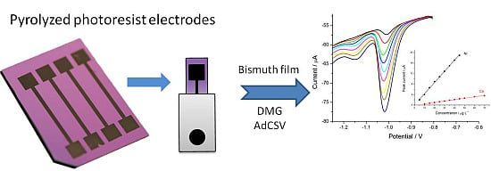
Pyrolyzed Photoresist Carbon Electrodes for Trace Electroanalysis of Nickel(II)

1
Department of Molecular Sciences and Nanosystems, University Ca' Foscari of Venice, S. Marta 2137, Venice I-30123, Italy
2
Veneto Nanotech, Via delle Industrie 5, Venice-Marghera 30175, Italy
*
Author to whom correspondence should be addressed.
Chemosensors 2015, 3(2), 157-168; https://doi.org/10.3390/chemosensors3020157
Submission received: 18 March 2015 / Revised: 29 April 2015 / Accepted: 8 May 2015 / Published: 15 May 2015
(This article belongs to the Special Issue Electrochemical Sensors for Environmental and Food Analysis)

Abstract

:
Novel pyrolyzed photoresist carbon electrodes for electroanalytical applications have been produced by photolithographic technology followed by pyrolysis of the photoresist. A study of the determination of Ni(II) dimethylglyoximate (Ni-DMG) through adsorptive cathodic stripping voltammetry at an in situ bismuth-modified pyrolyzed photoresist electrode (Bi-PPCE) is reported. The experimental conditions for the deposition of a bismuth film on the PPCE were optimized. The Bi-PPCE allowed the analysis of trace concentrations of Ni(II), even in the presence of Co(II), which is the main interference in this analysis, with cathodic stripping square wave voltammograms characterized by well-separated stripping peaks. The calculated limits of detection (LOD) were 20 ng∙L−1 for Ni(II) alone and 500 ng∙L−1 in the presence of Co(II). The optimized method was finally applied to the analysis of certified spring water (NIST1640a).

Graphical Abstract

1. Introduction

Carbon is available in nature in a variety of allotropes that exhibit a wide range of mechanical, chemical, electrical and electrochemical properties based on the underlying microstructure [1]. Graphite and glassy carbon (GC) are the more widely-investigated allotropic forms of carbons for electrochemical applications [1,2]. Indeed, carbon is a commonly-used solid electrode material due to its wide potential window, mechanical stability, low cost and applicability to a wide range of redox systems. Carbon’s chemical inertness and stability in acidic and basic media make it an attractive material for the deposition of metal thin films, such as mercury [3], gold [4] and, more recently, bismuth, antimony [5,6], metal nanoparticles [7,8] and polymers [9,10], employed for electroanalysis. Microfabrication of low-cost and disposable electrodes has received attention recently due to the development of electrochemical sensors, such as the thick-film screen-printed carbon electrodes prepared using commercially available carbon inks. The fabrication of microelectrode arrays is fundamental for electroanalytical studies in biological media, in particular for in vivo analysis, and in such media, carbon is the most attractive electrode material [11].
A new kind of carbon electrode, namely pyrolyzed photoresist carbon electrodes (PPCE), was first proposed by Madou et al. [12,13] and is conveniently fabricated employing simple and inexpensive batch fabrication methodologies, namely UV photolithography followed by pyrolysis. The latter is carried out in a flow of N2 gas with a standard temperature ramp-up of about 10 °C∙min−1 and a one-hour dwell time at the maximum temperature of 900 °C, followed by the natural cooling of the furnace. These electrodes, whose fabrication procedure was recently optimized [14], have an electrochemical behavior similar to the one displayed by the classical glassy carbon substrates, with the advantage of being inexpensive and having the possibility for a customized design.
Bismuth-modified electrodes were proposed for the first time by Wang et al. in 2000 [15] as an alternative to the toxic mercury as an electrodic surface. Compared to other “environmental-friendly” electrode materials, such as silver [16], iridium [17], several alloys [18] and different configurations of carbon [19], bismuth displays better electroanalytical performance and, therefore, has been commonly accepted as a replacement for mercury.
Bismuth electrodes have been successfully applied in anodic [20,21,22] and cathodic or adsorptive stripping voltammetry (AdCSV) [23,24]. The latter technique relies on the interfacial accumulation of a complex of the target metal on the surface of the working electrode; this is an interesting approach, especially when the analyte cannot form an amalgam/alloy [25,26,27].
In particular, for nickel analysis, several electrochemical methods are proposed in the literature [24,28,29]. The majority of these studies were performed using the ex situ prepared bismuth film electrode and dimethylglyoxime (DMG) as the complexing agent [30,31,32]. Alves et al. proposed a solid bismuth vibrating electrode for the determination of Ni(II) and Co(II) [33]. Recently, the use of an in situ prepared bismuth film electrode (BiFE) [29,34] and an antimony film electrode (SbFE) [35] for measuring Ni(II) and Co(II) ions was proposed, with the advantage of providing a simplified, faster and, yet, sensitive and reliable procedure together with a fresh electrode surface for each consecutive measurement.
In this paper, Bi-PPCEs are applied for the first time to the AdCSV of Ni(II); the applicability of the proposed method was confirmed by the analysis of a Ni(II) certified reference material of natural water, namely NIST-1640a, from the National Institute of Standards & Technology, USA.

2. Experimental Section

2.1. Reagents and Materials

Standard solutions of Bi(III), Ni(II) and Co(II) (1000 mg∙L−1) were provided by Merck and diluted as required with 0.01 mol∙L−1 HCl. In addition, 0.1 mol∙L−1 ammonia buffer (pH 9.0) was prepared from analytical grade reagents (Sigma Aldrich). Furthermore, 1 mol∙L−1 potassium sodium tartrate and 0.01 mol∙L−1 DMG solutions were prepared from Sigma reagents. SU-8, an epoxy-based negative photoresist, and its developer were from MicroChem Inc., MA (San Diego, CA, USA).
The NIST-1640a-certified reference standard was provided by the National Institute of Standards & Technology of the United States. It consists of spring water acidified with 2% HNO3 with mass fractions and mass concentrations assigned for 22 elements. The certified concentration of Ni(II) is 25.12 ± 0.12 µg∙L−1.

2.2. Apparatus

Voltammetric measurements were performed using a CHI440 electrochemical workstation at room temperature (22 °C). A three-electrode single-compartment electrochemical cell (20 mL) was used for all experiments with a PPCE working electrode, a platinum coil counter electrode and a Ag/AgCl (KCl saturated) reference electrode. All of the potentials in this work are referred to this reference electrode.

2.3. Procedure

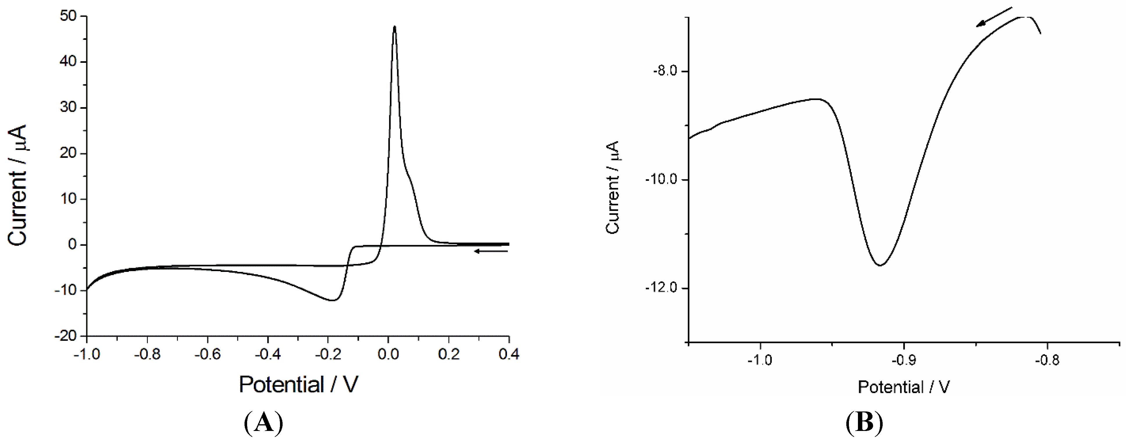
2.3.1. PPCE Preparation

A negative tone epoxy-based photoresist, SU-8, was photopatterned employing standard UV photolithography and pyrolyzed as reported previously [14]. Briefly, the wafers were dehydrated for 30 min at 120 °C right before the application of the photoresist. The photoresist was applied manually on the wafers and then spun, reaching the desired thickness following the manufacturer’s specifications [36]. After deposition, the casting solvent was evaporated from the photoresist in the soft or pre-bake step, which is usually performed at 95 °C on a hot plate. The baking of the photoresist in a convection oven would evaporate initially the solvent present on the top surface, thus hindering the evaporation of the solvent from the bulk of the polymer. The resist is then exposed to UV light (360 nm), which activates the catalyst for the cross-linking reaction. After a post-exposure bake, which is needed to fully polymerize the SU-8, the patterned features are developed. In this step, the un-polymerized SU-8 is dissolved upon immersion in a developer agent. The patterned structures are carbonized using a three-step pyrolysis process in an open-ended alumina-tube furnace. In the first step, the samples are heated under N2 (flow rate: 2000 sccm) at 300 °C for 1 h. The temperature is then ramped at a rate of 10 °C∙min−1 up to 900 °C. The samples are kept at the final pyrolysis temperature for 1 h before cooling. The electrodes were insulated with a Monokote® (Topflite, Champaign, IL, USA) film in order to define the geometric area to be exposed (0.07 cm2) to the solution during the electrochemical measurements (see [14] for details).
After preparation, every single electrode was characterized and validated by studying the cyclic voltammetric behavior of Fe(CN)64− used as the redox probe, as described previously [14]. It was observed that electrodes with peak-to-peak separation values (ΔEp = Epf − Epb, where Epf and Epb are the forward and backward peak potentials, respectively) comprised between 60 and 120 mV provide comparable results in terms of measured stripping current (i.e., within 5% of variability).

2.3.2. Analytical Procedure

The determination of Ni(II) at an in situ modified Bi-PPCE was carried out following a procedure previously described by Mardegan et al. [34] for the determination of Ni at gold electrodes modified with a bismuth film. Briefly, the PPCE was dipped into a 0.1-mol∙L−1 ammonium buffer solution containing 10 mg∙L−1 Bi(III) in the form of its complex with tartrate and 1 × 10⁻⁵ mol∙L−1 dimethylglyoxime (DMG) as the complexing agent. Tartrate was selected in order to avoid the precipitation of bismuth at the pH value necessary for Ni(II) and Co(II) determination [37].
A two-step accumulation protocol was followed: the application of a potential of −1.1 V for 60 s (in situ deposition of bismuth) followed by a second potential step of −0.8 V for 120 s (accumulation of the Ni-DMG or Co-DMG complex) under stirring condition. After 15 s of equilibration time, a cathodic stripping square-wave voltammogram (SWV) was recorded (from −0.8 V to −1.3 V) with a frequency of 25 Hz, a potential step of 5 mV and an amplitude of 50 mV. After each measurement, a cleaning step for the stripping of bismuth film was applied by holding the potential at +0.5 V for 120 s in the same solution. Each determination is the average of at least three consecutive measurements.

3. Results and Discussion

3.1. AdCSV of Ni(II) at Bi Modified PPCE

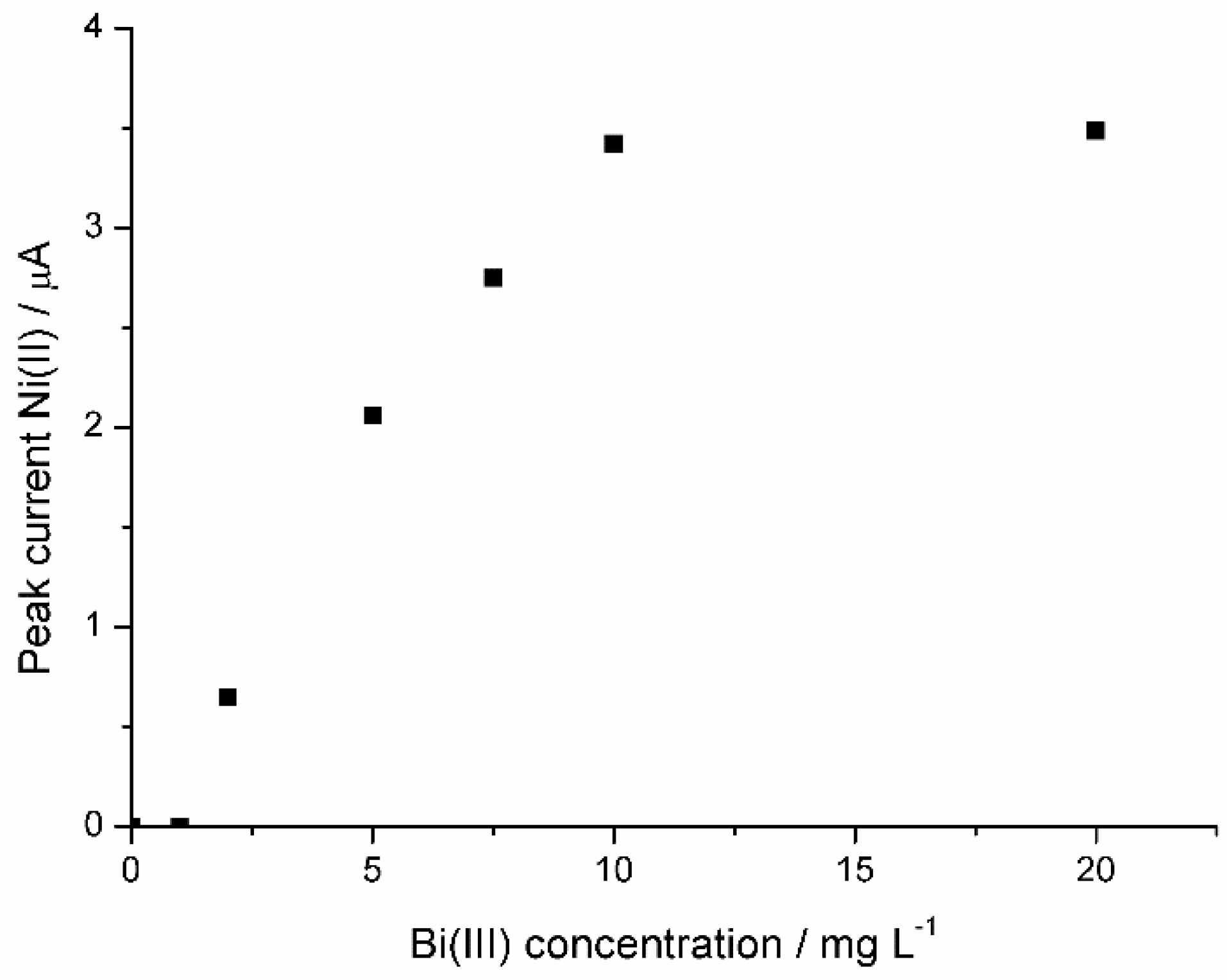
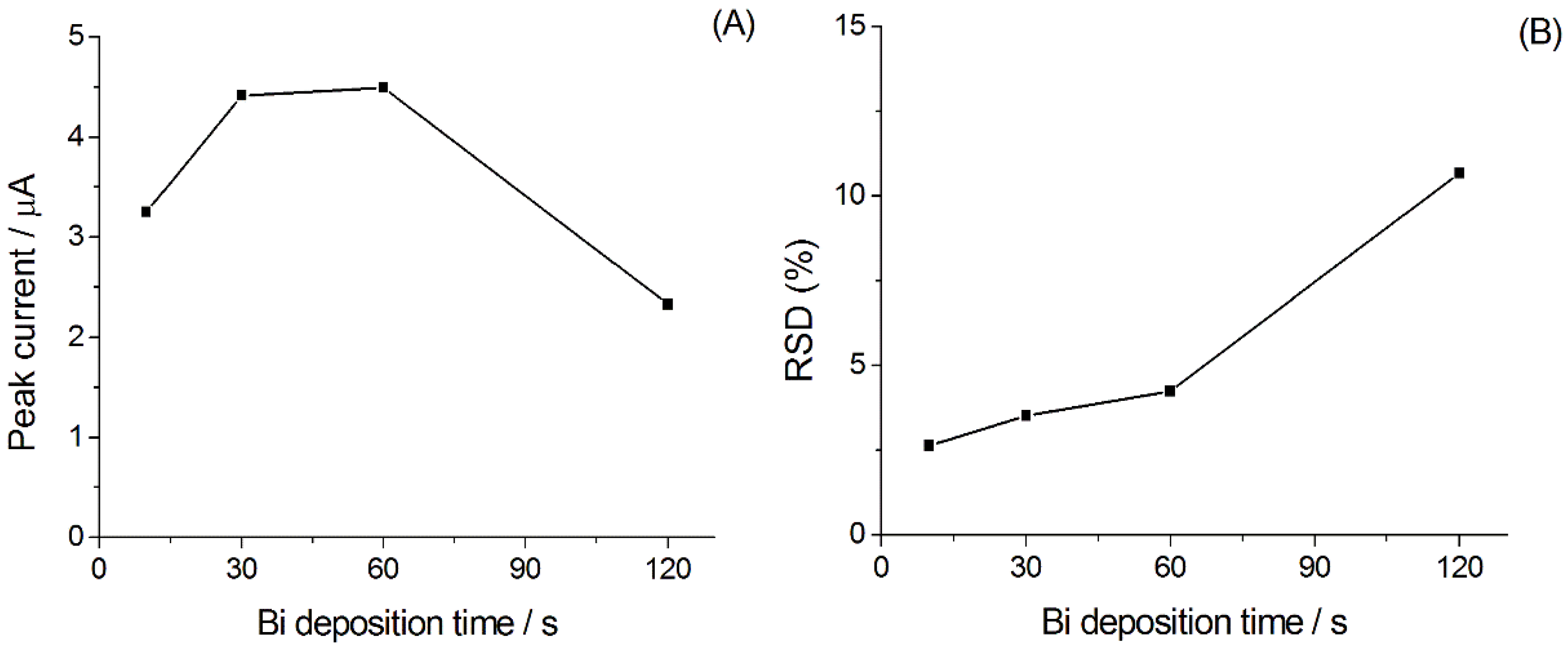
3.1.1. Optimization of the Experimental Parameters for Ni(II) Analysis

Following the procedure previously proposed for the determination of Ni at gold electrodes modified with a bismuth film [34], the study of the experimental conditions at PPC electrodes was carried out. Cyclic voltammetry was used to define the potential region for the deposition and dissolution of bismuth on PPCEs. The cyclic voltammogram recorded in 1 mmol∙L−1 Bi(III) acetate buffer solution at a scan rate of 10 mV s−1 is shown in Figure 1A. It is characterized by a couple of associated cathodic and anodic peaks at −0.18 V and +0.04 V and a crossover between the cathodic and anodic branches. The presence of the crossover is diagnostic for the formation of bismuth nuclei on the electrode surface [38].
The mechanism for the adsorption and cathodic stripping voltammetry of M(II)-DMG2, where M(II) is either Ni(II) or Co(II), follows a two-step route, in which the complex is firstly adsorbed on the electrode surface, and then, the application of cathodic potential scan results in the reduction of the metal complex following the scheme proposed by Ma et al [39]:
M(II)-DMG2 (sol) → M(II)-DMG2 (ads)
M(II)-DMG2 (ads) + ne → M(II)-DMG2n−
The reaction Equation (2) corresponds to the bielectronic reduction of the metal ion or to the four electron reduction of the ligand, or both; the ligand reduction is pH dependent. For more details on this process, see [39].
In the optimized procedure, the deposition of bismuth was fixed at a potential of −1.1 V, according to the literature [34]. In this step, Bi was deposited on the PPCE surface, whereas Ni(II)-(DMG)2 complex present in the solution was not adsorbed, since the adsorption occurs in the potential range of −0.8 and −0.6 V, while the reduction of the adsorbed complex is at ca. −0.95 V [34]. Figure 1B shows a typical AdCS voltammogram of Ni(II) 5 µg∙L−1 at an in situ prepared Bi-PPCE in 0.1 M ammonia buffer solution containing 10 mg∙L−1 Bi(III), 1 × 10⁻⁵ mol∙L−1 DMG and 0.01 mol∙L−1 tartrate. The concentration of DMG used here was optimized in previous works for the analysis with gold and GC electrodes [24,34]. The voltammogram is characterized by a well-defined cathodic peak potential at −0.92 V. The optimization of the experimental conditions was carried out on the basis of this behavior.
Figure 1. (A) Cyclic voltammogram recorded at a pyrolyzed photoresist electrode (PPCE) in 1 mmol∙L−1 Bi(NO3)3, 0.1 mol∙L−1 acetate buffer (pH 4.5) solution. Scan rate 10 mV s−1. (B) Typical adsorptive stripping voltammetry (AdCSV) of Ni(II) at an in situ prepared Bi-PPCE in 5 µg∙L−1 Ni(II) in 0.1 M ammonia buffer solution containing 10 mg∙L−1 Bi(III), 1 × 10⁻⁵ mol∙L−1 DMG and 0.01 mol∙L−1 tartrate. Deposition at −1.1 V for 60 s followed by accumulation at −0.8 V for 120 s; equilibration time of 15 s. Square wave parameters: frequency 25 Hz, potential step 5 mV, amplitude 50 mV.
Chemosensors 03 00157 g001
The effect of the bismuth concentration on the Ni(II) analysis was evaluated at a fixed deposition time of 60 s at the potential of −1.1 V, as suggested for the Bi deposition on gold electrodes [34]. During this step, Bi was deposited on the PPCE electrode surface, whereas the Ni(II)-DMG complex was not adsorbed. The concentration of Bi(III) in the measurement solution was changed in the range from 0 to 20 mg∙L−1, and its effect upon the Ni(II) stripping signal is shown in Figure 2. The stripping peak current increased when increasing the Bi(III) concentration from 2.5 mg∙L−1 up to reaching an approximately constant value at a Bi(III) concentration ≥10 mg∙L−1. At Bi-PPCE, the Ni(II) signal is observed at a Bi(III) concentration lower than at the Bi-Au electrode [34]; this behavior is due to the higher overpotential for hydrogen evolution observed at carbon with respect to gold electrodes [22]. The concentration of 10 mg∙L−1 of Bi(III) was then chosen to carry out nickel determination.
Figure 2. Effect of Bi(III) concentration on the Ni(II) peak current. Concentration of Ni(II): 10 µg∙L−1; accumulation conditions: 60 s at −0.8 V. Other experimental conditions as in Figure 1.
Chemosensors 03 00157 g002
The optimization of the bismuth deposition time was carried out at a fixed bismuth concentration (10 mg∙L−1) and a deposition potential of −1.1 V. The Bi(III) deposition time was studied in the range from 10 to 120 s, and its effect on the Ni(II) stripping signal is shown in Figure 3A. The highest peak current was recorded between 30 and 60 s of deposition, while for longer times, the signal decreases. Figure 3B shows the relative standard deviation (RSD) for a set of 10 independent measurements at each deposition time. It can be noted that for a deposition time up to 60 s, the RSD is lower than 5%, increasing remarkably for longer times. These results suggest that the deposition in 10 mg∙L−1 Bi(III) for 60 s represents the best conditions for the determination of Ni(II) with Bi-PPCEs. Such conditions were used in the following determinations of Ni(II).
Figure 3. (A) Effect of Bi(III) deposition time on the Ni(II) peak current; (B) effect of Bi(III) deposition time on the Ni(II) peak relative standard deviation (RSD). Concentration of Ni(II): 10 µg∙L−1, accumulation time at −0.8V 60 s. Other experimental conditions as in Figure 1.
Chemosensors 03 00157 g003

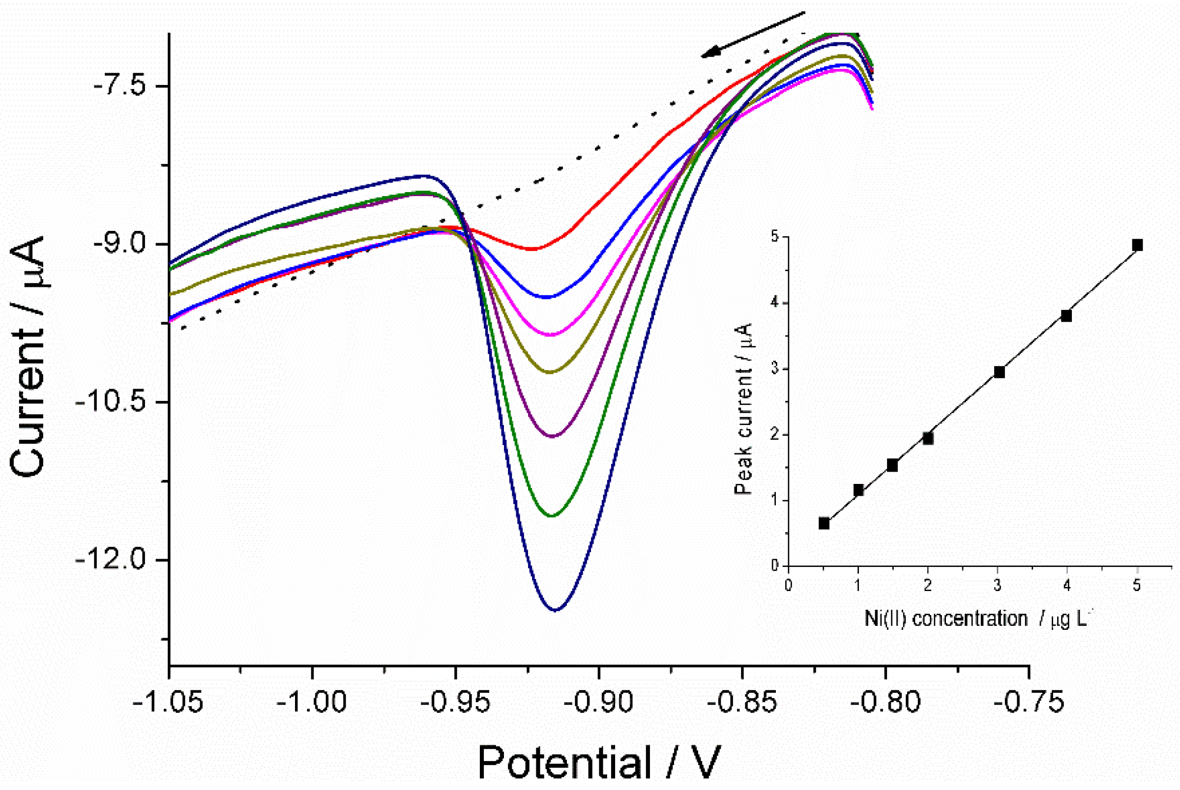
3.1.2. Calibration Plot

Figure 4 shows the AdCSVs recorded at a Bi-PPCE in the above optimized conditions for increasing concentrations of Ni(II) in the range from 0 to 5.0 µg∙L−1. According to [34], an accumulation time of the complex of 120 s represents a good compromise between sensitivity and analysis time.The reduction peak of the Ni-DMG complex, observed at around −0.9 V, increases linearly with the Ni concentration (see inset in Figure 4). A detection limit (LOD) of 20 ng∙L−1 was calculated by using the 3σb/m criterion, where σb is the standard deviation of the blank and m is the slope of the calibration plot. The peak current values obtained using different electrodes were reproducible within 5%. This value is comparable to the one obtained with bulk gold substrate and lower with respect to other electroanalytical methods reported in the literature [29,30,31,32,33,34].
Figure 4. AdCSVs at Bi-PPCE for increasing concentration of Ni(II) in the range of 0.5 to 5 µg∙L−1 in 0.1 M ammonia buffer solution containing 10 mg∙L−1 Bi(III), 1 × 10⁻⁵ mol∙L−1 dimethylglyoximate (DMG) and 0.01 mol∙L−1 tartrate. The dotted line was recorded in the absence of Ni(II). Deposition at −1.1 V for 60 s followed by accumulation at −0.8 V for 120 s; equilibration time of 15 s. Other experimental conditions as in Figure 1.
Chemosensors 03 00157 g004

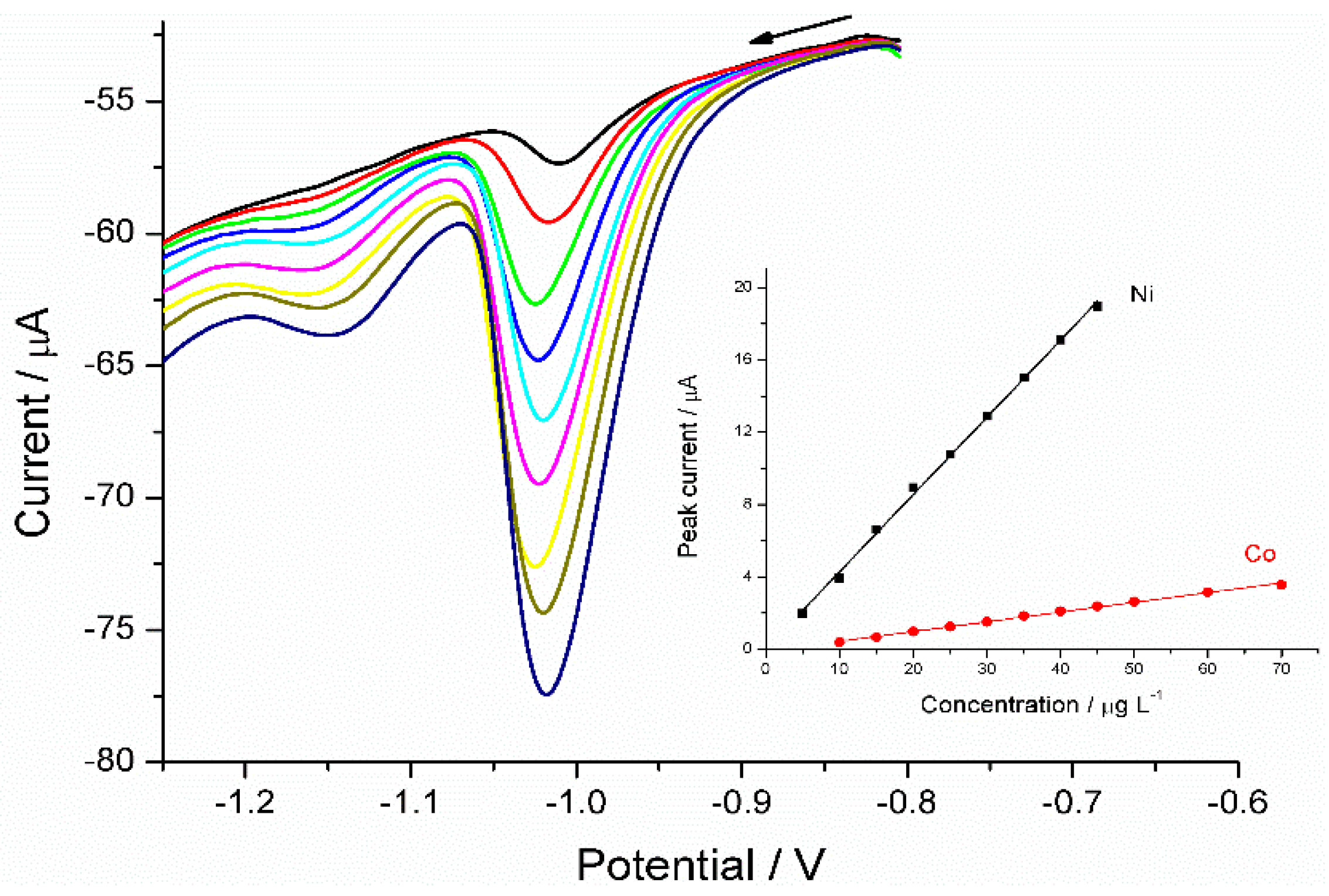
3.1.3. Adsorptive Stripping of Ni(II) in the presence of Co(II)

It is well known that DMG complexes other metal cations, such as Co(II) [37,40]; for this reason, cobalt is usually considered an interference in the determination of nickel. In order to verify the possible interference of Co(II), the above described analytical procedure was applied for analyzing Ni(II) in the presence of Co(II).
As shown in the voltammograms in Figure 5, the signals of both the analytes furnish two well-resolved stripping peaks at −1.050 V for Ni and −1.150 V for Co. A cathodic shift on the peak potential of Ni(II) stripping was formerly observed by other researchers [24]. A linear increase of the peak current is observed for both analytes in the range from 5 to 45.0 µg∙L−1 and 10 to 70 µg∙L−1, and the coefficients of determination (r2) of the linear fitting are 0.9992 and 0.9983, for Ni(II) and Co(II), respectively (see the inset in Figure 5). The calculated LODs (3σ/m) were 0.5 µg∙L−1 for Ni(II) and 2.5 µg∙L−1 for Co(II); this difference can be ascribed mainly to the lower sensitivity towards Co(II). These results confirm that Ni can be detected even in the presence of Co. Moreover, in principle, the method could be used to detect also Co, however with relatively low sensitivity.
Figure 5. AdCSVs at Bi-PPCE for increasing concentration of Ni(II) and Co(II) in 0.1 M ammonia buffer solution (pH = 9.0), 10 mg∙L−1 Bi(III), 1 × 10⁻⁵ mol∙L−1 DMG and 0.01 mol∙L−1 potassium sodium tartrate. Deposition at −1.1 V for 60 s followed by accumulation at −0.8 V for 60 s. Other parameters as in Figure 1.
Chemosensors 03 00157 g005

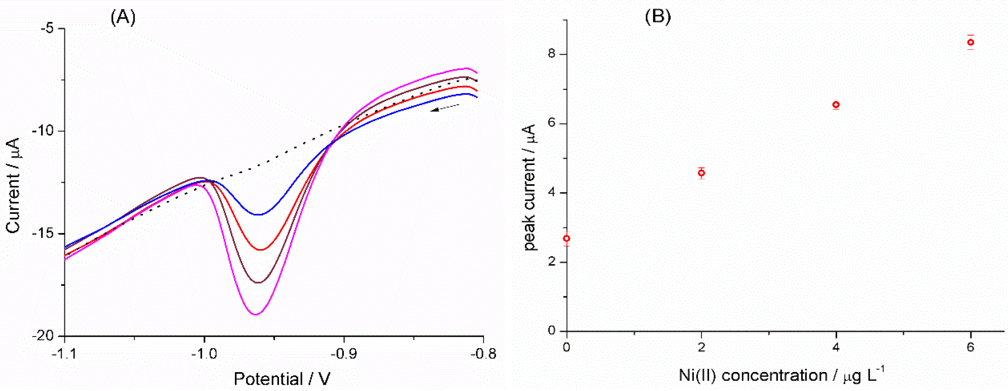
3.1.4. Analysis of Ni(II) in Certified Reference Material (NIST-1640a)

To evaluate the accuracy of the proposed method, Ni(II) was determined in a certified reference material (NIST-1640a). Two milliliters of NIST-1640a were diluted 1:10 with 0.1 mol∙L−1 ammonium buffer solution containing 0.01 mol∙L−1 sodium potassium tartrate, 1 × 10⁻⁵ mol∙L−1 DMG and 10 mg∙L−1 Bi(III). The AdCSVs recorded are characterized by a cathodic stripping peak at ca. −0.95V (see Figure 6A), which increased linearly with the additions of Ni(II), as shown in Figure 6B, and the linear fitting presented an r2 of 0.9999. Each value in the calibration plot is the average of four measurements. The calculated sample concentration was 26.3 ± 0.6 µg∙L−1 for Ni(II), which is in satisfactory agreement with the certified value of 25.12 ± 0.12 µg∙L−1.
Figure 6. (A) AdCSVs recorded at Bi-PPCE in a solution of NIST-1640a certified reference material and successive standard additions. Experimental conditions as in Figure 5. (B) Relevant standard additions plot.
Chemosensors 03 00157 g006

4. Conclusions

Pyrolyzed photoresist carbon electrodes (PPCE) were conveniently fabricated employing simple and inexpensive batch fabrication methodologies, UV photolithography followed by pyrolysis. PPCEs are good substrates for the deposition of bismuth, and the obtained Bi-PPCE display excellent analytical performances for trace nickel analyses. The nickel determination on Bi-PPCE is characterized by a LOD of 20 ng∙L−1, which, in the presence of Co, a common interference in Ni analysis, increases to 500 ng∙L−1. Application to analysis of real water samples is demonstrated.

Acknowledgments

We thank Marc Madou and Rahul Kamath (University of California, Irvine) for providing us with the PPCE. Financial support by the Veneto Region and the Ministry of Education, University and Research, MIUR, (Project PRIN 2010AXENJ8) is gratefully acknowledged.

Author Contributions

All authors contributed to conceive and design the experiments. A.M. worked in the preparation of the PPCE; A.M. and M.C. optimized the procedure and built the analytical curves; P.S. and L.M.M. supervised the experimental activity; L.M.M. wrote the manuscript. All authors discussed the results and commented on the manuscript.

Conflicts of Interest

The authors declare no conflict of interest.

References

  1. Kinoshita, K. Carbon Electrochemical and Physicochemical Properties; John Wiley & Sons: New York, NY, USA, 1988. [Google Scholar]
  2. Taylor, R.J.; Humffray, A.A. Electrochemical studies on glassy carbon electrodes: I. Electron transfer kinetics. J. Electroanal. Chem. Interfacial Electrochem. 1973, 42, 347–354. [Google Scholar] [CrossRef]
  3. Florence, T.M. Anodic stripping voltammetry with a glassy carbon electrode mercury-plated in situ. J. Electroanal. Chem. Interfacial Electrochem. 1970, 27, 273–281. [Google Scholar] [CrossRef]
  4. Sun, Y.C.; Mierzwa, J.; Yang, M.H. New method of gold-film electrode preparation for anodic stripping voltammetric determination of arsenic (III and V) in seawater. Talanta 1997, 44, 1379–1387. [Google Scholar] [CrossRef] [PubMed]
  5. Svancara, I.; Prior, C.; Hocevar, S.B.; Wang, J. A decade of bismuth-based electrodes in electroanalysis. Electroanalysis 2010, 22, 1405–1420. [Google Scholar] [CrossRef]
  6. Hocevar, S.B.; Svancara, I.; Ogorevc, B.; Vytras, K. Antimony film electrode for electrochemical stripping analysis. Anal. Chem. 2007, 79, 8639–8643. [Google Scholar] [CrossRef] [PubMed]
  7. Dai, S.; Nekrassova, O.; Hyde, M.E.; Compton, R.G. Anodic stripping voltammetry of arsenic(III) using gold nanoparticle-modified electrodes. Anal. Chem. 2004, 76, 5924–5929. [Google Scholar] [CrossRef] [PubMed]
  8. Dai, X.; Compton, R.G. Direct electrodeposition of gold nanoparticles onto indium tin oxide film coated glass: Application to the detection of arsenic(III). Anal. Sci. 2006, 22, 567–570. [Google Scholar] [CrossRef] [PubMed]
  9. Ugo, P.; Moretto, L.M.; Mazzocchin, G.A. Voltammetric determination of trace mercury in chloride media at glassy carbon electrodes modified with polycationic ionomers. Anal. Chim. Acta 1995, 305, 74–82. [Google Scholar]
  10. Ugo, P.; Moretto, L.M. Ion-exchange voltammetry at polymer-coated electrodes: Principles and analytical prospects. Electroanalysis 1995, 7, 1105–1113. [Google Scholar] [CrossRef]
  11. Zachek, M.K.; Takmakov, P.; Moody, B.; Wightman, R.M.; McCarty, G.S. Simultaneous decoupled detection of dopamine and oxygen using pyrolyzed carbon microarrays and fast-scan cyclic voltammetry. Anal. Chem. 2009, 81, 6258–6265. [Google Scholar] [CrossRef] [PubMed]
  12. Kim, J.; Song, X.; Kinoshita, K.; Madou, M.; White, R. Electrochemical Studies of Carbon Films from Pyrolyzed Photoresist. J. Electrochem. Soc. 1998, 145, 2314–2319. [Google Scholar] [CrossRef]
  13. Ranganathan, S.; McCreery, R.; Majji, S.M.; Madou, M. Photoresist derived carbon for microelectromechanical systems and electrochemical applications. J. Electrochem. Soc. 2000, 147, 277–282. [Google Scholar] [CrossRef]
  14. Mardegan, A.; Kamath, R.; Sharma, S.; Scopece, P.; Ugo, P.; Madou, M. Optimization of carbon electrodes derived from epoxy-based photoresist. J. Electrochem. Soc. 2013, 160, B132–B137. [Google Scholar] [CrossRef] [Green Version]
  15. Wang, J.; Lu, J.; Hocevar, S.B.; Farias, P.A.M.; Ogorevc, B. Bismuth-coated carbon electrodes for anodic stripping voltammetry. Anal. Chem. 2000, 72, 3218–3222. [Google Scholar] [CrossRef] [PubMed]
  16. Mikkelsen, Ø.; Schrøder, K.H. An Oscillating and renewing silver electrode for cadmium and lead detection in differential pulse stripping voltammetry. Electroanalysis 2001, 13, 687–692. [Google Scholar]
  17. Nolan, M.A.; Kounaves, S.P. Microfabricated array of iridium microdisks as a substrate for direct determination of Cu2+ or Hg2+ using square-wave anodic stripping voltammetry. Anal. Chem. 1999, 71, 3567–3573. [Google Scholar] [CrossRef]
  18. Mikkelsen, Ø.; Skogvold, S.M.; Schrøder, K.H.; Gjerde, M.I.; Aarhaug, T.A. Evaluation of solid electrodes for use in voltammetric monitoring of heavy metals in samples from metallurgical nickel industry. Anal. Bioanal. Chem. 2003, 377, 322–326. [Google Scholar]
  19. Švancara, I.; Matoušek, M.; Sikora, E.; Schachl, K.; Kalcher, K.; Vytřas, K. Carbon paste electrodes plated with a gold film for the voltammetric determination of mercury(II). Electroanalysis 1997, 9, 827–833. [Google Scholar] [CrossRef]
  20. Charalambous, A.; Economou, A. A study on the utility of bismuth-film electrodes for the determination of In(III) in the presence of Pb(II) and Cd(II) by square wave anodic stripping voltammetry. Anal. Chim. Acta 2005, 547, 53–58. [Google Scholar] [CrossRef]
  21. Švancara, I.; Baldrianová, L.; Tesařová, E.; Hočevar, S.B.; Elsuccary, S.A.A.; Economou, A.; Sotiropoulos, S.; Ogorevc, B.; Vytřas, K. Recent advances in anodic stripping voltammetry with bismuth-modified carbon paste electrodes. Electroanalysis 2006, 18, 177–185. [Google Scholar] [CrossRef]
  22. Mardegan, A.; Dal Borgo, S.; Scopece, P.; Moretto, L.M.; Hočevar, S.B.; Ugo, P. Bismuth modified gold nanoelectrode ensemble for stripping voltammetric determination of lead. Electrochem. Commun. 2012, 24, 28–31. [Google Scholar] [CrossRef] [Green Version]
  23. Bučková, M.; Gründler, P.; Flechsig, G.U. Adsorptive stripping voltammetric detection of daunomycin at a bismuth bulk electrode. Electroanalysis 2005, 17, 440–444. [Google Scholar] [CrossRef]
  24. Hutton, E.A.; Hočevar, S.B.; Ogorevc, B.; Smyth, M.R. Bismuth film electrode for simultaneous adsorptive stripping analysis of trace cobalt and nickel using constant current chronopotentiometric and voltammetric protocol. Electrochem. Commun. 2003, 5, 765–769. [Google Scholar] [CrossRef]
  25. Paneli, M.G.; Voulgaropoulos, A. Applications of adsorptive stripping voltammetry in the determination of trace and ultratrace metals. Electroanalysis 1993, 5, 355–373. [Google Scholar] [CrossRef]
  26. Brett, C.M.A.; Oliveira Brett, A.M.C.F.; Pereira, J.L.C. Adsorptive stripping voltammetry of cobalt and nickel in flow systems at wall-jet electrodes. Electroanalysis 1991, 3, 683–689. [Google Scholar] [CrossRef]
  27. Wang, J. Analytical Electrochemistry; Wiley-VCH: Verlag, Germany, 2006. [Google Scholar]
  28. Hutton, E.A.; Ogorevc, B.; Hočevar, S.B.; Smyth, M.R. Bismuth film microelectrode for direct voltammetric measurement of trace cobalt and nickel in some simulated and real body fluid samples. Anal. Chim. Acta 2006, 557, 57–63. [Google Scholar] [CrossRef]
  29. Korolczuk, M.; Rutyna, I.; Tyszczuk, K. Adsorptive stripping voltammetry of Nickel at an in-situ plated Bismuth film electrode. Electroanalysis 2010, 22, 1494–1498. [Google Scholar] [CrossRef]
  30. Wang, J.; Lu, J. Bismuth film electrodes for adsorptive stripping voltammetry of trace nickel. Electrochem. Commun. 2000, 2, 390–393. [Google Scholar] [CrossRef]
  31. Ruhlig, D.; Schulte, A.; Schuhmann, W. An electrochemical robotic system for routine cathodic adsorptive stripping analysis of Ni2+ ion release from corroding NiTi shape memory alloys. Electroanalysis 2006, 18, 53–58. [Google Scholar] [CrossRef]
  32. Piankova, L.A.; Malakhova, N.A.; Stozhko, N.Y.; Brainina, K.Z.; Murzakaev, A.M.; Timoshenkova, O.R. Bismuth nanoparticles in adsorptive stripping voltam-metry of nickel. Electrochem. Commun. 2011, 13, 981–984. [Google Scholar] [CrossRef]
  33. Alves, G.M.S.; Magalhães, J.M.C.S.; Soares, H.M.V.M. Simultaneous determination of nickel and cobalt, using a solid bismuth vibrating electrode, by adsorptive cathodic stripping voltammetry. Electroanalysis 2013, 25, 1247–1255. [Google Scholar] [CrossRef]
  34. Mardegan, A.; Dal Borgo, S.; Scopece, P.; Moretto, L.M.; Hocevar, S.B.; Ugo, P. Simultaneous adsorptive cathodic stripping voltammetric determination of Nickel(II) and Cobalt(II) at the in-situ bismuth-modified gold electrode. Electroanalysis 2013, 25, 2471–2479. [Google Scholar] [CrossRef] [Green Version]
  35. Sopha, H.; Jovanovski, V.; Hocevar, S.B.; Ogorevc, B. In-situ plated antimony film electrode for adsorptive cathodic stripping voltammetric measurement of trace nickel. Electrochem. Commun. 2012, 20, 23–25. [Google Scholar] [CrossRef]
  36. SU-8 2000 permanent epoxy negative photoresist processing guidelines for:SU-8 2025, SU-8 2035, SU-8 2050 and SU-8 2075. Available online: http://www.microchem.com/pdf/SU-82000DataSheet2025thru2075Ver4.pdf (accessed on 12 May 2015).
  37. Korolczuk, M.; Moroziewicz, A.; Grabarczyk, M. Determination of subnanomolar concentrations of cobalt by adsorptive stripping voltammetry at a bismuth film electrode. Anal. Bioanal. Chem. 2005, 382, 1678–1682. [Google Scholar] [CrossRef] [PubMed]
  38. Yang, M.; Hu, Z. Electrodeposition of bismuth onto glassy carbon electrodes from nitrate solutions. J. Electroanal. Chem. 2005, 583, 46–55. [Google Scholar] [CrossRef]
  39. Ma, F.; Jagner, D.; Renman, L. Mechanism for the electrochemical stripping reduction of the nickel and cobalt dimethylglyoxime complexes. Anal. Chem. 1997, 69, 1782–1784. [Google Scholar] [CrossRef] [PubMed]
  40. Rutyna, I.; Korolczuk, M. Catalytic adsorptive stripping voltammetry of cobalt in the presence of nitrite at an in situ plated bismuth film electrode. Electroanalysis 2011, 23, 637–641. [Google Scholar]

