Review

Yeast Gup1(2) Proteins Are Homologues of the Hedgehog Morphogens Acyltransferases HHAT(L): Facts and Implications

1
CBMA—Centre of Molecular and Environmental Biology, University of Minho, Campus de Gualtar, 4710-054 Braga, Portugal
2
School of Biology, Faculty of Biological Sciences, University of Leeds, Leeds LS2 9JT, UK
*
Author to whom correspondence should be addressed.
J. Dev. Biol. 2016, 4(4), 33; https://doi.org/10.3390/jdb4040033
Submission received: 1 July 2016 / Revised: 25 October 2016 / Accepted: 27 October 2016 / Published: 5 November 2016
(This article belongs to the Collection Hedgehog Signaling in Embryogenesis)

Abstract

:
In multiple tissues, the Hedgehog secreted morphogen activates in the receiving cells a pathway involved in cell fate, proliferation and differentiation in the receiving cells. This pathway is particularly important during embryogenesis. The protein HHAT (Hedgehog O-acyltransferase) modifies Hh morphogens prior to their secretion, while HHATL (Hh O-acyltransferase-like) negatively regulates the pathway. HHAT and HHATL are homologous to Saccharomyces cerevisiae Gup2 and Gup1, respectively. In yeast, Gup1 is associated with a high number and diversity of biological functions, namely polarity establishment, secretory/endocytic pathway functionality, vacuole morphology and wall and membrane composition, structure and maintenance. Phenotypes underlying death, morphogenesis and differentiation are also included. Paracrine signalling, like the one promoted by the Hh pathway, has not been shown to occur in microbial communities, despite the fact that large aggregates of cells like biofilms or colonies behave as proto-tissues. Instead, these have been suggested to sense the population density through the secretion of quorum-sensing chemicals. This review focuses on Gup1/HHATL and Gup2/HHAT proteins. We review the functions and physiology associated with these proteins in yeasts and higher eukaryotes. We suggest standardisation of the presently chaotic Gup-related nomenclature, which includes KIAA117, c3orf3, RASP, Skinny, Sightless and Central Missing, in order to avoid the disclosure of otherwise unnoticed information.

Graphical Abstract

1. Introduction

The yeast Saccharomyces cerevisiae is a recognised model to enlighten higher eukaryotic molecular processes, including the ones underlying human pathologies, which are very far from microbial life [1]. Intuitively, we associate yeasts, as microbes, to a planktonic form of life. However, microbes in the wild live mostly as large communities of cells forming biofilms or colonies, the biology of which remains largely unknown [2]. A colony or a biofilm displays a proto-tissue complex behaviour [3]. In these communities, cells organise spatially, morphologically and functionally to ensure the survival of the group. This implies the outlying orchestrated differentiation and death of cells to complete an efficient colonisation of the substrate, be it the pulp of a fruit or the surface of medical devices.
Within the large multicellular communities of yeast, cells behave similarly to their higher eukaryotic counterparts. They are born, grow larger and age while replicating until they senesce and die. Alternatively, they may die young, following an apoptotic cell death programme [4,5], allowing the supply of nutrients to the inner layers of the group [4], located further from the nutrient richer environment. Additionally, yeast cells may differentiate, shifting from yeast into true or pseudo-hyphae. These differ morphologically and physiologically [6,7,8]. The differentiation shift promotes an invasive behaviour generally associated with strain virulence [9]. Therefore, in contrast to planktonic growth, the survival strategy is collective [2] and resembles tissues from higher eukaryotes. Accordingly, large populations of yeast cells are imbedded in an extracellular matrix (ECM) composed of polysaccharides [10] and a large proteome [11], including many representatives of higher eukaryote ECM key proteins [11].
In higher eukaryotes, long distance communication between cells is achieved by secreted signalling proteins, namely from the Hedgehog (Hh) or Wingless (Wnt) pathways. These ligands interact with specific membrane-resident receptors that trigger a downstream signalling pathway in the receiving cell. No yeast cell-to-cell molecular communication vehicle has been recognised so far, although ammonia gradients have been suggested to perform a role in the collective orchestration of cell behaviour [12]. Moreover, the secretion of quorum-sensing small chemicals has been described in yeasts and suggested to perform a role in the control of population density [13,14]. Concomitantly, neither Hh- or Wnt-like pathways have been described in yeasts.
The maturation of the Hedgehog secreted morphogens involves intein self-splicing and C- and N-terminal lipidation (reviewed by [15]). In S. cerevisiae, there is only one recognised self-splicing protein that has a Hedgehog-like Hint domain, the endonuclease VDE (PI-SceI), which is required for gene conversion during meiosis [16]. VDE derives from the splicing of Vma1, the alpha subunit of the V-ATPase [17], whose human homologue has a role in human breast cancer cell invasiveness [18]. More importantly, the highly homologous yeast proteins Gup1 and Gup2, discovered by our group in 2000 [19], have two homologues in higher eukaryotes that are responsible for the Hh-secreted morphogen N-terminal palmitoylation (HHAT/Gup2) [20,21] and the negative regulation of the pathway (HHATL/Gup1) [22]. Gup1 and Gup2 are members of the MBOAT superfamily of membrane bound O-acyltransferases [23,24] and are present in all eukaryotic genomes sequenced to date. Their designation as GUP (Glycerol Uptake Protein) comes from their influence on the performance of glycerol active transport [19,25], which is actually accomplished by Stl1 [26], meaning that Gup proteins were often mistaken for the actual permease (e.g., [27]). In animal cells, the nomenclature is not consistent. In mouse, human or fly cells, both Gup1 and Gup2 are known by numerous aliases (Table 1). Such ambiguity generated the dispersion of experimental data on these proteins, as well as the under-interpretation of their roles. In yeasts, the Gup proteins have been implicated in a vast number of phenotypes from very diverse but fundamental biological/molecular processes, which will be revised in this work. In mammals and fly, the experimental data are scarcer and more recent. Here, we review the information available from S. cerevisiae and from another model yeast, the human commensal/opportunistic pathogen Candida albicans, and complement this with the role of Gup homologues in the regulation of Hedgehog ligand secretion in higher eukaryotes. Moreover, data on amino acid sequence comparison throughout model organisms are presented and a harmonising nomenclature is suggested.

2. Yeast Gup1 and Gup2 Proteins

2.1. Gup1 and Gup2 Are Members of the MBOAT Superfamily

S. cerevisiae GUP1 was first associated with the recovery of glycerol from the medium for osmoregulation purposes [19]. In accordance, glycerol active transport is deficient in GUP1-deleted mutants [19,25]. However, this is an indirect effect, resulting from the involvement of Gup1 in lipid raft formation [37]. Raft disassembly causes Pma1 H+-ATPase plasma membrane misdistribution [27], decreasing the appropriate proton motive force for active transporters like Stl1 to work properly [37]. Gup1 has a close homologue Gup2. The two proteins share a high degree of similarity (77%) and identity (57%) [19]. Gup1 and Gup2 are members of the MBOAT superfamily of multispanning membrane-bound O-acyltransferases, a superfamily that was first suggested based exclusively on sequence similarity between a small group of proteins [23]. Besides Gup proteins from S. cerevisiae, the MBOAT superfamily includes proteins as diverse as mammalian acyl-CoA:cholesterol acyltransferases (ACAT), fly palmitoyltransferases (Porcn and Nessy), Arabidopsis thaliana diglyceride acyltransferases (DGAT), bacteria Bacillus subtilis, Staphylococcus aureus and Pseudomonas aeruginosa d-alanyltransferases (DltB) and alginate O-acetyltransferases (AlgI) [23]. All of these proteins share a highly conserved histidine residue localised in a hydrophobic domain (His447 in yeast Gup1), as well as another residue of histidine, asparagine or aspartic acid, localised 30–50 amino acids upstream, within a hydrophilic region (His411 in the yeast Gup1). These conserved positions specify the active centre of these enzymes. The MBOAT superfamily is presently subdivided into three functionally different subgroups. One group includes enzymes involved in neutral lipid biosynthesis (ACATs and DGATs), another group includes proteins involved in phospholipid remodelling (lysophosphatidate acyltransferases—LPATs) and a third group includes the enzymes implicated in protein acylation (Porcupine (from the Wnt pathway), HHAT(L) (from the Hh pathway) and Ghrelin O-acyltransferase—GOAT (from the insulin regulatory pathway)) [38]. Gup1, although included in the third group, has also been included in the LPATs group due to the multiple biological and molecular roles assigned to it. In the mammalian HHAT(L), the highly conserved MBOAT signature amino acid residues are different. The yeast Gup1 His411/His447 and Gup2 His461/His498 positions are occupied by aspartic acid and histidine in HHAT and by aspartic acid and leucine in HHATL, respectively. Interestingly, the substitution of His447 by a leucine in S. cerevisiae Gup1 caused loss of phenotype [39], raising the question whether mammalian HHATL actually functions as an acyltransferase [22]. The roles attributed to Gup1 in all cellular models are for now exclusively biological/phenomenological, not biochemical. In contrast, the HHAT from high eukaryotes has a fully recognised enzymatic function in the N-palmitoylation of the Hh-secreted signals that can be reproduced in vivo and in vitro [21,40]. The yeast Gup2 shares with Gup1 the two MBOAT conserved histidines in the amino acid sequence positions 460 and 496.
According to the most common algorithms used to calculate the hydrophobicity of amino acid sequences, the Gup1 and Gup2 proteins both have 10 well-defined transmembrane domains (TMD). This number of TMD implicates that Gup1 terminals are both located on the same side of the membrane. However, the Gup1 C- and N-terminal domains are localised, respectively, in the periplasmic and cytosolic sides of the membrane [41], which is a rather uncommon topology. HHAT from mammals has a re-entrant loop stabilised by a lipid attachment, a palmitoylation of the MBOAT aspartic acid signature (Asp339) [42]. This re-entrant loop actually comprises both MBOAT signature amino acids (M. musculus Asp339 and His374; H. sapiens Asp340 and His377). S. cerevisiae Gup1 and Gup2 and C. albicans Gup1 present a partially hydrophobic region, located between TMD 7 and 8 (using the TMD prediction engine available at [43]). The correspondent sequence aligns perfectly with the sequence from the M. musculus HHAT re-entrant loop, showing several conserved several amino acid residues. In silico palmitoylation prediction of Gup amino acid sequence (using the prediction engine available at [44]) indicates a fully conserved cysteine residue in that region: S. cerevisiae Gup1 Cys396 and Gup2 Cys445; C. albicans Gup1 Cys417; M. musculus HHAT Cys324 and HHATL Cys326; H. sapiens HHAT Cys325 and HHATL Cys326; and D. melanogaster RASP Cys326. This degree of conservation, in addition to the possibility that a palmitoylation is involved, suggests that Gup proteins might require the MBOAT signature residues to be embedded in or at the surface of the membrane, whether this is obtained by Cys or Asp palmitoylation as in mammalian HHAT [42]. This possibility will have to be studied in the future.

2.2. Gup1 and Gup2 Subcellular Localisation

In S. cerevisiae, Gup1 protein co-localises with markers of the plasma membrane, the endoplasmic reticulum (ER) and the mitochondria [19]. Gup2 shares the first two locations [45]. The more prominent localisation of Gup1 appears to be the plasma membrane, as revealed by GFP tagging at either N- or -C-terminal [41]. The plasma membrane localisation of Gup1 is dependent on the secretory pathway proteins Sec6-4 [41] and End-3-mediated excision [26,41]. In opposition, in mammalian cells (mouse myocytes cell line), the Gup1 orthologue localises predominantly in the cytosol and only residually in the ER closer to the nucleus [34].

2.3. GUP1 and GUP2 Expression in Yeast

The yeast GUP2 promoter displays consensus sequences for 65 transcription factors (TF), and GUP1 for 19 TF (Table 2; [46]). These include major players in yeast transcription control, like the general stress regulators Msn2 and Msn4, the glucose-repression controller Mig1, Ste12 from pheromone response and mating and Gcn4 from nitrogen-associated regulation. Three TFs are predicted to regulate both promoters identically (Tec1, Mig3 and Rap1), and one—Ash1—is predicted to activate GUP1 and inhibits GUP2 transcription (Table 2).
The presence of numerous TF consensus sequences in yeast GUP1/2 promoters could reflect a complex transcription regulation, yet GUP1 and GUP2 are almost invariantly expressed in several different conditions. This is the case for cells actively growing on glucose (repression conditions—fermentation) and on glycerol or ethanol (de-repression conditions—respiration) [47], as well as in cells adapted to osmotic stress conditions created by high amounts of salt [47,48], or subjected to osmotic shock by high amounts of sorbitol [49]. These results suggest that the expression of these genes might be constitutive [47]. Nevertheless, this cannot be the case since GUP1 transcription duplicates upon NaCl shock [50]. Additionally, at the level of the protein, Gup1 is stably internalised in an End3-dependent manner upon sudden shift from glycerol to glucose [26,41]. Glycerol has multiple and complex roles in yeast cells. It is a respired poor carbon source whose metabolism requires induction and relief from glucose-repression. It functions as an osmolyte (reviewed by [51]), a lipid synthesis precursor [52], a pendulum of the NAD(H) redox balancing cytosol/mitochondrial shuttle [53], and is necessary for maintaining the signalling competent state of the cells [54]. The fact that Gup1 is retrieved from the plasma membrane upon glycerol-to-glucose transition, and overexpressed upon salt shock when glycerol accumulation is required for cell survival [51], establishes a connection between Gup1 and glycerol-associated regulation, which nature keeps unknown.
GUP2 is generally less expressed than GUP1 [47], and gup2∆ null mutants do not present any marked phenotype in response to carbon sources (glucose/glycerol) or stress (salts, ethanol, weak acids and high temperature). In contrast, gup1∆ mutants are moderately sensitive to all stresses [28] with the exception of high temperature, to which gup1∆ is highly sensitive [28,55]. The expression of GUP2 (not GUP1) has been shown to increase considerably upon ultraviolet irradiation [56], leading the authors to suggest Gup2 as part of the Early Genotoxic Response, i.e., the response to metal poisoning.
Up-regulation of Gup1 induces the up-regulation of several genes resident in the ER and Golgi, from the secretory pathway and lipid synthesis [39]. Kar2, a key protein from the Unfolded Protein Response (UPR) signalling pathway, stands out for its 24-fold increased expression [38]. Up-regulation of Gup1 further induces proliferation of membranous cisternal structures, with 1–7 per cell, physically separated from each other. This phenomenon is dependent on Ire1, another protein from the UPR pathway. ER, Golgi and itinerant proteins are present in these structures. Gup1 was also found, appearing to diffuse in and out of these structures [39]. Remarkably, the production of these membrane structures is dependent on the H447 MBOAT signature amino acid.

3. Yeast Phenotypes Associated with the Deletion of GUP1

3.1. Phenotypes Emerging from Genome-Wide Yeast Screenings

The extensive list of phenotypes associated with the deletion of GUP1 comprises many that were obtained in genome-wide screening. These include cytoskeleton polarisation and bud site selection patterns [57,58], secretory and endocytic pathways [59], vacuole morphology [59] and anaerobic growth [60]. Other high throughput studies cannot be presently rationalised, namely Askree et al. [61], which mentions Gup1 as an important protein in the maintenance of proper telomere length. Importantly, many of these studies show the resistance of the gup1∆ mutant to pharmaceutical drugs such as the anti-tumoral imatinib [62], the cytostatic cisplatin [63], the anti-inflammatory ibuprofen [64], the immunosuppressant FK506 [65] and the antimicrobial thymol [66]. Further resistance associated with GUP1 deletion includes lithium [67] and arsenic [68]. This large list, by itself, suggests an equally large complexity of Gup1 functions. In contrast, GUP2 has been referred to in only two HTP screenings [69,70]. The few evidences show that GUP2 expression is increased in response to ethanol stress in wine fermentative conditions/strains.

3.2. Cell Wall Integrity and Biogenesis

As mentioned above, gup1∆ is very sensitive to high temperature [28,55]. Generally, the sensitivity of yeast to high temperature is correlated with major defects in cell wall. Consistently, the deletion of GUP1 in S. cerevisiae causes an increase in the cell wall content of β-1,3-glucans (+25%) [28] and chitin (+90%) [28,71], and a decrease in the amount of mannoproteins (−70%) [28]. These large differences are responsible for a deficient wall morphology and sensitivity to wall-perturbing agents [28]. In spite of these defects, the Cell Wall Integrity (CWI)/PKC pathway is signalling normally in the gup1∆ mutant, as seen by MAPK (Slt2) (Figure 1A) dually phosphorylated state [28,72] after induction of the pathway by hypo-osmotic shock [28] or high temperature, or by NaCl or ethanol stress [73].
The CWI/PKC pathway controls not only the biogenesis of cell wall constituents and their remodelling, but also the associated organisation of the actin cytoskeleton and secretory pathway (reviewed by [74]). Unlike mammalian cells, S. cerevisiae only has one protein kinase C (Pkc1) that responds to the G-protein Rho1, the yeast homologue of RhoA. Rho1 activates Pkc1 besides other effectors. This allows the coordinated control of the actin cytoskeleton, exocytosis, membrane fluidity and the synthesis of wall glucans through transcription regulation in response to nutrients and stress [75]. Rho1 in turn is activated by Rom2/1 GDP/GTP exchange factors. These respond directly at least to a couple of wall stress sensors, Wsc1 and Mid2, and are further activated by the ligation of PI(4,5)P2 (phosphoinositol biphosphate) and/or of Slm1/2 proteins. The latter are targets for TORC2 (Target of Rapamycin 2 (TOR2) Complex) phosphorylation [76]. TORC2 also directly activates Pkc1, inducing the PKC pathway response. Importantly, Rho1 and consequently the PKC pathway are also activated by the α-factor plasma membrane receptor Ste2, a physical partner of Gup2 (Saccharomyces Genome Database). Rho1 is a signalling node, regulating many proteins and being under the effect of many others (including Kog1 from TORC1 (Target of Rapamycin 1 (TOR1) Complex) [77]), and it acts on actin cytoskeleton, thus causing cellular shape/polarity and filamentation [78].
TORC2 is redundant with TORC1 in the control of major cellular functions, but the cell wall composition and integrity and the polarisation of the actin cytoskeleton associated with cell growth, division and morphogenesis is only dependent on TORC2 [79,80]. Even though TOR proteins are very similar phosphatidylinositol kinase (PIK)-related kinases, only TOR1 is sensitive to inhibition by caffeine or the immunosuppressant rapamycin (reviewed by [80]). The gup1∆ mutant, both alone and in combination with gup2∆, is resistant to rapamycin [73]. On the other hand, gup1∆ is equally sensitive to caffeine as the wild type strain but, unlike this last, its sensitivity is remediable with sorbitol [28].
The mechanisms of action of caffeine and rapamycin are poorly understood. Both induce Rho1 activation and depend on this induction to inactivate the TORC1 pathway [77]. Caffeine induces the activation of Slt2 in a Tor1/Rom2 dependent manner [81] and elicits a transient decrease in the intracellular levels of cAMP, concomitantly inhibiting the Ras/cAMP pathway [81]. Moreover, caffeine supposedly activates the Pkc1 cascade through Tor1-mediated signalling [81]. Rapamycin, on the other hand, represses rRNA transcription and induces arrest of translation and cell cycle at G1. Furthermore, it causes glycogen accumulation, sporulation and autophagy [82]. These effects demand the complexation of rapamycin with Fpr1 (yeast equivalent of the immunophilin FKBP12) and subsequently with TOR1 [83]. Resistance to rapamycin in S. cerevisiae, can be provided by Fap1, which compets with Fpr1 [83,84]. However, the way in which Gup1 might be involved remains obscure.
Bearing in mind that the deletion of GUP1 causes defects in cell wall [73], membrane [37,55] and endocytosis [59], the cause of resistance to rapamycin could be the inability of the drug to enter the cell. Rapamycin is practically insoluble in water and poorly soluble in ethanol or glycerol [85]. This, together with its large molecular mass (≈900 Da), suggests an endocytic entrance. Additionally, gup1∆ is also resistant to the calcineurin inhibitor FK506 [65]. In mammalian cells, this immunosuppressant drug binds to FKBP12 inhibiting the Ca2+ signalling. Actually, FKBP12 bridges FK506, calcineurin and IP3 (inositol 3-phosphate) receptor, promoting the dephosphorylation of the latter, thereby modulating Ca2+ flux and signalling [86]. In yeast, FK506 affects the Ca2+-mediated response to high temperature, high NaCl-osmotic stress and α-factor pheromone [87]. High temperatures and high sodium, as discussed above, cause severe phenotypes in gup1∆ [19,28], while α-factor secretion is higher in gup1∆ [59].
The regulatory roles of phosphoinositides in yeast are not as well understood as in higher eukaryotes [88]. Still, their synthesis and turnover pathways are genetically characterised and the effects of some of the enzymes on major cellular processes have been recognised. At least seven unique second-messenger molecules are generated that regulate growth, differentiation, cytoskeletal rearrangements, membrane trafficking and protein vacuole sorting [88]. The phosphoinositide signalling operates in yeast as a star-type pathway, i.e., by cross-talking with numerous other pathways controlling their associated transcription, thus promoting the orchestrated cellular response to numerous stimuli. Major effectors associated with phosphoinosides in yeast belong to TORC2 and PKC pathways.
Yeast TOR pathways respond to carbon and nitrogen availability [89]. In silico analysis of the promoter regions of both GUP1 and GUP2 unveiled a series of putative regulatory sequences, from which the presence of repeated consensus sequences for Gcn4, a major transcription factor that responds to amino acid starvation and nitrogen in the dependence of TORC1, stands out [90]. Accordingly, wild-type strain and gcn4∆ mutant expressing lacZ reporter constructions for GUP1 and GUP2 promoter regions showed that β-galactosidase activity depended on Gcn4 for full expression of the GUP1 but not GUP2 in synthetic medium. If the medium is supplemented with 0.5% ammonium sulphate and 15 mM glycerol, the expression of Gup1 in gcn4∆ reverts to the values of wild-type strain [73]. The gup1∆ mutant dual dependence on nitrogen source and glycerol suggests an intricate position of the Gup1 protein in the midst of crossroads of N and C regulatory pathways. Moreover, the operationality of PKC signalling observed in the gup1∆ mutant suggests that the TORC2 pathway is not affected by the function of Gup1 protein. However, as discussed below, many of the cellular functions controlled by TORC2 are defective in the absence of GUP1. Besides wall damage-associated phenotypes, the most striking of these is the altered polarity during division exhibited by gup1∆ mutant [57,58].
The gup1∆ was found to display profoundly aberrant random bud site selection [57,58,76]. This implies a large defect on the proper establishment of cytoskeleton polarity [91,92], which is known to depend on Rho1/Pkc1 signalling [78]. It is as well dependent on other pathways associated with the multiple roles of the signalling node Cdc42 [93] (Figure 1B), including the Sho1 branch of the HOG pathway [94]. Cdc42 has the same fundamental role in mammalian cells (Figure 1B). This intricate crosstalk implicates the coordinated control of actin cytoskeleton remodelling for directional growth (elongation), which is mandatory for cell division through budding or mating. However, more importantly, underlies the morphogenic switch into filamentation that mediates invasion and adhesion [6,95].

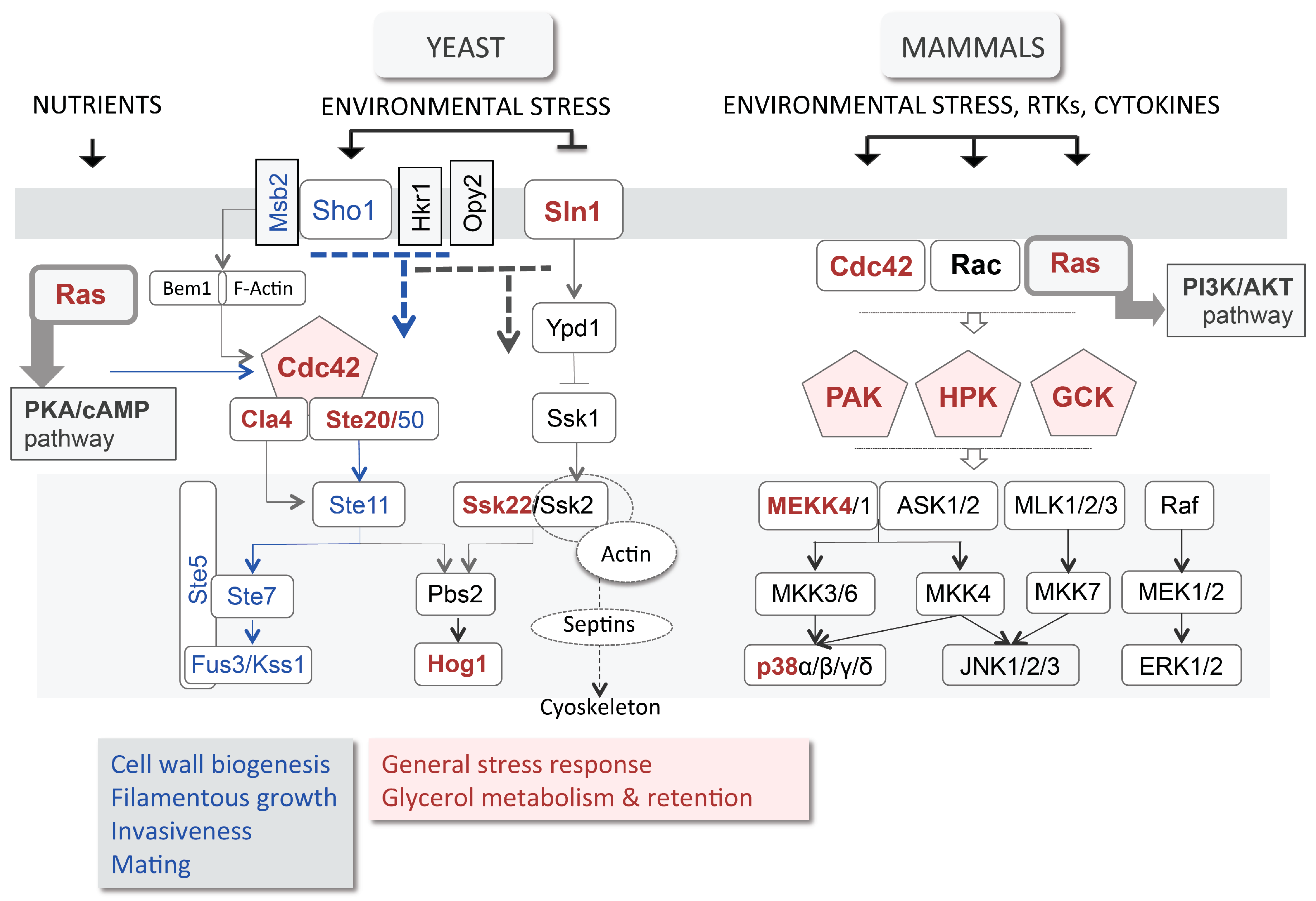
3.3. High Osmolarity Glycerol (HOG) Pathway

In spite of the GUP1 deletion, the CWI/PKC pathway is working properly at the level of Slt2 phosphorylation when stimulated by hypotonic shock [28]. Nevertheless, the wall of the mutant is severely affected. Two other pathways contribute to the wall integrity and remodelling: the Sho1 branch of the HOG pathway, and the Long-Chain Base (LCB)/sphingolipids YPK pathway (Figure 1A). HOG stands for High Osmolarity Glycerol, a name derived from control of the production and accumulation of the yeast osmolyte glycerol in response to high osmotic stress (reviewed by [51]). The HOG pathway controls many other processes such as polarity, adhesion and invasiveness, filamentation (i.e., differentiation), mating and cell wall biogenesis (reviewed by [96]). Importantly, the HOG pathway is functionally equivalent to the mammalian Wnt/p38 stress responsive MAPK pathway (Figure 2) [97,98]. Null mutants from HOG pathway have been successfully complemented by murine [99] or human p38 and associated kinases [100,101]. The p38 pathway is a non-canonical branch of Wnt MAPK, as are the JNK and ERK5 branches. Nevertheless, it has been shown to crosstalk with the Wnt canonical ERK1/2/β-catenin branch [102]. Actually, the four Wnt branches (ERK1/2, p38, JNK and ERK5) are all partially redundant in the control of growth/proliferation, i.e., cell cycle progression, differentiation, adhesion, migration and apoptosis in response to growth factors, cytokines and stress (reviewed by [103,104]). The HOG pathway Sho1 sensor functions in an exceptionally complex manner [105]. It binds to effectors that deliver different responses: Msb2 binds to Cdc42, activating the Ste11/Ste7/Fuss3/Kss1 filamentation/mating and cell wall biogenesis pathway, and Hkr1 or Opy2 that activate the HOG pathway (Figure 1 and Figure 2) [105,106].
The strains without GUP1 are unable to grow under high osmotic stress [19,28]. This could indicate a malfunction of the HOG pathway and a concomitant deficient production and/or accumulation of glycerol. Hyperosmotic stress also stimulates the CWI/PKC pathway [107], a fact that is not surprising since the adaptation to high osmotic stress implicates changes in cell volume and turgor that demand remodelling of the cell wall. The simultaneous activation of HOG and CWI/PKC pathways can also result from other stimuli, such as high temperature [108] and the action of zymolyase, an enzyme cocktail with β1,3-glucanase activity [109,110]. gup1∆ is extremely sensitive to both of these stimuli [28]. The effect of zymolyase is particularly severe, namely in comparison with lyticase, another wall disrupting enzyme [28]. This severity could be the consequence of the increased percentage of β1,3-glucans in the mutant cell wall. The malfunction of HOG rather than CWI/PKC pathway was also inferred from the pronounced phenotype of gup1∆ mutant in the presence of Calcofluor White [28] compared to Congo Red, which caused a negligible defect [111]. While some insults to the cell wall like Congo Red only trigger CWI/PKC [81,112], others trigger both HOG and CWI/PKC pathways [107,108,109,110]. In spite of the intricate interplay between both pathways (reviewed by [107]) the phenotypes caused by GUP1 deletion associated with osmoregulation [19] and the cell wall [28] are consistent with a relation of Gup1 with both pathways.

3.4. Plasma Membrane Composition and Associated Signalling

Gup1 is implicated in several membrane and lipids associated phenotypes [37,55,60,113]. The signalling pathway associated with complex lipids, the LCB/sphingolipid YPK pathway (partially depicted in Figure 1A), is not very well studied in yeasts, and no parallel has been established with the mammalian counterpart. The activation of Ypk1/2 is dependent on phosphorylation by Pkh1/2 kinases [114,115] or by TORC2 [115,116]. Pkh1/2 responds to LCBs and is implicated in membrane trafficking events. These include endocytosis through Pkc1-associated actin remodelling [114], protein phosphorylation [117], the trafficking of glycosylphosphatidylinositol (GPI)-anchored proteins, and the cell surface expression of amino acid permeases [118]. In mammalian cells, sphingosine acts as an inhibitor of PKC, or as activator of casein kinase II, Pkcζ, Pak1 (p21-activated kinase) and PDK1 (phosphoinositide-dependent kinase 1) (reviewed by [119]).
The lipid membrane composition and rafts content have implications in membrane fluidity/rigidity. In this regard, membrane fluidity homeostasis has been suggested to be under the control of a novel pathway that involves Rho1 effector [75]. According to these authors, the overlap between the CWI pathway with the membrane fluidity homeostasis pathway includes at least six components: Rho1, Pkc1, Bck1, Mkk1, Mkk2, Slt2 and Swi6. This is comparable to the overlap between mating response, pseudohyphal growth and the HOG pathways [107]. Membrane fluidity depends ultimately on the amount of complex lipids. Accordingly, the depletion of sphingolipids elicits the activation of TORC2 through Ypk1/2 and Orm1/2 that in turn stimulate their synthesis (Figure 1A) [120].
Down-regulation of TORC2-Ypk1 signalling allows cell survival under high osmotic stress independently of HOG pathway activation [121]. TORC2-Ypk1 signalling operates through closing of the glycerol channel Fps1, by blocking its phosphorylation by Ypk1. This is crucial to maintain the Fps1 channel open in the absence of stress [121]. This TORC2-Ypk1-mediated control occurs independently of the control of the channel by Hog1 [122]. Moreover, the enzyme responsible for deviating the glycolytic flux towards glycerol production Gpd1 (cytosolic glycerol 3 phosphate dehydrogenase) is also a target for Ypk1/2 [122], while its mitochondrial isogene Gpd2, from the glycerol NAD(H) shuttle, is under the direct control of glucose and is activated by phosphorylation via the AMP-activated kinase Snf1 [122]. Moreover, glycerol consumption and active uptake through Stl1 permease [26] is severely affected by the absence of PKC1 [123]. This strengthens the notion that glycerol is a major driver in maintaining the signalling competent state of cells, as suggested before [54], namely in the crossroads of responses to nutrients availability and stress through TORC2, sphingolipids, CWI/PKC and HOG pathways. The defects caused by GUP1 deletion appear in the centre of this puzzle, suggesting a pivotal role in signalling for the Gup1 protein.
LCB/sphingolipids form Detergent-Resistant Microdomains (DRM) in the plasma membrane, also known as lipid rafts due to their particular molecular/physicochemical properties. These properties derive from the predominantly saturated hydrocarbon tails of the sphingolipids and the high concentration of sterols: cholesterol in animal cells (e.g., [124]) and ergosterol in yeasts [125,126]. In yeast, as in mammalian cells, lipid rafts participate in protein sorting (contributing to the functions of transporters, pumps, sensors, receptors or G-proteins), dynamically including or excluding proteins [127,128,129]. Importantly, the components of lipid rafts are also actively involved in signalling and in stabilization of actin cytoskeleton–membrane interactions and therefore polarity [128,129].
In yeast, many proteins are known to functionally localise in lipid rafts forming a complex patchwork [128], which biogenesis is still poorly known. The first proteins found in yeast membrane microdomains were the H+-ATPase pump Pma1 [130] and the amino acid permease Can1 [131]. Presently, almost 300 proteins have been found to differentially localise into lipid rafts [128], including many permeases involved in ion homeostasis, water, glycerol or drugs transport as well as sugar or nitrogen transport/sensing, cell wall metabolism, stress response, signalling, flocculation, bud site selection and eisosome formation. Some are GPI-anchored like the β1,3-glucanosyltransferase Gas1 [125], while others are prenylated, or farnesylated and palmitoylated like Ras2 [132], but most just have transmembrane domains (see [128] for a comprehensive list). Different types of membrane lipid microdomains were identified and designated identified on the basis of the proteins they harbour: Membrane Compartment occupied by Pma1 (MCP), Can1 (MCC) [131] or TORC2 (MCT) [133].
The plasma membrane localisation of Gup1 [19,41] and Gup2 [45] has not been associated with raft residence [128]. Still, a patchy distribution of the labelled Gup1 protein was observed by electron microscopy [41]. Importantly, gup1∆ is affected in lipid rafts integrity and assembly [37]. This was evidenced by the even, not punctate, distribution of sterols in the mutant plasma membrane, as well as the 40% lower amounts of DRM recovered [37]. Additionally, Pma1 (H+ pump known to reside in lipid rafts, specifically MCPs [128]) is absent from gup1∆ DRMs, and the amounts of Gas1 (the most abundant yeast GPI-anchored protein) are residual, suggesting that rafts were unselectively affected by GUP1 deletion [37]. Accordingly, Gup1 interferes in sterol and sphingolipids synthesis, as evidenced by respectively increased and decreased resistance of the mutant to inhibitors of specific biosynthetic steps [37].
The yeast cell is a facultative anaerobic organism that can efficiently endure environments with virtually inexistent oxygen. In these conditions, the yeast cell does not synthesise sterols. Instead, it must take up sterols from the environment using the Pdr11 ATP-binding cassette (ABC) multidrug transporter [134], another permease known to locate in lipid rafts [128]. GUP1 was found to be crucial for survival in anaerobiosis [63]. In gup1Δ, not only is the biosynthesis of sterols affected, but also the uptake of sterols is impaired, underlying its inability to grow in anaerobic conditions [60].
Yeast lipid rafts assembly, unlike mammalian counterparts, takes place in the ER, only subsequently stepping into the Golgi [125]. Moreover, MCPs and MCCs are likely to segregate differently, since Pma1 is only incorporated into the rafts outside the ER [130]. ER resident proteins targeted to sphingolipids/ergosterol-poor membranes, such as the vacuole membrane, are not involved in rafts assembling [129]. Therefore, the defect in rafts assembly generated by GUP1 deletion must occur in the ER (one of Gup1 localisations) and may induce the segregation of specific membrane/protein assemblages towards their correct location. Another group of results concurs for a clear implication of the Gup1 protein in the secretory/endocytic pathways. In yeast, the vacuole performs the role of mammalian lysosomes. Proteins are addressed to the vacuole for degradation by the same pathway as carboxypeptidase Y (CPY), through the late endosome (reviewed by [135]). GUP1 was identified as one of the genes required for efficient sorting of CPY to the vacuole, as well as for the maintenance of this organelle regular size and morphology [59]. Its deletion also causes the excessive secretion of α-factor [59], the ER-resident protein Pdi1 [28], and the GPI-anchored β1,3-glucanosyltransferase Gas1 [37].
The deletion of GUP1, besides affecting sterols and sphingolipids synthesis and impairing sterols uptake, also causes changes in the regular concentrations of major lipid types: a high increase in diacyl- and triacylglycerol (Figure 3A) and a decrease in phospholipids [55]. Fatty acids synthesis, on the other hand, remains normal [37]. However, Gup1 is not recognised as an enzyme from lipid biosynthesis pathways. In this regard, Bosson and co-workers [113] proposed the involvement of Gup1 in the GPI synthesis/remodelling process, which occurs at the level of the ER in S. cerevisiae [136,137]. The anchors of mature GPI-anchored proteins of S. cerevisiae contain either ceramide or diacylglycerol with a C26 fatty acid in the sn2 position [138,139]. Gup1 was proposed to be the enzyme that adds C26 fatty acids to the sn2 position of lyso-phosphatidylinositol (lyso-PI)-containing GPI anchors (Figure 3A,B) [113]. Accordingly, the GUP1 deleted mutant, in contrast to the wild-type strain, was shown to accumulate lyso-PI and to contain phosphatidylinositol tails with C16 and C18 fatty acids instead of C26, all found in much smaller amounts compared to wild type (Figure 3B) [113]. The direct involvement of Gup1 as an enzyme in this process is nevertheless controversial. The wild-type GPI-anchor types can still be found in the mutant [113]. Concomitantly, it was suggested that Cwh43 was able to catalyse a bypass to the Gup1-dependent step in inositol phosphoceramide (IPC) tail maturation (Figure 3B) [140,141], using indifferently PI or Lyso-PI as substrates (Figure 3B). Moreover, the metabolic step associated with Gup1 is preceded by the transformation of PI into lyso-PI by Per1 (Figure 3B) [139]. In gup1∆ mutants, the lyso-PI harbours different fatty acid chains, suggesting that the Per1-dependent step is affected by the deletion of GUP1 as well [113]. Altogether, it is prudent to restrain from naming Gup1 as a mere GPI remodellase, since this classification does not take into account the multiple phenotypes associated with the protein, neither does it consider the localisation of Gup1 in the plasma membrane and its association with the mitochondria. In view of the complexity associated with Gup1 protein so far, the effects observed in GPI anchor synthesis could be indirect. GPI-anchor remodelling, if confirmed to be a direct function of Gup1 as an enzyme, will be among the other functions. The enzymes that catalyse the last steps of PI(4,5)P2 synthesis have associated phenotypes common to the gup1∆ mutant (Figure 4) (Saccharomyces Genome Database). This could provide the rationale for considering the possibility of Gup1 acting upstream of these enzymes, at the level of PI production (Figure 3). A change in PI availability would provide a reason for the changes observed at the level of the ceramide and diacylglycerol types (Figure 3B). The inosite metabolism was though never studied in this mutant.

3.5. Cell Death

Apoptosis is the most common process of Programmed Cell Death (PCD) in eukaryotes. It is vital for the fast elimination of useless or injured cells and for the differential development of tissues and organs. The existence of PCD processes in yeasts is presently unarguable [142]. As mentioned above, multicellular aggregates of microbial cells, like colonies or biofilms, are spatially organised and require the specialization of cells differentially localised to ensure supply of nutrients and water to the whole cell ensemble [143]. PCD occurs in a differentiated way within these aggregates [4]. The underlying rationale is that older cells of the colony centre that have already multiplied extensively, dying can contribute to the survival of adjacent cells with limited access to nutrients and water [4].
Several types of stimuli, both endogenous and exogenous, are known to cause yeast cells to enter a PCD process. H2O2 and acetic acid are major exogenous triggers commonly used to induce apoptosis in yeast (reviewed by [142]). However, several additional agents have been reported to induce apoptotic phenotypes, namely, salt stress, ethanol, heavy metals, UV radiation and high temperatures. Endogenous triggers on the other hand, include defects in N-glycosylation, chromatid cohesion, mRNA stability and ubiquitination (reviewed by [142]). Moreover, DNA damage (resulting from oxygen metabolism and ROS generation) and replication failure can stimulate the activation of yeast cell death programmes. Apoptosis may also play a role in ageing, replicatively [144] or chronologically [145,146].
Gup1 is required for several cellular processes that are related to apoptosis, namely the above discussed rafts integrity and stability [37], lipid metabolism [55], including GPI anchor correct remodelling [113], mitochondrial [60] and vacuole [59,147] functions and actin dynamics [57,58]. Accordingly, the deletion of GUP1 induces the hypersensitivity of yeast cells to two known apoptosis inducing conditions [148], chronological life span (CLS) [149] and acetic acid (reviewed by [150]). Two apparently contradictory works suggest that the mutation decreases [148], or extends [147] CLS. In this last work, replicative life span (RLS) is also described to increase in the absence of GUP1. The main difference between both studies resides in the amino acid availability in the yeast growth medium, which was four times higher in the study by Li et al. [147] than in the one by Tulha et al. [148]. Considering the above-mentioned suggestions that Gup1 might be implicated in ammonium and TOR signalling, which is controlled by nitrogen/amino acid abundance [79], the mutant will likely respond differently to apoptosis induction in either cultivation condition.
On the other hand, in the presence of lethal concentrations of acetic acid, gup1Δ unlike the wild type undergoes a necrotic-like cell death process [148]. This is inferred from the absence of typical apoptotic features: (a) maintenance of the membrane integrity, (b) phosphatidylserine externalisation, (c) depolarisation of mitochondrial membrane, and (d) chromatin condensation. Moreover, preliminary assays on autophagy inducing conditions, i.e., nitrogen starvation, show that the deletion of GUP1 causes a clear decrease of autophagic activity as determined by GFP-Atg8 (autophagy-related 8) degradation assay [45,151].

3.6. Differentiation and Morphology

The Hedgehog pathway has well-established roles in the development and maintenance of high eukaryote differentiation. In yeasts, changes in morphology and differentiation occur at two very distinct levels. Individual cells can differentiate by changing shape, shifting from yeasts to very elongated polarised cells of true or pseudo hyphae. Colonies can display multiple 3D shapes [152] that relate with the ability to invade and adhere. At this level, Candida species, namely C. albicans, have been more studied than S. cerevisiae, mainly due to their pathogenicity.
C. albicans is a commensal constituent of normal human microflora that acts as an opportunistic pathogen causing infections such as denture stomatitis, thrush and urinary tract infections, but can also provoke more severe systemic infections, in particular in immunocompromised individuals [153,154]. Among the underlying virulence mechanisms, the most well studied so far is probably the morphological switch from budding yeast to filamentous fungus, which, together with cell surface expression of adhesins and invasins (reviewed by [153]), allows colonisation of host tissues and their aggressive invasion, eventually overcoming the endothelial barrier [155]. The infection-associated yeast fast reproduction promotes formation of biofilms, not only on human and other organism tissues, but also on inert surfaces from clinical devices [154,156], constituting a major threat for hospitalised patients, in particular if immunocompromised.
C. albicans has only one GUP orthologue: caGUP1. caGup1 has been implicated in C. albicans virulence [29]. Accordingly, the cagup1Δ null mutant [157] was strongly affected in the ability to develop hyphae, to adhere, invade and form biofilm, and its colonies exhibited aberrant morphology/differentiation patterns [29]. Interestingly, M. musculus HHATL cDNA was able to complement the cagup1Δ null mutant morphogenic defects associated with colony invasive growth (Figure 5) [29]. Preliminary data suggests that the hyphae development defects of the mutant can be reverted although not completely by S. cerevisiae GUP1, as well as the human HHATL and D. melanogaster RASP(HHAT) [158]. This partial reversal corresponds to a notorious cell elongation into pseudohyphae instead of true hyphae. The amino acid sequences of C. albicans and M. musculus Gup proteins share only 20% similarity, whereas S. cerevisiae and C. albicans GUP1 sequences share 58%. Moreover, as mentioned above, the two key MBOAT amino acid residues differ in yeast Gup1/2 and mammalian HHAT(L). It remains to be seen in the future how far the high eukaryotes HHAT(L) ability goes in complementing yeast phenotypes, namely using alternative expression strategies and if the substitutions in the MBOAT residues play a role.
Similarly to S. cerevisiae, deletion of GUP1 in C. albicans modifies ergosterol distribution at the level of plasma membrane [29]. Among various lipids of yeast membranes, ergosterol has been used as target of the most common antifungals like polyenes and azoles [159,160]. In S. cerevisiae, deletion of GUP1 causes high resistance to ergosterol synthesis inhibitors including azoles [37]. Likewise, C. albicans cagup1Δ null mutant displayed increased resistance to fluconazole, ketoconazole and clotrimazole [29].
Another important phenotype of the cagup1Δ null mutant is the delayed ability to form biofilm [29]. The formation of biofilms, as the formation of colonies, derives from extensive colonisation of the environment, generating multicellular structures that allow regular feeding of all cells, comparably to a proto-tissue. Traditionally, C. albicans or other closely related yeasts have been extensively used to study biofilm formation. S. cerevisiae can be used to mimic biofilm by promoting its growth onto large communities such as giant colonies or mats [2,161,162,163]. Analogously to the tissues from higher eukaryotes, these multicellular aggregates depend on the production and secretion of extracellular matrix (ECM) [163]. In S. cerevisiae, this was shown to be made of two different polysaccharides [10] and to harbour a large and abundant proteome composed of 684 well-identified proteins belonging to very different functional classes [11]. The deletion of GUP1 in S. cerevisiae provokes a profound difference in the composition of the secretome, yielding a sludgy-textured ECM that lost one of the polysaccharides present in wild-type ECM and harbours approximately 15% less proteins, 26% of which are not found in the wild-type ECM [10,11]. Additionally, DIGE analysis identified as well common proteins though present in statistically different amounts [11]. Among the missing proteins, there are some key regulators of metabolic pathways, namely Fbp1, Pyc2 and Pdc5/6 from glycolysis/gluconeogenesis/fermentation; important effectors in cytoskeleton organization like Tub2, Ent2/3, Rvs161 or VPS; stress response and secretory pathway proteins like Vma6/7, Vph1 or Ctt1 and Ecm4/38; and proteins involved in ubiquitination and sumoylation [11]. Moreover, the selective presence in the wild-type ECM of alpha-saccharides remodelling enzymes, such as Glc3 1,4-α-glucan branching enzyme, Mnn2 α-1,2-mannosyltransferase and Sga1 α-glucoamylase, suggest the presence of α-based polysaccharides in opposition to the cell wall beta sugars backbone. These seem to be important for ECM texture as inferred from the absence of these enzymes in gup1∆ mutant ECM [11]. On the other hand, the mutant ECM comprises a great number of proteins involved in cell wall remodelling that were not found in the wild-type ECM, including the homologous GPI-anchored putative mannosidases Dcw1 and Dfg1, required for cell wall biosynthesis, Utr2, Kre6 and Krt2 proteins involved in the biosynthesis of β-glucans, Pir1 and Pir2 proteins involved in stabilization of the cell wall, and GPI-anchored protease Yps1 required for cell wall growth and maintenance. Clearly GUP1 considerably affects the secretion of both glycosides and proteins into the yeast ECM.

4. Proteins Interacting with Yeast Gup1 and Gup2

Although much work is still necessary to fully identify the full interactome of Gup1/2 proteins in yeast, these have putative physical partners, predicted by whole genome screenings, or ascertained in particular single-gene studies in the case of Gup1 (Table 3). Gup2 partners include interesting proteins (Table 3), like Ste2, the receptor for α-factor, a yeast pheromone that initiates signalling response to mating by interacting both with the pheromone and a heterotrimeric G-protein [164]. On the other hand, Gup1 co-localises with Mep2 [165] and Por1 [45] and immunoprecipitates with Por1 and Pil1 [45]. Pil1 is a major constituent of eisosomes, and Por1 is the mitochondrial VDAC (voltage-dependent anion channel). The nature of these two last interactions remains unknown for the time being. Biochemically, Por1 interacts with Sfk1, a suppressor of the calcineurin inhibitor immunosuppressant drug FK506, by which it modulates the mitochondrial ionic balance [166]. gup1∆ is resistant to FK506 [65], suggesting that Gup1 might be necessary for the interaction of Por1 with Sfk1, which would therefore be impaired from inhibiting FK506 in the mutant.
Mep2 belongs to a highly conserved Mep/Amt/Rh superfamily that also includes the Rh blood-group human antigens [167]. There are three well-characterised Mep-type ammonium transporters in S. cerevisiae, Mep1, 2 and 3 [168]. S. cerevisiae Mep2 and its positive regulator Npr1 kinase [169,170], and C. albicans Mep1 and Mep2 [171], are indispensable for differentiation, i.e., for true or pseudo-hyphae formation respectively. Mep2 has accordingly been associated with TORC1 signalling, implicating in CLS and ageing [172]. Moreover, Mep2 promotes activation of the PKA pathway by ammonium on nitrogen-starved cells [173]. GUP1 is required for proper distribution of Mep2 over the plasma membrane, while another protein that also putatively interacts with Gup1, Vma4 (Table 3), is required for Mep2 expression [165]. In gup1∆, Mep2 is localised in patches instead of homogenously over the entire plasma membrane, while both transport and signalling associated to Mep2 function are increased [165]. Although these authors suggest that the effect of Gup1 on Mep2 might be indirect in view of the multiple phenotypes associated with GUP1 gene involving plasma membrane, rafts and wall, it seems more likely that Gup1 directly negatively regulates Mep2. This would justify the higher Mep2-mediated transport and signalling observed in the strain deleted in GUP1 (with Mep2 uninhibited). However, Mep2 is released naturally onto the wild-type yeast ECM [11], suggesting that the picture is much more complex.

5. Gup1/2 Homologues from High Eukaryotes

5.1. Hedgehog Pathway

Yeast Gup1 and its close homologue Gup2 share significant sequence homology with proteins belonging to higher eukaryotes, from Caenorhabditis elegans, to Drosophila melanogaster, to mammals. All of those proteins, identical to Gup1/2 from yeast [23], were identified as members of the membrane bound O-acyltransferase (MBOAT) family based on their sequence homology. They all share two highly conserved regions of amino acids: one hydrophobic region containing an invariant histidine and a region composed of moderately hydrophobic amino acids that surround a well-conserved asparagine/aspartic acid residue [23]. The only exception found so far is the mammalian homologue of Gup1 HHATL (Hedgehog acyltransferase-like), in which the homologous regions are maintained, but the conserved histidine residue is replaced with a leucine. Thanks to the high degree of conservation of these two regions, at least 11 members of the MBOAT family have been identified so far in the human genome [38].
MBOAT family proteins also share a common topology. They all have 8–12 TMDs and localise in the ER or Golgi [38,181,182]. HHAT was first predicted to have eight TMDs [40], but this topology seems inconsistent when compared to other members of the MBOAT family. Specifically, the MBOAT signature amino acid residues would be located in the cytoplasm, which would be a feature unique to HHAT among all the other MBOAT members [42]. Recently, the topology of human HHAT has been further investigated using different prediction software, and the results obtained by two independent research groups showed that HHAT contains 10 TMDs and two re-entrant loops. The invariant His is located in the lumenal side of the endoplasmic reticulum, whereas the Asp residue is on the opposite side [42,183]. Conceivably, HHATL should have a similar structure, due to the high sequence similarity.
Most members of the MBOAT family catalyse the transfer of long chain fatty acids to hydroxyl groups of other lipophilic molecules [184,185,186], or the formation of wax esters [187]. They are categorised into three subgroups based on their biochemical reaction, due to the variety of processes in which they are involved [38]. Mammalian HHAT and its fly counterpart, together with Porcupine and Ghrelin O-acyltransferase (GOAT), belong to the group involved in the acylation of secreted proteins [38,182]. In particular, HHAT is the protein responsible for the N-palmitoylation of the Hedgehog morphogens [41,42] and Spitz (also known as Torpedo or DER), a fly protein homologous to TGF-alpha, that either acts as a secreted or as a cell-bound EGFR ligand [42]. M. musculus and human HHATL, as mentioned above, have the invariant His necessary for the palmitoylation activity of HHAT replaced by a Leu [40,42]. It is, therefore, expected to not act as an acyltransferase, although it was found to co-localise with both HHAT and Sonic in the ER [21]. It was shown that HHATL interacts directly with Shh decreasing the efficiency of Shh palmitoylation [22], being proposed as a negative regulator of the HHAT-driven N-palmitoylation of Shh and of the Hh pathway. However, the precise mechanism of action has yet to be described.
In mammals as well as in Drosophila, HHAT promotes Hedgehog secreted morphogens N-palmitoylation [20,21,22,40]. Hh morphogens are diffusible proteins involved in long distance cell-cell communication, playing fundamental roles in embryonic development, patterning and cellular differentiation [188,189,190,191], as well as in adult tissue homeostasis and cancer [192]. Proper signalling is achieved by a specific gradient of Hh morphogens concentration from the cells that produce it to those destined to bind them [169,193,194,195]. Hh post-translational lipophilic modifications have a key role in maintaining the protein concentration gradient, influencing the long range signalling [196,197], which depends on the formation of diffusible multimeric forms [198,199,200]. On the other hand, secretion of Hh morphogens might operate through several mechanisms. One is the formation of specific fat body-derived diffusible lipoprotein particles [196,201], which in Drosophila largely depends on lipid rafts [202,203,204]. Another mechanism is an exosome-mediated pathway [205,206]. Exosomes derive from the endocytic compartment where vesicles are packed [207]. Yeasts also produce and secrete exosomes [208,209], which proteome was characterised in C. albicans [210,211]. Still there is no idea regarding their biological roles.
In mammalian cells, Hedgehog responsive cells activate transcription through Gli1. This occurs in the Hh responding cells by the triangulation Hh ligand-Ptch1-Smo. Gli1 can be activated by Smo-independent mechanisms, increasing Hh signalling even in the absence of the Hh ligand. This non-canonical activation of Gli1 was described in association with oesophageal adenocarcinoma [212]. In that case, Gli1 is controlled by a TNFα-derived TORC1/S6K1 pathway. TORC1 promotes the phosphorylation of Gli1 (at S384) by S6K1, freeing it from SuFu and easing the entrance to the nucleus. Additionally, TORC2 promotes the stabilisation of Gli1 by keeping it phosphorylated (at S230) through the action of AKT [212,213]. AKT commands a star-type pathway and demands intracellular signalling by phosphoinosides, from which the designation of PI3K-PKB/AKT pathway (reviewed by [214]). Other possible cross-talks with Hedgehog have been suggested to operate at the level of Gli1 activation: MAPK/ERK [215] and KRAS [216].

5.2. HHAT(L) Proteins Expression and Associated Pathologies

Palmitoylation at the N-terminal domain of Hedgehog proteins is necessary for the proper Hh signalling at both long and short range [217]. It was shown in vitro that palmitoylated forms of Shh are 40–160-fold more active compared to unmodified Shh [218]. Palmitoylated Sonic Hh is required for the formation of multimeric complexes that are fundamental for extracellular migration [199]. Therefore, the proper functionality of HHAT is fundamental for maintenance of the correct Hh function. This should stand for the Hh pathway function, namely during embryogenesis. Accordingly, HHAT loss of function by a single G287V missense mutation resulted in serious sex development defects [219]. In general, Hh signalling malfunction, causes severe and often life-threatening developmental disorders such as holoprosencephaly, craniofacial defects and skeletal malformations [31,220,221,222].
In human tissues, the HHAT(L) picture appears somewhat more complex than predicted [22]. The HHATL gene is expressed in very few tissues and in different amounts: heart > skeletal muscle > brain [34,35,223]. Actually, the gene maps to a locus of dilated cardiomyopathy (CMD1E/OMIM 601154), a disorder characterised by ventricular dilation and impaired systolic function, resulting in congestive heart failure and arrhythmia, eventually causing premature death that also harbours another gene encoding a cardiac muscle Na+ channel (SCN5A/HH1). Despite this association, the absence of HHATL disease-specific mutations in a cohort of 48 patients [34], leaves no clear indication that HHATL might be responsible for the disease.
It has been estimated that 25% of all human tumours required Hh signalling to maintain tumour cell viability [167]. Corrupted Hh signalling, namely through over-activation, underlies many types of cancer, including small and non-small cell lung cancer, squamous cell and basal cell carcinomas and myeloid leukaemias [224,225,226,227,228]. The importance of Hh signal palmitoylation on this regard has recently been demonstrated. The knockdown of HHAT caused in vitro reduced proliferation and invasiveness of human pancreatic ductal adenocarcinoma [229]. Therefore, it is conceivable that blocking the Hedgehog pathway using HHAT inhibitors may block tumour progression [230,231]. Moreover, HHATL transfected cells presented lower capability of cell proliferation, invasion and tumorigenicity, supporting the notion that the protein might function as a tumour suppressor [36]. This function is compatible with the negative regulation of HHAT-mediated Hh palmitoylation described for HHATL [22]. The reason why this gene is only expressed in few tissues appears intriguing. Other works reported the expression of KIAA1173 (another GUP1/HHATL alias—Table 1) in normal skin [232] and nasopharyngeal mucosa [36], and its extreme down-regulation in cells of skin squamous cell carcinoma [232]. Still, the KIAA1173 protein is N-172 amino acids shorter than the cardiac isoform above-mentioned [34], suggesting that the HHATL ORF might undergo extensive RNA or protein splicing and perform different roles in different tissues.
The HHAT gene has also been associated with type 2 diabetes [233]. Its changed expression was found in patients undergoing bariatric surgery [233], further suggesting a correlation with glucose homeostasis and therefore obesity. Diabetes mellitus and obesity are closely related [234], through deficient glycerol consumption by glycerol kinase [235] and deficient tissue regulation of glycerol transport by aquaglyceroporins [236]. This once again raises the notion that in yeasts Gup1 implicates in glycerol transport, consumption and production for osmoregulation purposes [19,28] and that it apparently connects nutrient regulation to morphogenic-related Hedgehog-like phenotypes, through a signalling entanglement between the CWI/Pkc1, TORC2/YPK TORC1/Pkh1/2 and HOG/Sho1 pathways.

6. Similarity Analysis of the GUP/HHAT(L) Amino Acid Sequences

Gup proteins from both yeast and higher eukaryotes belong to the MBOAT superfamily of membrane-bound O-acyltransferases, although only the mammalian HHAT has been assigned a function as an enzyme performing the palmitoylation of the Sonic Hedgehog ligand [21]. All eukaryotes have at least one such gene/protein. Therefore, the databases present an extremely large number of sequences designated as Gup or HHAT(L). This generalised presence does not necessarily mean that the amino acid sequence is highly conserved. Multiple sequence alignment similarity matrices were built using Clustal Omega [237], based on the accession numbers available at UniProt database and confronting them with NCBI available sequences (Supplementary File S1). These were grouped in a simple manner: yeasts, filamentous fungi, mammals, fish, teleostomi, reptiles, birds, amphibian, insects, nematodes, testudines and arthopoda (Supplementary File S2). A few isolated organisms presenting GUP/HHAT(L) sequences were grouped as others in Supplementary File S2. Gup1/HHATL is absent from nematodes and insects, while Gup2/HHAT is absent from reptiles, amphibian, testudines, teleostomi and Zea mays, the only plant where a Gup amino acid sequence was so far identified. Within each matrix, the percentage of similarity varied between two extreme values (shadowed in File S2). Notably, Gup1/HHATL sequences from nematodes are the most diverse. Generally, Gup1/HHATL sequences are less diverse than the Gup2/HHAT, which is perceptible in the respective cladograms (Figure S1A,B) built using Mega 7 (Molecular Evolutionary Genetics Analysis) [238]. Still, the difference in similarity between the Gup1/HHATL and Gup2/HHAT sequences is very small. As an example, we present in Table 4 the similarity matrix between the better studied Gup proteins: S. cerevisiae Gup1 and Gup2, C. albicans Gup1, M. musculus HHATL and HHAT, H. sapiens HHATL and HHAT and D. melanogaster RASP/HHAT. The sequence similarity presented is very high. M. musculus HHAT shares 20% similarity with yeast Gup2 and 19.3% with yeast Gup1, while HHATL shares 21.9% similarity with yeast Gup2 and 20.7% with yeast Gup1. The frontier between proteins considered HHAT and HHATL is very narrow. Moreover, some sequences actually appear to be too different from the yeast Gup proteins to even be considered as such (Supplementary File S2).

7. Conclusions

Paracrine signalling has not yet been recognised as occurring in microbial communities. However, the notion that microbes multicellular aggregates can be regarded as tissues, suggested for the first time by Shapiro in 1988 (reviewed by [239]), predicts that each individual cell does not live exclusively in response to environmental stimuli, as happens in planktonic life, but must involve cell–cell communication. It remains unclear whether this may happen through a diffusible chemical, like ammonia [12,240,241] or the quorum-sensing chemicals [13,14], or through a peptide signal like in higher Eukaryotes. The large number and diverse proteome from the yeast ECM [11,210,211] hampers the identification of a putative peptide signal. Additionally, the also high number of proteins identified as being differently expressed in the GUP1-deleted mutant ECM compared to the wt strain, does not allow the suggestion of a candidate [11]. Nevertheless, the fact that all Eukaryotes present a Gup/HHAT(L) protein suggests a conserved mechanism in which these proteins may participate. One should, however, not discard the hypothesis that proteins from the same group might perform very different roles in different organisms. The plethora of phenotypes from the yeast gup1∆ mutant, and the multiple localisation of the Gup1 protein, actually favour this as having multiple roles.
The most prominent Gup1 and Gup2 localisation is the plasma membrane [19,41,45]. In mammalian or fly cells, HHAT is almost exclusively located in the ER, while the localisation HHATL is still scarcely documented. Data suggest that yeast Gup1 is, or locates at, a hub between CWI/Pkc1, TORC2/YPK, TORC1/Pkh1/2 and HOG/Sho1 pathways, controlling response to nutrients and stress and morphogenic-related Hedgehog-like phenotypes. Therefore, it is not surprising that GUP1 deletion-associated phenotypes cover a vast number of pathways controlling basic processes of yeast life (summarised in Figure 6), namely associated with growth and development and that the deletion of the gene does not, in spite of its central role, become lethal. Altogether only fitness appears to be decreased in most of the environmental conditions studied (temperature, pH, stress and so on).
Importantly, most of the phenotypes caused in yeast by the deletion of GUP1 or GUP2 are associated with the GUP1 deletion, in opposition of mammalian and fly cells in which case the information available almost exclusively regards Gup2/HHAT (Figure 6). This does not mean that Gup2 has no function in yeasts. In mammals, the Gup2/HHAT is the enzyme that performs the palmitoylation of the Hh signal [21], while Gup1/HHATL acts as an inhibitor of that palmitoylation [21] through an as yet unknown mechanism. If that would be the case for yeasts, Gup2 could be performing a role that is inhibited by Gup1 and, therefore, all of the phenotypes associated with the deletion of GUP1 would actually derive from the uninhibited function of Gup2.
A last comment goes to nomenclature. It is important that Gup proteins are not further taken as glycerol transporters. This mistaken allusion has equally appeared in the literature dedicated to yeasts or mammalian cells. Due to the poor understanding of these proteins, the Gup1 from S. cerevisiae has even been cloned and heterologously expressed as if it were the actual glycerol permease (e.g., [27]). Moreover, careful naming would be recommended in the fields of research using high eukaryotes, in view of the extreme similarity between HHAT and HHATL and between these and the original S. cerevisiae Gup1 and Gup2 proteins. In order to avoid the misguidance generated by the multiple designations (Table 1) we propose a single notification. Based on the examples of the Ras and TOR proteins, for which the name used in the higher eukaryotes and the numeration of yeasts were combined, we propose that HHAT1 and HHAT2 are consistently adopted hereon to be designated Gup1/HHATL and Gup2/HHAT, regardless of the biological system under study.
A lot is still required to unveil the true nature and biological function of the Gup proteins. The results so far contribute to stress that studies based on either yeasts or higher eukaryotes are equally important and complement themselves. In particular, in the near future, the search for the molecular partners/targets of the Gup proteins in yeast will be important, along with an understanding of why Gup1/HHATL is only present and important in a few adult human tissues, the physiology of which does not straightforwardly relate to the Hedgehog pathway.

Supplementary Materials

The following are available online at www.mdpi.com/2221-3759/4/4/33/s1.

Acknowledgments

This work was supported by the strategic programme UID/BIA/04050/2013 (POCI-01-0145-FEDER-007569) funded by national funds through the FCT I.P. and by the ERDF through the COMPETE2020—Programa Operacional Competitividade e Internacionalização (POCI); and by Marie Curie Initial Training Network GLYCOPHARM (PITN-GA-2012-317297). Giulia Cazzanelli is PhD grantee from GLYCOPHARM. Joana Tulha is PhD grantee SFRH/BD/76025/2011 from FCT (Fundação para a Ciência e Tecnologia) from the Portuguese Ministry of Science, Technology and Higher Education. Ricardo Franco-Duarte is post-doc grantee UMINHO/BPD/54/2015. We thank Fábio Faria-Oliveira presently a post-doc at the University of Göteborg, Sweden, for critical reading of the manuscript, and Pedro Ferraz, PhD student from our group at CBMA for all the text corrections and kind availability and support.

Author Contributions

Cândida Lucas, Giulia Cazzanelli and Joana Tulha wrote the paper. Célia Ferreira read, corrected and commented the manuscript. Ricado Franco-Duarte performed all the amino acids sequences analyses.

Conflicts of Interest

The authors declare no conflict of interest.

Nomenclature

According to the rules from Yeast scientific nomenclature, genes, proteins and mutants are presented in, capitals/italic (ex.: GUP1), title case (ex.: Gup1), and lower case/italics (ex.: gup1∆), respectively. In the case of other model organisms, proteins and genes are presented as in the corresponding literature.

References

  1. Verduyckt, M.; Vignaud, H.; Bynens, T.; van den Brande, J.; Franssens, V.; Cullin, C.; Winderickx, J. Yeast as a model for Alzheimer’s disease: Latest studies and advanced strategies. Meth. Mol. Biol. 2016, 1303, 197–215. [Google Scholar]
  2. Brückner, S.; Mösch, H.U. Choosing the right lifestyle: Adhesion and development in Saccharomyces cerevisiae. FEMS Microbiol. Rev. 2012, 36, 25–58. [Google Scholar] [CrossRef] [PubMed]
  3. Čáp, M.; Štepánek, L.; Harant, K.; Váchová, L.; Palková, Z. Cell differentiation within a yeast colony: Metabolic and regulatory parallels with a tumor-affected organism. Mol. Cell 2012, 46, 436–448. [Google Scholar] [CrossRef] [PubMed]
  4. Váchová, L.; Palková, Z. Physiological regulation of yeast cell death in multicellular colonies is triggered by ammonia. J. Cell Biol. 2005, 169, 711–717. [Google Scholar] [CrossRef] [PubMed]
  5. Váchová, L.; Palková, Z. Caspases in yeast apoptosis-like death: Facts and artefacts. FEMS Yeast Res. 2007, 7, 12–21. [Google Scholar] [CrossRef] [PubMed]
  6. Turrà, D.; Nordzieke, D.; Vitale, S.; El Ghalid, M.; di Pietro, A. Hyphal chemotropism in fungal pathogenicity. Semin. Cell Dev. Biol. 2016, 57, 69–75. [Google Scholar] [CrossRef] [PubMed]
  7. St’ovíček, V.; Váchová, L.; Kuthan, M.; Palková, Z. General factors important for the formation of structured biofilm-like yeast colonies. Fungal Genet. Biol. 2010, 47, 1012–1022. [Google Scholar] [CrossRef] [PubMed]
  8. Váchová, L.; Kucerová, H.; Devaux, F.; Ulehlová, M.; Palková, Z. Metabolic diversification of cells during the development of yeast colonies. Environ. Microbiol. 2009, 11, 494–504. [Google Scholar] [CrossRef] [PubMed]
  9. Zupan, J.; Raspor, P. Invasive growth of Saccharomyces cerevisiae depends on environmental triggers: A quantitative model. Yeast 2010, 27, 217–228. [Google Scholar] [PubMed]
  10. Faria-Oliveira, F.; Carvalho, J.; Belmiro, C.L.; Ramalho, G.; Pavão, M.; Lucas, C.; Ferreira, C. Elemental biochemical analysis of the polysaccharides in the extracellular matrix of the yeast Saccharomyces cerevisiae. J. Basic Microbiol. 2015, 55, 685–694. [Google Scholar] [CrossRef] [PubMed]
  11. Faria-Oliveira, F.; Carvalho, J.; Ferreira, C.; Hernáez, M.L.; Gil, C.; Lucas, C. Quantitative differential proteomics of yeast extracellular matrix: There is more to it than meets the eye. BMC Microbiol. 2015, 15, 271. [Google Scholar] [CrossRef] [PubMed]
  12. Palková, Z.; Váchová, L. Ammonia signaling in yeast colony formation. Int. Rev. Cytol. 2003, 225, 229–272. [Google Scholar] [PubMed]
  13. Chen, H.; Fujita, M.; Feng, Q.; Clardy, J.; Fink, G.R. Tyrosol is a quorum sensing molecule in Candida albicans. Proc. Nat. Acad. Sci. USA 2004, 101, 5048–5052. [Google Scholar] [CrossRef] [PubMed]
  14. Sprague, G.F.; Winans, S.C. Eukaryotes learn how to count: Quorum sensing by yeast. Genes Dev. 2006, 20, 1045–1049. [Google Scholar] [CrossRef] [PubMed]
  15. Guerrero, I.; Kornberg, T.B. Hedgehog and its circuitous journey from producing to target cells. Semin. Cell Dev. Biol. 2014, 33, 52–62. [Google Scholar] [CrossRef] [PubMed]
  16. Fukuda, T.; Nagai, Y.; Ohya, Y. Molecular mechanism of VDE-initiated intein homing in yeast nuclear genome. Adv. Biophys. 2004, 38, 215–232. [Google Scholar] [CrossRef]
  17. Chong, S.; Shao, Y.; Paulus, H.; Benner, J.; Perler, F.B.; Xu, M.Q. Protein splicing involving the Saccharomyces cerevisiae VMA intein. The steps in the splicing pathway, side reactions leading to protein cleavage, and establishment of an in vitro splicing system. J. Biol. Chem. 1997, 271, 22159–22168. [Google Scholar]
  18. McGuire, C.; Cotter, K.; Stransky, L.; Forgac, M. Regulation of V-ATPase assembly and function of V-ATPases in tumor cell invasiveness. Biochim. Biophys. Acta 2016, 1857, 1213–1218. [Google Scholar] [CrossRef] [PubMed]
  19. Hölst, B.; Lunde, C.; Lages, F.; Oliveira, R.; Lucas, C.; Kielland-Brandt, M.C. GUP1 and its close homologue GUP2, encoding multimembrane-spanning proteins involved in active glycerol uptake in Saccharomyces cerevisiae. Mol. Microbiol. 2000, 37, 108–124. [Google Scholar] [CrossRef] [PubMed] [Green Version]
  20. Chamoun, Z.; Mann, R.K.; Nellen, D.; von Kessler, D.P.; Bellotto, M.; Beachy, P.A.; Basler, K. Skinny Hedgehog, an acyltransferase required for palmitoylation and activity of the Hedgehog signal. Science 2001, 293, 2080–2084. [Google Scholar] [CrossRef] [PubMed]
  21. Buglino, J.A.; Resh, M.D. Hhat is a palmitoylacyltransferase with specificity for N-palmitoylation of Sonic Hedgehog. J. Biol. Chem. 2008, 283, 22076–22088. [Google Scholar] [CrossRef] [PubMed]
  22. Abe, Y.; Kita, Y.; Niikura, T. Mammalian Gup1, a homolog of Saccharomyces cerevisiae glycerol uptake/transporter 1, acts as a negative regulator for N-terminal palmitoylation of Sonic hedgehog. FEBS J. 2008, 275, 318–331. [Google Scholar] [CrossRef] [PubMed]
  23. Hofmann, K. A superfamily of membrane-bound O-acyltransferases with implications for Wnt signaling. Trends Biochem. Sci. 2000, 26, 111–112. [Google Scholar] [CrossRef]
  24. Neves, L.; Oliveira, R.; Lucas, C. Yeast orthologues associated with glycerol transport and metabolism. FEMS Yeast Res. 2004, 5, 51–62. [Google Scholar] [CrossRef] [PubMed] [Green Version]
  25. Neves, L.; Lages, F.; Lucas, C. New insights on glycerol transport in Saccharomyces cerevisiae. FEBS Lett. 2004, 565, 160–162. [Google Scholar] [CrossRef] [PubMed]
  26. Ferreira, C.; van Voorst, F.; Martins, A.; Neves, L.; Oliveira, R.; Kielland-Brandt, M.; Lucas, C.; Brandt, A. A member of the sugar transporter family, Stl1p is the glycerol/H+ symporter in Saccharomyces cerevisiae. Mol. Biol. Cell 2005, 16, 2068–2076. [Google Scholar] [CrossRef] [PubMed]
  27. Yu, K.O.; Kim, S.W.; Han, S.O. Engineering of glycerol utilization pathway for ethanol production by Saccharomyces cerevisiae. Bioresour. Technol. 2010, 101, 4157–4161. [Google Scholar] [CrossRef] [PubMed]
  28. Ferreira, C.; Silva, S.; van Voorst, F.; Aguiar, C.; Kielland-Brandt, M.C.; Brandt, A.; Lucas, C. Absence of Gup1p in Saccharomyces cerevisiae results in defective cell wall composition, assembly, stability and morphology. FEMS Yeast Res. 2006, 6, 1027–1038. [Google Scholar] [CrossRef] [PubMed]
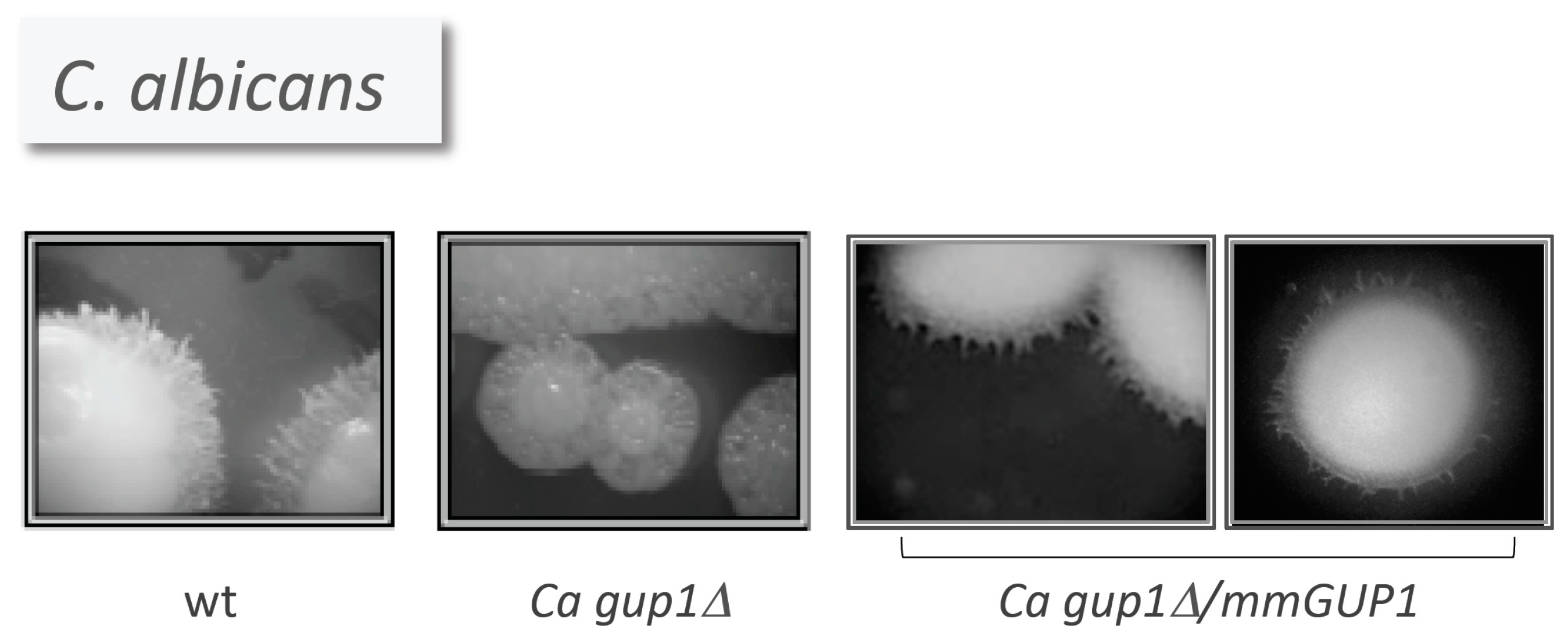
  29. Ferreira, C.; Silva, S.; Faria-Oliveira, F.; Pinho, E.; Henriques, M.; Lucas, C. Candida albicans virulence and drug-resistance requires the O-acyltransferase Gup1p. BMC Microbiol. 2010, 10, 238. [Google Scholar] [CrossRef] [PubMed] [Green Version]
  30. Micchelli, C.A.; The, I.; Selva, E.; Mogila, V.; Perrimon, N. Rasp, a putative transmembrane acyltransferase, is required for Hedgehog signaling. Development 2002, 129, 843–851. [Google Scholar] [PubMed]
  31. Lee, J.D.; Treisman, J.E. Sightless has homology to transmembrane acyltransferases and is required to generate active Hedgehog protein. Curr. Biol. 2001, 11, 1147–1152. [Google Scholar] [CrossRef]
  32. Amanai, K.; Jiang, J. Distinct roles of Central Missing and Dispatched in sending the Hedgehog signal. Development 2001, 128, 5119–5127. [Google Scholar] [PubMed]
  33. NCBI—National Center for Biotechnological Information. Available online: http://www.ncbi.nlm.nih.gov/ (assessed on 28 October 2016).
  34. Soejima, H.; Kawamoto, S.; Akai, J.; Miyoshi, O.; Arai, Y.; Morohka, T.; Matsuo, S.; Niikawa, N.; Kimura, A.; Okubo, K.; et al. Isolation of novel heart-specific genes using the Body Map database. Genomics 2001, 74, 115–120. [Google Scholar] [CrossRef] [PubMed]
  35. Zhang, S.Q.; Peng, H.; Jiang, H.Y.; Hu, H.; Zhang, J.H.; Yao, K.T.; Zhao, T. Cloning of human KIAA1173 gene and biological characterization of transfected 6–10B cells. Di Yi Jun Yi Da Xue Xue Bao 2005, 25, 1216–1220. [Google Scholar] [PubMed]
  36. Zhang, S.Q.; Peng, H.; Song, L.Y.; Li, X.M.; Jiang, H.Y.; Yao, K.T.; Zhao, T. Detection of KIAA1173 gene expression in nasopharyngeal carcinoma tissues and cell lines on tissue microarray. Ai Zheng 2005, 24, 1322–1326. [Google Scholar] [PubMed]
  37. Ferreira, C.; Lucas, C. The yeast O-acyltransferase Gup1p interferes in lipid metabolism with direct consequences on the sphingolipid-sterol-ordered domains integrity/assembly. Biochim. Biophys. Acta 2008, 1778, 2648–2653. [Google Scholar] [CrossRef] [PubMed]
  38. Chang, C.C.Y.; Sun, J.; Chang, T.-Y. Membrane-bound O-acyltransferases (MBOATs). Front. Biol. 2011, 6, 177–182. [Google Scholar]
  39. Bleve, G.; di Sansebastiano, G.P.; Grieco, F. Over-expression of functional Saccharomyces cerevisiae GUP1, induces proliferation of intracellular membranes containing ER and Golgi resident proteins. Biochim. Biophys. Acta 2011, 1808, 733–744. [Google Scholar] [CrossRef] [PubMed]
  40. Buglino, J.A.; Resh, M.D. Identification of conserved regions and residues within Hedgehog acyltransferase critical for palmitoylation of Sonic Hedgehog. PLoS ONE 2010, 5, e11195. [Google Scholar] [CrossRef] [PubMed]
  41. Bleve, G.; Zacheo, G.; Cappello, M.S.; Dellaglio, F.; Grieco, F. Subcellular localization and functional expression of the glycerol uptake protein 1 (GUP1) of Saccharomyces cerevisiae tagged with green fluorescent protein. Biochem. J. 2005, 390, 145–155. [Google Scholar] [CrossRef] [PubMed]
  42. Konitsiotis, A.D.; Jovanović, B.; Ciepla, P.; Spitaler, M.; Lanyon-Hogg, T.; Tate, E.W.; Magee, A.I. Topological analysis of Hedgehog acyltransferase, a multipalmitoylated transmembrane protein. J. Biol. Chem. 2015, 290, 3293–3307. [Google Scholar] [CrossRef] [PubMed]
  43. Transmembrane Helix Prediction. Available online: http://www.cbs.dtu.dk/services/TMHMM/ (assessed on 15 July 2016).
  44. CSS-Palm. Prediction of Palmitoylation Site. Available online: http://csspalm.biocuckoo.org (assessed on 15 July 2016).
  45. Tulha, J.; Lucas, C.; CBMA-Centre of Molecular and Environmental Biology, University of Minho, Braga, Portugal. Unpublished work. 2016.
  46. YEASTRACT, Yeast Search for Transcriptional Regulators and Consensus Tracking. Available online: http://www.yeastract.com/ (assessed on 15 July 2016).
  47. Oliveira, R.; Lucas, C. Expression studies of GUP1 and GUP2, genes involved in glycerol active transport in Saccharomyces cerevisiae, using semi-quantitative RT-PCR. Curr. Genet. 2004, 46, 140–146. [Google Scholar] [CrossRef] [PubMed] [Green Version]
  48. Posas, F.; Chambers, J.R.; Heyman, J.A.; Hoeffler, J.P.; de Nadal, E.; Ariño, J. The transcriptional response of yeast to saline stress. J. Biol. Chem. 2000, 275, 17249–17255. [Google Scholar] [CrossRef] [PubMed]
  49. Rep, M.; Krantz, M.; Thevelein, J.M.; Hohmann, S. The transcriptional response of Saccharomyces cerevisiae to osmotic shock. Hot1p and Msn2p/Msn4p are required for the induction of subsets of high osmolarity glycerol pathway-dependent genes. J. Biol. Chem. 2000, 275, 8290–8300. [Google Scholar] [CrossRef] [PubMed]
  50. Yale, J.; Bohnert, H.J. Transcript expression in Saccharomyces cerevisiae at high salinity. J. Biol. Chem. 2001, 276, 15996–16007. [Google Scholar] [CrossRef] [PubMed]
  51. Hohmann, S. Osmotic stress signaling and osmoadaptation in yeasts. Microbiol. Mol. Biol. Rev. 2002, 66, 300–372. [Google Scholar] [CrossRef] [PubMed]
  52. Henry, S.A.; Kohlwein, S.D.; Carman, G.M. Metabolism and regulation of glycerolipids in the yeast Saccharomyces cerevisiae. Genetics 2012, 190, 317–349. [Google Scholar] [CrossRef] [PubMed]
  53. Larsson, C.; Påhlman, I.-L.; Ansell, R.; Rigoulet, M.; Adler, L.; Gustafsson, L. The importance of the glycerol 3-phosphate shuttle during aerobic growth of Saccharomyces cerevisiae. Yeast 1988, 14, 347–357. [Google Scholar] [CrossRef]
  54. Siderius, M.; van Wuytswinkel, O.; Reijenga, K.A.; Kelders, M.; Mager, W.H. The control of intracellular glycerol in Saccharomyces cerevisiae influences osmotic stress response and resistance to increased temperature. Mol. Microbiol. 2000, 36, 1381–1390. [Google Scholar] [CrossRef] [PubMed]
  55. Oelkers, P.; Tinkelenberg, A.; Erdeniz, N.; Cromley, D.; Billheimer, T.; Sturley, S.L. A lecithin cholesterol acyltransferase-like gene mediates diacylglycerol esterification in yeast. J. Biol. Chem. 2000, 275, 15609–15612. [Google Scholar] [CrossRef] [PubMed]
  56. Dardalhon, M.; Lin, W.; Nicolas, A.; Averbeck, D. Specific transcriptional responses induced by 8-methoxypsoralen and UVA in yeast. FEMS Yeast Res. 2007, 7, 866–878. [Google Scholar] [CrossRef] [PubMed]
  57. Ni, L.; Snyder, M. A genomic study of the bipolar bud site selection pattern in Saccharomyces cerevisiae. Mol. Biol. Cell 2001, 12, 2147–2170. [Google Scholar] [CrossRef] [PubMed]
  58. Casamayor, A.; Snyder, M. Bud-site selection and cell polarity in budding yeast. Curr. Opin. Microbiol. 2002, 5, 179–186. [Google Scholar] [CrossRef]
  59. Bonangelino, C.J.; Chavez, E.M.; Bonifacino, J.S. Genomic screen for vacuolar protein sorting genes in Saccharomyces cerevisiae. Mol. Biol. Cell 2002, 13, 2486–2501. [Google Scholar] [CrossRef] [PubMed]
  60. Reiner, S.; Micolod, D.; Zellnig, G.; Schneiter, R. A genomewide screen reveals a role of mitochondria in anaerobic uptake of sterols in yeast. Mol. Biol. Cell 2006, 17, 90–103. [Google Scholar] [CrossRef] [PubMed]
  61. Askree, S.H.; Yehuda, T.; Smolikov, S.; Gurevich, R.; Hawk, J.; Coker, C.; Krauskopf, A.; Kupiec, M.; McEachern, M.J. A genome-wide screen for Saccharomyces cerevisiae deletion mutants that affect telomere length. Proc. Natl. Acad. Sci. USA 2004, 101, 8658–8663. [Google Scholar] [CrossRef] [PubMed]
  62. Dos Santos, S.C.; Sá-Correia, I. Genome-wide identification of genes required for yeast growth under imatinib stress: Vacuolar H+-ATPase function is an important target of this anticancer drug. OMICS 2009, 13, 185–198. [Google Scholar] [CrossRef] [PubMed]
  63. Liao, C.; Hu, B.; Arno, M.J.; Panaretou, B. Genomic screening in vivo reveals the role played by vacuolar H+ ATPase and cytosolic acidification in sensitivity to DNA-damaging agents such as cisplatin. Mol. Pharmacol. 2007, 71, 2416–2425. [Google Scholar] [CrossRef] [PubMed]
  64. Tucker, C.L.; Fields, S. Quantitative genome-wide analysis of yeast deletion strain sensitivities to oxidative and chemical stress. Comp. Funct. Genom. 2004, 5, 216–224. [Google Scholar] [CrossRef] [PubMed]
  65. Viladevall, L.; Serrano, R.; Ruiz, A.; Domenech, G.; Giraldo, J.; Barceló, A.; Ariño, J. Characterization of the calcium-mediated response to alkaline stress in Saccharomyces cerevisiae. J. Biol. Chem. 2004, 279, 43614–43624. [Google Scholar] [CrossRef] [PubMed]
  66. Darvishi, E.; Omidi, M.; Bushehri, A.A.; Golshani, A.; Smith, M.L. Thymol antifungal mode of action involves telomerase inhibition. Med. Mycol. 2013, 51, 826–834. [Google Scholar] [CrossRef] [PubMed]
  67. Zhao, J.; Lin, W.; Ma, X.; Lu, Q.; Ma, X.; Bian, G.; Jiang, L. The protein kinase Hal5p is the high-copy suppressor of lithium-sensitive mutations of genes involved in the sporulation and meiosis as well as the ergosterol biosynthesis in Saccharomyces cerevisiae. Genomics 2010, 95, 290–298. [Google Scholar] [CrossRef] [PubMed]
  68. Zhou, X.; Arita, A.; Ellen, T.P.; Liu, X.; Bai, J.; Rooney, J.P.; Kurtz, A.D.; Klein, C.B.; Dai, W.; Begley, T.J.; et al. A genome-wide screen in Saccharomyces cerevisiae reveals pathways affected by arsenic toxicity. Genomics 2009, 94, 294–307. [Google Scholar] [CrossRef] [PubMed]
  69. Rossouw, D.; Bauer, F.F. Comparing the transcriptomes of wine yeast strains: Toward understanding the interaction between environment and transcriptome during fermentation. Appl. Microbiol. Biotechnol. 2009, 84, 937–954. [Google Scholar] [CrossRef] [PubMed]
  70. Ma, M.; Liu, L.Z. Quantitative transcription dynamic analysis reveals candidate genes and key regulators for ethanol tolerance in Saccharomyces cerevisiae. BMC Microbiol. 2010, 10, 169. [Google Scholar] [CrossRef] [PubMed]
  71. Lesage, G.; Shapiro, J.; Specht, C.A.; Sdicu, A.M.; Ménard, P.; Hussein, S.; Tong, A.H.; Boone, C.; Bussey, H. An interactional network of genes involved in chitin synthesis in Saccharomyces cerevisiae. BMC Genet. 2005, 6, 8. [Google Scholar] [CrossRef] [PubMed] [Green Version]
  72. De Nobel, H.; Ruiz, C.; Martin, H.; Morris, W.; Brul, S.; Molina, M.; Klis, F.M. Cell wall perturbation in yeast results in dual phosphorylation of the Slt2/Mpk1 MAP kinase and in an Slt2-mediated increase in FKS2-lacZ expression, glucanase resistance and thermotolerance. Microbiology 2000, 46, 2121–2132. [Google Scholar] [CrossRef] [PubMed]
  73. Ferreira, C. Identification and characterisation of the glycerol/H+ symporter in Saccharomyces cerevisiaie and the involvement of related genes in the cell wall integrity. Ph.D. Thesis, University of Minho, Braga, Portugal, September 2005. Available online: http://repositorium.sdum.uminho.pt/ (assessed on 28 October 2016). [Google Scholar]
  74. Levin, D.E. Regulation of cell wall biogenesis in Saccharomyces cerevisiae: The cell wall integrity signaling pathway. Genetics 2011, 189, 1145–1175. [Google Scholar] [CrossRef] [PubMed]
  75. Lockshon, D.; Olsen, C.P.; Brett, C.L.; Chertov, A.; Merz, A.J.; Lorenz, D.A.; van Gilst, M.R.; Kennedy, B.K. Rho signaling participates in membrane fluidity homeostasis. PLoS ONE 2012, 7, e45049. [Google Scholar] [CrossRef] [PubMed]
  76. Audhya, A.; Loewith, R.; Parsons, A.B.; Gao, L.; Tabuchi, M.; Zhou, H.; Boone, C.; Hall, M.N.; Emr, S.D. Genome-wide lethality screen identifies new PI4,5P2 effectors that regulate the actin cytoskeleton. EMBO J. 2004, 23, 3747–3757. [Google Scholar] [CrossRef] [PubMed]
  77. Yan, G.; Lai, Y.; Jiang, Y. The TOR complex 1 is a direct target of Rho1 GTPase. Mol. Cell 2012, 45, 743–753. [Google Scholar] [CrossRef] [PubMed]
  78. Perez, P.; Rincón, S.A. Rho GTPases: Regulation of cell polarity and growth in yeasts. Biochem. J. 2010, 426, 243–253. [Google Scholar] [CrossRef] [PubMed]
  79. Loewith, R.; Hall, M.N. Target of Rapamycin (TOR) in nutrient signaling and growth control. Genetics 2011, 189, 1177–1201. [Google Scholar] [CrossRef] [PubMed] [Green Version]
  80. Jacinto, E.; Lorberg, A. TOR regulation of AGC kinases in yeast and mammals. Biochem. J. 2008, 410, 19–37. [Google Scholar] [CrossRef] [PubMed]
  81. Kuranda, K.; Leberre, V.; Sokol, S.; Palamarczyk, G.; François, J. Investigating the caffeine effects in the yeast Saccharomyces cerevisiae brings new insights into the connection between TOR, PKC and Ras/cAMP signalling pathways. Mol. Microbiol. 2006, 61, 1147–1166. [Google Scholar] [CrossRef] [PubMed]
  82. Hardwick, J.S.; Kuruvilla, F.G.; Tong, J.K.; Shamji, A.F.; Schreiber, S.L. Rapamycin-modulated transcription defines the subset of nutrient-sensitive signaling pathways directly controlled by the Tor proteins. Proc. Natl. Acad. Sci. USA 1999, 96, 14866–14870. [Google Scholar] [CrossRef] [PubMed]
  83. Lorenz, M.C.; Heitman, J. TOR mutations confer rapamycin resistance by preventing interaction with FKBP12-rapamycin. J. Biol. Chem. 1995, 270, 27531–27537. [Google Scholar] [PubMed]
  84. Kunz, J.; Loeschmann, A.; Deuter-Reinhard, M.; Hall, M.N. FAP1, a homologue of human transcription factor NF-X1, competes with rapamycin for binding to FKBP12 in yeast. Mol. Microbiol. 2000, 37, 1480–9143. [Google Scholar] [CrossRef] [PubMed]
  85. Simamora, P.; Alvarez, J.M.; Yalkowsky, S.H. Solubilization of rapamycin. Int. J. Pharm. 2001, 213, 25–29. [Google Scholar] [CrossRef]
  86. Cameron, A.M.; Nucifora, F.C., Jr.; Fung, E.T.; Livingston, D.J.; Aldape, R.A.; Ross, C.A.; Snyder, S.H. FKBP12 binds the inositol 1,4,5-trisphosphate receptor at leucine-proline (1400–1401) and anchors calcineurin to this FK506-like domain. J. Biol. Chem. 1997, 272, 27582–27588. [Google Scholar] [CrossRef] [PubMed]
  87. Yoshimoto, H.; Saltsman, K.; Gasch, A.P.; Li, H.X.; Ogawa, N.; Botstein, D.; Brown, P.O.; Cyert, M.S. Genome-wide analysis of gene expression regulated by the Calcineurin/Crz1p signaling pathway in Saccharomyces cerevisiae. J. Biol. Chem. 2002, 277, 31079–31088. [Google Scholar] [CrossRef] [PubMed]
  88. Odorizzi, G.; Babst, M.; Emr, S.D. Phosphoinositide signaling and the regulation of membrane trafficking in yeast. Trends Biol. Sci. 2000, 25, 229–235. [Google Scholar] [CrossRef]
  89. Staschke, K.A.; Dey, S.; Zaborske, J.M.; Palam, L.R.; McClintick, J.N.; Pan, T.; Edenberg, H.J.; Wek, R.C. Integration of general amino acid control and target of rapamycin (TOR) regulatory pathways in nitrogen assimilation in yeast. J. Biol. Chem. 2010, 285, 16893–16911. [Google Scholar] [CrossRef] [PubMed]
  90. Valenzuela, L.; Aranda, C.; González, A. TOR modulates GCN4-dependent expression of genes turned on by nitrogen limitation. J. Bacteriol. 2001, 183, 2331–2334. [Google Scholar] [CrossRef] [PubMed]
  91. Schuldiner, M.; Collins, S.R.; Thompson, N.J.; Denic, V.; Bhamidipati, A.; Punna, T.; Ihmels, J.; Andrews, B.; Boone, C.; Greenblatt, J.F.; et al. Exploration of the function and organization of the yeast early secretory pathway through an epistatic miniarray profile. Cell 2005, 123, 507–519. [Google Scholar] [CrossRef] [PubMed]
  92. Burston, H.E.; Maldonado-Báez, L.; Davey, M.; Montpetit, B.; Schluter, C.; Wendland, B.; Conibear, E. Regulators of yeast endocytosis identified by systematic quantitative analysis. J. Cell Biol. 2009, 185, 1097–1110. [Google Scholar] [CrossRef] [PubMed]
  93. Martin, S.G. Spontaneous cell polarization: Feedback control of Cdc42 GTPase breaks cellular symmetry. Bioessays 2015, 37, 1193–1201. [Google Scholar] [CrossRef] [PubMed]
  94. Raitt, D.C.; Posas, F.; Saito, H. Yeast Cdc42 GTPase and Ste20 PAK-like kinase regulate Sho1-dependent activation of the Hog1 MAPK pathway. EMBO J. 2000, 19, 4623–4631. [Google Scholar] [CrossRef] [PubMed]
  95. Styles, E.; Youn, J.Y.; Mattiazzi Usaj, M.; Andrews, B. Functional genomics in the study of yeast cell polarity: Moving in the right direction. Philos. Trans. R. Soc. B 2016, 368, 20130118. [Google Scholar] [CrossRef] [PubMed]
  96. Hohmann, S. An integrated view on a eukaryotic osmoregulation system. Curr. Genet. 2015, 61, 373–382. [Google Scholar] [CrossRef] [PubMed]
  97. Gordon, M.D.; Nusse, R. Wnt signaling: Multiple pathways, multiple receptors, and multiple transcription factors. J. Biol. Chem. 2006, 281, 22429–22433. [Google Scholar] [CrossRef] [PubMed]
  98. Cuadrado, A.; Nebreda, A.R. Mechanisms and functions of p38 MAPK signalling. Biochem. J. 2010, 429, 403–417. [Google Scholar] [CrossRef] [PubMed]
  99. Han, J.; Lee, J.D.; Bibbs, L.; Ulevitch, R.J. A MAP kinase targeted by endotoxin and hyperosmolarity in mammalian cells. Science 1994, 265, 808–811. [Google Scholar] [CrossRef] [PubMed]
  100. Kumar, S.; McLaughlin, M.M.; McDonnell, P.C.; Lee, J.C.; Livi, G.P.; Young, P.R. Human mitogen-activated protein kinase CSBP1, but not CSBP2, complements a hog1 deletion in yeast. J. Biol. Chem. 1995, 270, 29043–29046. [Google Scholar] [PubMed]
  101. Takekawa, M.; Posas, F.; Saito, H. A human homolog of the yeast Ssk2/Ssk22 MAP kinase kinase kinases, MTK1, mediates stress-induced activation of the p38 and JNK pathways. EMBO J. 1997, 16, 4973–4982. [Google Scholar] [CrossRef] [PubMed]
  102. Bikkavilli, R.K.; Feigin, M.E.; Malbon, C.C. p38 mitogen-activated protein kinase regulates canonical Wnt–β-catenin signaling by inactivation of GSK3β. J. Cell Sci. 2008, 121, 3598–3607. [Google Scholar] [CrossRef] [PubMed]
  103. Gallo, K.A.; Johnson, G.L. Mixed-lineage kinase control of JNK and p38 MAPK pathways. Nat. Rev. 2002, 3, 663–672. [Google Scholar] [CrossRef] [PubMed]
  104. The Wnt Homepage. Available online: http://web.stanford.edu/group/nusselab/cgi-bin/wnt/ (assessed on 8 August 2016).
  105. Tatebayashi, H.; Yamamoto, K.; Nagoya, M.; Takayama, T.; Nishimura, A.; Sakurai, M.; Momma, T.; Saito, H. Osmosensing and scaffolding functions of the oligomeric four-transmembrane domain osmosensor Sho1. Nat. Commun. 2015, 6, 6975. [Google Scholar] [CrossRef] [PubMed]
  106. Nishimura, A.; Yamamoto, K.; Oyama, M.; Kozuka-Hata, H.; Saito, H.; Tatebayashi, K. Scaffold protein Ahk1, which associates with Hkr1, Sho1, Ste11, and Pbs2, inhibits cross talk signaling from the Hkr1 osmosensor to the Kss1 mitogen-activated protein kinase. Mol. Cell Biol. 2016, 36, 1109–1123. [Google Scholar] [CrossRef] [PubMed]
  107. Rodríguez-Peña, J.M.; García, R.; Nombela, C.; Arroyo, J. The high-osmolarity glycerol (HOG) and cell wall integrity (CWI) signalling pathways interplay: A yeast dialogue between MAPK routes. Yeast 2010, 27, 495–502. [Google Scholar] [CrossRef] [PubMed]
  108. Winkler, A.; Arkind, C.; Mattison, C.P.; Burkholder, A.; Knoche, K.; Ota, I. Heat stress activates the yeast high-osmolarity glycerol mitogen-activated protein kinase pathway, and protein tyrosine phosphatases are essential under heat stress. Eukaryot. Cell 2002, 1, 163–173. [Google Scholar] [CrossRef] [PubMed]
  109. Alonso-Monge, R.; Real, E.; Wojda, I.; Bebelman, J.P.; Mager, W.H.; Siderius, M. Hyperosmotic stress response and regulation of cell wall integrity in Saccharomyces cerevisiae share common functional aspects. Mol. Microbiol. 2001, 41, 717–730. [Google Scholar] [CrossRef] [PubMed]
  110. Bermejo, C.; Rodríguez, E.; García, R.; Rodríguez-Peña, J.M.; de la Concepción, M.L.R.; Rivas, C.; Arias, P.; Nombela, C.; Posas, F.; Arroyo, J. The sequential activation of the yeast HOG and SLT2 pathways is required for cell survival to cell wall stress. Mol. Biol. Cell 2008, 19, 1113–1124. [Google Scholar] [CrossRef] [PubMed]
  111. Faria-Oliveira, F. First molecular and biochemical characterization of the extracelular matrix of Saccharomyces cerevisiae. Ph.D. Thesis, University of Minho, Braga, Portugal, September 2013. Available online: http://repositorium.sdum.uminho.pt/ (assessed on 28 October 2016). [Google Scholar]
  112. García, R.; Bermejo, C.; Grau, C.; Pérez, R.; Rodríguez-Peña, J.M.; Francois, J.; Nombela, C.; Arroyo, J. The global transcriptional response to transient cell wall damage in Saccharomyces cerevisiae and its regulation by the cell integrity signaling pathway. J. Biol. Chem. 2004, 279, 15183–15195. [Google Scholar] [CrossRef] [PubMed]
  113. Bosson, R.; Jaquenoud, M.; Conzelmann, A. GUP1 of Saccharomyces cerevisiae encodes an O-acyltransferase involved in remodeling of the GPI anchor. Mol. Biol. Cell 2006, 17, 2736–2645. [Google Scholar] [CrossRef] [PubMed]
  114. Friant, S.; Lombardi, R.; Schmelze, T.; Hall, M.N.; Riezman, H. Sphingolipid base signalling via PKh kinases is required for endocytosis in yeast. EMBO J. 2001, 20, 6783–6792. [Google Scholar] [CrossRef] [PubMed]
  115. Roelants, F.M.; Torrance, P.D.; Bezman, N.; Thorner, J. Pkh1 and Pkh2 differentially phosphorylate and activate Ypk1 and Ykr2 and define protein kinase modules required for maintenance of cell wall integrity. Mol. Biol. Cell 2002, 13, 3005–3028. [Google Scholar] [CrossRef] [PubMed]
  116. Roelants, F.M.; Torrance, P.D.; Thorner, J. Differential roles of PDK1- and PDK2-phosphorylation sites in the yeast AGC kinases Ypk1, Pck1 and Sch9. Nat. Rev. Microbiol. 2004, 150, 3289–3304. [Google Scholar]
  117. Friant, S.; Zanolari, B.; Riezman, H. Increased protein kinase or decreased PP2A activity bypasses sphigoid base requirement in endocytosis. EMBO J. 2000, 19, 2834–2844. [Google Scholar] [CrossRef] [PubMed]
  118. Dupré, S.; Haguenauer-Tsapis, R. Raft partitioning of the yeast uracil permease during trafficking along the endocytic pathway. Traffic 2003, 4, 83–96. [Google Scholar] [CrossRef] [PubMed]
  119. Neubauer, H.A.; Pitson, S.M. Roles, regulation and inhibitors of sphingosine kinase 2. FEBS J. 2013, 280, 5317–5336. [Google Scholar] [CrossRef] [PubMed]
  120. Roelants, F.M.; Breslow, D.K.; Muir, A.; Weissman, J.S.; Thornera, J. Protein kinase Ypk1 phosphorylates regulatory proteins Orm1 and Orm2 to control sphingolipid homeostasis in Saccharomyces cerevisiae. Proc. Natl. Acad. Sci. USA 2011, 108, 19222–19227. [Google Scholar] [CrossRef] [PubMed]
  121. Muir, A.; Roelants, F.M.; Timmons, G.; Leskoske, K.L.; Thorner, J. Down-regulation of TORC2-Ypk1 signaling promotes MAPK-independent survival under hyperosmotic stress. eLife 2015, 4, e09336. [Google Scholar] [CrossRef] [PubMed]
  122. Lee, J.; Reiter, W.; Dohnal, I.; Gregori, C.; Beese-Sims, S.; Kuchler, K.; Ammerer, G.; Levin, D.E. MAPK Hog1 closes the S. cerevisiae glycerol channel Fps1 by phosphorylating and displacing its positive regulators. Genes Dev. 2013, 27, 2590–2601. [Google Scholar] [CrossRef] [PubMed]
  123. Gomes, K.N.; Freitas, S.M.; Pais, T.M.; Fietto, J.L.; Totola, A.H.; Arantes, R.M.; Martins, A.; Lucas, C.; Schuller, D.; Casal, M.; et al. Deficiency of Pkc1 activity affects glycerol metabolism in Saccharomyces cerevisiae. FEMS Yeast Res. 2005, 5, 767–776. [Google Scholar] [CrossRef] [PubMed] [Green Version]
  124. Van Meer, G. Lipid traffic in animal cells. Annu. Rev. Cell Biol. 1989, 5, 247–275. [Google Scholar] [CrossRef] [PubMed]
  125. Bagnat, M.; Keranen, S.; Shevchenko, A.; Simons, K. Lipid rafts function in biosynthetic delivery of proteins to the cell surface in yeast. Proc. Natl. Acad. Sci. USA 2000, 97, 3254–3259. [Google Scholar] [CrossRef] [PubMed]
  126. Jacobson, K.; Mouritsen, O.G.; Anderson, R.G.W. Lipid rafts: At a crossroad between cell biology and physics. Nat. Cell Biol. 2007, 9, 7–14. [Google Scholar] [CrossRef] [PubMed]
  127. Mollinedo, F. Lipid raft involvement in yeast cell growth and death. Front. Oncol. 2012, 2, 140. [Google Scholar] [CrossRef] [PubMed] [Green Version]
  128. Spira, F.; Mueller, N.S.; Beck, G.; von Olshausen, P.; Beig, J.; Wedlich-Söldner, R. Patchwork organization of the yeast plasma membrane into numerous coexisting domains. Nat. Cell Biol. 2012, 14, 640–648. [Google Scholar] [CrossRef] [PubMed]
  129. Bagnat, M.; Simons, K. Lipid rafts in protein sorting and cell polarity in budding yeast Saccharomyces cerevisiae. Biol. Chem. 2002, 383, 1475–1480. [Google Scholar] [CrossRef] [PubMed]
  130. Bagnat, M.; Chang, A.; Simons, K. Plasma membrane proton ATPase Pma1p requires raft association for surface delivery in yeast. Mol. Biol. Cell 2001, 12, 4129–4138. [Google Scholar] [CrossRef] [PubMed]
  131. Malinska, K.; Malinsky, J.; Opekarova, M.; Tanner, W. Visualization of protein compartmentation within the plasma membrane of living yeast cells. Mol. Biol. Cell 2003, 14, 4427–4436. [Google Scholar] [CrossRef] [PubMed]
  132. Kuroda, Y.; Suzuki, N.; Kataoka, T. The effect of posttranslational modifications on the interaction of Ras2 with adenylyl cyclase. Science 1993, 259, 683–686. [Google Scholar] [CrossRef] [PubMed]
  133. Berchtold, D.; Walther, T. TORC2 Plasma membrane localization is essential for cell viability and restricted to a distinct domain. Mol. Biol. Cell 2009, 20, 1565–1575. [Google Scholar] [CrossRef] [PubMed]
  134. Li, Y.; Prinz, W.A. ATP-binding cassette (ABC) transporters mediate nonvesicular, raft-modulated sterol movement from the plasma membrane to the endoplasmic reticulum. J. Biol. Chem. 2004, 279, 45226–45234. [Google Scholar] [CrossRef] [PubMed]
  135. Odorizzi, G.; Cowles, C.R.; Emr, S.D. The AP-3 complex: A coat of many colours. Trends Cell Biol. 1998, 8, 282–288. [Google Scholar] [CrossRef]
  136. Bosson, R.; Guillas, I.; Vionnet, C.; Roubaty, C.; Conzelmann, A. Incorporation of ceramides into Saccharomyces cerevisiae glycosylphosphatidylinositol-anchored proteins can be monitored in vitro. Euk. Cell 2009, 8, 306–314. [Google Scholar] [CrossRef] [PubMed]
  137. Fujita, M.; Watanabe, R.; Jaensch, N.; Romanova-Michaelides, M.; Satoh, T.; Kato, M.; Riezman, H.; Yamaguchi, Y.; Maeda, Y.; Kinoshita, T. Sorting of GPI-anchored proteins into ER exit sites by p24 proteins is dependent on remodeled GPI. J. Cell Biol. 2011, 194, 61–75. [Google Scholar] [CrossRef] [PubMed]
  138. Sipos, G.; Reggiori, F.; Vionnet, C.; Conzelmann, A. Alternative lipid remodelling pathways for glycosylphosphatidylinositol membrane anchors in Saccharomyces cerevisiae. EMBO J. 1997, 16, 3494–3505. [Google Scholar] [CrossRef] [PubMed]
  139. Fujita, M.; Umemura, M.; Yoko-o, T.; Jigami, Y. PER1 is required for GPI-phospholipase A2 activity and involved in lipid remodeling of GPI-anchored proteins. Mol. Biol. Cell 2006, 17, 5253–5264. [Google Scholar] [CrossRef] [PubMed]
  140. Umemura, M.; Fujita, M.; Yoko-o, T.; Fukamizu, A.; Jigami, Y. Saccharomyces cerevisiae CWH43 is involved in the remodelling of the lipid moiety of GPI anchors to ceramides. Mol. Biol. Cell 2007, 18, 4304–4316. [Google Scholar] [CrossRef] [PubMed]
  141. Yoko-o, T.; Ichikawa, D.; Miyagishi, Y.; Kato, A.; Umemura, M.; Takase, K.; Ra, M.; Ikeda, K.; Taguchi, R.; Jigami, Y. Determination and physiological roles of the glycosylphosphatidylinositol lipid remodelling pathway in yeast. Mol. Microbiol. 2013, 88, 140–155. [Google Scholar] [CrossRef] [PubMed]
  142. Carmona-Gutierrez, D.; Eisenberg, T.; Büttner, S.; Meisinger, C.; Kroemer, G.; Madeo, F. Apoptosis in yeast: Triggers, pathways, subroutines. Cell Death Differ. 2010, 17, 763–773. [Google Scholar] [CrossRef] [PubMed]
  143. Palková, Z.; Váchová, L. Yeast cell differentiation: Lessons from pathogenic and non-pathogenic yeasts. Semin. Cell Dev. Biol. 2016, 57, 110–119. [Google Scholar] [CrossRef] [PubMed]
  144. Laun, P.; Pichova, A.; Madeo, F.; Fuchs, J.; Ellinger, A.; Kohlwein, S.; Dawes, I.; Fröhlich, K.U.; Breitenbach, M. Aged mother cells of Saccharomyces cerevisiae show markers of oxidative stress and apoptosis. Mol. Microbiol. 2001, 39, 1166–1173. [Google Scholar] [CrossRef] [PubMed]
  145. Fabrizio, P.; Pletcher, S.D.; Minois, N.; Vaupel, J.W.; Longo, V.D. Chronological aging-independent replicative life span regulation by Msn2/Msn4 and Sod2 in Saccharomyces cerevisiae. FEBS Lett. 2004, 557, 136–142. [Google Scholar] [CrossRef]
  146. Herker, E.; Jungwirth, H.; Lehmann, K.A.; Maldener, C.; Fröhlich, K.U.; Wissing, S.; Büttner, S.; Fehr, M.; Sigrist, S.; Madeo, F. Chronological aging leads to apoptosis in yeast. J. Cell Biol. 2004, 164, 501–507. [Google Scholar] [CrossRef] [PubMed]
  147. Li, B.; Skinner, C.; Castello, P.R.; Kato, M.; Easlon, E.; Xie, L.; Li, T.; Lu, S.P.; Wang, C.; Tsang, F.; et al. Identification of potential calorie restriction-mimicking yeast mutants with increased mitochondrial respiratory chain and nitric oxide levels. J. Aging Res. 2011, 2011, 673185. [Google Scholar] [CrossRef] [PubMed]
  148. Tulha, J.; Faria-Oliveira, F.; Costa, C.; Lucas, C.; Ferreira, C. Saccharomyces cerevisiae Gup1p is required for a programmed cell death process. BMC Microbiol. 2012, 12, 80. [Google Scholar] [CrossRef] [PubMed]
  149. Fabrizio, P.; Longo, V.D. Chronological aging-induced apoptosis in yeast. Biochim. Biophys. Acta 2008, 1783, 1280–1285. [Google Scholar] [CrossRef] [PubMed]
  150. Guaragnella, N.; Antonacci, L.; Passarella, S.; Marra, E.; Giannattasio, S. Achievements and perspectives in yeast acetic acid-induced programmed cell death pathways. Biochem. Soc. Trans. 2011, 39, 1538–1543. [Google Scholar] [CrossRef] [PubMed]
  151. Shintani, T.; Klionsky, D.J. Autophagy in health and disease: A double-edged sword. Science 2004, 306, 990–995. [Google Scholar] [CrossRef] [PubMed]
  152. Granek, J.A.; Magwene, P.M. Environmental and genetic determinants of colony morphology in yeast. PLoS Genet. 2010, 6, e1000823. [Google Scholar] [CrossRef] [PubMed]
  153. Mayer, F.L.; Wilson, D.; Hube, B. Candida albicans pathogenicity mechanisms. Virulence 2013, 4, 119–128. [Google Scholar] [CrossRef] [PubMed] [Green Version]
  154. Eggimann, P.; Pittet, D. Candida colonization index and subsequent infection in critically ill surgical patients: 20 years later. Intensive Care Med. 2014, 40, 1429–1448. [Google Scholar] [CrossRef] [PubMed]
  155. Cheng, S.C.; Joosten, L.A.; Kullberg, B.J.; Netea, M.G. Interplay between Candida albicans and the mammalian innate host defense. Infect. Immun. 2012, 80, 1304–1313. [Google Scholar] [CrossRef] [PubMed]
  156. Nobile, C.J.; Johnson, A.D. Candida albicans biofilms and human disease. Annu. Rev. Microbiol. 2015, 69, 71–92. [Google Scholar] [CrossRef] [PubMed]
  157. Kayingo, G.; Martins, A.; Andrie, R.; Neves, L.; Lucas, C.; Wong, B. A permease encoded by STL1 is required for active glycerol uptake by Candida albicans. Microbiology 2009, 155, 1547–1557. [Google Scholar] [CrossRef] [PubMed]
  158. Armada, R. Human, mouse, fly and yeast GUP1 orthologues in Candida albicans. Master’s Thesis, University of Minho, Braga, Portugal, September 2011. Available online: http://repositorium.sdum.uminho.pt/ (assessed on 28 October 2016). [Google Scholar]
  159. Pemán, J.; Cantón, E.; Espinel-Ingroff, A. Antifungal drug resistance mechanisms. Expert Rev. Anti Infect. Ther. 2009, 7, 453–460. [Google Scholar] [CrossRef] [PubMed]
  160. Sanglard, D.; Ischer, F.; Parkinson, T.; Falconer, D.; Bille, J. Candida albicans mutations in the ergosterol biosynthetic pathway and resistance to several antifungal agentes. Antimicrob. Agents Chemother. 2003, 47, 2040–2412. [Google Scholar] [CrossRef]
  161. Reynolds, T.B.; Fink, G.R. Bakers’ yeast, a model for fungal biofilm formation. Science 2001, 291, 878–881. [Google Scholar] [CrossRef] [PubMed]
  162. Bojsen, R.K.; Andersen, K.S.; Regenberg, B. Saccharomyces cerevisiae—A model to uncover molecular mechanisms for yeast biofilm biology. FEMS Immunol. Med. Microbiol. 2012, 65, 169–182. [Google Scholar] [CrossRef] [PubMed]
  163. Faria-Oliveira, F.; Carvalho, J.; Belmiro, C.L.; Martinez-Gomariz, M.; Hernaez, M.L.; Pavão, M.; Gil, C.; Lucas, C.; Ferreira, C. Methodologies to generate, extract, purify and fractionate yeast ECM for analytical use in proteomics and glycomics. BMC Microbiol. 2014, 14, 244. [Google Scholar] [CrossRef] [PubMed]
  164. Alvaro, C.G.; O’Donnell, A.F.; Prosser, D.C.; Augustine, A.A.; Goldman, A.; Brodsky, J.L.; Cyert, M.S.; Wendland, B.; Thorner, J. Specific α-arrestins negatively regulate Saccharomyces cerevisiae pheromone response by down-modulating the G-protein-coupled receptor Ste2. Mol. Cell Biol. 2014, 34, 2660–2681. [Google Scholar] [CrossRef] [PubMed]
  165. Van Zeebroeck, G.; Kimpe, M.; Vandormael, P.; Thevelein, J.M. A split-ubiquitin two-hybrid screen for proteins physically interacting with the yeast amino acid transceptor Gap1 and ammonium transceptor Mep2. PLoS ONE 2011, 6, e24275. [Google Scholar] [CrossRef] [PubMed]
  166. Butcher, R.A.; Schreiber, S.L. A small molecule suppressor of FK506 that targets the mitochondria and modulates ionic balance in Saccharomyces cerevisiae. Chem. Biol. 2003, 10, 521–531. [Google Scholar] [CrossRef]
  167. Andrade, S.L.; Einsle, O. The Amt/Mep/Rh family of ammonium transport proteins. Mol. Membr. Biol. 2007, 24, 357–365. [Google Scholar] [CrossRef] [PubMed]
  168. Marini, A.-M.; Soussi-Boudekou, S.; Vissers, S.; André, B. A family of ammonium transporters in Saccharomyces cerevisiae. Mol. Cell Biol. 1997, 17, 4282–4293. [Google Scholar] [CrossRef] [PubMed]
  169. Boeckstaens, M.; André, B.; Marini, A.M. The yeast ammonium transport protein Mep2 and its postivie regulator, the Npr1 kinase, play an importante role in normal and pesudohyphal growth on various nitrogen media through retrieval of excreted ammonium. Mol. Microbiol. 2007, 64, 534–546. [Google Scholar] [CrossRef] [PubMed]
  170. Boeckstaens, M.; André, B.; Marini, A.M. Distinct transport mechanisms in yeast ammonium transport/sensor proteins of the Mep/Amt/Rh family and impact on filamentation. J. Biol. Chem. 2008, 283, 21362–21370. [Google Scholar] [CrossRef] [PubMed]
  171. Neuhäuser, B.; Dunkel, N.; Satheesh, S.V.; Morschhäuser, J. Role of the Npr1 kinase in ammonium transport and signaling by the ammonium permease Mep2 in Candida albicans. Euk. Cell 2011, 10, 332–342. [Google Scholar] [CrossRef] [PubMed]
  172. Powers, R.W., III; Kaeberlein, M.; Caldwell, S.D.; Kennedy, B.K.; Fields, S. Extension of chronological life span in yeast by decreased TOR pathway signaling. Genes Dev. 2006, 20, 174–184. [Google Scholar] [CrossRef] [PubMed]
  173. Thevelein, J.M.; Geladé, R.; Holsbeeks, I.; Lagatie, O.; Popova, Y.; Rolland, F.; Stolz, F.; van de Velde, S.; van Dijck, P.; Vandormael, P.; et al. Nutrient sensing systems for rapid activation of the protein kinase A pathway in yeast. Biochem. Soc. Trans. 2005, 33, 253–256. [Google Scholar] [CrossRef] [PubMed]
  174. Miller, J.P.; Lo, R.S.; Ben-Hur, A.; Desmarais, C.; Stagljar, I.; Noble, W.S.; Fields, S. Large-scale identification of yeast integral membrane protein interactions. Proc. Natl. Acad. Sci. USA 2005, 102, 12123–12128. [Google Scholar] [CrossRef] [PubMed]
  175. Ptacek, J.; Devgan, G.; Michaud, G.; Zhu, H.; Zhu, X.; Fasolo, J.; Guo, H.; Jona, G.; Breitkreutz, A.; Sopko, R.; et al. Global analysis of protein phosphorylation in yeast. Nature 2005, 438, 679–684. [Google Scholar] [CrossRef] [PubMed]
  176. Hasegawa, Y.; Irie, K.; Gerber, A.P. Distinct roles for Khd1p in the localization and expression of bud-localized mRNAs in yeast. RNA 2008, 14, 2333–2347. [Google Scholar] [CrossRef] [PubMed]
  177. Schlecht, U.; Miranda, M.; Suresh, S.; Davis, R.W.; St Onge, R.P. Multiplex assay for condition-dependent changes in protein-protein interactions. Proc. Natl. Acad. Sci. USA 2012, 109, 9213–9218. [Google Scholar] [CrossRef] [PubMed]
  178. Batisse, J.; Batisse, C.; Budd, A.; Boettcher, B.; Hurt, E. Purification of nuclear poly(A)-binding protein Nab2 reveals association with the yeast transcriptome and a messenger ribonucleoprotein core structure. J. Biol. Chem. 2009, 284, 34911–34917. [Google Scholar] [CrossRef] [PubMed]
  179. Snider, J.; Hanif, A.; Lee, M.E.; Jin, K.; Yu, A.R.; Graham, C.; Chuk, M.; Damjanovic, D.; Wierzbicka, M.; Tang, P.; et al. Mapping the functional yeast ABC transporter interactome. Nat. Chem. Biol. 2013, 9, 565–572. [Google Scholar] [CrossRef] [PubMed]
  180. Johansson, M.J.; He, F.; Spatrick, P.; Li, C.; Jacobson, A. Association of yeast Upf1p with direct substrates of the NMD pathway. Proc. Natl. Acad. Sci. USA 2007, 104, 20872–20877. [Google Scholar] [CrossRef] [PubMed]
  181. Chang, S.C.; Magee, A.I. Acyltransferases for secreted signalling proteins. Mol. Membr. Biol. 2009, 26, 104–113. [Google Scholar] [CrossRef] [PubMed]
  182. Masumoto, N.; Lanyon-Hogg, T.; Rodgers, U.R.; Konitsiotis, A.D.; Magee, A.I.; Tate, E.W. Membrane bound O-acyltransferases and their inhibitors. Biochem. Soc. Trans. 2015, 43, 246–252. [Google Scholar] [CrossRef] [PubMed]
  183. Matevossian, A.; Resh, M.D. Membrane topology of hedgehog acyltransferase. J. Biol. Chem. 2014, 290, 2235–2243. [Google Scholar] [CrossRef] [PubMed]
  184. Cases, S.; Smith, S.J.; Zheng, Y.W.; Myers, H.M.; Lear, S.R.; Sande, E.; Novak, S.; Collins, C.; Welch, C.B.; Lusis, A.J.; et al. Identification of a gene encoding an acyl CoA:diacylglycerol acyltransferase, a key enzyme in triacylglycerol synthesis. Proc. Natl. Acad. Sci. USA 1998, 95, 13018–13023. [Google Scholar] [CrossRef] [PubMed]
  185. Chang, C.; Lee, C.Y.; Chang, E.T.; Cruz, J.C.; Levesque, M.C.; Chang, T.Y. Recombinant acyl-CoA:cholesterol acyltransferase-1 (ACAT-1) purified to essential homogeneity utilizes cholesterol in mixed micelles or in vesicles in a highly cooperative manner. J. Biol. Chem. 1998, 273, 35132–35141. [Google Scholar] [CrossRef] [PubMed]
  186. Stahl, U.; Stalberg, K.; Stymne, S.; Ronne, H. A family of eukaryotic lysophospholipid acyltransferases with broad specificity. FEBS Lett. 2008, 582, 305–309. [Google Scholar] [CrossRef] [PubMed]
  187. Yen, C.L.; Monetti, M.; Burri, B.J.; Farese, R.V., Jr. The triacylglycerol synthesis enzyme DGAT1 also catalyzes the synthesis of diacylglycerols, waxes, and retinyl esters. J. Lipid Res. 2005, 46, 1502–1511. [Google Scholar] [CrossRef] [PubMed]
  188. Echelard, Y.; Epstein, D.J.; St-Jacques, B.; Shen, L.; Mohler, J.; McMahon, J.A.; McMahon, A.P. Sonic hedgehog, a member of a family of putative signaling molecules, is implicated in the regulation of CNS polarity. Cell 1993, 75, 1417–1430. [Google Scholar] [CrossRef]
  189. Ma, C.; Zhou, Y.; Beachy, P.A.; Moses, K. The segment polarity gene hedgehog is required for progression of the morphogenetic furrow in the developing Drosophila eye. Cell 1993, 75, 927–938. [Google Scholar] [CrossRef]
  190. Heemskerk, J.; DiNardo, S. Drosophila hedgehog acts as a morphogen in cellular patterning. Cell 1994, 76, 449–460. [Google Scholar] [CrossRef]
  191. Ericson, J.; Muhr, J.; Placzek, M.; Lints, T.; Jessell, T.M.; Edlund, T. Sonic hedgehog induces the differentiation of ventral forebrain neurons: A common signal for ventral patterning within the neural tube. Cell 1995, 81, 747–756. [Google Scholar] [CrossRef]
  192. Nybakken, K.; Perrimon, N. Hedgehog signal transduction: Recent findings. Curr. Opin. Genet. Dev. 2002, 12, 503–511. [Google Scholar] [CrossRef]
  193. Gritli-Linde, A.; Lewis, P.; McMahon, A.P.; Linde, A. The whereabouts of a morphogen: Direct evidence for short- and graded long-range activity of hedgehog signaling peptides. Dev. Biol. 2001, 236, 364–386. [Google Scholar] [CrossRef] [PubMed]
  194. Stamataki, D.; Ulloa, F.; Tsoni, S.V.; Mynett, A.; Briscoe, J. A gradient of Gli activity mediates graded Sonic Hedgehog signaling in the neural tube. Genes Dev. 2005, 19, 626–641. [Google Scholar] [CrossRef] [PubMed]
  195. Fuccillo, M.; Joyner, A.L.; Fishell, G. Morphogen to mitogen: The multiple roles of hedgehog signalling in vertebrate neural development. Nat. Rev. Neurosci. 2006, 7, 772–783. [Google Scholar] [CrossRef] [PubMed]
  196. Panaková, D.; Sprong, H.; Marois, E.; Thiele, C.; Eaton, S. Lipoprotein particles are required for Hedgehog and Wingless signalling. Nature 2005, 435, 58–65. [Google Scholar] [CrossRef] [PubMed]
  197. Callejo, A.; Culi, J.; Guerrero, I. Patched, the receptor of Hedgehog, is a lipoprotein receptor. Proc. Natl. Acad. Sci. USA 2008, 105, 912–917. [Google Scholar] [CrossRef] [PubMed]
  198. Zeng, X.; Goetz, J.A.; Suber, L.M.; Scott, W.J.; Schreiner, C.M.; Robbins, D.J. A freely diffusible form of Sonic hedgehog mediates long-range signalling. Nature 2001, 411, 716–720. [Google Scholar] [CrossRef] [PubMed]
  199. Chen, M.H.; Li, Y.J.; Kawakami, T.; Xu, M.; Chuang, P.T. Palmitoylation is required for the production of a soluble multimeric Hedgehog protein complex and long-range signaling in vertebrates. Genes Dev. 2004, 18, 641–659. [Google Scholar] [CrossRef] [PubMed]
  200. Goetz, J.A.; Singh, S.; Suber, L.M.; Kull, F.J.; Robbins, D.J. A highly conserved amino-terminal region of sonic hedgehog is required for the formation of its freely diffusible multimeric form. J. Biol. Chem. 2006, 281, 4087–4093. [Google Scholar] [CrossRef] [PubMed]
  201. Steinhauer, J.; Treisman, J.E. Lipid-modified morphogens: Functions of fats. Curr. Opin. Genet. Dev. 2009, 19, 308–314. [Google Scholar] [CrossRef] [PubMed]
  202. Burke, R.; Nellen, D.; Bellotto, M.; Hafen, E.; Senti, K.A.; Dickson, B.J.; Basler, K. Dispatched, a novel sterol-sensing domain protein dedicated to the release of cholesterol-modified hedgehog from signaling cells. Cell 1999, 99, 803–815. [Google Scholar] [CrossRef]
  203. Katanaev, V.L.; Solis, G.P.; Hausmann, G.; Buestorf, S.; Katanayeva, N.; Schrock, Y.; Stuermer, C.A.; Basler, K. Reggie-1/Flotillin-2 promotes secretion of the long-range signalling forms of Wingless and Hedgehog in Drosophila. EMBO J. 2008, 27, 509–521. [Google Scholar] [CrossRef] [PubMed] [Green Version]
  204. Palm, W.; Swierczynska, M.M.; Kumari, V.; Ehrhart-Bornstein, M.; Bornstein, S.R.; Eaton, S. Secretion and signaling activities of lipoprotein-associated hedgehog and non-sterol-modified hedgehog in flies and mammals. PLoS Biol. 2013, 11, e1001505. [Google Scholar] [CrossRef] [PubMed]
  205. Liegeois, S.; Benedetto, A.; Garnier, J.M.; Schwab, Y.; Labouesse, M. The V0-ATPase mediates apical secretion of exosomes containing Hedgehog-related proteins in Caenorhabditis elegans. J. Cell Biol. 2006, 173, 949–961. [Google Scholar] [CrossRef] [PubMed]
  206. Vyas, N.; Walvekar, A.; Tate, D.; Lakshmanan, V.; Bansal, D.; Lo Cicero, A.; Raposo, G.; Palakodeti, D.; Dhawan, J. Vertebrate Hedgehog is secreted on two types of extracellular vesicles with different signaling properties. Nat. Sci. Rep. 2014, 4, 7357. [Google Scholar] [CrossRef] [PubMed]
  207. Raposo, G.; Stoorvogel, W. Extracellular vesicles: Exosomes, microvesicles, and friends. J. Cell Biol. 2013, 200, 373–383. [Google Scholar] [CrossRef] [PubMed]
  208. Oliveira, D.L.; Nakayasu, E.S.; Joffe, L.S.; Guimarães, A.J.; Sobreira, T.J.P.; Nosanchuk, J.D.; Cordero, R.J.B.; Frases, S.; Casadevall, A.; Almeida, I.C.; et al. Biogenesis of extracellular vesicles in yeast. Many questions and few answers. Commun. Integr. Biol. 2010, 3, 533–535. [Google Scholar] [CrossRef] [PubMed]
  209. Oliveira, D.L.; Nakayasu, E.S.; Joffe, L.S.; Guimarães, A.J.; Sobreira, T.J.P.; Nosanchuk, J.D.; Cordero, R.J.B.; Frases, S.; Casadevall, A.; Almeida, I.C.; et al. Characterization of yeast extracellular vesicles: Evidence for the participation of different pathways of cellular traffic in vesicle biogenesis. PLoS ONE 2010, 5, e11113. [Google Scholar] [CrossRef] [PubMed]
  210. Gil-Bona, A.; Llama-Palacios, A.; Parra, C.M.; Vivanco, F.; Nombela, C.; Monteoliva, L.; Gil, C. Proteomics unravels extracellular vesicles as carriers of classical cytoplasmic proteins in Candida albicans. J. Prot. Res. 2015, 14, 142–153. [Google Scholar] [CrossRef] [PubMed]
  211. Gil-Bona, A.; Monteoliva, L.; Gil, C. Global proteomic profiling of the secretome of Candida albicans ecm33 cell wall mutant reveals the involvement of Ecm33 in Sap2 secretion. J. Prot. Res. 2015, 14, 4270–4281. [Google Scholar] [CrossRef] [PubMed]
  212. Wang, Y.; Ding, Q.; Yen, C.J.; Xia, W.; Izzo, J.G.; Lang, J.Y.; Li, C.W.; Hsu, J.L.; Miller, S.A.; Wang, X.; et al. The crosstalk of mTOR/S6K1 and Hedgehog pathways. Cancer Cell 2012, 21, 374–387. [Google Scholar] [CrossRef] [PubMed]
  213. Stecca, B.; Mas, C.; Clement, V.; Zbinden, M.; Correa, R.; Piguet, V.; Beermann, F.; Ruiz, I.; Altaba, A. Melanomas require Hedgehog-GLI signaling regulated by interactions between GLI1 and the RAS-MEK/AKT pathways. Proc. Natl. Acad. Sci. USA 2007, 104, 5895–5900. [Google Scholar] [CrossRef] [PubMed]
  214. Hemmings, B.A.; Restuccia, D.F. PI3K-PKB/AKT pathway. Cold Spring Harb. Perspect. Biol. 2012, 4, a011189. [Google Scholar] [CrossRef] [PubMed]
  215. Seto, M.; Ohta, M.; Asaoka, Y.; Ikenoue, T.; Tada, M.; Miyabayashi, K.; Mohri, D.; Tanaka, Y.; Ijichi, H.; Tateishi, K.; et al. Regulation of the hedgehog signaling by the mitogen-activated protein kinase cascade in gastric cancer. Mol. Carcinog. 2009, 48, 703–712. [Google Scholar] [CrossRef] [PubMed]
  216. Nolan-Stevaux, O.; Lau, J.; Truitt, M.L.; Chu, G.C.; Hebrok, M.; Fernández-Zapico, M.E.; Hanahan, D. GLI1 is regulated through Smoothened-independent mechanisms in neoplastic pancreatic ducts and mediates PDAC cell survival and transformation. Genes Dev. 2009, 23, 24–36. [Google Scholar] [CrossRef] [PubMed]
  217. Lee, J.D.; Kraus, P.; Gaiano, N.; Nery, S.; Kohtz, J.; Fishell, G.; Loomis, C.A.; Treisman, J.E. An acylatable residue of Hedgehog is differentially required in Drosophila and mouse limb development. Dev. Biol. 2001, 233, 122–136. [Google Scholar] [CrossRef] [PubMed]
  218. Pepinsky, R.B.; Zeng, C.; Wen, D.; Rayhorn, P.; Baker, D.P.; Williams, K.P.; Bixler, S.A.; Ambrose, C.M.; Garber, E.A.; Miatkowski, K.; et al. Identification of a palmitic acid-modified form of human Sonic hedgehog. J. Biol. Chem. 1998, 273, 14037–14045. [Google Scholar] [CrossRef] [PubMed]
  219. Callier, P.; Calvel, P.; Matevossian, A.; Makrythanasis, P.; Bernard, P.; Kurosaka, H.; Vannier, A.; Thauvin-Robinet, C.; Borel, C.; Mazaud-Guittot, S.; et al. Loss of function mutation in the palmitoyltransferase HHAT leads to syndromic 46,XY disorder of sex development by impeding Hedgehog protein palmitoylation and signaling. PLoS Genet. 2014, 10, e1004340. [Google Scholar] [CrossRef] [PubMed]
  220. Cooper, M.K.; Porter, J.A.; Young, K.E.; Beachy, P.A. Teratogen-mediated inhibition of target tissue response to Shh signaling. Science 1998, 280, 1603–1607. [Google Scholar] [CrossRef] [PubMed]
  221. Hahn, H.; Christiansen, J.; Wicking, C.; Zaphiropoulos, P.G.; Chidambaram, A.; Gerrard, B.; Vorechovsky, I.; Bale, A.E.; Toftgard, R.; Dean, M.; et al. A mammalian patched homolog is expressed in target tissues of sonic hedgehog and maps to a region associated with developmental abnormalities. J. Biol. Chem. 1996, 271, 12125–12128. [Google Scholar] [CrossRef] [PubMed]
  222. Incardona, J.P.; Roelink, H. The role of cholesterol in Shh signaling and teratogen-induced holoprosencephaly. Cell Mol. Life Sci. 2000, 57, 1709–1719. [Google Scholar] [CrossRef] [PubMed]
  223. The Human Protein Atlas. Available online: http://www.proteinatlas.org/ (assessed on 17 August 2016).
  224. Karhadkar, S.S.; Bova, G.S.; Abdallah, N.; Dhara, S.; Gardner, D.; Maitra, A.; Isaacs, J.T.; Berman, D.M.; Beachy, P.A. Hedgehog signalling in prostate regeneration, neoplasia and metastasis. Nature 2004, 431, 707–712. [Google Scholar] [CrossRef] [PubMed]
  225. Feldmann, G.; Dhara, S.; Fendrich, V.; Bedja, D.; Beaty, R.; Mullendore, M.; Karikari, C.; Alvarez, H.; Iacobuzio-Donahue, C.; Jimeno, A.; et al. Blockade of hedgehog signaling inhibits pancreatic cancer invasion and metastases: A new paradigm for combination therapy in solid cancers. Cancer Res. 2007, 67, 2187–2196. [Google Scholar] [CrossRef] [PubMed]
  226. Yauch, R.L.; Gould, S.E.; Scales, S.J.; Tang, T.; Tian, H.; Ahn, C.P.; Marshall, D.; Fu, L.; Januario, T.; Kallop, D.; et al. A paracrine requirement for hedgehog signalling in cancer. Nature 2008, 455, 406–410. [Google Scholar] [CrossRef] [PubMed]
  227. Tian, H.; Callahan, C.A.; DuPree, K.J.; Darbonne, W.C.; Ahn, C.P.; Scales, S.J.; de Sauvage, F.J. Hedgehog signaling is restricted to the stromal compartment during pancreatic carcinogenesis. Proc. Natl. Acad. Sci. USA 2009, 106, 4254–4259. [Google Scholar] [CrossRef] [PubMed]
  228. Jacob, L.; Lum, L. Deconstructing the Hedgehog pathway in development and disease. Science 2007, 318, 66–68. [Google Scholar] [CrossRef] [PubMed]
  229. Konitsiotis, A.D.; Chang, S.C.; Jovanović, B.; Ciepla, P.; Masumoto, N.; Palmer, C.P.; Tate, E.W.; Couchman, J.R.; Magee, A.I. Attenuation of hedgehog acyltransferase-catalyzed Sonic Hedgehog palmitoylation causes reduced signaling, proliferation and invasiveness of human carcinoma cells. PLoS ONE 2014, 9, e89899. [Google Scholar] [CrossRef] [PubMed]
  230. Matevossian, A.; Resh, M.D. Hedgehog acyltransferase as a target in estrogen receptor positive, HER2 amplified, and tamoxifen resistant breast cancer cells. Mol. Cancer 2015, 14, 72. [Google Scholar] [CrossRef] [PubMed]
  231. Petrova, E.; Matevossian, A.; Resh, M.D. Hedgehog acyltransferase as a target in pancreatic ductal adenocarcinoma. Oncogene 2015, 34, 263–268. [Google Scholar] [CrossRef] [PubMed]
  232. Zhang, S.Q.; Tian, X.; Luo, Y.W.; Zhong, D.Q.; Huang, Z.M.; Zhang, X.B. Expression, clinical and pathological significance of KIAA1173 gene in skin squamous cell carcinoma. Zhonghua Yi Xue Za Zhi 2010, 90, 1243–1246. [Google Scholar] [PubMed]
  233. Berisha, S.Z.; Serre, D.; Schauer, P.; Kashyap, S.R.; Smith, J.D. Changes in whole blood gene expression in obese subjects with type 2 diabetes following bariatric surgery: A pilot study. PLoS ONE 2011, 6, e16729. [Google Scholar] [CrossRef] [PubMed]
  234. Al-Goblan, A.; Al-Alfi, M.; Khan, M.Z. Mechanism linking diabetes mellitus and obesity. Diab. Metabol. Synd. Obes. Targ. Ther. 2014, 7, 587–591. [Google Scholar] [CrossRef] [PubMed]
  235. Rahib, L.; MacLennan, N.K.; Horvath, S.; Liao, J.C.; Dipple, K.M. Glycerol kinase deficiency alters expression of genes involved in lipid metabolism, carbohydrate metabolism, and insulin signaling. Eur. J. Hum. Genet. 2007, 15, 646–657. [Google Scholar] [CrossRef] [PubMed]
  236. Maeda, N.; Funahashi, T.; Shimomura, I. Metabolic impact of adipose and hepatic glycerol channels aquaporin 7 and aquaporin 9. Nat. Clin. Pract. Endocrinol. Metab. 2008, 4, 627–634. [Google Scholar] [CrossRef] [PubMed]
  237. Sievers, F.; Wilm, A.; Dineen, D.; Gibson, T.J.; Karplus, K.; Li, W.; Lopez, R.; McWilliam, H.; Remmert, M.; Söding, J.; et al. Fast, scalable generation of high-quality protein multiple sequence alignments using Clustal Omega. Mol. Syst. Biol. 2011, 7, 539. Available online: www.ebi.ac.uk/Tools/msa/clustalo/ (assessed on 8 July 2016). [Google Scholar] [CrossRef] [PubMed]
  238. Kumar, S.; Stecher, G.; Tamura, K. MEGA7: Molecular Evolutionary Genetics Analysis version 7.0 for bigger datasets. Mol. Biol. Evol. 2016, 33, 1870–1874. [Google Scholar] [CrossRef] [PubMed]
  239. Shapiro, J.A. Thinking about bacterial populations as multicellular organisms. Annu. Rev. Microbiol. 1998, 52, 81–104. [Google Scholar] [CrossRef] [PubMed]
  240. Palková, Z.; Janderová, B.; Gabriel, J.; Zikánová, B.; Pospísek, M.; Forstová, J. Ammonia mediates communication between yeast colonies. Nature 1997, 390, 532–536. [Google Scholar] [CrossRef] [PubMed]
  241. Palková, Z.; Forstová, J. Yeast colonies synchronise their growth and development. J. Cell Sci. 2000, 113, 1923–1928. [Google Scholar] [PubMed]
Figure 1. Major players implicated in the phenotypes associated with the deletion of GUP1 in S. cerevisiae and C. albicans (grey boxes). Plain arrows indicate established interactions; dashed arrows indicate possible interactions. Pentagons refer to nodal proteins exerting multiple signalling, affecting positively or negatively many other proteins. (A) The Sho1 downward cascade belongs to the HOG (High Osmolarity Glycerol) pathway, and the Rom/Rho cascade constitutes the Cell Wall Integrity—CWI/PKC pathway. Crosstalk between HOG, PKC, the complex lipids and TORC1/2 signalling pathways relevant for GUP1 associated phenotypes is shown; (B) Centrality of Cdc42 phosphorylation (pink boxes) in the control of pathways ultimately promoting differentiation, polarity, growth and reproduction. The central role of one protein shared by many pathways is designated in the text as star-type pathway.
Jdb 04 00033 g001
Figure 2. Parallelism between MAPK signalling cascades in yeast and in mammalian cells. In bold: proteins that functionally complement yeast mutants. In orange: proteins that are identical in yeast and mammalian pathways. In blue: proteins involved in the yeast phenotypes affected by GUP1 deletion. Pentagons refer to nodal proteins exerting multiple signalling, affecting positive or negatively many other proteins (star-type pathway). Required nomenclature disambiguation: PAK: P21 Activated Kinase; HPK: Histidine Protein Kinase (Sln1 is also a histidine-aspartate phosphotransferase/kinase); GCK: Germinal Center Kinase; MEKK4 = MTK1.
Jdb 04 00033 g002
Figure 3. (A) Key lipid synthesis pathways from S. cerevisiae. In red are shown the types of lipids which amounts are affected by the GUP1 deletion. Steps in GPI anchor synthesis occurring in S. cerevisiae wild-type strain (B) and in gup1∆ mutant (C). Gas1 is an example of a GPI anchored protein that is found excessively liberated into the medium in the mutant cultures. Red arrows indicate the step in which Bosson et al. (2006) suggest that Gup1 acts as an acyltransferase. GPI anchor backbone is composed of mannose ( Jdb 04 00033 i001
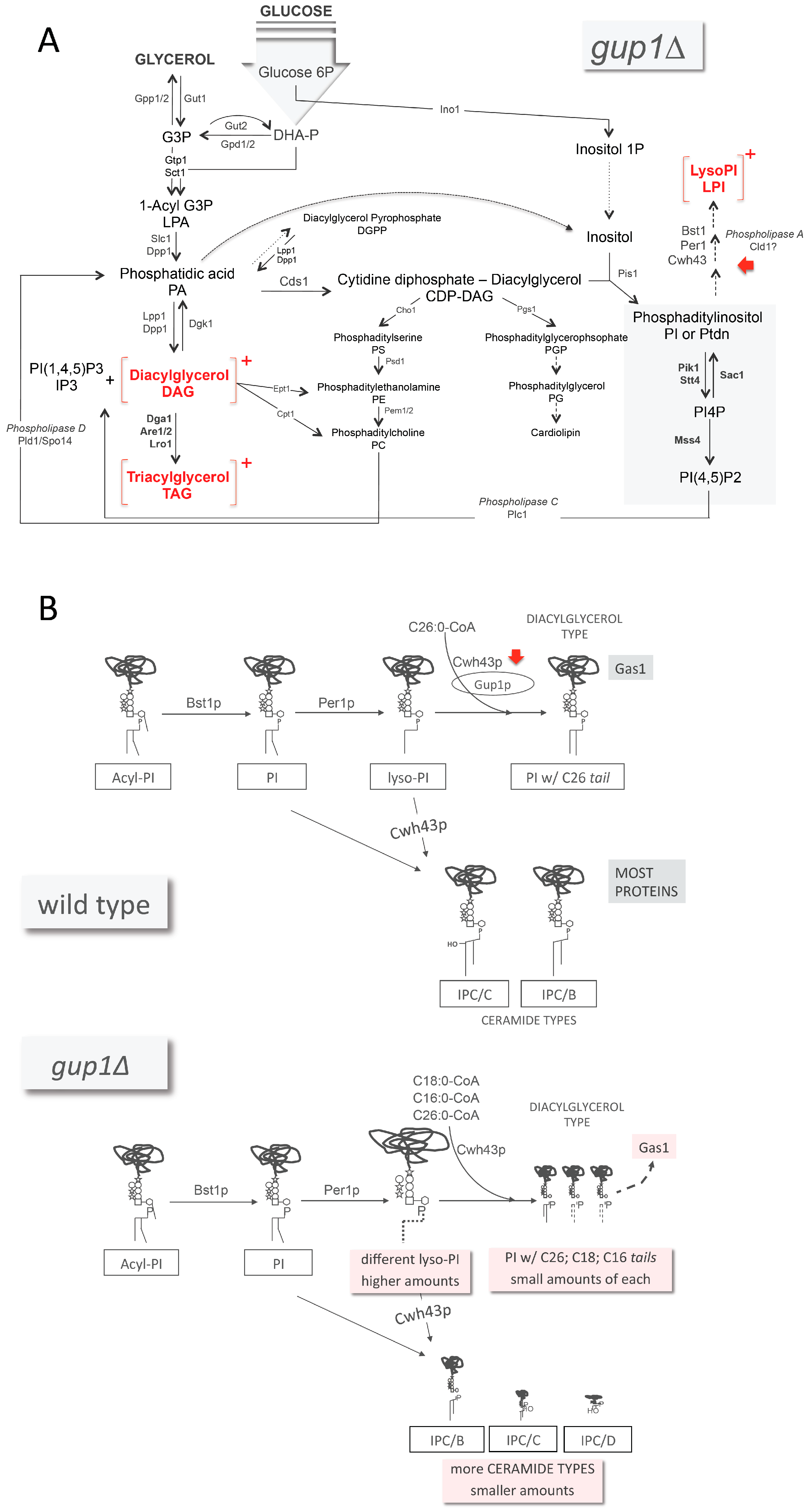
), glucosamine (✩) and ethanolamine-P (☐). Dark thick scrawls represent peptide chains. PI: phosphatidylinositol; IPC: inositol phosphoceramide. Types B, C and D were defined from bands obtained using TLC [136].
Jdb 04 00033 g003
Figure 4. Phenotypes associated to the enzymes known to catalyse the steps from PI to PI(4,5)P2 in S. cerevisiae. These phenotypes are also associated with the GUP1 deletion, with the exception of mitophagy. * Preliminary results [45].
Jdb 04 00033 g004
Figure 5. Mus musculus HHATL complementation of C. albicans colonies hyphae defect caused by the double deletion of GUP1 alleles [29,156]. Pictures of colony borders were taken after 3 days of incubation in spider medium at 37 °C.
Jdb 04 00033 g005
Figure 6. Phenotypes and biological processes associated to the Gup proteins in yeasts (S. cerevisiae and C. albicans) and in the higher eukaryotes M. musculus, H. sapiens and D. melanogaster. Clipart was downloaded from the free access supplier http://etc.usf.edu/clipart/home/.
Jdb 04 00033 g006
Table 1. Gup1 and Gup2 aliases in the literature and databases.
AliasesOrganismKey References
Gup1Gup2Saccharomyces cerevisiae[19,28]
Gup1-Candida albicans[29]
-RASPDrosophila melanogaster[30]
Skinny Hedgehog[20]
Sightless[31]
Central Missing[32]
HHATLHHATMus musculus[22]
HHATLHHATHomo sapiens[33]
c3orf3[34]
KIAA117[35,36]
MBOAT3 *[33]
MSTP002 #[33]
OACT3 *[33]
* Nomenclature shared with Xenopus laevis and X. tropicalis; # Nomenclature shared with X. tropicalis.
Table 2. Documented transcription factors that function as regulators of Gup1 and Gup2 [46]. Activ.: activator; Inhib.: inhibitor.
Predicted Function in GUP1 and GUP2 Transcription Regulation
GUP1 GUP2Identical RegulationOpposite Regulation
Activ.Inhib.BothActiv.Inhib.BothActiv.Inhib.BothActiv. GUP1 + Inhib. GUP2Inhib. GUP1 + Activ. GUP2
Ash1Abf1Rap1Ecm22Sfp1Ace2Aft1Tec1Mig3Rap1Ash1none
Met4Cac2Spt23Gat4Snf2Ash1Cbf1
Tec1Cup9Yrm1Gcr2Snf6Bas1Cin5
Gal4 Gis1Sok2Fhl1Plm2
Hir3 Gln3Spt10Gcn4Pho4
Mig3 Gzf3Spt2Gcr2Rap1
Sfp1 Hap5Ste12Mig3Rfx1
Sum1 Hda1Sut1Msn4Sko1
Uls1 Hms1Swi3Rif2Xbp1
Isw2Taf14Set2Yap6
Mbp1Tec1Swi5Msn2
Mcm1Tup1Tbf1Swi4
Mga1Uls1
Mig1Uc2
Mot3Urc2
Nrg1Xbp1
Rgm1Yhp1
Rox1Yox1
YR015C
Observations: Ash1: TF inhibitor of HO transcription; Mig3: TR with a major role in catabolite repression and ethanol response; inactivated by phosphorylation by Snf1 or Mec1 pathway; Rap1: Essential TR for many loci: transcription activation and repression, chromatin silencing, and telomere length maintenance; Tec1: TF targeting filamentation genes and Ty1 expression; activity dependent on association with Ste12; Ste12 activation of most filamentation gene promoters depends on Tec1.
Table 3. Gup1 and Gup2 ascertained and predicted physical partners.
PartnerAssayType of Study Key ReferenceAssigned Function
Gup1Fet3PCAHTP[174]Iron-O2-oxidoreductase; multicopper oxidase that oxidizes ferrous (Fe2+) to ferric iron (Fe3+) for subsequent cellular uptake by transmembrane permease Ftr1.
Frk1BAHTP[175]Protein kinase of unknown cellular role; interacts with rRNA transcription and ribosome biogenesis factors, and the long chain fatty acyl-CoA synthetase Faa3p.
Hek2ACHTP[176]RNA-binding protein involved in asymmetric localization of ASH1 mRNA; represses translation of ASH1 mRNA, an effect reversed by Yck1-dependent phosphorylation; regulates telomere position effect and length.
Mep22H/Co-LSingle study[165]NH4+ permease; regulation of pseudohyphal growth; expression regulated by nitrogen catabolite repression (NCR).
Msc7PCAHTP[177]Cytoplasmic protein of unknown function.
Nab2ACHTP[178]Nuclear poly(A)-binding protein; required for nuclear mRNA export and tail length control.
Pil1Co-IPSingle study[45]Eisosome core component; detected in phosphorylated state in mitochondria; phosphorylated upon Pkc1 hyperactivation in a Slt2p MAPK-dependent fashion.
Por1Co-IP/Co-LSingle study[45]Mitochondrial voltage-dependent anion channel (VDAC); required for maintenance of mitochondrial osmotic stability and membrane permeability; couples the glutathione pools of the intermembrane space and the cytosol.
Sat4BAHTP[175]Ser/Thr protein kinase involved in salt tolerance; functions in regulation of Trk1-Trk2 potassium transporter.
Vtc4PCAHTP[174]Vacuolar membrane polyP polymerase; subunit of the vacuolar transporter chaperone (VTC) complex; regulates membrane trafficking; role in non-autophagic vacuolar fusion.
YHL042WPCAHTP[174]Putative protein of unknown function.
Gup2Aqy1PCAHTP[174]Spore-specific aquaporin.
Aus1PCAHTP[179]Plasma membrane sterol transporter of the ATP-binding cassette family; required, along with Pdr11, for uptake of exogenous sterols and their incorporation into the plasma membrane.
Hsp30PCAHTP[174]Negative regulator of the H(+)-ATPase Pma1; stress-responsive; induced by heat shock, ethanol, weak organic acid, glucose limitation, and entry into stationary phase.
Ifa38PCAHTP[174]Microsomal beta-keto-reductase; mutants exhibit reduced VLCFA synthesis, accumulate high levels of dihydrosphingosine, phytosphingosine and medium-chain ceramides.
Nam7ACHTP[180]ATP-dependent RNA helicase.
Pdr10PCAHTP[179]ATP-binding cassette (ABC) transporter; multidrug transporter involved in the pleiotropic drug resistance network; regulated by Pdr1p and Pdr3p.
Pho88PCAHTP[174]Probable membrane protein involved in phosphate transport; role in the maturation of secretory proteins.
Sss1PCAHTP[174]Subunit of the Sec61 translocation complex (Sec61-Sss1-Sbh1); this complex forms a channel for passage of secretory proteins through the ER membrane.
Ste2PCAHTP[174]Receptor for α-factor pheromone; interacts with both pheromone and a heterotrimeric G-protein to initiate the signaling response that leads to mating.
HTP: high throughput; PCA: protein complementation assay; AC: affinity capture (RNA); BA: biochemical activity; 2H: two-hybrid; Co-IP: co-immunoprecipitation; Co-L: co-localization.
Table 4. Similarity matrix between the most studied GUP proteins.
C. albicans SC5314 Gup1S. cerevisiae S288c Gup1S. cerevisiae S288c Gup2D. melanogaster Gup2/RASPM. musculus Gup1/HHATLH. sapiens Gup1/HHATLM. musculus Gup2/HHATH. sapiens Gup2/HHAT
C. albicans SC5314 Gup1100.0057.6447.0216.6319.1518.4518.4918.77
S. cerevisiae S288c Gup157.64100.0056.6117.8420.7020.4519.3019.80
S. cerevisiae S288c Gup247.0256.61100.0017.8020.9220.2020.0021.07
D. melanogaster Gup2/RASP16.6317.8417.80100.0021.6221.1425.9426.58
M. musculus Gup1/HHATL19.1520.7020.9221.62100.0090.0728.1627.92
H. sapiens Gup1/HHATL18.4520.4520.2021.1490.07100.0028.3327.86
M. musculus Gup2/HHAT18.4919.3020.0025.9428.1628.33100.0082.91
H. sapiens Gup2/HHAT18.7719.8021.0726.5827.9227.8682.91100.00

