Article

The In Vitro Anti-Proliferative Interaction of Flavonoid Quercetin and Toxic Metal Cadmium in the 1321N1 Human Astrocytoma Cell Line

Department of Pharmaceutical Chemistry, Faculty of Pharmacy, Kuwait University, P.O. Box 24923, Safat 13110, Kuwait
*
Author to whom correspondence should be addressed.
Sci. Pharm. 2018, 86(3), 36; https://doi.org/10.3390/scipharm86030036
Submission received: 12 June 2018 / Revised: 4 September 2018 / Accepted: 5 September 2018 / Published: 10 September 2018

Abstract

:
Cadmium (Cd) is a toxic heavy metal occurring in the environment as an industrial pollutant. The systematic accumulation of Cd in the human body may lead to major health problems. Quercetin (QE) is a natural flavonoid widely distributed in plants and is a part of human diet. Many studies have demonstrated the multiple benefits of QE to humans in protecting cells of our bodies. The aim of this study was to investigate the effect of QE and Cd on the proliferation of astrocytoma 1321N1 cells. Results indicated that the simultaneous exposure of the cells to 200 µM QE and 16 μM Cd significantly reduced cell viability to 6.9 ± 1.6% with respect to vehicle-treated cells. Other experiments of QE pre-treatment followed by the exposure to Cd alone or with QE indicated significant but decreased ability of QE or Cd to reduce proliferation of the cells compared to their co-incubation. Our study suggested a synergetic anti-proliferative interaction of Cd and QE in malignantly transformed cells. This adds new information regarding the biological effects of QE.

1. Introduction

Cadmium (Cd) is a toxic nonessential heavy metal which was classified as a human carcinogen by the National Toxicology Program. The major sources of exposure to Cd are occupations in metal industries and battery factories [1]. Cd represents a widely dispersed environmental pollutant through the air, mining and smelting, phosphate fertilizers and presence in sewage sludge. Cd is also present in almost all food and tobacco leaves [2]. Cd has a biological half-life of 10–30 years [2,3]. The human body has a limited capability of dealing with the toxic Cd and with transforming it to harmless products [1]. Thus, exposure to Cd is associated with major health problems that include osteoporosis, pulmonary dysfunction, hypertension, nephropathy, neurotoxicity, and cancer [3].
Several studies reported the notable risk factors of exposure to Cd in the relation to pulmonary, prostate, kidney, pancreas, breast and urinary cancers [4]. Cd induces genetic instabilities by various pathways. It is shown that Cd induces oxidative stress in various cell cultures or in intact animals by depleting the levels of glutathione and other antioxidant sulfhydryl-containing proteins together with reducing the activities of enzymes preventing oxidation. This results in enhancement of the production of reactive oxygen species (ROS), including superoxide ion, hydrogen peroxide, and hydroxyl radicals. These radicals, in turn, increase lipid peroxidation and result in DNA damage [5]. Additionally, Cd interacts with a variety of DNA repair processes, including base excision repair, nucleotide excision repair, DNA mismatch repair, double-strand break repair and cell-cycle arrest [6]. Moreover, it is demonstrated that Cd induces epigenetic changes through induction of DNA hypomethylation [7], histone modifications and non-coding RNA expression [8].
Quercetin (3,3,4,5,7-pentahydroxyflavone) (QE) belongs to naturally occurring flavonoids and is widely distributed in plants [9]. It is present in many components of the human diet: onions, peppers, cranberries, blueberries, apples, cherries, grapes and many others [10]. Many studies have demonstrated multiple benefits of QE to the human. The most prominent ones are anti-aging, anti-inflammatory, anti-diabetic, anti-obesity, angiopreventive, chemopreventive and anti-cancer properties [11]. In its anti-carcinogenic action, QE directly inhibits signaling pathways mediating oncogenic transformation [12]. This can lead to the apoptotic death of cancer cells through QE effects on the mitochondria [13]. QE can also inhibit signaling pathways involved in cancer progression [14]. Studies involving malignant cell lines of different origins have revealed that most sensitive cells to QE are those originating from blood, brain, lung, uterine, salivary gland and melanoma tissues. Additionally, it is demonstrated that QE has a selective cytotoxicity against more aggressive cells compared to slowly growing cells. This suggests that the cells with a higher extent of malignant transformation are preferentially targeted by QE [15]. As a chemopreventive agent, QE has the potential to stop, reverse or delay tumorigenesis at early stages [16], for example by suppressing generation of ROS [17].
Several studies have been performed to investigate the protective potentials of QE in animals that were exposed to the toxic Cd. These studies focused mainly on the anti-oxidative action of QE, which ameliorate the Cd-induced oxidative stress. It is shown that Cd-induced oxidative stress in the mouse liver model is eliminated by administration of QE [18]. QE can markedly stimulate the expressions of the hepatic metallothionein proteins and nitric acid synthase (NOS) that were reduced in Wistar rats due to exposure to Cd [19]. However, the most sensitive organs to Cd toxicity are the kidneys because of Cd accumulation in them [3]. QE can prevent nephrotoxicity caused by Cd by maintaining the appropriate levels of renal NOS and cyclooxygenase-2 in Wistar rats [20] and it also markedly reduces the tubular lesions [21]. QE also attenuates the renal biochemical and pathological alterations in rats exposed to Cd [22]. The in-vitro simultaneous treatment of rat proximal tubular cells with QE and Cd provides protection of the cells against the mitochondrial oxidative damage and degradation by Cd [23] and inhibited autophagy caused by Cd in mouse kidney [9]. Additionally, the significant apoptotic effect of Cd on the germ cells of Institute of Cancer Research (ICR) mice is prevented by QE [24]. Similar data have been obtained with granulosa cells derived from chicken ovarian follicles [25]. The intraperitoneal injection of QE into rats protects them from the testicular injury caused by prior injection of Cd [26]. The administration of QE into experimental animals restores the serum concentrations of reproductive hormones and results in the prevention of apoptosis in follicular cells due to the exposure to Cd [27].
Data have also been published regarding QE having a protective effect against Cd-induced alterations in brain cholinergic signaling and neurotoxicity in rats [28] and regarding the ability of QE to reverse the Cd-related cognitive impairment effect in female mice that were exposed to Cd during their lactating period [29]. QE protects experimental animals from memory impairment and from anxiogenic behavior induced by Cd by preventing alterations in the activities of enzymes present in rats’ cerebral cortex, hippocampus, and hypothalamus [30,31,32]. QE also significantly reduces the neuronal injury in the frontal cortex of rats exposed to Cd [33]. QE also possesses the ability to restore alterations in various enzymatic activities in serum of experimental rats exposed to Cd as show on the activity of serum creatine kinase-muscle/brain (MB), transaminases, alkaline phosphatase, and lactate dehydrogenase [34]. Additionally, a pre-treatment of experimental rats with QE prevents Cd-induced changes in a lipid profile and of lipoproteins [35].
The aim of this study was to investigate the interaction of Cd and QE in malignant astrocytoma 1321N1 cells. QE is recognized for its protecting properties in normal cells where it diminishes the effects of the toxic pollutant Cd. The importance of exploring QE’s effect on malignant cells exposed to Cd seems to be highly significant. We aimed at clarifying whether QE also protects malignant cells from the presence of heavy metal Cd or the nature of QE–Cd interaction in malignant cells is different from that in normal cells.

2. Materials and Methods

Unless otherwise specified, all materials were purchased from Sigma-Aldrich (St. Louis, MO, USA).

2.1. Cell Culture

The human brain astrocytoma 1321N1 cell line was purchased and obtained from the Health Protection Agency Culture Collection (Porton Down, Salisbury, UK), and it was grown in culture flasks (Corning Life Sciences, Tewksbury, MA, USA) containing Dulbecco’s Modified Eagle’s Medium/Ham’s Nutrient Mixture F12 supplemented with 10% fetal calf serum, 1% l-glutamine (Gibco, ThermoFischer Scientific, Grand Island, NY, USA) and 1% penicillin/streptomycin (Gibco). The cell culture was maintained and treated at 37 °C in a humidified atmosphere containing 5% CO2. Due to a slow growth rate of these cells, the experiments were performed with cells in the log growth phase.

2.2. Stock Solutions and Concentrations Used for Treating the Cells

Stock solutions of QE and CdI2 were prepared by dissolving in the vehicle Dimethyl sulfoxide (DMSO). Concentrations of stock solutions were calculated to always obtain 0.5% of DMSO as the final concentration of this vehicle in the culture medium. The selection of the experimental QE and Cd concentrations was made based on the data published earlier [36,37] and based on the experiments performed in our laboratory. Treatment, pre-treatment or co-treatment of cells with QE was done at 100 or 200 µM of QE [36], whereas the cells were exposed to 16 µM Cd [37].

2.3. Treatments of the 1321N1 Cells with QE and Cd

The effects of QE and Cd on cell viability were evaluated in the 1321N1 cells by the commonly used the 3-(4,5-dimethylthiazol-2-yl)-2,5-diphenyl-tetrazolium bromide (MTT) assay, which depends on the mitochondrial dehydrogenase-mediated conversion of water-soluble yellow MTT to a water-insoluble blue formazan product [38]. Incubation of the cells with individual or combined QE and Cd was performed with or without pre-incubation with QE. The experimental conditions of the incubations are summarized in Table 1.
The 1321N1 cells were seeded overnight in a 96-well plate at the density of 2 × 10⁴ cell/well leaving a lane containing medium only to serve as a blank. On the next day, the cells were incubated in medium containing the vehicle DMSO or QE and/or Cd. At the end of the incubation times (Table 1), the cells were incubated for 5 h with 500 µg/mL MTT reagent at 37 °C. Afterwards, the media were removed and 150 µL DMSO was added to dissolve formazan crystals and the absorbance values of the colored formazan solutions were measured at 540 nm using a plate reader (BioTek, Highland Lake, VT, USA). Mean absorbance values were calculated across 4 wells for each treatment. Blank’s mean absorbance was subtracted from all other absorbance values and the results were expressed as percentages of cell viability with respect to vehicle-treated cells from independent experiments.
Data were analyzed by two-way analysis of variance (ANOVA) followed by Bonferroni multiple comparison test using the GraphPad Prism software package (version 7, La Jolla, CA, USA). p Values of ≤0.05 were considered significant. Graphs are presented as bars of mean ± SEM (Standard Error for the mean) values versus the incubation times.

3. Results

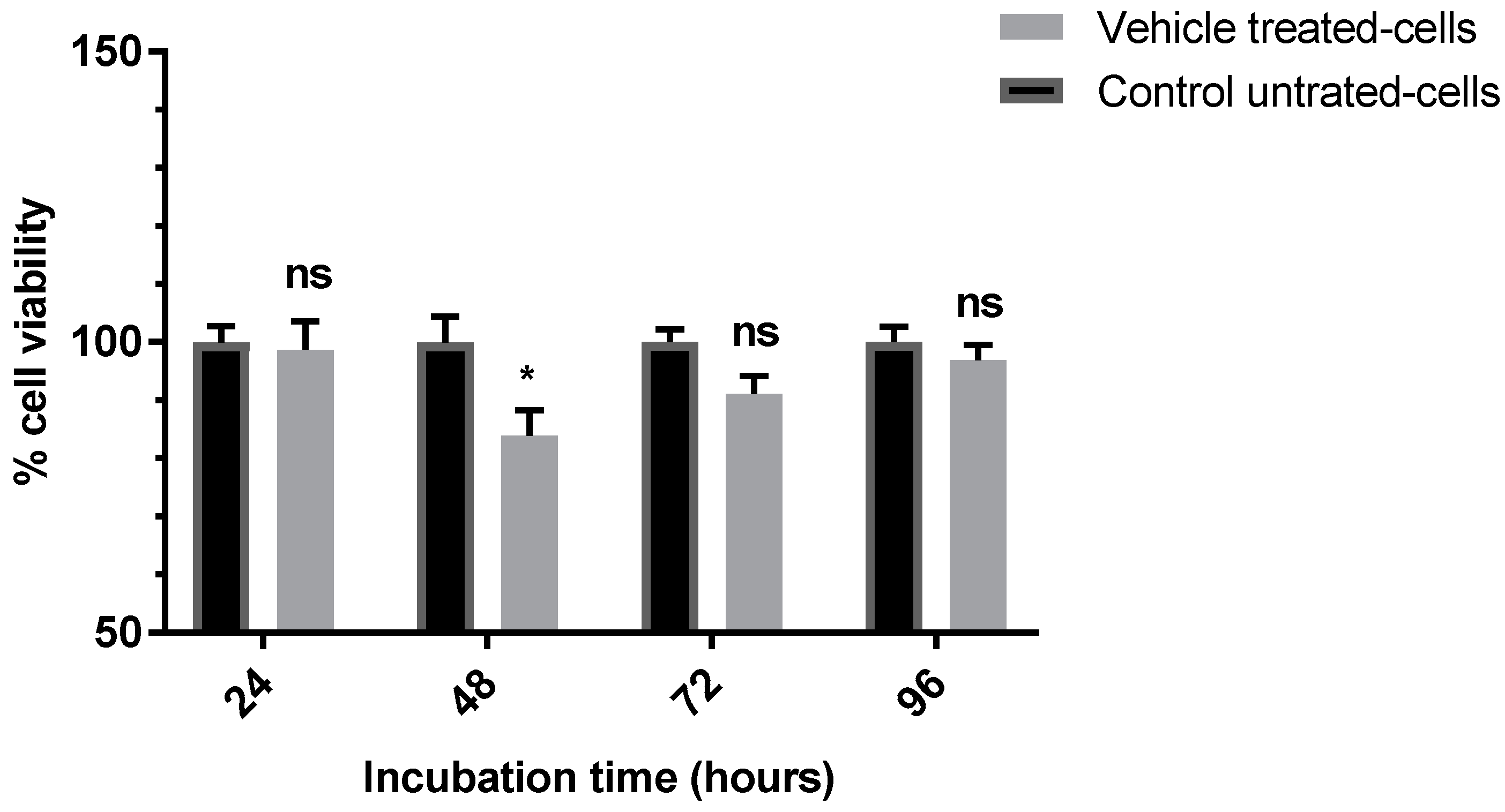
3.1. Evaluation of the Anti-Proliferative Effect of the Vehicle (0.5%) DMSO

To eliminate the effect that the vehicle (0.5% final concentration of DMSO in the tissue medium) has on the viability of 1321N1 cells, the cells were incubated with 0.5% DMSO in the medium for 24, 48, 72 and 96 h. After evaluation of cells viability by MTT assay, the effect of 0.5% DMSO was found not to be significant at 24-, 72- and 96-h time points as it did not affect the cell viability (98.7 ± 5.0%, 91.1 ± 3.1% and 96.9 ± 2.7%, respectively) with respect to control cells (p > 0.9999, 0.0711 and > 0.9999 respectively) (Figure 1). However, incubating cells with DMSO for 48 h slightly (83.88 ± 4.39% cell viability) but significantly (p = 0.0102) affected cell viability (Figure 1). This effect of DMSO at 48 h treatment appears to be transient as the cell viability was recovered at 72-h time point (91.10 ± 3.10% cell viability). However, it was confirmed that 1321N1 cells can tolerate up to 0.5% DMSO for 96 h. The stock solutions of QE and Cd were always prepared in such way that they attained 0.5% of the DMSO final concentration (in cellular cultures) in all evaluated experiments.
Consequently, all cell viability data for cells exposed to QE and/or Cd were corrected for the effect of 0.5% DMSO at the appropriate time point.

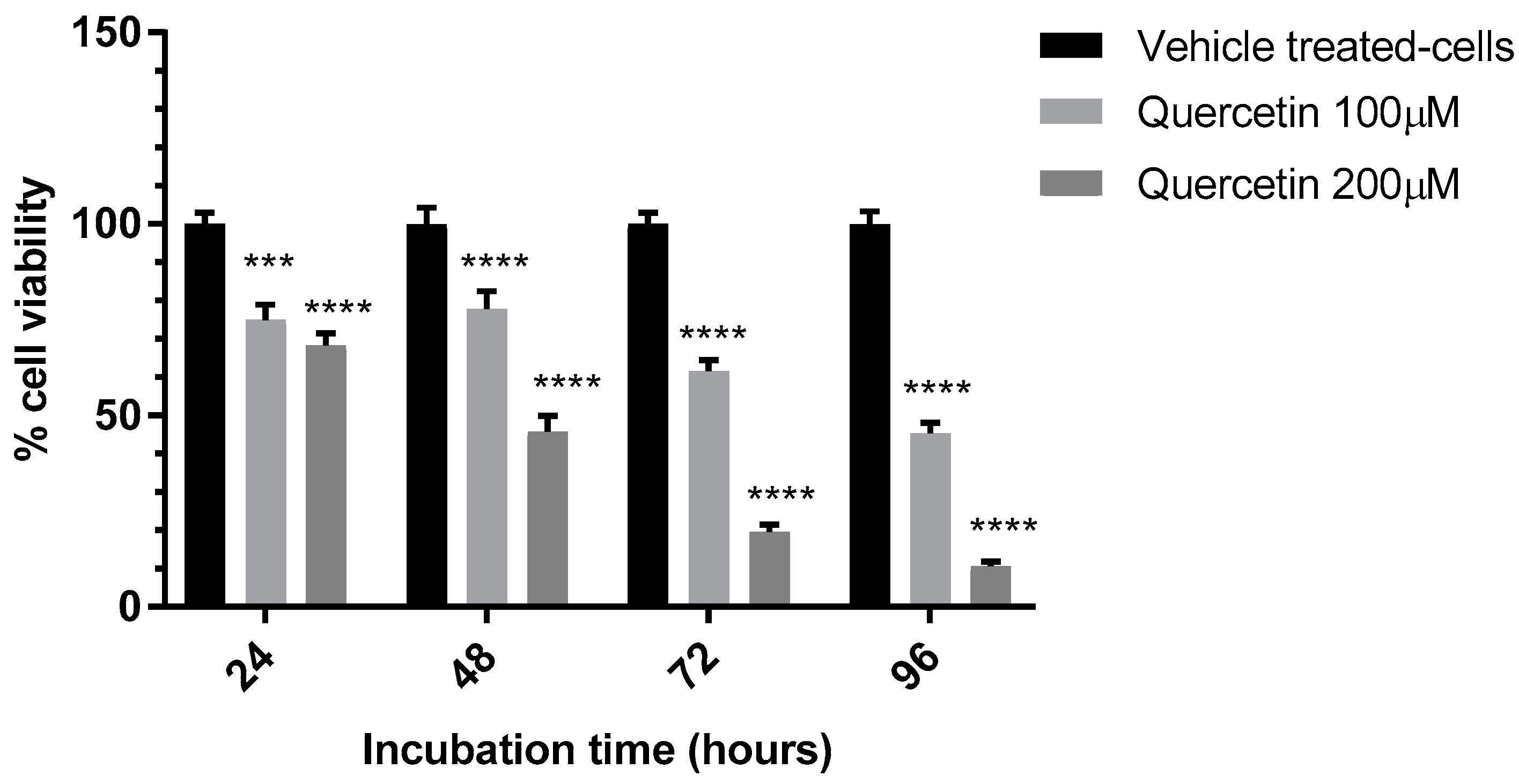
3.2. Evaluation of the Anti-Proliferative Effect of QE

The anti-proliferative effect of QE on 1321N1 cells was evaluated using data from experiments with different incubation times of cells exposed to QE. The experimental concentrations of QE were 100 and 200 μM, respectively. After treatment with either vehicle or QE, cells viability was determined by MTT assay.
As shown in Figure 2, treating 1321N1 cells with 100 μM QE significantly reduced cellular viability in a time-dependent manner. Cells were exposed to 100 μM QE for 24, 48, 72 and 96 h and the cell viability was significantly decreased at all the used time points (74.9 ± 4.0%, 77.9 ± 4.6%, 61.5 ± 2.9% and 45.3 ± 2.8% of the original cell viability, respectively). The p values were <0.0001 for the first three time points and 0.0007 for the time point of 96 h. The cell viability values were reduced even more when the cells were exposed to 200 μM QE. The cell viability values at the same time points were 68.3 ± 3.2%, 45.8 ± 4.6%, 19.5 ± 1.9% and 10.6 ± 1.2% of the original cell viability, respectively (with respect to vehicle treated-cells (p < 0.0001 for all time points) (Figure 2).

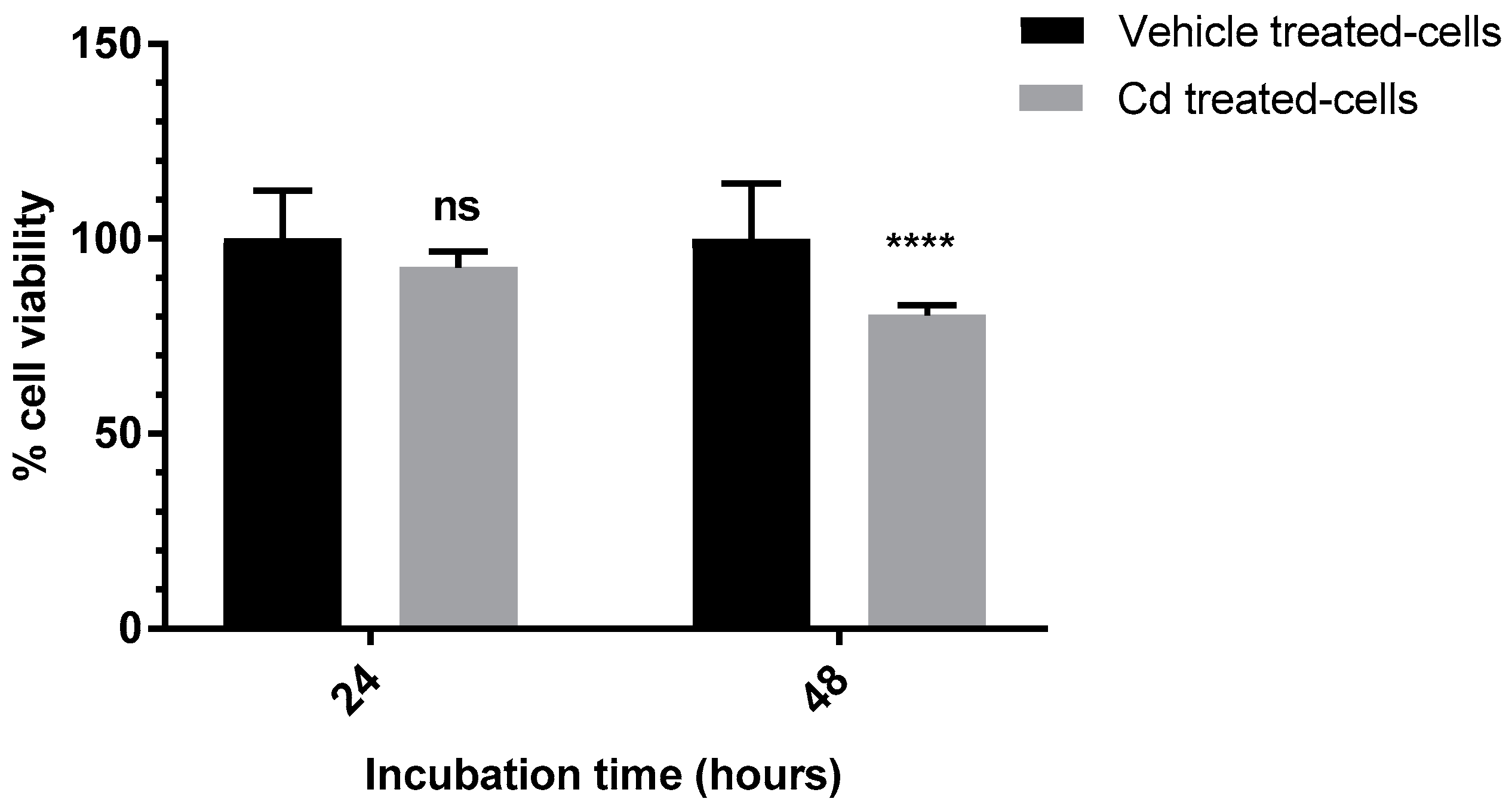
3.3. Evaluation of the Anti-Proliferative Effect of Cd

The anti-proliferative effect of Cd on 1321N1 cells was evaluated at incubation times of 24 and 48 h. Similar to QE, the salt CdI2 was dissolved in DMSO at the concentration calculated to attain 16 μM Cd concentration at 0.5% final concentration of DMSO in tissue culture (the 96-well plate). As shown in Figure 3, treating the cells with Cd for 24 h did not significantly affect cellular viability (92.7 ± 4.0%) in comparison to vehicle treated-cells (p = 0.1298). Contrarily, treating the cells with Cd for 48 h resulted in a significant reduction in cell viability (80.5 ± 2.4%) in comparison to cells treated only with the vehicle (p < 0.0001) (Figure 3).

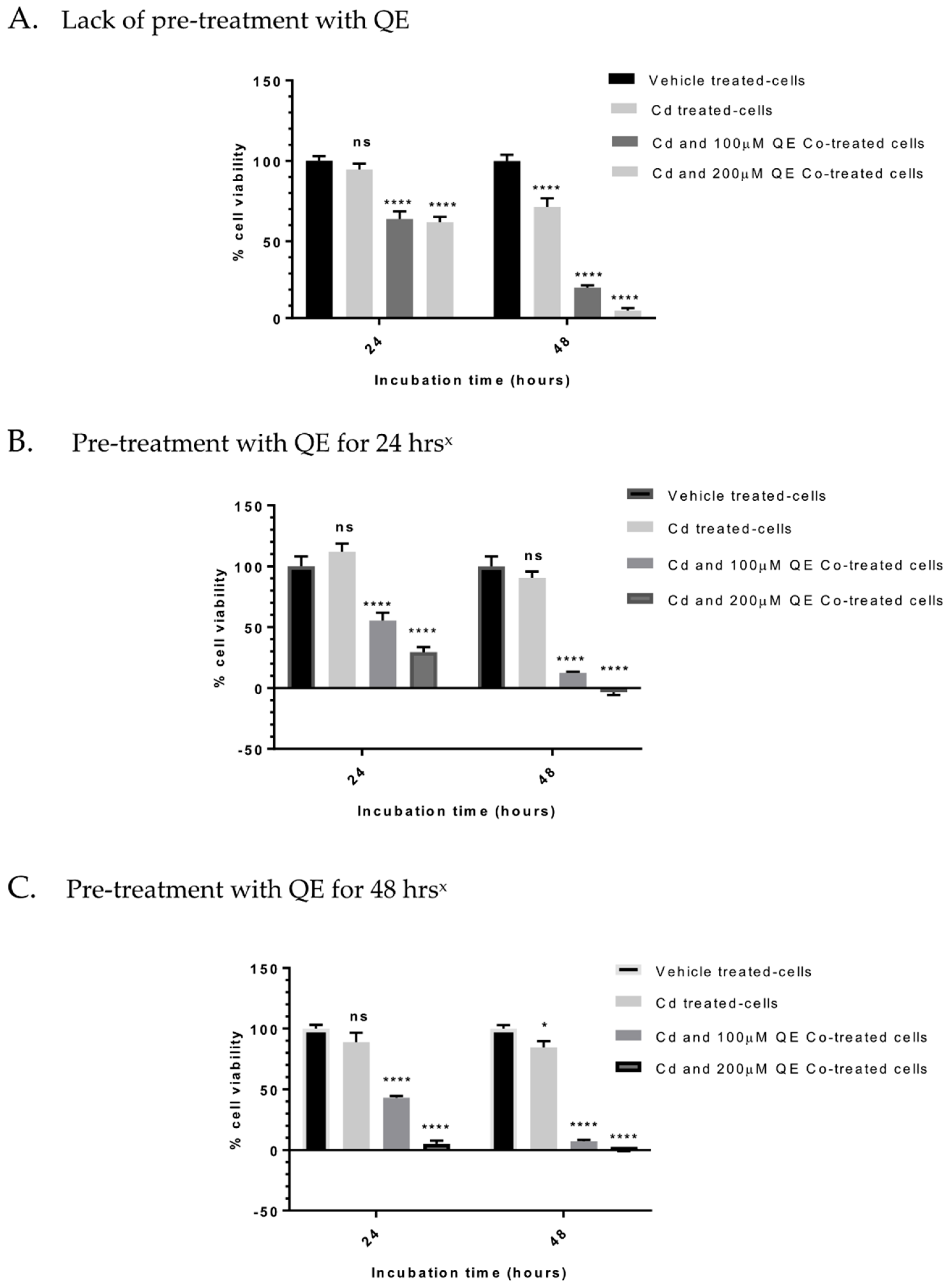
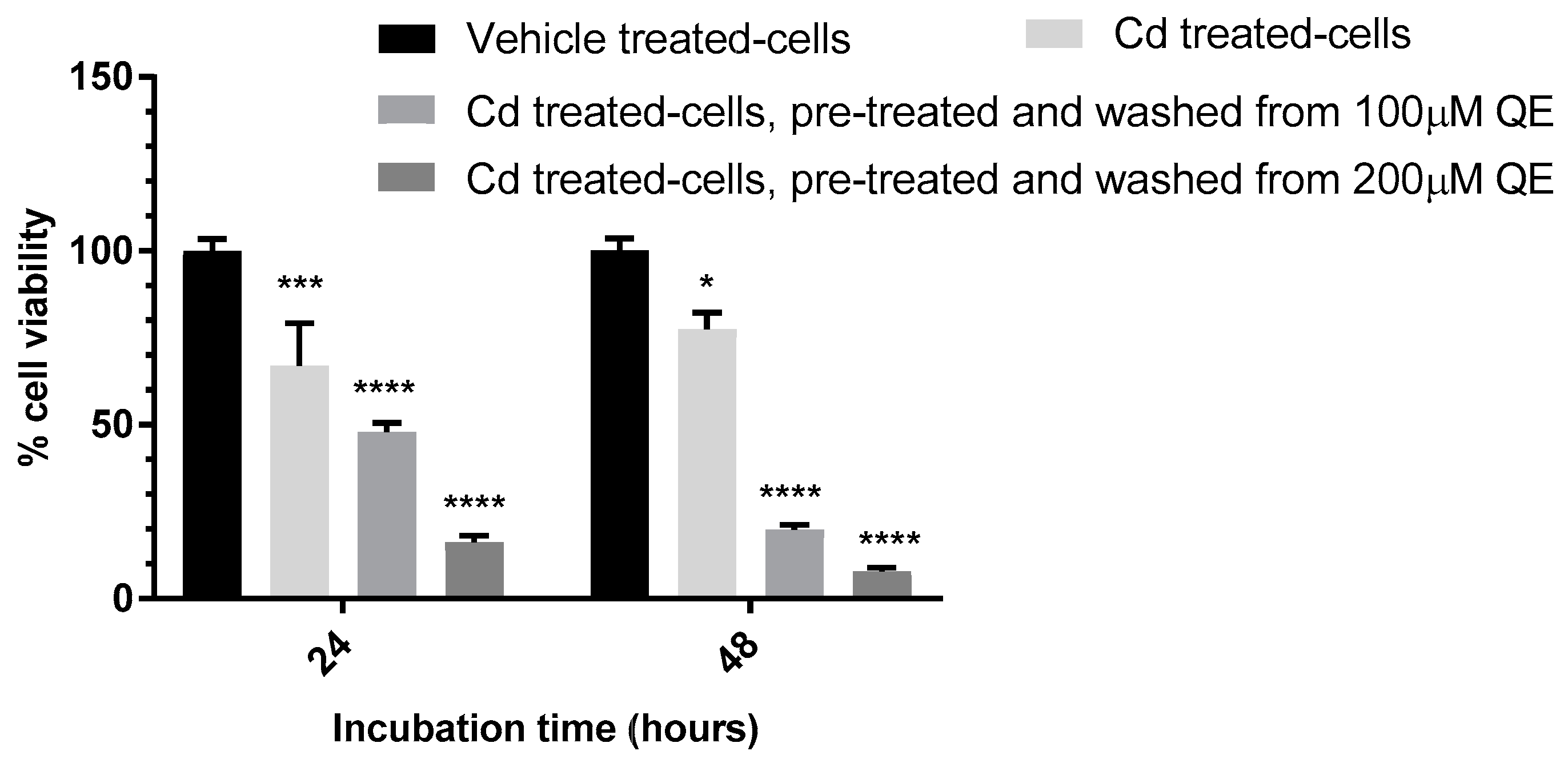
3.4. Evaluation of the Anti-Proliferative Effect of QE and Cd in Co-Treatment

The 1321N1 cells were co-treated with QE and Cd or they were pre-treated with QE and then co-treated with QE and Cd (Figure 4). As shown in Figure 4A, co-treatment of the cells with QE and Cd for 24 and 48 h significantly reduced cell viability. When the cells were treated with Cd only, the reduction of viability observed was not significant after 24 h of the Cd exposure but became significant after 48 h (24 h: 94.7 ± 3.7% cell viability, p > 0.999; 48 h: 71.4 ± 5.4% cell viability, p < 0.0001; Figure 4A). Co-treatment of cells with Cd (16 μM) and QE (100 or 200 μM of QE) for 24 h resulted in the same decrease of viability at both used concentrations of QE (64.0 ± 4.8 and 62.1 ± 3.1% cell viability, respectively, p < 0.0001). However, incubation for 48 h showed the different extent of QE and Cd effects on cell viability (21.4 ± 1.3% and 6.9 ± 1.6% cell viabilities of 100 and 200 μM QE, respectively with the same concentration of Cd, p < 0.0001) (Figure 4A).
Figure 4B shows the higher effect on cell viability induced by pre-treating cells with QE prior to co-treatment with QE and Cd. Compared to the non-significant effect of treatment for 24 h with Cd alone (111.9 ± 6.8% cell viability, p = 0.9047), the pre-treatment with QE for the same time followed by co-treatment with Cd and 100 or 200 μM of QE for 24 h significantly reduced cell viability (55.5 ± 6.4% and 29.6 ± 3.9%, respectively, p < 0.0001) (Figure 4B). Further extension of co-treatment time up to 48 h under the same experimental conditions led to a higher and significant reduction of cell viability with 100 μM of QE (12.4 ± 1.0% cell viability, p < 0.0001), and negative cell viability value at 200 μM QE (−3.4 ± 2.4%, p < 0.0001) (Figure 4B). In contrast, under the otherwise same experimental conditions, treatment of the cells with Cd alone affected cell viability only insignificantly (90.4 ± 5.3% cell viability, p > 0.999) (Figure 4B).
A higher and significant reduction in cell viability was observed when the cells were pre-treated with QE for 48 h and co-treated with Cd and QE for 24 h with concentrations of QE being 100 μM (43.1 ± 1.4% cell viability, p < 0.0001) or 200 μM where 200 μM resulted in negligible cell viability (5.0 ± 2.7%, p < 0.0001) (Figure 4C). As shown in Figure 4C, the extension of co-treatment with Cd and QE to 48 h (under the otherwise same experimental conditions), resulted in a negligible cell viability at 100 μM QE (7.2 ± 1.2%, p < 0.0001) and negative cell viability value at 200 μM QE (−0.5 ± 0.5%, p < 0.0001) (Figure 4C). Contrarily, under the same experimental conditions exposing the cells to Cd alone for 24 h resulted in non-significant reduction in cell viability (89.0 ± 7.6%, p = 0.2583) and in a significant reduction effect (84.6 ± 5.2% cell viability, p = 0.0337) for 48 h.
In subsequent experiments, the anti-proliferative effect of QE was evaluated after its removal from the tissue culture to see whether QE entered the cells in the tissue culture. The experiment was carried out by washing off QE after pre-treating cells for 48 h. Pre-treated cells were then exposed to Cd and cell viability was compared to that of cells exposed to Cd without pre-treatment with QE. As shown in Figure 5, despite washing off QE from the cells, the anti-proliferative effect of QE remained for 24 and up to 48 h when the cells were treated with Cd. Exposing cells for 24 h to Cd alone significantly reduced cell viability (66.9 ± 12.2%, p = 0.0002) (Figure 5), whereas exposing cells to Cd after removing 100 and 200 μM QE from the cell culture resulted in significantly higher reduction of cell viability (47.9 ± 2.6% and 16.2 ± 2.0%, respectively, p < 0.0001) (Figure 5). In contrast to this, exposing cells for 48 h to Cd alone had significant but not so prominent effect on cell viability compared to only 24 h exposure (77.5 ± 4.7% cell viability, p = 0.0178) (Figure 5). The exposure to Cd for 48 h after washing off the cells pre-treated with 100 µM QE resulted in much higher and significant effect on cell viability (19.8 ± 1.4%, p < 0.0001), and a negligible value when the cells pre-treated with 200 µM of QE (7.8 ± 1.1% cell viability, p < 0.0001) (Figure 5).
To ensure that the reduction in the cell viability after washing off was due to QE rather than the washing-out process, similar experiments were carried out in which QE was washed off, but re-added and co-incubated with Cd. The obtained cells viability was very close to those reported in Figure 4C (data not shown). Cells pre-treated with QE for 48 h and then treated with Cd and QE for 24 and 48 h without washing off QE (Figure 4C) has similar viability to cell subjected to a washing-off process. This confirmed that the remained anti-proliferative effect of QE for up to 48 h is due to the intracellular QE as extracellular QE was washed off. The data confirm that washing off QE does not affect its anti-proliferative effect.

4. Discussion

Cd is a toxic industrial pollutant significantly affecting human health. The environmental presence of Cd is a major concern because of its long biological half-life of up to 30 years and limited metabolic detoxification and excretion. Cd was classified as “carcinogenic to humans” (group I) by the International Agency for Research on Cancer (IARC) [39]. Exposure to Cd induces the production of ROS resulting in an increased lipid peroxidation and DNA damage. Consequently, exposure to Cd is potentially genotoxic and can result in epigenetic abnormalities, i.e., reduced DNA methylation.
QE is a natural substance—a flavonoid widely distributed in plants. The scientific literature contains data on its potential therapeutic utilization. QE may produce anti-aging, anti-inflammatory, anti-diabetic, anti-cancer, cancer-chemopreventive and other health-related effects. Despite the promising anti-cancer activity of QE in vitro, further investigations are needed to overcome its limited bioavailability and to identify the actual active molecular species, be it QE itself or its metabolites [15,40].
Several studies have been performed to investigate the protective effect of QE against toxicities induced by exposure to Cd. To the best of our knowledge, apart from Wang et al. study [23], thus far, all of these studies have been conducted in intact healthy animals and no information on the interaction of QE and Cd in malignant cells was obtained. Results of pre- or simultaneous treatment of normal cells with QE showed a significant protective potential of QE against Cd toxicity. This was documented in the liver, kidney, reproductive system and Central Nervous System (CNS). QE acts as an anti-oxidant that increases the expression of antioxidant enzymes and thus prevents lipid peroxidation enhanced by Cd, ameliorates the biochemical and pathological alterations caused by Cd and inhibits apoptotic cell death caused by Cd cytotoxicity. In the present study, we explored the effect of the flavonoid QE in a model of human brain astrocytoma 1321N1 cells exposed to Cd. Treatment with QE in our study was prior or simultaneous to Cd exposure where the effects on cell viabilities were compared to those of QE or Cd alone.
In our study, treatment with QE significantly reduced the viability of malignant 1321N1 cells. This effect was clearly concentration- and time-dependent. At both experimental concentrations of QE (100 and 200 μM), the percentages of cell viability significantly decreased with increasing the duration of the incubation time from 24 h up to 96 h. In line with our study, published data [41] indicate that the anti-proliferative effect observed in 132N1 cells was due to treatment with QE at concentrations above 50 μM [41]. Similar to our results obtained on the human astrocytoma 1321N1 cells, the sensitivity of breast cancer MCF-7 cells to QE is reported [36] with the IC50 value of QE being 37 μM at 24 h. On the other hand, the lack of anti-proliferative effect of QE in breast cancer MDA-MB-231 cells is documented even at 100 μM of QE [36]. This is the QE concentration used in our study.
Exposing 1321N1 cells to 16 μM Cd for 24 h did not significantly reduce cell viability. This effect is slightly increased and became significant after 48 h exposure to Cd. These data were processed and final values are based on data from several individual experiments (n = 40). However, by going through each of these experiments, some inconsistencies were determined. Data available in the scientific literature [42] demonstrate the adaptive and protective cellular response to Cd by exposing 1321N1 cells to non-cytotoxic concentration (5–10 μM) of Cd for 24 h. Cd in these experiments stimulate the expression of anti-oxidant enzymes NAD(P)H:quinone oxidoreductase and haem oxygenase. The expression and nuclear accumulation of the transcription factor Nrf2, which regulates the expression of these enzymes, is also stimulated [43]. These results agree with our data as no significant reduction in cell viability is observed when 1321N1 cells are exposed to Cd for 24 h in most experiments. Thus, the results we obtained at 24 h Cd exposure could be explained by stimulation of the repair mechanism in these cells that may have the potential to recover from the toxic effect of Cd. In contrast, the failure of such repair process was indicated in one experiment (Figure 5) that showed that exposing the cells to Cd for 24 h significantly reduced cell viability. Similarly, as demonstrated in one experiment (Figure 4B), carrying out the same experiment for 48 h did not exert a significant reducing effect on cell viability. This can be compared with data where a significant viability reduction was recorded for 48 h in other experiments. The lack of the significant reduction in cell viability by exposing 1321N1 cells to Cd for 48 h may be due to the presence of a higher number of cells undergoing multiplication during the longer incubation time, which leads to a dilution in the toxic intracellular Cd level.
In line with our results, the published IC50 value of Cd in 1321N1 cells is determined to be 19.92 µM [42]. This result indicates that IC50 of higher than 16 µM (used in our study) is needed for significant cellular viability alteration in the 1321N1 cells. Another study that used 1321N1 cells [37] reports that 16 µM Cd is enough to induce a high reduction effect in U937-derived macrophage cells (14.14 ± 2.42% cell viability), showing the high sensitivity of these cells to the toxic effect of Cd [37]. Additionally, the cellular exposure to Cd (in the form of CdI2) can be potentially genotoxic [44,45,46,47].
Treating the Cd-exposed 1321N1 cells with 100 and 200 µM QE significantly reduced cell viability under different experimental conditions. At first, cells were simultaneously exposed and treated with Cd and QE for 24 and 48 h. A similar reduction of cell viability was observed after 24 h for both QE concentrations. A significantly higher decrease in cell viability was observed with both QE concentrations after 48 h. However, pre-treating the cells with QE prior to the co-exposure with Cd further enhanced the reduction of cell viability. Pre-treatment with QE for 24 h followed by 24 h co-treatment with Cd and QE induced a high reduction of cellular viability. A prolongation of co-exposure to QE and Cd up to 48 h resulted in an additional significant decrease in cellular viability that becomes negligible at 100 µM QE. Based on our calculation, the experiment using 200 µM QE gave a negative value of cellular viability. Consequently, no cellular viability was retained.
By extending the pre-treatment time with QE to 48 h, the co-exposure with Cd further increased the reduction of cell viability. As discussed in the previous paragraph, cell viability by the co-exposure to Cd with QE for 24 h significantly decreased and became negligible when the concentration of QE used was 200 μM. When the co-exposure was prolonged to 48 h, cell viability at 100 μM of QE also became negligible and of a “negative” value at 200 μM of QE (as explained in the previous paragraph). Obtained data indicate the benefit of longer QE pre-treatment. This, when followed by QE–Cd co-treatment of 132N1, cells resulted in a highly significant effect on cell viability. There is a need for malignant cells to be exposed to QE for a reasonable time to achieve its accumulation within the 132N1 cells and to produce a significant reduction of viability. Then, the action of QE and Cd together may lead to a significant decrease of cell viability that is higher than the effects of these two agents individually. Our experiments show that the QE and Cd act in a synergistic manner on the 132N1 cells under used experimental conditions (Table 2). The column “Viability” (24 and 48 h) contains experimental findings. The column “Combined effect calculated” contains results calculated by subtracting both effects of individual quercetin and of Cd alone from 100 (percentage of viability): (100 − viability decrease by QE alone − viability decrease by Cd alone):
CE = 100 − (VDQE + VDCd),
where CE is combined effect calculated, VDQE is decrease of viability caused by QE alone, and VDCd is decrease of viability caused by Cd alone.
Combination index (CI) calculations shown in Table 2 are based on the quantitative definition by Chou [48]. The effect of drug combination is the ratio or combination index (CI) of the observed and expected drugs combination effect. The additive effect is observed when CI = 1, synergism occurs at CI < 1, and antagonism takes place at CI > 1 in drug combinations.
An additional experiment was performed to determine the place of QE action—inside or outside of the 132N1 cells. A pre-treatment with QE for 48 h was followed by washing off QE. The obtained significant decrease in cellular viability reflected the anti-proliferative effect of the intracellular QE in the 1321N1 cells when the removal of extracellular QE was performed. It was also determined that washing off QE from the cells did not eliminate its significant anti-proliferative effect when exposing these cells to Cd. However, recorded cellular viability was a little higher compared to that when both QE and Cd are present in a cell culture.
The findings described above were further confirmed under the same experimental conditions when the washing-off procedure was performed but QE was re-added to the cell culture. Viability data were similar to those of cells pre-treated for 48 h with QE and then co-treated with QE and Cd for 24 or 48 h. This led us to the conclusion that the significant reduction in cell viability was due to the retained anti-proliferative effect of QE and not to the QE washing-off procedure itself (that might damage the cells). This is also supported by the fact that the decreased viability persists for more than 48 h when cells would surely recover from the stress of washing-off procedure. It has been shown [49] that QE can protect 1321N1 cells against oxidative stress induced by 10 mM acrolein but not against the much lower concentration of 1 mM acrolein. It also cannot protect cells against acetaldehyde [49]. This indicates the specificity of the protective effect of QE. Another interesting published effect of QE [37] is an increase in cellular viability of U937 macrophage cells that were exposed to Cd (16 µM) and simultaneously treated with varying (2–16 µM) QE concentration [37].

5. Conclusions

It is obvious that, based on the numerous data available in the scientific literature, QE possesses protective effects in tissues of healthy animals exposed to toxic Cd. In such situation, QE prevents cytotoxicity of Cd and maintains normal hemostasis. In contrast, our results obtained in vitro conditions using a model of Cd-exposed 1321N1 astrocytoma cells reveal that QE does not protect but exerts an anti-proliferative effect on these cancer cells. On the other hand, Cd itself is less cytotoxic to these 1321N1 cells. However, QE and Cd co-exposure induces a significant synergistic anti-proliferative effect in these cells. This effect is concentration- and time-dependent. It is possible that the 1321N1 cells lost their ability to benefit from QE presence in their environment during their malignant transformation. Thus, our study demonstrates a synergetic anti-proliferative interaction of Cd and QE in malignantly transformed cells, although the used concentrations of QE are high and do not have pharmacological relevance at this moment. However, our study brings new and important information regarding the QE and Cd interaction inside the 1321N1 astrocytoma cells and information regarding the biological effects of quercetin.

Author Contributions

Conceptualization, N.A.A.-H. and L.N.; Data curation, N.A.A.-H.; Formal analysis, N.A.A.-H. and L.N.; Funding acquisition, N.A.A.-H.; Investigation, N.A.A.-H. and S.A.A.; Methodology, N.A.A.-H. and L.N.; Resources, N.A.A.-H.; Supervision, N.A.A.-H.; Validation, S.A.A.; Writing—original draft, N.A.A.-H.; and Writing—review and editing, N.A.A.-H. and L.N.

Acknowledgment

We would like to thank Willias Masocha, Department of Pharmacology and Therapeutics, Faculty of Pharmacy, Kuwait University for his help and assistance in the statistical analysis of our data. The work was supported by the Faculty of Pharmacy, Kuwait University.

Conflicts of Interest

The authors declare no conflict of interest.

References

  1. Arita, A.; Costa, M. Epigenetics in metal carcinogenesis: Nickel, arsenic, chromium and cadmium. Metallomics 2009, 1, 222–228. [Google Scholar] [CrossRef] [PubMed]
  2. Jarup, L.; Akesson, A. Current status of cadmium as an environmental health problem. Toxicol. Appl. Pharmacol. 2009, 238, 201–208. [Google Scholar] [CrossRef] [PubMed]
  3. Ellis, J.K.; Athersuch, T.J.; Thomas, L.D.; Teichert, F.; Perez-Trujillo, M.; Svendsen, C.; Spurgeon, D.J.; Singh, R.; Järup, L.; Bundy, J.G.; et al. Metabolic profiling detects early effects of environmental and lifestyle exposure to cadmium in a human population. BMC Med. 2012, 10, 61. [Google Scholar] [CrossRef] [PubMed] [Green Version]
  4. Huff, J.; Lunn, R.M.; Waalkes, M.P.; Tomatis, L.; Infante, P.F. Cadmium-induced cancers in animals and in humans. Int. J. Occup. Environ. Health 2007, 13, 202–212. [Google Scholar] [CrossRef] [PubMed]
  5. Liu, J.; Qu, W.; Kadiiska, M.B. Role of oxidative stress in cadmium toxicity and carcinogenesis. Toxicol. Appl. Pharmacol. 2009, 238, 209–214. [Google Scholar] [CrossRef] [PubMed] [Green Version]
  6. Filipic, M. Mechanisms of cadmium induced genomic instability. Mutat. Res. 2012, 733, 69–77. [Google Scholar] [CrossRef] [PubMed]
  7. Singh, K.P.; Kumari, R.; Pevey, C.; Jackson, D.; DuMond, J.W. Long duration exposure to cadmium leads to increased cell survival, decreased DNA repair capacity, and genomic instability in mouse testicular Leydig cells. Cancer Lett. 2009, 279, 84–92. [Google Scholar] [CrossRef] [PubMed]
  8. Venza, M.; Visalli, M.; Biondo, C.; Oteri, R.; Agliano, F.; Morabito, S.; Caruso, G.; Caffo, M.; Teti, D.; Venza, I. Epigenetic effects of cadmium in cancer: Focus on melanoma. Curr. Genom. 2014, 15, 420–435. [Google Scholar] [CrossRef] [PubMed]
  9. Yuan, Y.; Ma, S.; Qi, Y.; Wei, X.; Cai, H.; Dong, L.; Lu, Y.; Zhang, Y.; Guo, Q. Quercetin inhibited cadmium-induced autophagy in the mouse kidney via inhibition of oxidative stress. J. Toxicol. Pathol. 2016, 29, 247–252. [Google Scholar] [CrossRef] [PubMed] [Green Version]
  10. Harnly, J.M.; Doherty, R.F.; Beecher, G.R.; Holden, J.M.; Haytowitz, D.B.; Bhagwat, S.; Gebhardt, S. Flavonoid content of U.S. fruits, vegetables, and nuts. J. Agric. Food Chem. 2006, 54, 9966–9977. [Google Scholar] [CrossRef] [PubMed]
  11. D’Andrea, G. Quercetin: A flavonol with multifaceted therapeutic applications? Fitoterapia 2015, 106, 256–271. [Google Scholar] [CrossRef] [PubMed]
  12. Chirumbolo, S. Quercetin in cancer prevention and therapy. Integr. Cancer Ther. 2013, 12, 97–102. [Google Scholar] [CrossRef] [PubMed]
  13. Gorlach, S.; Fichna, J.; Lewandowska, U. Polyphenols as mitochondria-targeted anticancer drugs. Cancer Lett. 2015, 366, 141–149. [Google Scholar] [CrossRef] [PubMed]
  14. Bruning, A. Inhibition of mTOR signalingby quercetin in cancer treatment and prevention. Anticancer Agents Med. Chem. 2013, 13, 1025–1031. [Google Scholar] [CrossRef] [PubMed]
  15. Sak, K. Site-specific anticancer effects of dietary flavonoid quercetin. Nutr. Cancer 2014, 66, 177–193. [Google Scholar] [CrossRef] [PubMed]
  16. Russo, G.L.; Russo, M.; Spagnuolo, C.; Tedesco, I.; Bilotto, S.; Iannitti, R.; Palumbo, R. Quercetin: A pleiotropic kinase inhibitor against cancer. Cancer Treat. Res. 2014, 159, 185–205. [Google Scholar] [CrossRef] [PubMed]
  17. Maurya, A.K.; Vinayak, M. Modulation of PKC signaling and induction of apoptosis through suppression of reactive oxygen species and tumor necrosis factor receptor 1 (TNFR1): Key role of quercetin in cancer prevention. Tumour Biol. 2015, 36, 8913–8924. [Google Scholar] [CrossRef] [PubMed]
  18. Zargar, S.; Siddiqi, N.J.; Al Daihan, S.K.; Wani, T.A. Protective effects of quercetin on cadmium fluoride induced oxidative stress at different intervals of time in mouse liver. Acta Biochim. Pol. 2015, 62, 207–213. [Google Scholar] [CrossRef] [PubMed]
  19. Vicente-Sanchez, C.; Egido, J.; Sanchez-Gonzalez, P.D.; Perez-Barriocanal, F.; Lopez-Novoa, J.M.; Morales, A.I. Effect of the flavonoid quercetin on cadmium-induced hepatotoxicity. Food Chem. Toxicol. 2008, 46, 2279–2287. [Google Scholar] [CrossRef] [PubMed]
  20. Morales, A.I.; Vicente-Sanchez, C.; Jerkic, M.; Santiago, J.M.; Sanchez-Gonzalez, P.D.; Perez-Barriocanal, F.; López-Novoa, J.M. Effect of quercetin on metallothionein, nitric oxide synthases and cyclooxygenase-2 expression on experimental chronic cadmium nephrotoxicity in rats. Toxicol. Appl. Pharmacol. 2006, 210, 128–135. [Google Scholar] [CrossRef] [PubMed]
  21. Morales, A.I.; Vicente-Sanchez, C.; Sandoval, J.M.; Egido, J.; Mayoral, P.; Arevalo, M.A.; Fernández-Tagarro, M.; López-Novoa, J.M.; Pérez-Barriocanal, F. Protective effect of quercetin on experimental chronic cadmium nephrotoxicity in rats is based on its antioxidant properties. Food Chem. Toxicol. 2006, 44, 2092–2100. [Google Scholar] [CrossRef] [PubMed]
  22. Renugadevi, J.; Prabu, S.M. Quercetin protects against oxidative stress-related renal dysfunction by cadmium in rats. Exp. Toxicol. Pathol. 2010, 62, 471–481. [Google Scholar] [CrossRef] [PubMed]
  23. Wang, L.; Lin, S.Q.; He, Y.L.; Liu, G.; Wang, Z.Y. Protective effects of quercetin on cadmium-induced cytotoxicity in primary cultures of rat proximal tubular cells. Biomed. Environ. Sci. 2013, 26, 258–267. [Google Scholar] [CrossRef] [PubMed]
  24. Bu, T.; Mi, Y.; Zeng, W.; Zhang, C. Protective effect of quercetin on cadmium-induced oxidative toxicity on germ cells in male mice. Anat. Rec. 2011, 294, 520–526. [Google Scholar] [CrossRef] [PubMed]
  25. Jia, Y.; Lin, J.; Mi, Y.; Zhang, C. Quercetin attenuates cadmium-induced oxidative damage and apoptosis in granulosa cells from chicken ovarian follicles. Reprod. Toxicol. 2011, 31, 477–485. [Google Scholar] [CrossRef] [PubMed]
  26. Kanter, M.; Aktoz, T.; Aktas, C.; Ozen, F.; Yarali, O.; Kanter, B. Role of quercetin in cadmium-induced oxidative stress, testicular damage, and apoptosis in rats. Anal. Quant. Cytopathol. Histpathol. 2016, 38, 45–51. [Google Scholar] [PubMed]
  27. Nna, V.U.; Usman, U.Z.; Ofutet, E.O.; Owu, D.U. Quercetin exerts preventive, ameliorative and prophylactic effects on cadmium chloride—Induced oxidative stress in the uterus and ovaries of female Wistar rats. Food Chem. Toxicol. 2017, 102, 143–155. [Google Scholar] [CrossRef] [PubMed]
  28. Gupta, R.; Shukla, L.P.; Chandravanshi, L.P.; Srivastava, P.; Dhuriya, Y.K.; Shanker, J.; Singh, M.P.; Pant, A.B.; Khanna, V.K. Protective role of quercetin in cadmium-induced cholinergic dysfunctions in rat brain by modulating mitochondrial integrity and MAP kinase signaling. Mol. Neurobiol. 2017, 54, 4560–4583. [Google Scholar] [CrossRef] [PubMed]
  29. Halder, S.; Kar, R.; Galav, V.; Mehta, A.K.; Bhattacharya, S.K.; Mediratta, P.K.; Banerjee, B.D. Cadmium exposure during lactation causes learning and memory-impairment in F1 generation mice: Amelioration by quercetin. Drug Chem. Toxicol. 2016, 39, 272–278. [Google Scholar] [CrossRef] [PubMed]
  30. Abdalla, F.H.; Cardoso, A.M.; Pereira, L.B.; Schmatz, R.; Goncalves, J.F.; Stefanello, N.; Fiorenza, A.M.; Gutierres, J.M.; Serres, J.D.; Zanini, D.; et al. Neuroprotective effect of quercetin in ectoenzymes and acetylcholinesterase activities in cerebral cortex synaptosomes of cadmium-exposed rats. Mol. Cell. Biochem. 2013, 381, 1–8. [Google Scholar] [CrossRef] [PubMed]
  31. Abdalla, F.H.; Schmatz, R.; Cardoso, A.M.; Carvalho, F.B.; Baldissarelli, J.; de Oliveira, J.S.; Rosa, M.M.; Goncalves Nunes, M.A.; Rubin, M.A.; da Cruz, I.B.; et al. Quercetin protects the impairment of memory and anxiogenic-like behavior in rats exposed to cadmium: Possible involvement of the acetylcholinesterase and Na+,K+-ATPase activities. Physiol. Behav. 2014, 135, 152–167. [Google Scholar] [CrossRef] [PubMed]
  32. Kanter, M.; Unsal, C.; Aktas, C.; Erboga, M. Neuroprotective effect of quercetin against oxidative damage and neuronal apoptosis caused by cadmium in hippocampus. Toxicol. Ind. Health 2016, 32, 541–550. [Google Scholar] [CrossRef] [PubMed]
  33. Unsal, C.; Kanter, M.; Aktas, C.; Erboga, M. Role of quercetin in cadmium-induced oxidative stress, neuronal damage, and apoptosis in rats. Toxicol. Ind. Health 2015, 31, 1106–1115. [Google Scholar] [CrossRef] [PubMed]
  34. Milton Prabu, S.; Muthumani, M.; Shagirtha, K. Quercetin potentially attenuates cadmium induced oxidative stress mediated cardiotoxicity and dyslipidemia in rats. Eur. Rev. Med. Pharmacol. Sci. 2013, 17, 582–595. [Google Scholar] [PubMed]
  35. Prabu, S.M.; Shagirtha, K.; Renugadevi, J. Amelioration of cadmium-induced oxidative stress, impairment in lipids and plasma lipoproteins by the combined treatment with quercetin and alpha-tocopherol in rats. J. Food Sci. 2010, 75, T132–T140. [Google Scholar] [CrossRef] [PubMed]
  36. Ranganathan, S.; Halagowder, D.; Sivasithambaram, N.D. Quercetin suppresses Twist to induce apoptosis in MCF-7 breast cancer cells. PLoS ONE 2015, 10, e0141370. [Google Scholar] [CrossRef] [PubMed]
  37. Okoko, T.; Ere, D. Some bioactive potentials of two biflavanols isolated from Garcinia kola on cadmium-induced alterations of raw U937 cells and U937-derived macrophages. Asian Pac. J. Trop. Med. 2013, 6, 43–48. [Google Scholar] [CrossRef]
  38. Carmichael, J.; DeGraff, W.G.; Gazdar, A.F.; Minna, J.D.; Mitchell, J.B. Evaluation of a tetrazolium-based semiautomated colorimetric assay: Assessment of chemosensitivity testing. Cancer Res. 1987, 47, 936–942. [Google Scholar] [PubMed]
  39. IARC Working Group on the Evaluation of Carcinogenic Risks to Humans. Arsenic, metals, fibres, and dusts. In IARC Monographs Evaluating Carcinogenic Risks to Humans; IARC: Lyon, France, 2012; Volume 100, pp. 11–465. [Google Scholar]
  40. Dajas, F. Life or death: Neuroprotective and anticancer effects of quercetin. J. Ethnopharmacol. 2012, 143, 383–396. [Google Scholar] [CrossRef] [PubMed]
  41. Santel, T.; Pflug, G.; Hemdan, N.Y.; Schafer, A.; Hollenbach, M.; Buchold, M.; Hintersdorf, A.; Lindner, I.; Otto, A.; Bigl, M.; et al. Curcumin inhibits glyoxalase 1: A possible link to its anti-inflammatory and anti-tumor activity. PLoS ONE 2008, 3, e3508. [Google Scholar] [CrossRef] [PubMed]
  42. Lawal, A.O.; Ellis, E. Differential sensitivity and responsiveness of three human cell lines HepG2, 1321N1 and HEK 293 to cadmium. J. Toxicol. Sci. 2010, 35, 465–478. [Google Scholar] [CrossRef] [PubMed] [Green Version]
  43. Lawal, A.O.; Ellis, E.M. Nrf2-mediated adaptive response to cadmium-induced toxicity involves protein kinase C delta in human 1321N1 astrocytoma cells. Environ. Toxicol. Pharmacol. 2011, 32, 54–62. [Google Scholar] [CrossRef] [PubMed]
  44. Chopikashvili, L.V.; Bobyleva, L.A.; Shapiro, A.N. The genetic action of ammonium molybdate and cadmium chloride and iodide. Tsitol. Genet. 1991, 25, 63–67. (In Russian) [Google Scholar] [PubMed]
  45. Rabkova, L.M. On the use of cadmium iodide and semicarbazide Hc1 in the treatment of bladder cancers. Vop. Onkol. 1963, 9, 125–126. (In Russian) [Google Scholar]
  46. Vilner, L.S. Data on the results of treatment, at the united hospital of the Moscow district of Leningrad, of cancer patients with cadmium iodide and semicarbazide Hc1. Vop. Onkol. 1963, 9, 120–125. (In Russian) [Google Scholar]
  47. Kruglova, G.V. Results of the clinical trial of treating cancer patients with cadmium iodide and semicarbazide Hc1. Vop. Onkol. 1963, 9, 104–113. (In Russian) [Google Scholar]
  48. Chou, T.C. Drug combination studies and their synergy quantification using the Chou-Talalay method. Cancer Res. 2010, 70, 440–446. [Google Scholar] [CrossRef] [PubMed]
  49. Gupta, A.; Ellis, M.E.; Oduse, K.A. The roles of phytochemicals in red wine as a protective agent against alcohol damage. Int. Food Res. J. 2013, 20, 1191–1197. [Google Scholar]
Figure 1. The percentages of cell viability of 1321N1 cells treated with 0.5% DMSO. Cells at the density of 1 × 10⁵ cell/well were incubated with DMSO for up to 96 h. Cells were then subjected to MTT assay and the resulting absorbance values are expressed as a percentage of the values obtained with control, i.e., untreated-cells. The bars represent mean ± SEM (n = 8 for 24 h, 16 for 48 h, 32 for 72 h and 24 for 96 h). * p = 0.0102; ns, not significant.
Scipharm 86 00036 g001
Figure 2. The anti-proliferative effect of QE treated 1321N1 cells. Cells at the density of 2 × 10⁴ cell/well were incubated with 100 and 200 μM for up to 96 h. Cells were then subjected to MTT assay and the resulting absorbance values are expressed as percentages with respect to vehicle-treated cells. The bars represent mean ± SEM (n = 8 for 24 h, 16 for 48 h, 24 for 72 h and 16 for 96 h). *** p = 0.0007 and **** p < 0.0001.
Scipharm 86 00036 g002
Figure 3. The anti-proliferative effect of Cd on 1321N1 cells. Cells at the density of 2 × 10⁴ cell/well were incubated with 16 μM Cd for up to 48 h. Cells were then subjected to MTT assay and the resulting absorbance values are expressed as percentages with respect to vehicle-treated cells. The bars represent mean ± SEM (n = 40). **** p < 0.0001; ns, not significant.
Scipharm 86 00036 g003
Figure 4. The anti-proliferative effect of the co-treatment of 1321N1 cells with QE and Cd. Cells seeding density at the start of the experiments was 2 × 10⁴ cell/well. At the end of treatment conditions stated in (AC), cells cultures were evaluated for their viability by MTT assay and the resulting absorbance values are expressed as percentages with respect to vehicle-treated cells. The bars represent mean ± SEM (n = 8). * p = 0.0337, and **** p < 0.0001; ns, not significant. x The pre-treatment with QE was used only for cells later co-treated with QE and Cd together.
Scipharm 86 00036 g004
Figure 5. The anti-proliferate effect on 1321N1 cells treated with Cd with or without pre-treatment and washing-off of QE. Cells at the density of 2 × 10⁴ cell/well were pre-treated with 100 or 200 μM of QE for 48 h. QE was washed off and cells were subsequently treated with Cd for 24 or 48 h. Cells cultures were then evaluated using MTT assay and the resulting absorbance values are expressed as percentages with respect to vehicle-treated cells. The bars represent mean ± SEM (n = 8). * p = 0.0178 and *** p = 0.0002. Cd and QE bars have significant **** p values of < 0.0001.
Scipharm 86 00036 g005
Table 1. Combined incubations of 1321N1 cells with QE and/or Cd.
QE Pre-IncubationPre-Incubation with QE Cd Incubations for 24 and 48 hQE and Cd Incubations for 24 and 48 hRemarks
----
24 h--
48 h--
48 h-QE washed off with HBSS * or medium
* Hanks buffer saline solution. -: Not applicable; √: applicable.
Table 2. Evaluation of the QE and Cd combined effect on the viability of 132N1 astrocytoma cells in vitro.
Concentration of Quercetin (QE) and/or Cadmium (Cd)ViabilityCombination Index (CI) ***
24 h48 h24 h48 h
QE 100 μM74.9 ± 4.077.9 ± 4.6
QE 200 μM68.3 ± 3.245.8± 4.2
Cd 16 μM94.7 ± 3.771.4 ± 5.4
QE 100 μM + Cd 16 μM64.0 ± 4.8 *21.4 ± 1.3 *0.910.50
QE 200 μM + Cd 16 μM62.1 ± 3.1 **6.9 ± 1.6 *1.010.41
* Statistically significant (synergistic) effect; ** additive effect; *** as defined in [48].

