Non-Targeted Secondary Metabolite Profile Study for Deciphering the Cosmeceutical Potential of Red Marine Macro Alga Jania rubens—An LCMS-Based Approach


1
Department of Earth & Environmental Science, Krantiguru Shyamji Krishna Verma Kachchh University, Bhuj-Kachchh 370001, Gujarat, India
2
Discipline of Marine Biotechnology & Ecology, CSIR-Central Salt and Marine Chemicals Research Institute, Gijubhai Badheka Marg, Bhavnagar 364002, Gujarat, India
*
Author to whom correspondence should be addressed.
Cosmetics 2017, 4(4), 45; https://doi.org/10.3390/cosmetics4040045
Submission received: 13 September 2017
/
Revised: 21 October 2017
/
Accepted: 23 October 2017
/
Published: 30 October 2017
(This article belongs to the Special Issue Plant Extracts in Skin Care Products)
Abstract
:This study aims to unveil the cosmeceutical traits of Jania rubens by highlighting its mineral composition, antioxidant potential, and presence of bioactive molecules using non-targeted metabolite profiling. This study showed that among minerals, (macro), Ca (14790.33 + 1.46 mg/100 g dry weight (DW)) and in (micro) Fe (84.93 + 0.89 mg/100 g DW) was the highest. A total of 23 putative metabolites in the +ESI (Electrospray Ionization) mode of LCMS-TOF (Liquid Chromatography Mass Spectrometry-Time of Flight) were detected. Two anthocyanins—malonylshisonin and 4′′′-demalonylsalvianin (m/z 825.19; anti-aging, antioxidant, anticancer properties) were detected. Two flavonoids, viz, medicocarpin and agecorynin C, 4′-O-methylglucoliquiritigenin—a flavonoid-7-O-glycoside, and 5,6,7,8,3′,4′,5′-heptamethoxyflavone, a polymethoxygenated flavone (m/z 415.15), were detected. Maclurin 3-C-(2″,3″,6″-trigalloylglucoside) (m/z 863.15) (antioxidant, antimicrobial and anticancer traits) and theaflavonin (m/z 919.18), belonging to the class of theaflavins (whitening and anti-wrinkle agent), were obtained. Pharmacologically active metabolites like berberrubin (m/z 305.1; antitumor activity), icaceine (m/z 358.24; anticonvulsant properties), agnuside (m/z 449.15; constituent for treatment of premenstrual syndrome), γ-coniceine (m/z 108.12; formulations to treat breast cancer), eremopetasitenin B2, and eremosulphoxinolide A (m/z 447.18; therapeutic effect of allergy and asthma) were observed. 6-O-Methylarmillaridin (m/z 445.18) (antimicrobial and antifungal) and simmondsin 2-ferulate, (m/z 534.21) (insecticidal, antifungal and antifeedant) were detected. Aromatic lignans, viz, 8-Acetoxy-4′-methoxypinoresinol, sesartemin, and cubebinone (m/z 413.16), in addition to an aromatic terpene glycoside, tsangane L3 glucoside (m/z 357.23), were detected. Zizybeoside I, benzyl gentiobioside, and trichocarposide were also detected. The determination of antioxidant potential was performed through assays such as like DPPH (2,2-diphenyl-1-picrylhydrazyl), FRAP (Ferric Ion Reducing Antioxidant Power), ABTS (2,2′-azino-bis(3-ethylbenz-thiazoline-6-sulfonic acid)), and total antioxidants. Therefore, this study progresses the probability for the inclusion of J. rubens as an ingredient in modern day cosmetic formulations.

1. Introduction
“Cosmeceuticals” is a term derived from cosmetics and pharmaceuticals, indicating that a specific product contains certain active ingredients [1] with drug-like benefits that enhance or protect the appearance of human body [2]. Cosmeceuticals have medicinal benefits that affect the biological functioning of skin, depending upon the type of functional ingredients they contain. Marine macro algae have gained much attention nowadays in the context of cosmeceutical product development, owing to their remarkably rich bioactive composites. Bioactive components from such macro algal origins exhibit varied functional roles such as secondary metabolites, and these properties can thus be harnessed for the development of cosmeceuticals [3]. These secondary metabolites include polyphenols, alkaloids, cyclic peptide, phlorotannins, diterpenoids, polyketides, sterols, quinones, glycerols and polysaccharides [4]. They are not directly involved in any primary functions viz; growth and development, unlike the primary metabolites (amino acids, organic acids, lipids and other compounds). Instead, they play precise roles in providing protection against infections caused by ultraviolet radiation and pathogens [5]. Their other roles include pigmentation and reproduction [6]. Secondary metabolites can be distinguished based on their chemical structure, precursor molecules or synthetic pathways. A complete set of both primary as well as the secondary metabolites comprise the metabolome. Metabolomics is thus the understanding of such complex metabolic networks, as represented by the identification and quantification of all available metabolites of the corresponding biological system [7].
Skin ageing and wrinkle formation can also be caused by reactive oxygen species (ROS) which are produced due to oxidative stress [8]. Herbal cosmeceutical preparations are quite popular among customers, as these agents are mostly perceived by customers as safe, non-toxic, and exhibiting strong antioxidant activity. Such antioxidants act as guardians to counteract oxidative stress generated by excess ROS formation. Phenol and flavonoids in particular, are important classes of natural antioxidants. Consequently, there is a rise in exploring unique and effective antioxidants of botanical origin that can quench free radicals and ROS, in order to defend the skin from oxidative damages.
Currently, several studies have opened up insights emphasizing upon the role of biological activities of marine algae in promoting health, skin and beauty products. Until now, a few marine organisms have been explored for screening of some cosmeceutical compounds. For instance, Ecklonia cava, a phaeophyte, has already proven itself to be an interesting candidate based on its phlorotannin (eckol and dieckol) contents [9,10]. Apparently, an aqueous extract of another phaeophyte, Macrocystis pyrifera, has been found to foster the synthesis of a major component of the extracellular matrix of the skin called the hyaluronic acid [11]. Another report stated that the methanolic extract of a red alga named Corallina pilulifera, lowered the manifestation of MMP-2 and -9 (induced by UV radiations) in dermal fibroblast of humans [12]. An edible brown alga has been reported to cause a substantial reduction of cutaneous progerin, resulting in the stimulation of collagen production via its lipophilic extract [13]. Several other species like Schizymenia dubyi, Endarachne binghamiae, Sargassum siliquastrum and Ecklonia cava have already been documented to be virtuous natural alternatives for skin lightening. Furthermore, the red alga Corallina ellongata is used as a source for the extraction of phycoerythrin and certain other proteins, which have gained significance in therapeutics, immunodiagnostic as well as cosmetics [14].
Jania rubens, a benthic red marine macro alga belonging to the Corallinaceae family, possesses unique structural and functional features. It is described as an alga that is heavily infused with biochemically precipitated CaCO3, in the form of calcite, through the phenomenon of biomineralization. A strong antifouling activity against mussels has been reported from this alga, strengthening the knowledge of a chemical defense mechanism of this alga [15]. J. rubens has been reported to protect itself against epibionts using a physical antifouling phenomena without the release of any chemicals bearing antifouling properties [16]. It has been studied for its antimicrobial potential [17] cytotoxic activity [18] and its antibacterial properties against human pathogens [19]. J. rubens (L.) Lamx has also been a source for the isolation of seven brominated diterpenes of the parguerene and isoparguerene series having marked antitumor activity [20]. It has also been a source for the isolation of xylogalactans, which have industrially applications as thickeners and gelling agents [21]. A Jania rubens extract is being proposed for formulating slimming cosmetics, as it apparently promotes the elimination of fats and the synthesis of collagen for smoothing out cellulite. This claim however, is not substantiated by the scientific literature. In addition to this, J. rubens extract is also being proposed for skin conditioning formulations. But to the best of our knowledge, this is the first comprehensive scientific study to assess its cosmeceutical virtues. Unveiling the secondary metabolite profile of such a resourceful marine algae would be quite interesting, as it may open new avenues for designing algal bio refinery for obtaining high value added products useful for future cosmetic applications. Therefore, the present study aims at undertaking a comprehensive study of J. rubens from the perspective of decoding its secondary metabolite framework, antioxidant potential, and biomineralization attributes for its possible inclusion as a cosmeceutical ingredient in future formulations.
2. Materials and Methods
2.1. Sample Collection
Jania rubens was collected from the Pingleshwar region along the Kachchh coast (23°54′ N, 68°48′ E), in Gujarat, India. Manual harvesting of the algal thalli was undertaken during the month of December, and the collected algal biomass was identified and confirmed by the experts at CSIR-CSMCRI (Council of Scientific and Industrial Research-Central Salt and Marine Chemicals Research Institute), Bhavnagar, India. The biomass was then cleaned and shade-dried.
2.2. Mineral Content Analysis
The mineral content estimation for this study was performed according to the protocol of Santoso et al. [22]. A 100 mg of dried seaweed sample was weighed and 5 mL of concentrated nitric acid was added to it. This mixture was allowed to stand overnight. A total of 1 mL concentrated perchloric acid and 100 μL of sulfuric acid were then added to the sample. This was then followed by heating until the emission of white smoke was not observed. A total of 5 mL 2% HCl was used to dissolve the digested sample. The sample was further filtered using a 0.22 μm membrane filter. Inductively coupled plasma atomic emission spectroscopy (ICP-AES) (Perkin-Elmer, Optima 2000, Waltham, MA, USA) was then used for performing the mineralogical analysis of the micro- as well as macro-elements present in J. rubens.
2.3. Extraction and Identification of Metabolites
50 mg macro algal sample (DW) was macerated using liquid N2, followed by the addition of chilled aqueous methanol (70%, v/v) for the determination of secondary metabolites, as per De Vos et al. [23]. The mixture was then vortexed and incubated in an ultrasonic water bath (MRC, Holon, Israel) for 1 h at frequency 40 kHz (25 °C). Thereafter, it was subjected to centrifugation for 10 min at 16,000× g at 25 °C. The supernatant was collected and filtered using a 0.25 μm membrane. The secondary metabolite analysis was done using Liquid Chromatography coupled with Time of Flight Mass Spectrometry (Micromass, Waters, Milford, MA, USA). The identification of these metabolites was performed by comparing the LC-TOF MS/MS peaks to the free METLIN database [24]. The source and desolvation temperatures were adjusted to 110 and 200 °C, respectively. A total of 2.5 kV was applied to the electrospray capillary, with the cone voltage kept as 25 V, and with nitrogen used as the collision gas. The filtered sample was then directly injected using a syringe pump to the ESI-MS at a flow rate of 50 μL min−1. The extracted metabolites were examined in positive-ion ESI/MS-MS mode. The scanning range was 0–1000 m/z, with an acquisition rate of 0.25 s, and an inter-scan delay of 0.1 s. For the peak integration, the background of each spectrum was subtracted, the data was smoothed and centered, and peaks were integrated using Mass Lynx software version 4.0 (Micromass, Waters, Milford, MA, USA).
2.4. Estimation of Non-Enzymatic Antioxidant Potential
The determination of the antioxidant potential of the J. rubens extract was measured as per Gupta et al. [25] with slight modification. 10 g of the dry sample was milled and then extracted twice with 50 mL of MeOH at room temperature for 48 h. The mixture was then centrifuged at 10,000× g for 15 min. This extraction step was repeated twice. The supernatants were then collected, combined, filtered, and evaporated to dryness using a rotary evaporator (Buchi Rota vapor R-200, Flawil, Switzerland) under vacuum at 37 °C (150–100 mbar) to give the methanolic extract (dark green mass). The extract was then used to define the total flavonoid content, total phenolic content as well as the antioxidant potential. All the tests were performed in triplicates and the values have been expressed as mean ± SD.
2.4.1. Total Phenolic Content
The total phenolic content in this study was determined following the protocol as per Lim et al. [26]. A total of 2.9 mL of Milli Q water and 0.5 mL of Folin-Ciocalteu’s reagent were mixed with the seaweed extract. After 10 min of incubation, 1.5 mL of 20% sodium carbonate solution was added. All the contents were mixed carefully and allowed to stand for 1 h under dark conditions at room temperature. The absorbance was measured at 725 nm. The total phenolic content was calculated based on a standard curve of phloroglucinol.
2.4.2. Total Flavonoid Content (TFC)
The determination of total flavonoid content was conducted according to Zhishen et al. [27]. Briefly, the methanolic extract of J. rubens was added to 5% (v/v) NaNO2 (0.3 mL) and incubated for 5 min at room temperature. Thereafter, 2 mL NaOH (1 M) and 0.3 mL AlCl3 (10%, v/v) were added. The absorbance was recorded at 510 nm. TFC was expressed as mg quercetin equivalents (QE)/g extracts (DW).
2.5. DPPH Radical Scavenging Assay
A simple yet sensitive procedure used for the antioxidant studies in natural product chemistry is the DPPH radical scavenging assay. The preparation of the working solution was done by diluting the DPPH stock solution (0.024%, w/v in methanol) until an absorbance of (0.98 ± 0.02) was achieved at 517 nm. The assay was performed using a 96-well plate with five different concentrations of the seaweed extract, ranging from 1250–2500 μg/mL. The extracts were mixed with the DPPH working solution and incubated in dark, for facilitating the color change from purple to yellow upon absorption of hydrogen from the extracts. All the concentrations were tested in triplicates. Ascorbic acid was used as the standard. The absorbance was noted at 517 nm and the scavenging potential of the extracts was calculated using the following equation:
2.6. Ferric Ion Reducing Antioxidant Power (FRAP) Assay
For this study, stock solutions of 10 mM Tripyridyltriazine (TPTZ) solution in 40 mM hydrochloric acid, 300 mM acetate buffer with pH 3.6 (16 mL glacial acetic acid + 3.1 g sodium acetate trihydrate), and 20 mM ferric chloride hexahydrate solution were prepared. A fresh working solution was prepared each time, by mixing 25 mL acetate buffer, 2.5 mL FeCl3·6H2O, and 2.5 mL TPTZ solution. Before use, this mixture was heated. J. rubens extracts with different concentrations (1250–2500 μg/mL) were allowed to react with the FRAP solution in the dark, in a 96-well titer plate for 30 min. The formation of colored products, ferrous tripyridyltriazine complex was observed and the readings were noted at an absorbance of 593 nm. The results have were expressed in terms of mg Ascorbic Acid Equivalents/g seaweed (DW).
2.7. ABTS Scavenging Activity
The scavenging activity of 2,2′-azino-bis(3-ethylbenz-thiazoline-6-sulfonic acid (ABTS) was determined by using different concentrations of seaweed extracts (1250–2500 μg·mL−1). A working solution was prepared by mixing 50% of Reagent 2 (2.6 mM Potassium persulfate) + 50% of Reagent 1 (7.4 mM ABTS). In order to facilitate the formation of 2,2′-azino-bis(3-ethylbenzothiazoline-6-sulfonic acid) ABTS cations, this mixture was incubated in the dark for 12 h. This assay was based on the capacity of the extract (with different concentrations) to scavenge these ABTS cations. An aliquot of 1 mL from this prepared solution was added to 59 mL methanol and the optical density (OD) value was set as 1.1 at an absorbance of 734 nm. The test samples were then made to react with this solution in a 96-well micro titer plate and further incubated for 2 h in dark. The absorbance was then noted down at 734 nm against the blank, using trolox as a standard, and the scavenging activity was determined. The inhibition (%) was calculated as per the formula:
2.8. Total Antioxidant Assay
The total antioxidant activity of the seaweed extracts with different concentration (1250–2500 μg·mL−1) was assessed as per Prieto et al. [28] with certain modifications. The protocol was standardized for a 96-well plate method, for ease. The total antioxidant reagent was prepared by mixing 0.6 M H2SO4 + 28 mM sodium phosphate + 4 mM ammonium molybdate. The samples were mixed with the prepared reagent, and kept at 95 °C for 90 min. The absorbance was recorded at 695 nm. Trolox was used as a standard. The standard curve of the trolox solution was prepared following a similar procedure.
3. Results
3.1. Proximate Composition
The in-depth mineral composition analysis of J. rubens was done in order to decipher its cosmeceutical potential. The investigated algal species (in triplicate) contained a good quantity of macro-elements (Na, K, Ca and Mg), as well as microelements (Fe, Mn, Zn, Cu, Mo, and Ni) on a dry weight basis (Table 1). The total quantities of the macro minerals (Na + K + Ca + Mg) obtained were 18,860.74 ± 1.60 mg/100 g DW, while that for micro minerals (Fe + Cu + Zn + Mn + Ni) was 88.15 ± 0.89 mg/100 g DW. Among the macro minerals, Ca content was found to be present in the highest amount (14790.33 ± 1.46 mg/100 g DW), while K content was 627.33 ± 1.52 mg/100 g DW, the lowest whereas among the micro minerals, Fe content was the highest (84.93 ± 0.89 mg/100 g DW) and Cu content was the lowest (0.63 ± 0.05 mg/100 g DW). Other macro minerals such as Na and Mg contents were 1215 ± 1.00 mg/100 g DW and 2228.08 ± 2.09 mg/100 g DW respectively. Micro minerals such as Zn and Mn were present in trace quantities (1.35 ± 0.12 mg/100 g DW and 1.23 ± 0.15 mg/100 g DW) respectively, while Ni showed a value below detectable levels.
3.2. Total Phenol Content
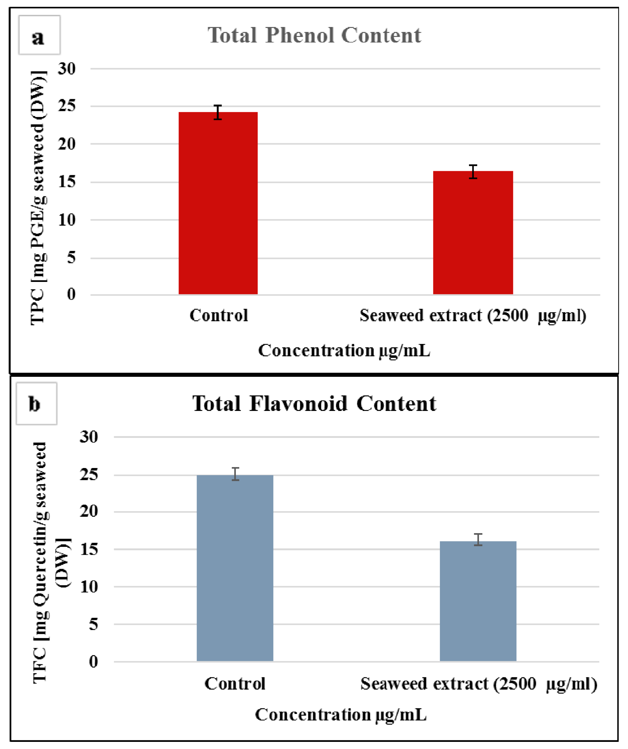
The presence of natural antioxidants is not simply restricted to terrestrial sources. Seaweeds have also proven to be reliable and rich sources of natural antioxidant compounds [29]. The radical scavenging properties of the macro algal species have been reported to be associated with the phenolic compounds present within them [30]. Phenolics possess unique redox properties that contribute in adsorbing as well as neutralizing free radicals, decompose peroxides, and quench singlet and/or triplet oxygen. The antioxidant activity of phenolics primarily owe their properties to such features. Figure 1a represents the total phenol content of the methanolic extract (highest concentration) of J. rubens as determined by using the Folin-Ciocalteu reagent and expressed as mg phloroglucinol equivalents (PGE) per g of seaweed extract, on a dry weight basis. A high total phenolic content was observed to be present in the methanolic extract of J. rubens, although this was not higher than the commercial control (Phloroglucinol) used at the same concentration.
3.3. Total Flavonoid Content
Flavonoids comprise the principal class of polyphenols. They can scavenge practically all known ROS, depending on their structure. They possess the capability to not only scavenge free radicals, but also inhibit the enzymes responsible for free radical production, and also chelate metal ions like iron and copper. The antioxidative assets of the flavonoids may be thus be credited to such mechanisms. Figure 1b represents the total flavonoid content of J. rubens. Quercetin was used as a standard, and the total flavonoid content was expressed as mg quercetin equivalents (QE) per g of the seaweed extract on dry weight basis. The total flavonoid content of the methanolic extract of J. rubens was thus, found to be lower as compared to the standard when used at a similar concentration (Figure 1b).
3.4. DPPH Radical Scavenging Assay
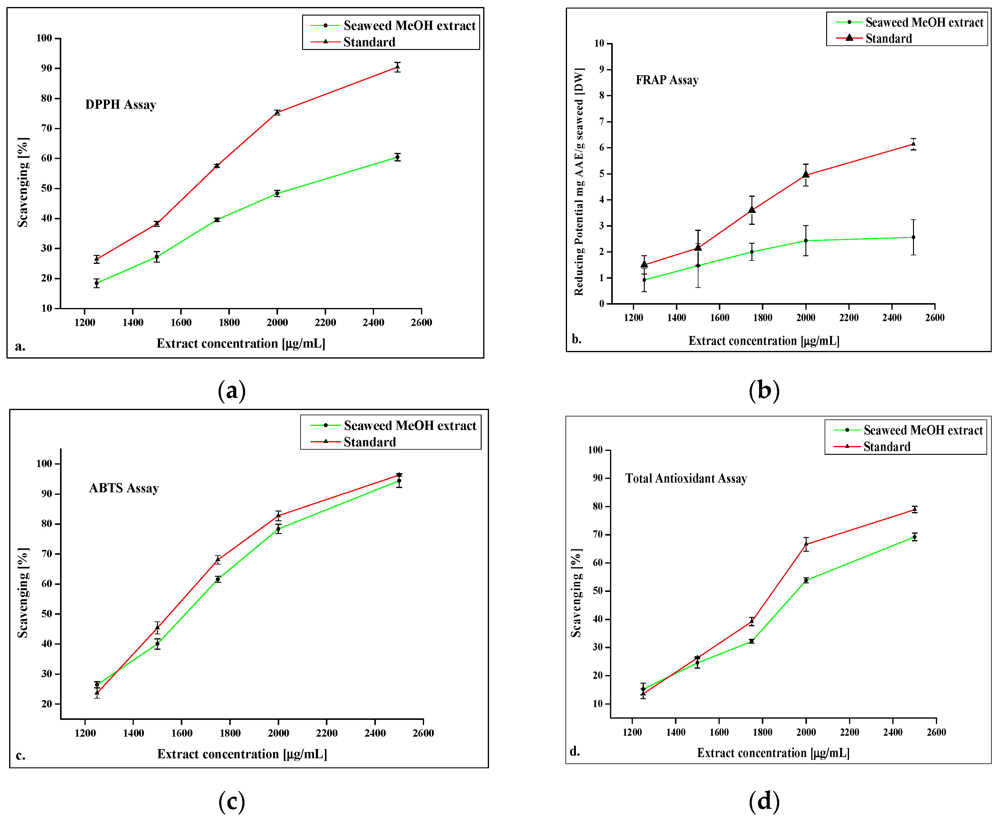
DPPH (1,1-diphenyl-2-picrylhydrazyl) behaves as a free radical donor, and is widely used to assess the radical scavenging potential of antioxidants from natural sources. It possesses a nitrogen free radical and is readily quenched by a free radical scavenger. The DPPH assay is exclusively used as a non-enzymatic antioxidant protocol in determining the radical-scavenging capacity of novel antioxidants of natural origin in organic environments that function as a proton radical scavengers or hydrogen donors. Radical quenching, which can be achieved either by single electron transfer or by hydrogen atoms, result in the neutralization of the DPPH radical. Upon reduction, color changes from purple to yellow with the absorption of hydrogen moiety from the antioxidant. This reaction is stoichiometric in nature, and the antioxidant effect can be easily measured by a decrease in ultra-violet (UV) absorption at 517 nm. The DPPH radical-scavenging capacity of the tropical seaweed J. rubens selected for the current study has been shown in Figure 2a. The scavenging activity of the sample is indicated by the degree of discoloration. The scavenging potential increased concomitantly with an increase in the extract concentration. The study showed scavenging activity >60% at the highest concentration of 2500 μg/mL. However, the value obtained was less when compared to the standard ascorbic acid value (90%) used at a similar concentration.
3.5. Ferric Ion Reducing Antioxidant Power (FRAP) Assay
The principal of the FRAP assay is based on the reduction of ferric-tripyridyltriazine (Fe3+-TPTZ) complex to ferrous tripyridyltriazine (Fe2+-TPTZ) by the antioxidants of the sample at low pH. Fe2+-TPTZ—the final end product, shows a blue color with an absorption maximum at 593 nm. The change in the absorbance is related to the antioxidant capacity of the seaweed extract. The reducing potential of J. rubens extract has been shown in Figure 2b. The reduction potential has been expressed as (μg/mL AAE/mL). The study showed a reduction potential value >50% at the highest concentration of 2500 μg/mL for the extract. However, the value obtained was less when compared to the standard ascorbic acid value (>60%) at the same concentration.
3.6. ABTS Scavenging Activity
The principal underlying this assay is that the ABTS is converted to its radical cation on the addition of potassium persulfate. The radical cation so formed is blue in color and absorbs light at 734 nm. This ABTS radical cation is reactive towards a majority of antioxidants which include thiols, phenolics and ascorbic acid. The blue colored ABTS radical cation is converted back to its colorless neutral form during the course of the reaction. The antioxidants in the sample reduce ABTS, preventing the color formation to a level that is proportional to their concentrations. The ABTS scavenging potential of the J. rubens extract is shown in Figure 2c. The results obtained at the highest concentration (2500 μg/mL) were quite comparable to that obtained for the standard (>90%) at similar concentration.
3.7. Total Antioxidant Assay
The total antioxidant capacity (TAC) was performed using the phosphomolybdenum method. The principle behind this protocol is the formation of Mo (V) by the reduction of Mo (VI) using the test extract/s, and consequent production of the green phosphate/Mo (V) complex at an acidic pH. It evaluates both fat-soluble as well as water-soluble antioxidants (total antioxidant capacity). The phosphomolybdenum method is a simple, cheap, and a good alternative to other laborious methods used for assessing the total antioxidant capacity. This assay was carried out in order to gain a broader understanding of the total antioxidant capacity exhibited by J. rubens, rather than determining the antioxidant potential of individual constituents. Moreover, it gives a clear understanding of the changes in the antioxidant activity in relation to the oxidative stress, and is widely used in a variety of complex mixtures used in pharmaceutical and cosmetic preparations. The results for the TAC have been expressed as scavenging %, as shown in Figure 2d. The total antioxidant capacity of the extract (>65%) was found to be lower as compared to the standard (>75%) at the same concentration (2500 μg/mL).
3.8. Metabolite Profiling
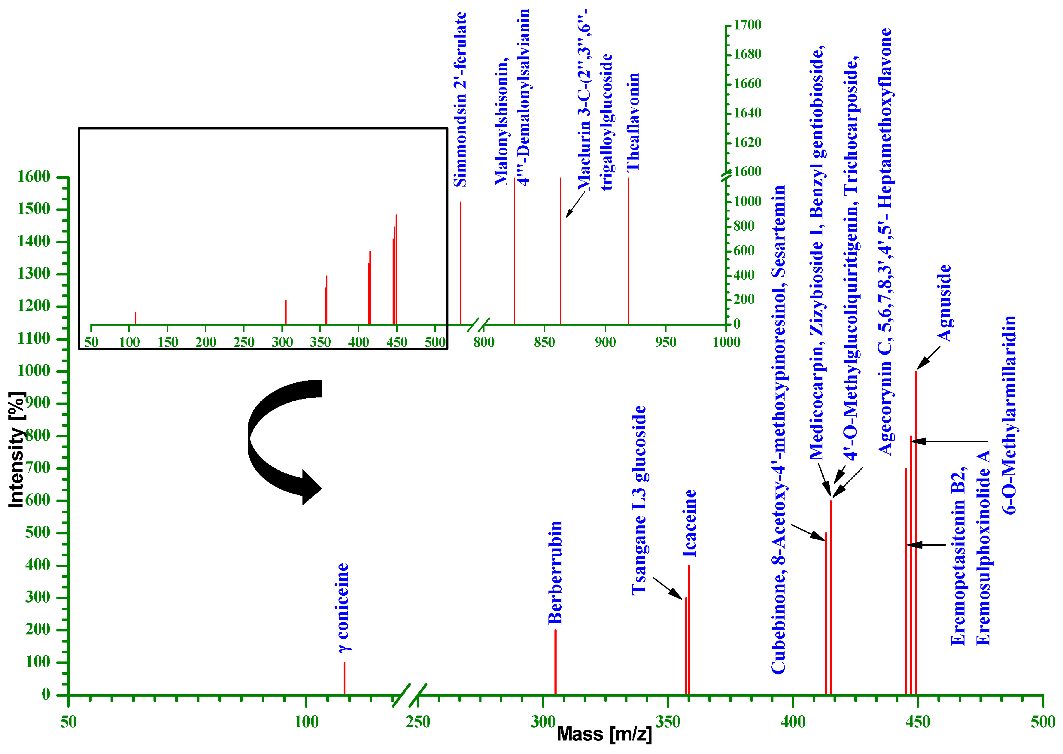
Using liquid chromatography-TOF-MS, around 23 different probable metabolites were encountered in the ESI positive mode (Figure 3). These secondary metabolites comprised of alkaloids, phenyl propanoid, furanoid lignans, flavonoids, tannin, terpene lactone, glycosyl and glycoside compounds, and malleolides and analogues, etc. A phytochemical piperidine alkaloid γ-coniceine (m/z 108.12), which is a precursor of several hemlock alkaloids, was detected. It is used in several formulations that are used to treat breast cancer [31]. Another alkaloid, berberrubin (m/z 305.1), bearing strong antitumor activity was also observed in the seaweed. A potential aromatic compound belonging to the class of terpene glycosides named tsangane L3 glucoside (prenol lipid containing a carbohydrate moiety glycosidically bound to a terpene backbone) with m/z 357.23, was detected. The industrial application of this compound is as a surfactant, as well as an emulsifier. An aromatic coumaric acid derivative named trichocarposide (m/z 415.15), was observed. Trichocarposide has been found to be one of the active ingredients in the methanolic extract of Salix martiana Leyb that exhibits strong DPPH antioxidant activity [32]. A diterpene-based alkaloid, icaceine (m/z 358.24), known to exhibit anticonvulsant properties, was detected. Two aromatic heteropolycyclic compounds viz; 8-Acetoxy-4′-methoxypinoresinol (a furanoid lignan, and a constituent of Olea europaea (olive), which is widely used in cosmetics and pharmaceutical industries), sesartemin (a phenylpropanoid and an inhibitor of cytochrome P450-linked oxygenase) [33] and a natural lignan compound cubebinone (an active constituent of Piper cubeba possessing anti-inflammatory as well as antiseptic properties), all with an (m/z 413.16), were detected during the study. Four flavonoids, viz, agecorynin C, medicocarpin, 4′-O-methylglucoliquiritigenin, and 5,6,7,8,3′,4′,5′-heptamethoxyflavone, a crude drug, viz, zizybeoside I, and an O-glycosyl compound named benzyl gentiobioside (all with an m/z 415.15) were detected. 6-O-Methylarmillaridin (m/z 445.18), belonging to the family of melleolides and analogues, was detected to be present in the current study.
An eremophilane-type sesquiterpene lactone, namely eremopetasitenin B2 (m/z 447.18)—an important biogenetic intermediate, and eremosulphoxinolide A—a terpene lactone (m/z 447.18), along with a terpenoid, viz, agnuside (m/z 449.15), an active constituent playing a role in the estrogenic activity for the potential treatment of premenstrual syndrome in women [34], were found to be present. Additionally, two anthocyanins malonylshisonin/4′′′-demalonylsalvianin (m/z 825.19) belonging to the class of flavonoids, were also detected which possess antioxidant and anticancer (chemo preventive) attributes. Maclurin 3-C-(2″,3″,6″-trigalloylglucoside) (m/z 863.15) belonging to the family of hydrolysable tannins, and the class of chemical entities known as phenolic glycosides, were identified. Theaflavonin (m/z 919.18), belonging to the class of organic compounds known as complex tannins, was detected. An aromatic coumaric acid ester derivative, simmondsin 2′-ferulate (m/z 534.21), was also observed to be present.
4. Discussion
Jania rubens is a versatile red marine macro algae possessing dichotomous ramifications and extraordinary properties of biomineralization. It is described as an alga that is heavily impregnated with biochemically precipitated CaCO3 in the form of calcite, through the phenomenon of biomineralization. This attribute makes the algae very tough and resistant to corrosion, making the coralline algae one of the most important structural elements of coral reefs. It has an extraordinary capability to bind trace minerals and elements found in seawater, which render it with the invaluable cosmetic virtues that it possesses. The occurrence of coralline algae in several types of habitats defines its reliability as a biological indicator of relative levels of phosphate pollution in seawater caused by human activity [35]. The identification of untargeted metabolites along with the study on the mineral composition, as well as the ROS scavenging capacity of this seaweed, revealed its potential to be used as a key ingredient in the cosmeceutical formulations, and provide useful insights into the metabolite constituents of this macro alga.
The health as well as the appeal of the skin depends on nutrition [36]. This species is a noble source of both macro- as well as micro-minerals. Macro-minerals (Ca, Na, Mg and K) were found to be present in significant amounts. Sodium (Na), plays a crucial role in the fluid maintenance, muscle contraction, enzyme operation, and osmo-regulation of the human body. Additionally, it also plays a critical role in skincare also, due to its anti-aging properties. This anti-aging feature can be attributed to its ability to fight the free radicals that accelerate the aging process. It is therefore incorporated as an active ingredient in a range of skincare products. It is also widely used in cleansers and moisturizers. Products for sensitive skin care also contain sodium, as it acts as a mild wetting agent. Thus, sodium in combination with other elements or alone, offers a number of benefits for the skin. Potassium (K), an essential macro mineral, keeps the skin hydrated and moisturized. Additionally, potassium prevents the skin from looking dull and cracked, as it supports the growth of new skin cells. Many skin care and cosmetic products use KOH (potassium hydroxide) as one of their composites. It maintains a balance in the pH level of the skin. Calcium (Ca) was found to be present in the highest amount as a macro mineral in this species. Ca has been proven to play an important role in skin homeostasis (self-replenishing process) and barrier function repair [37]. Due to this, the skin can shed and renew itself and maintain an appropriate lipid level. Calcium skin benefits include anti-aging properties, enabling a better resistance to irregularities like premature aging and fine wrinkling, through its important role in antioxidant production. Magnesium (Mg) possesses the capacity to cleanse the skin and detoxify the epidermis. It is quite effective in treating the areas of the skin that are prone to allergic reactions. Magnesium is very effective in reducing wrinkles and fine lines. It helps to combat acne or breakouts on the skin. Magnesium thus acts as a natural cellular protectant, fosters the restoration of cellular magnesium levels, facilitates effective and safe detoxification, provides relief from pains, spasms, aches and in turn, encourages healthy skin tissue growth.
Micro minerals also play an equally significant role in maintaining the youthfulness of skin. Iron (Fe) is a potential therapeutic target in the skin [38]. Copper (Cu) too plays an important role in skin care. It not only aids in the production of melanin which is responsible for the color of the skin and hair, but also helps in collagen production and skin regeneration, and increases the effect of antioxidants. Moreover, copper peptides (copper bound peptides) and copper gluconates (copper bound gluconic acid) are a promising treatment in skincare. Zinc (Zn) in the form of divalent zinc ions, provides an antioxidant photoprotector for skin. The benefits of either oral or topical zinc in the treatment of acne, possibly through anti-inflammatory effects, has been studied previously [39]. “Prolidase”, an enzyme that is necessary for collagen production—an essential structural component of the skin—requires manganese (Mn) as a co-factor. Manganese is thus an important mineral for everyday skin health, as it plays a specific role in collagen production. In addition to this, manganese aids in the protection of skin against oxygen-related damages, as well as against damages caused due to exposure to UV light by functioning as an antioxidant. J. rubens possesses all of these vital macro- as well as micro-minerals crucial for providing protection against pre-mature aging and maintaining the youthfulness of the skin.
One of the most fundamental entities that are known to be involved in human skin aging process are the ROS. The factors that are responsible for ROS generation within the skin include both, the intrinsic sources such as endogenous oxidative metabolism as well as the extrinsic sources such as UV radiation. The role of secondary metabolites is thus important for combating against ROS, as they are considered to be important radical scavengers and efficient antioxidants that possess biological activities [40]. Phytochemicals (secondary metabolites), including flavonoids, phenolic acids, and polyphenols, are potent antioxidants and are an essential part of human well-being. Antioxidants are important as a part of an anti-ageing skin support program because antioxidants help protect the skin from the toxic effects of free radicals that would otherwise impair and destroy healthy skin cells [41]. Scavenging properties and antioxidant potential depend on the content of both polyphenolics and flavonoids [40]. J. rubens possessed a good amount of both phenolic as well as flavonoid contents, and therefore showed a high antioxidant potential. The antioxidant activity was found to be proportional to the concentration of polyphenols and flavonoids. Five different methanolic concentrations were used to check the DPPH radical scavenging potential. The radical scavenging potential was lower compared to the standard ascorbic acid but was >60%. This result suggests the presence of free radical inhibitors acting as potential primary antioxidants. Therefore, J. rubens extract was found to contain metabolites that could function as hydrogen donors, thereby neutralizing the DPPH molecules. Moreover, the presence of metabolites like trichocarposide, that exhibit strong DPPH antioxidant activity, further supported the study. In the reducing power assay, the antioxidants donate an electron to stabilize the radicals and also break the free radical chain reaction. The ability of the different concentrations of the J. rubens extract to exhibit the reducing power in this investigation may be related to the presence of antioxidant phytochemicals. Reducing potential was found to be lower as compared to the commercial standard. Some of the cosmeceutical effects could be attributed to these compounds. The ABTS radical scavenging ability of the extract also exhibited a dose-dependent response. As the concentration of the methanolic extracts increased, ABTS radical scavenging as well as the total antioxidant activity also increased. In fact, the ABTS radical scavenging potential was found to be quite comparable to the standard value, while the latter was lower as compared to the standard.
Furthermore, about 23 different putative metabolites were identified in the present study using LC-TOF-MS/MS (Figure 3 and Supplementary Table S1). This study offers the first comprehensive scientific report on the untargeted metabolomics of J. rubens, highlighting its importance as an active cosmeceutical ingredient. Anthocyanins [42,43] provide protection against potentially effective agents, in order to avert the signs of skin aging. They have proven actions for giving protection to the skin from external injuries caused by UV radiation [44]. Additionally, anthocyanins not only possess strong antioxidant/anti-inflammatory activities, but also inhibit lipid peroxidation and inflammatory mediators; cyclooxygenase (COX)-1 and -2 [45]. They are also used as cosmetic colorants in many products [46]. The present study unveils the presence of two such anthocyanins, malonylshisonin and 4′′′-demalonylsalvianin, which may prove to be potential flavonoids for cosmetic industries. A complex oxidation product belonging to the natural class of flavonoid theaflavins, named theaflavonin, was detected. Theaflavins are potent antioxidant polyphenols already used in skin care product preparation, in order to impart effects such as whitening and the removal of freckles. [47]. Moreover, theaflavin in the skin care products shows excellent color stability. Therefore, they can be easily applied to other cosmetic preparations as well. Flavonoids have been found to possess antioxidant, anti-allergic, anti-viral, anti-aging, anti-carcinogenic, and anti-inflammatory properties. Moreover, they are also used in the treatment of skin aging, as they apparently contribute in the improvement of skin elasticity, skin hydration, regulation of oil gland secretion, and collagen content [48]. A set of flavonoids, namely agecorynin C (KEGG C14942), medicocarpin (KEGG C16223), and 5,6,7,8,3′,4′,5′-heptamethoxyflavone (KEGG C14953), have been detected in this study. Benzyl gentiobioside, belonging to the class of O-glycosyl compounds, was also detected.
Cosmetic formulations also possess natural as well as synthetic additives that confer aroma, which in turn masks unpleasant chemical odors. This study also revealed the presence of many natural aromatic compounds. One of them was trichocarposide—an aromatic coumaric ester derivative that is an essential constituent of Populus balsamifera (balsam poplar), which is highly sought after for its essential oil in various aroma therapies, was detected from J. rubens extract. It is known to nourish the skin and relax the mind. Additionally, 4′-O-Methylglucoliquiritigenin—a compound belonging to flavonoid-7-O-glycosides, was also detected. It is one of the constituent of the roots of Glycyrrhiza uralensis (Chinese licorice). The pharmacological significance of licorice includes its antimicrobial [49], antiviral [50], and antitumor [51], as well as its anti-inflammatory [52] properties. Zizybeoside I (KEGG C17564), detected during the present study, is usually found to be present in Zizyphus jujuba (Chinese date), and belongs to the family of Dihexoses (disaccharides containing two hexose carbohydrates). Zizyphus jujuba is commonly used for face nourishment and beautification, for battling oxidation and aging, as an antitumor agent, for resisting fatigue, and also for the relaxation of mind [53,54,55]. In fact, Chinese jujube is also commonly employed in Chinese beverages and as a food additive [56].
Eremopetasitenin B2 and eremosulphoxinolide A, isolated from the fresh rhizomes of Petasites japonicus, which has been used for its therapeutic effect on allergy and asthma in Korea and European countries, was also detected [57,58]. Simmondsin 2-ferulate, a glucoside with proven insecticidal, antifeedant, and antifungal activities [59] was noted to be present. Simmondsin 2′-ferulate is usually obtained from Simmondsia chinensis (jojoba). Lubricant, pharmaceutical and cosmetic industries hold a good market for Jojoba oil [60]. Jojoba seeds have also been shown to possess anti-inflammatory activity [61].
Terpenes comprise one of the major secondary metabolites, with diverse categories including monoterpenoids, diterpenoids, triterpenoids, and sesquiterpenoids. Terpenes have been found to possess anti-inflammatory, antitumor, antibacterial, antioxidant, and hepatoprotective activities from various pharmacological studies [62]. Tsangane L3 glucoside (prenol lipid containing a carbohydrate moiety glycosidically bound to a terpene backbone)—a potential aromatic compound belonging to the class of terpene glycosides, was detected in the present study. Another diterpene-based alkaloid, named icaceine (m/z 358.24), known to prevent or reduce the severity of epileptic seizures and agnuside, an iridoid glycoside possessing hepatoprotective properties [63], anti-inflammatory activity [64] antioxidant [65], and analgesic effects [66], were found to be present in the methanolic extract of J. rubens. Additionally, two alkaloids viz; γ-coniceine (KEGG C10138) which acts as a local analgesic [67], and berberrubin (HMDB30266), a protoberberine, known for exhibiting antitumor properties, were detected during the study [68].
Lignans act as both antioxidants and phytoestrogens [69]. Other salient traits, viz, anti-cancerous, anti-inflammatory, insecticidal, anti-hypertensive, hypocholesterolemic, anti-asthma activities, and hepatoprotective biological activities of lignans have also been previously reported [70]. Lignans derived from flax have not only been described as being useful in preventing osteoporosis and certain cancers, but also as anti-viral agents and fungicides. The role of lignans in plant defense was suggested by their pronounced antimicrobial, antifungal, antiviral, and antioxidant properties [71,72,73,74]. The glycosylated lignans obtained from flax seeds are known to inhibit the production of melanin, thereby playing a role in bleaching the skin [75]. They also act as agents for increasing the catalase and fibroblast activity, for inhibiting UV-induced wrinkles, for the synthesis of hyaluronic acid, and for strengthening the elasticity of the skin. Further, the prevention of formation of wrinkles, thereby assisting with maintaining the firmness of the skin by providing relief through scavenging OH radicals, has also been proven outcomes for glycosylated lignans derived from sesame. These are also well known as antioxidants, and may be used in moisturizing and protective creams [76]. The retinol-based anti-ageing compositions contain lignan known as nordihydroguaiaretic acid, which is an antioxidant. A furanoid lignan 8-acetoxy-4′-methoxypinoresinol (HMDB33277), and two natural lignin compounds, sesartemin (KEGG C10884) and cubebinone, (HMDB33259) were observed to be present in the methanolic extract of J. rubens. Certain melleolide analogs have shown to exhibit anti-microbial as well as antifungal activities [77,78]. 6-O-methylarmillaridin, a melleolide sesquiterpene was one of the bioactive metabolites encountered during the present study.
A number of brown seaweeds viz; Fucus vesiculosus (Fucoidan) [79], Pelvetia wrightii [80], Laminaria digitata (Minerals, proteins and carbohydrates) [81], are frequently used in slimming and anticellulite formulations. Macro algal constituents like flavonoids, phlorotannins, quercetin (flavonoid belonging to the class of flavonols) etc. act as lipolytic agents. Sargassum polycystum extracts (ethanolic, ethyl acetate and hexane) have been studied for their skin whitening properties [82]. The biochemical composition of these active fractions have revealed the presence of terpenoids, flavonoids, phenols, saponins, tannins etc. Moreover, antioxidants favor the skin health by reducing hyperpigmentation. It has been reported that macro algae have emerged with mechanisms to respond against the hazardous effects of UV-A and UV-B by producing phenolic compounds, UV-absorbing mycosporine-like amino acid (MAAs), or carotenoids [79]. Such MAAs act as a UV shield, and the antioxidant constituents present in the red algae (Rhodophytes) act as effective photoprotectors. Similarly, in the case of microalgae, an extract from Chlorella vulgaris has been reported to induce collagen synthesis in skin, thereby preventing wrinkle formation [83]. A protein-rich extract from Arthrospira has been reported to exert tightening of the skin, thereby protecting the early signs of skin aging [83]. The current study too has revealed the presence of many such bioactive metabolites in J. rubens, which may open up new avenues for the advancement in cosmetic compositions.
5. Conclusions
The study reveals that the red marine macro alga, J. rubens, is a rich source of essential macro- as well as micro-minerals, natural antioxidants, and bioactive metabolites with cosmeceutical potential. These features collectively make this coralline alga a promising candidate for its inclusion as an active ingredient in modern day cosmeceutical as well as pharmaceutical formulations/products utilized for skin conditioning, skin polishing, anti-ageing, and skin whitening. J. rubens can therefore be considered as a potential natural candidate for improving the quality of modern day cosmetics. However, further studies undertaking purification as well as clinical trials of the purified components, are required for its inclusion as an integral ingredient. The present study provides the base for future perspectives of considering J. rubens in the design of algal biorefineries and other eco-designs, to obtain large amounts of value-added products.
Supplementary Materials
The following are available online at www.mdpi.com/2079-9284/4/4/45/s1, Figure S1: Metabolite Profile of J. rubens extract using LCMS (ESI positive mode), Table S1: Putative metabolites identified in J. rubens extract and their possible function/application/role/importance.
Acknowledgments
Dhara Dixit (D.D.) sincerely thanks the Department of Science & Technology (DST), New Delhi, for awarding the Women Scientist Fellowship and rendering financial support (WOS-A/LS-388/2012) for undertaking this research. Dhara Dixit deeply acknowledges the support rendered by the Director, CSIR-CSMCRI for providing the infrastructure and the analytical facilities to conduct this study. The guidance extended by Avinash Mishra for LC-MS study is highly acknowledged. The field assistance extended for the collection of seaweed sample by Devesh K. Gadhavi (Deputy Director, Kutch Ecological Research Centre—A Division of The Corbett Foundation, Kachchh) is duly acknowledged. The author also thanks M.G. Thakkar (Head) & M.H. Trivedi (Asst. Prof.), Department of Earth & Environmental Science, Krantiguru Shyamji Krishna Verma (K.S.K.V.) Kachchh University for providing the administrative support for carrying out the work smoothly. The fellowship term is over and there is no fund for covering the costs to publish in open access.
Author Contributions
Dhara Dixit conceived and designed the experiments; Dhara Dixit performed the experiments; Dhara Dixit analyzed the data; C.R.K. Reddy contributed reagents/materials/analysis tools; Dhara Dixit and C.R.K. Reddy wrote the paper.
Conflicts of Interest
The authors declare no conflict of interest.
References
- Thomas, N.V.; Kim, S.K. Beneficial Effects of Marine Algal Compounds in Cosmeceuticals. Mar. Drugs 2013, 11, 146–164. [Google Scholar] [CrossRef] [PubMed]
- Kim, S.K.; Ravichandran, Y.D.; Khan, S.B.; Kim, Y.T. Prospectives of the cosmeceuticals derived from marine organisms. Biotechnol. Bioprocess Eng. 2008, 13, 511–523. [Google Scholar] [CrossRef]
- Wijesinghe, W.A.J.P.; Jeon, Y.J. Biological activities and potential cosmeceutical applications of bioactive components from brown seaweeds: A review. Phytochem. Rev. 2011, 10, 431–443. [Google Scholar] [CrossRef]
- Cabrita, M.; Vale, C.; Rauter, A. Halogenated compounds from marine algae. Mar. Drugs 2010, 8, 2301–2317. [Google Scholar] [CrossRef] [PubMed]
- Manach, C.; Scalbert, A.; Morand, C.; Rémésy, C.; Jiménez, L. Polyphenols: Food sources and bioavailability. Am. J. Clin. Nutr. 2004, 79, 727–747. [Google Scholar] [PubMed]
- Zern, T.L.; Fernandez, M.L. Cardio protective effects of dietary polyphenols. J. Nutr. 2005, 135, 2291–2294. [Google Scholar] [PubMed]
- Dunn, W.B.; Ellis, D.I. Metabolomics: Current analytical platforms and technologies. Trends Anal. Chem. 2005, 24, 285–294. [Google Scholar]
- Naidoo, K.; Machin, M.A.B. Oxidative Stress and Ageing: The Influence of Environmental Pollution, Sunlight and Diet on Skin. Cosmetics 2017, 4, 4. [Google Scholar] [CrossRef]
- Joe, M.J.; Kim, S.N.; Choi, H.Y.; Shin, W.S.; Park, G.M.; Kang, D.W.; Kim, Y.K. The inhibitory effects of eckol and dieckol from Ecklonia stolonifera on the expression of matrix metalloproteinase-1 in human dermal fibroblasts. Biol. Pharm. Bull. 2006, 29, 1735–1739. [Google Scholar] [CrossRef] [PubMed]
- Kim, M.M.; Van Ta, Q.; Mendis, E.; Rajapakse, N.; Jung, W.K.; Byun, H.G.; Jeon, Y.J.; Kim, S.K. Phlorotannins in Ecklonia cava extract inhibit matrix metalloproteinase activity. Life Sci. 2006, 79, 1436–1443. [Google Scholar] [CrossRef] [PubMed]
- Price, R.D.; Berry, M.G.; Navsaria, H.A. Hyaluronic acid: The scientific and clinical evidence. J. Plast. Reconstr. Aesthet. Surg. 2007, 60, 1110–1119. [Google Scholar] [CrossRef] [PubMed]
- Ryu, B.; Qian, Z.J.; Kim, M.M.; Nam, K.W.; Kim, S.K. Anti-photoaging activity and inhibition of matrix metalloproteinase (MMP) by marine red alga, Corallina pilulifera methanol extract. Radiat. Phys. Chem. 2009, 78, 98–105. [Google Scholar]
- Verdy, C.; Branka, J.E.; Mekideche, N. Quantitative assessment of lactate and progerin production in normal human cutaneous cells during normal ageing: Effect of an Alaria esculenta extract. Int. J. Cosmet. Sci. 2011, 33, 462–466. [Google Scholar] [CrossRef] [PubMed]
- Rossano, R.; Ungaro, N.; D’Ambrosio, A.; Liuzzi, G.M.; Riccio, P. Extracting and purifying R-phycoerythrin from Mediterranean red algae Corallina elongata. J. Biotechnol. 2003, 101, 289–293. [Google Scholar] [CrossRef]
- Joly, A.B. Generos de Algas Marinhas da Costa Atlantica Latino-Americana; Editora da Universidade de São Paulo: São Paulo, Brazil, 1967; p. 461. [Google Scholar]
- Bôas, V.; Bigio, A.; Figueiredo, M.A.D.O. Are anti-fouling effects in coralline algae species specific? Braz. J. Oceanogr. 2004, 52, 11–18. [Google Scholar] [CrossRef]
- Karabay-Yavasoglu, N.U.; Sukatar, A.; Ozdemir, G.; Horzum, Z. Antimicrobial activity of volatile components and various extracts of the red alga Jania rubens. Phytother. Res. 2007, 21, 153–156. [Google Scholar] [CrossRef] [PubMed]
- Ktari, L.; Blond, A.; Guyot, M. 16β-Hydroxy-5α-cholestane-3, 6-dione, a novel cytotoxic oxysterol from the red alga Jania rubens. Bioorg. Med. Chem. Lett. 2000, 10, 2563–2565. [Google Scholar] [CrossRef]
- El-Din, S.M.M.; El-Ahwany, A.M. El-Ahwany Bioactivity and phytochemical constituents of marine red seaweeds (Jania rubens, Corallina mediterranea and Pterocladia capillacea). J. Taibah Univ. Sci. 2016, 10, 471–484. [Google Scholar] [CrossRef]
- Awad, N.E. Bioactive brominated diterpenes from the marine red alga Jania rubens (L.) Lamx. Phytother Res. 2004, 18, 275–279. [Google Scholar] [CrossRef] [PubMed]
- Navarro, D.A.; Stortz, C.A. The system of xylogalactans from the red seaweed Jania rubens (Corallinales, Rhodophyta). Carbohydr. Res. 2008, 43, 2613–2622. [Google Scholar] [CrossRef] [PubMed]
- Santoso, J.; Gunji, S.; Yoshie-Stark, Y.; Suzuki, T. Mineral contents of Indonesian seaweeds and mineral solubility affected by basic cooking. Food Sci. Technol. Res. 2006, 12, 59–66. [Google Scholar] [CrossRef]
- De Vos, R.C.; Moco, S.; Lommen, A.; Keurentjes, J.J.; Bino, R.J.; Hall, R.D. Untargeted large-scale plant metabolomics using liquid chromatography coupled to mass spectrometry. Nat. Protoc. 2007, 2, 778–791. [Google Scholar] [CrossRef] [PubMed]
- Zhu, Z.J.; Schultz, A.W.; Wang, J.; Johnson, C.H.; Yannone, S.M.; Patti, G.J.; Siuzdak, G. Liquid chromatography quadrupole time-of-flight mass spectrometry characterization of metabolites guided by the METLIN database. Nat. Protoc. 2013, 8, 451–460. [Google Scholar] [CrossRef] [PubMed]
- Kumar, M.; Gupta, V.; Kumari, P.; Reddy, C.R.K.; Jha, B. Assessment of nutrient composition and antioxidant potential of Caulerpaceae seaweeds. J. Food Compos. Anal. 2011, 24, 270–278. [Google Scholar] [CrossRef]
- Lim, S.N.; Cheung, P.C.K.; Ooi, V.E.C.; Ang, P.O. Evaluation of antioxidative activity of extracts from brown seaweed, Sargassum siliquastrum. J. Agric. Food Chem. 2002, 50, 3862–3866. [Google Scholar] [CrossRef] [PubMed]
- Jia, Z.; Tang, M.; Wu, J. The determination of flavonoid contents in mulberry and their scavenging effects on superoxide radicals. Food Chem. 1999, 64, 555–559. [Google Scholar]
- Prieto, P.; Pineda, M.; Anguilar, M. Spectrophotometric quantitation of antioxidant capacity through the formation of a Phosphomolybdenum Complex: Specific application to the determination of Vitamin E. Anal. Biochem. 1999, 269, 337–341. [Google Scholar] [CrossRef] [PubMed]
- Patra, J.K.; Lee, S.-W.; Park, J.G.; Baek, K.-H. Antioxidant and Antibacterial Properties of Essential Oil Extracted from an Edible Seaweed Undaria Pinnatifida. J. Food Biochem. 2017, 41, e12278. [Google Scholar] [CrossRef]
- Duan, X.J.; Zhang, W.W.; Li, X.M.; Wang, B.G. Evaluation of antioxidant property of extract and fractions obtained from a red alga, Polysiphonia urceolata. Food Chem. 2006, 95, 37–43. [Google Scholar] [CrossRef]
- Usman, M.R.M.; Salgar, S.D.; Nagpal, N.; Shaikh, M.Z. Poisonous Herbal Plants: NA; Educreation Publishing: New Delhi, India, 2016. [Google Scholar]
- Fernandes, C.C.; de Carvalho Cursino, L.M.; de Abreu Pinheiro Novaes, J.; Demetrio, C.A.; Júnior, O.L.P.; Nunez, C.V.; do Amaral, I.L. Salicilatos isolados de folhas e talos de Salix martiana Leyb. (Salicaceae). Quim. Nova 2009, 32, 983–986. [Google Scholar] [CrossRef]
- Polya, G. Biochemical Targets of Plant Bioactive Compounds: A Pharmacological Reference Guide to Sites of Action and Biological Effects; CRC Press: Boca Raton, FL, USA, 2003; ISBN 0-415-30829-1. [Google Scholar]
- Ramakrishna, R.; Bhateria, M.; Singh, R.; Puttrevu, S.K.; Bhatta, R.S. Plasma pharmacokinetics, bioavailability and tissue distribution of agnuside following peroral and intravenous administration in mice using liquid chromatography tandem mass spectrometry. J. Pharm. Biomed. Anal. 2016, 125, 154–164. [Google Scholar] [CrossRef] [PubMed]
- Brown, V.; Ducker, S.C.; Rowan, K.S. The effect of orthophosphate concentration on the growth of articulated coralline algae (Rhodophyta). Phycologia 1977, 16, 125–131. [Google Scholar] [CrossRef]
- Boelsma, E.; Hendriks, H.F.J.; Roza, L. Nutritional skin care: Health effects of micronutrients and fatty acids1–3. Am. J. Clin. Nutr. 2001, 73, 853–864. [Google Scholar] [PubMed]
- Denda, M.; Fuziwara, S.; Inoue, K. Influx of Calcium and Chloride Ions into Epidermal Keratinocytes Regulates Exocytosis of Epidermal Lamellar Bodies and Skin Permeability Barrier Homeostasis. J. Investig. Dermatol. 2003, 121, 362–367. [Google Scholar] [CrossRef] [PubMed]
- Wright, J.A.; Richards, T.; Srai, S.K.S. The role of iron in the skin and cutaneous wound healing. Front. Pharmacol. 2014, 5, 156. [Google Scholar] [CrossRef] [PubMed]
- Solomons, N.W.; Ruz, M.; Duran, C.C. Putative therapeutic roles for zinc. In Zinc in Human Biology; Mills, C., Ed.; Springer: London, UK, 1989; pp. 297–321. [Google Scholar]
- Shahidi, F.; Ambigaipalan, P. Phenolics and polyphenolics in foods, beverages and spices: Antioxidant activity and health effects—A review. J. Funct. Foods 2015, 18, 820–897. [Google Scholar] [CrossRef]
- Salavkar, S.M.; Tamanekar, R.A.; Athawale, R.B. Antioxidants in skin ageing—Future of dermatology. Int. J. Green Pharm. 2011, 5, 161–168. [Google Scholar]
- Schreckinger, M.E.; Lotton, J.; Lila, M.A.; De Mejia, E.G. Berries from South America: A comprehensive review on chemistry, health potential, and commercialization. J. Med. Food 2010, 13, 233–246. [Google Scholar] [CrossRef] [PubMed]
- Afaq, F.; Zaid, M.A.; Khan, N.; Dreher, M.; Mukhtar, H. Protective effect of pomegranate-derived products on UVB-mediated damage in human reconstituted skin. Exp. Dermatol. 2009, 18, 553–561. [Google Scholar] [CrossRef] [PubMed]
- Afaq, F.; Khan, N.; Syed, D.N.; Mukhtar, H. Oral feeding of pomegranate fruit extract inhibits early biomarkers of UVB radiation-induced carcinogenesis in Skh-1 hairless mouse epidermis. Photochem. Photobiol. 2010, 86, 1318–1326. [Google Scholar] [CrossRef] [PubMed]
- Seeram, N.P.; Cichewicz, R.H.; Chandra, A.; Nair, M.G. Cyclooxygenase inhibitory and antioxidant compounds from crabapple fruits. J. Agric. Food Chem. 2003, 51, 1948–1951. [Google Scholar] [CrossRef] [PubMed]
- Khan, A.S. Flowering Plants: Structure and Industrial Products; Wiley: Hoboken, NJ, USA, 2017. [Google Scholar]
- Huiling, L.; Xiufang, Y.; Shikang, Z.; Rong, T.; Yunfei, T.; Junhao, K.; Xiaoqiang, C.; Yuping, G. Application of Theaflavin and Skin Care proDuct Containing Theaflavin. Patent No. CN103,083,199 A, 2013. [Google Scholar]
- Michalun, M.V.; Dinardo, J.C. Milady Skin Care and Cosmetic Ingredients Dictionary, 4th ed.; Cengage Learning: Independence, KY, USA, 2014. [Google Scholar]
- Ahn, S.J.; Cho, E.J.; Kim, H.J.; Park, S.N.; Lim, Y.K.; Kook, J.K. The antimicrobial effects of deglycyrrhizinated licorice root extract on Streptococcus mutans UA159 in both planktonic and biofilm cultures. Anaerobe 2012, 18, 590–596. [Google Scholar] [CrossRef] [PubMed]
- Adianti, M.; Aoki, C.; Komoto, M.; Deng, L.; Shoji, I.; Wahyuni, T.S. Anti-hepatitis C virus compounds obtained from Glycyrrhiza uralensis and other Glycyrrhiza species. Microbiol. Immunol. 2014, 58, 180–187. [Google Scholar] [CrossRef] [PubMed]
- Khan, R.; Khan, A.Q.; Lateef, A.; Rehman, M.U.; Tahir, M.; Ali, F. Glycyrrhizic acid suppresses the development of precancerous lesions via regulating the hyper proliferation, inflammation, angiogenesis and apoptosis in the colon of Wistar rats. PLoS ONE 2013, 8, e56020. [Google Scholar]
- Chandrasekaran, C.V.; Deepak, H.B.; Thiyagarajan, P.; Kathiresan, S.; Sangli, G.K.; Deepak, M. Dual inhibitory effect of Glycyrrhiza glabra (GutGardTM) on COX and LOX products. Phytomedicine 2011, 18, 278–284. [Google Scholar] [CrossRef] [PubMed]
- Fatemeh, V.; Mohsen, F.M.; Kazem, B. Evaluation of inhibitory effect and apoptosis induction of Zyzyphus jujube on tumor cell lines, an in vitro preliminary study. Cytotechnology 2008, 56, 105–111. [Google Scholar]
- Krings, U.; Berger, R.G. Antioxidative activity of some roasted foods. Food Chem. 2001, 72, 223–229. [Google Scholar] [CrossRef]
- Qi, H.M.; Zhang, Q.B.; Zhao, T.T.; Chen, R.; Zhang, H.; Niu, X.Z.; Li, Z. Antioxidative activity of different sulfate content derivatives of polysaccharide extracted from Ulva pertusa (Chlorophyta) in vitro. Int. J. Biol. Macromol. 2005, 37, 195–199. [Google Scholar] [CrossRef] [PubMed]
- Wang, S.; Zhang, J.; Zhang, Z.; Gao, W.; Yan, Y.; Li, X.; Liu, C. Identification of Chemical Constituents in the Extract and Rat Serum from Ziziphus Jujuba Mill by HPLC-PDA-ESI-MSn. Iran. J. Pharm. Res. 2014, 13, 1055–1063. [Google Scholar] [PubMed]
- Sok, D.E.; Oh, S.H.; Kim, Y.B.; Kang, H.G.; Kim, M.R. Neuroprotection by extract of Petasites japonicus leaves, a traditional vegetable, against oxidative stress in brain of mice challenged with kainic acid. Eur. J. Nutr. 2006, 45, 61–69. [Google Scholar] [CrossRef] [PubMed]
- Choi, O.B. Anti-allergic effects of Petasites japonicas. Korean J. Food Nutr. 2002, 5, 382–385. [Google Scholar]
- Abbassy, M.A.; Abdelgaleil, S.A.M.; Belal, A.S.H.; Rasoul, M.A.A.A. Insecticidal, antifeedant and antifungal activities of two glucosides isolated from the seeds of Simmondsia chinensis. Ind. Crop. Prod. 2007, 26, 345–350. [Google Scholar] [CrossRef]
- Arya, D.; Khan, S. A Review of Simmondsia chinensis (Jojoba) “The Desert Gold”: A Multipurpose Oil Seed Crop for Industrial Uses. J. Pharm. Sci. Res. 2016, 8, 381–389. [Google Scholar]
- Malty, R.H.; Naim, A.A.; Khalifa, A.E.; Azizi, M.M.A. Anti-inflammatory effects of jojoba liquid wax in experimental models. Pharmacol. Res. 2005, 51, 95–105. [Google Scholar]
- Yao, J.L.; Fang, S.M.; Liu, R.; Oppong, M.B.; Liu, E.W.; Fan, G.W.; Zhang, H. A Review on the Terpenes from Genus Vitex. Molecules 2016, 21, 1179. [Google Scholar] [CrossRef] [PubMed]
- Atta-ur-Rahman. Studies in Natural Products Chemistry, Bioactive Natural Products (Part L); Elsevier Science: Amsterdam, The Netherlands, 2005; Volume 32, p. 1268. [Google Scholar]
- Pandey, A.; Bani, S.; Satti, N.K.; Gupta, B.D.; Suri, K.A. Anti-arthritic activity of agnuside mediated through the down-regulation of inflammatory mediators and cytokines. Inflamm. Res. 2012, 61, 293–304. [Google Scholar] [CrossRef] [PubMed]
- Tiwari, N.; Luqman, S.; Masood, N.; Gupta, M.M. Validated high performance thin layer chromatographic method for simultaneousquantification of major iridoids in Vitex trifolia and their antioxidant studies. J. Pharm. Biomed. Anal. 2012, 61, 207–214. [Google Scholar] [CrossRef] [PubMed]
- Okuyama, E.; Fujimori, S.; Yamazaki, M.; Deyama, T. Pharmacologically active components of viticis fructus (Vitex rotundifolia). II. The components having analgesic effects. Chem. Pharm. Bull. 1998, 46, 655–662. [Google Scholar] [CrossRef] [PubMed]
- Nelly, A.; Annick, D.D.; Frederic, D. Plants used as remedies antirheumatic and antineuralgic in the traditional medicine of Lebanon. J. Ethnopharmacol. 2008, 120, 315–334. [Google Scholar]
- Jeon, Y.W.; Jung, J.W.; Kang, M.R.; Chung, I.K.; Lee, W.T. NMR Studies on Antitumor Drug Candidates, Berberine and Berberrubine. Bull. Korean Chem. Soc. 2002, 23, 391–394. [Google Scholar]
- Goyal, A.; Sharma, V.; Upadhyay, N.; Gill, S.; Sihag, M. Flax and flaxseed oil: An ancient medicine & modern functional food. J. Food Sci. Technol. 2014, 51, 1633–1653. [Google Scholar] [PubMed]
- Mao, J.; Yu, N.-J.; Yang, Y.; Zhao, Y.-M. Biological activities of dibenzyl butyrolactone lignans, Research advances. J. Int. Pharm. Res. 2014, 41, 275–281. [Google Scholar]
- Chandra, H.; Bishnoi, P.; Yadav, A.; Patni, B.; Mishra, A.P.; Nautiyal, A.R. Antimicrobial Resistance and the Alternative Resources with Special Emphasis on Plant-Based Antimicrobials—A Review. Plants 2017, 6, 16. [Google Scholar] [CrossRef] [PubMed]
- Windayani, N.; Rukayadi, Y.; Hakim, E.H.; Ruslan, K.; Syah, Y.M. Antifungal activity of lignans isolated from Phyllanthus myrtifoliu Moon against Fusarium oxysporum. Curr. Top. Phytochem. 2014, 12, 33–39. [Google Scholar]
- Elfahmi, N.V. Phytochemical and Biosynthetic Studies of Lignans, with a Focus on Indonesian Medicinal Plants. Ph.D. Thesis, University Library Groningen, Groningen, The Netherlands, 2006. [Google Scholar]
- Lin, X.; Zhou, L.; Li, T.; Brennan, C.; Fu, X.; Liu, R.H. Phenolic content, antioxidant and antiproliferative activities of six varieties of white sesame seeds (Sesamum indicum L.). RSC Adv. 2017, 7, 5751–5758. [Google Scholar] [CrossRef]
- Renault, B.; Catroux, P. Cosmetic Use of Lignans. Patent No. EP1,526,832 A1, 4 May 2005. [Google Scholar]
- Toshihiko, O.; Keiko, N.; Kyoko, S.; Takemoto, Y.; Kabushiki, K. Protein Composition Derived from Sesame Seed and Use Thereof. U.S. Patent 5,993,795 A, 30 November 1999. [Google Scholar]
- Midland, S.L.; Izac, R.R.; Wing, R.M.; Zaki, A.I.; Munnecke, D.E.; Sims, J.J. Melleolide, a new antibiotic from Armillaria mellea. Tetrahedron Lett. 1982, 23, 2515–2518. [Google Scholar] [CrossRef]
- Donnelly, D.M.; Abe, F.; Coveney, D.; Fukuda, N.; O’Reilly, J.; Polonsky, J.; Prangé, T. Antibacterial sesquiterpene aryl esters from Armillaria mellea. J. Nat. Prod. 1985, 48, 10–16. [Google Scholar] [CrossRef] [PubMed]
- Bedoux, G.; Hardouin, K.; Burlot, A.S.; Bourgougnon, N. Bioactive Components from Seaweeds: Cosmetic Applications and Future Development. Adv. Bot. Res. 2014, 71, 345–378. [Google Scholar]
- Rozkin, M.; Levina, M.N.; Efimov, V.S.; Usov, A.I. The anticoagulant and lipolysis-stimulating activity of polysaccharides from marine brown algae. Farmakol. Toksikol. 1991, 54, 40–42. [Google Scholar] [PubMed]
- Gedouin, A.; Vallee, R.; Morvan, P.Y. Use of Algae Extract to Stimulate the Oxygen Uptake by the Cells Having Lipolytic Effect to Produce ATP Molecules. Patent No. FR 2,879,098 A1, 16 June 2006. [Google Scholar]
- Chan, Y.Y.; Kim, K.H.; Cheah, S.H. Inhibitory effects of Sargassum polycystum on tyrosinase activity and melanin formation in B16F10 murine melanoma cells. J. Ethnopharmacol. 2011, 137, 1183–1188. [Google Scholar] [CrossRef] [PubMed]
- Spolaore, P.; Joannis-Cassan, C.; Duran, E.; Isambert, A. Commercial Applications of Microalgae. J. Biosci. Bioeng. 2006, 101, 201–211. [Google Scholar] [CrossRef] [PubMed] [Green Version]
Figure 1.
(a) Total phenol content and (b) Total flavonoid content of J. rubens extract at highest concentration. Value represents the mean + SE.
Figure 1.
(a) Total phenol content and (b) Total flavonoid content of J. rubens extract at highest concentration. Value represents the mean + SE.

Figure 2.
Scavenging and reducing activities of J. rubens extract. (a) DPPH (1,1-diphenyl-2-picrylhydrazyl) scavenging assay (b) FRAP (Ferric Ion Reducing Antioxidant Power) assay (c) ABTS (2,2′azino-bis(3 ethylbenzothiazoline-6-sulfonic acid) assay and (d) Total antioxidant assay. Value represents the mean ± SE.

Figure 3.
Metabolites detected from J. rubens extract using Liquid Chromatography–Mass Spectrometry analysis.
Figure 3.
Metabolites detected from J. rubens extract using Liquid Chromatography–Mass Spectrometry analysis.

Table 1.
Mineral composition study of J. rubens.
| Mineral Composition | |
|---|---|
| Macro Minerals (mg (100 g)−1 DW) | |
| Na | 1215 ± 1.00 |
| K | 627.33 ± 1.52 |
| Ca | 14,790.33 ± 1.46 |
| Mg | 2228.08 ± 2.09 |
| Na + K + Ca + Mg | 18,860.74 ± 1.60 |
| Micro Minerals (mg (100 g)−1 DW) | |
| Fe | 84.93 ± 0.89 |
| Cu | 0.63 ± 0.05 |
| Zn | 1.35 ± 0.12 |
| Mn | 1.23 ± 0.15 |
| Ni | Nd |
| Fe + Cu + Zn + Mn + Ni | 88.15 ± 0.89 |
| Total (Macro + Micro minerals) | 18,948.89 ± 1.06 (mg (100 g)−1 DW) |
© 2017 by the authors. Licensee MDPI, Basel, Switzerland. This article is an open access article distributed under the terms and conditions of the Creative Commons Attribution (CC BY) license (http://creativecommons.org/licenses/by/4.0/).
Share and Cite
MDPI and ACS Style
Dixit, D.; Reddy, C.R.K. Non-Targeted Secondary Metabolite Profile Study for Deciphering the Cosmeceutical Potential of Red Marine Macro Alga Jania rubens—An LCMS-Based Approach. Cosmetics 2017, 4, 45. https://doi.org/10.3390/cosmetics4040045
AMA Style
Dixit D, Reddy CRK. Non-Targeted Secondary Metabolite Profile Study for Deciphering the Cosmeceutical Potential of Red Marine Macro Alga Jania rubens—An LCMS-Based Approach. Cosmetics. 2017; 4(4):45. https://doi.org/10.3390/cosmetics4040045
Chicago/Turabian StyleDixit, Dhara, and C. R. K. Reddy. 2017. "Non-Targeted Secondary Metabolite Profile Study for Deciphering the Cosmeceutical Potential of Red Marine Macro Alga Jania rubens—An LCMS-Based Approach" Cosmetics 4, no. 4: 45. https://doi.org/10.3390/cosmetics4040045
Note that from the first issue of 2016, this journal uses article numbers instead of page numbers. See further details here.