Review

Skin Redox Balance Maintenance: The Need for an Nrf2-Activator Delivery System

1
The David and Ines Myers Skin Research Laboratory, The Institute for Drug Research, School of Pharmacy, The Hebrew University of Jerusalem, Jerusalem 9112100, Israel
2
Department of Chemical Engineering, Technion-Israel Institute of Technology, Technion City, Haifa 3200003, Israel
3
The Russell Berrie Nanotechnology Institute, Technion-Israel Institute of Technology, Haifa 32000, Israel
4
Department of Developmental Biology and Cancer Research, The Hebrew University Medical School, Ein-Karem Campus, Jerusalem 9112100, Israel
*
Author to whom correspondence should be addressed.
Cosmetics 2016, 3(1), 1; https://doi.org/10.3390/cosmetics3010001
Submission received: 8 November 2015 / Revised: 7 January 2016 / Accepted: 7 January 2016 / Published: 15 January 2016
(This article belongs to the Special Issue The Antioxidant Potential of the Skin)

Abstract

:
The skin, being the largest organ of the body, functions as a barrier between our body and the environment. It is consistently exposed to various exogenous and endogenous stressors (e.g., air pollutants, ionizing and non-ionizing irradiation, toxins, mitochondrial metabolism, enzyme activity, inflammatory process, etc.) producing reactive oxygen species (ROS) and physical damage (e.g., wounds, sunburns) also resulting in reactive oxygen species production. Although skin is equipped with an array of defense mechanisms to counteract reactive oxygen species, augmented exposure and continued reactive oxygen species might result in excessive oxidative stress leading to many skin disorders including inflammatory diseases, pigmenting disorders and some types of cutaneous malignancy. The nuclear factor erythroid 2-related factor 2 (Nrf2) is an emerging regulator of cellular resistance and of defensive enzymes such as the phase II enzymes. Induction of the Keap1–Nrf2 pathway may have a beneficial effect in the treatment of a large number of skin disorders by stimulating an endogenous defense mechanism. However, prolonged and enhanced activation of this pathway is detrimental and, thus, limits the therapeutic potential of Keap1–Nrf2 modulators. Here, we review the consequences of oxidative stress to the skin, and the defense mechanisms that skin is equipped with. We describe the challenges of maintaining skin redox balance and its impact on skin status and function. Finally, we suggest a novel strategy for maintenance of skin redox homeostasis by modulating the Keap1–Nrf2 pathway using nanotechnology-based delivery systems.

1. Introduction

Skin redox balance is a gentle equilibrium between reactive oxygen species (ROS) and their detoxification. Skin is a major target for oxidative stresses and damage [1,2] since it is continually exposed to ROS from the environment, as well as from endogenous sources. ROS are involved in basic cellular processes such as signal transduction, gene expression, and apoptosis [3,4,5,6,7] and possess both detrimental and beneficial roles. Skin ROS are involved in redox homeostatic maintenance and thus may be involved in the development of various skin diseases including inflammatory processes and cancer [8,9]. Therefore, under homeostatic conditions, their production and detoxification processes are tightly regulated by an arsenal of skin defense mechanisms [10,11]. However, these highly efficient skin defense mechanisms have a limited capacity, and can be stunned by a continuous efflux and accumulation of ROS resulting in an imbalance in skin redox [10]. A possible approach to attack ROS-mediated disorders for both preventive and treatment means, is based on targeting a cytoprotective signaling pathway, the Keap1–Nrf2 pathway which, in addition to other activities, regulates the antioxidant response maintaining skin redox balance [12]. The Keap1–Nrf2 pathway is of high significance in the health, repair and disease states of skin [12]. Pharmacological activation of the Keap1–Nrf2 pathway in skin was demonstrated to be beneficial for the protection of skin from acute UV damage and gamma-irradiation-induced dermatitis [12]. This approach was first suggested by Talalay and co-workers for protecting both mouse and human skin from UVB-induced erythema and inflammation by topically applying sulforaphane [13] to the skin. In addition, the pharmacological activation of this pathway was proven to provide protection from compounds toxic to skin such as inorganic arsenite [12]. Furthermore, Keap1–Nrf2 pathway activation has demonstrated its importance in a variety of skin diseases and disorders including skin cancer, psoriasis and atopic dermatitis [12]. However, long-term activation of this pathway could promote tumorigenesis and malignant progression. Therefore, its activation needs to be restricted [12] (for a review on this subject, please see [12]). In this review, we propose the possibility of controlling the activation of the Keap1–Nrf2 pathway to improve skin care by using the well-known nanotechnology-based dermal delivery system. Our approach supports the combination of the two diverse disciplines of cell biology and drug delivery, and the need for an Nrf2 activator delivery system in skin is emphasized. The concept of targeting specific signaling pathways with the aid of a nanotechnology-based delivery system in a variety of organs and tissues, in different pathologies or stress conditions is very promising and may lead to an innovative therapeutic development.

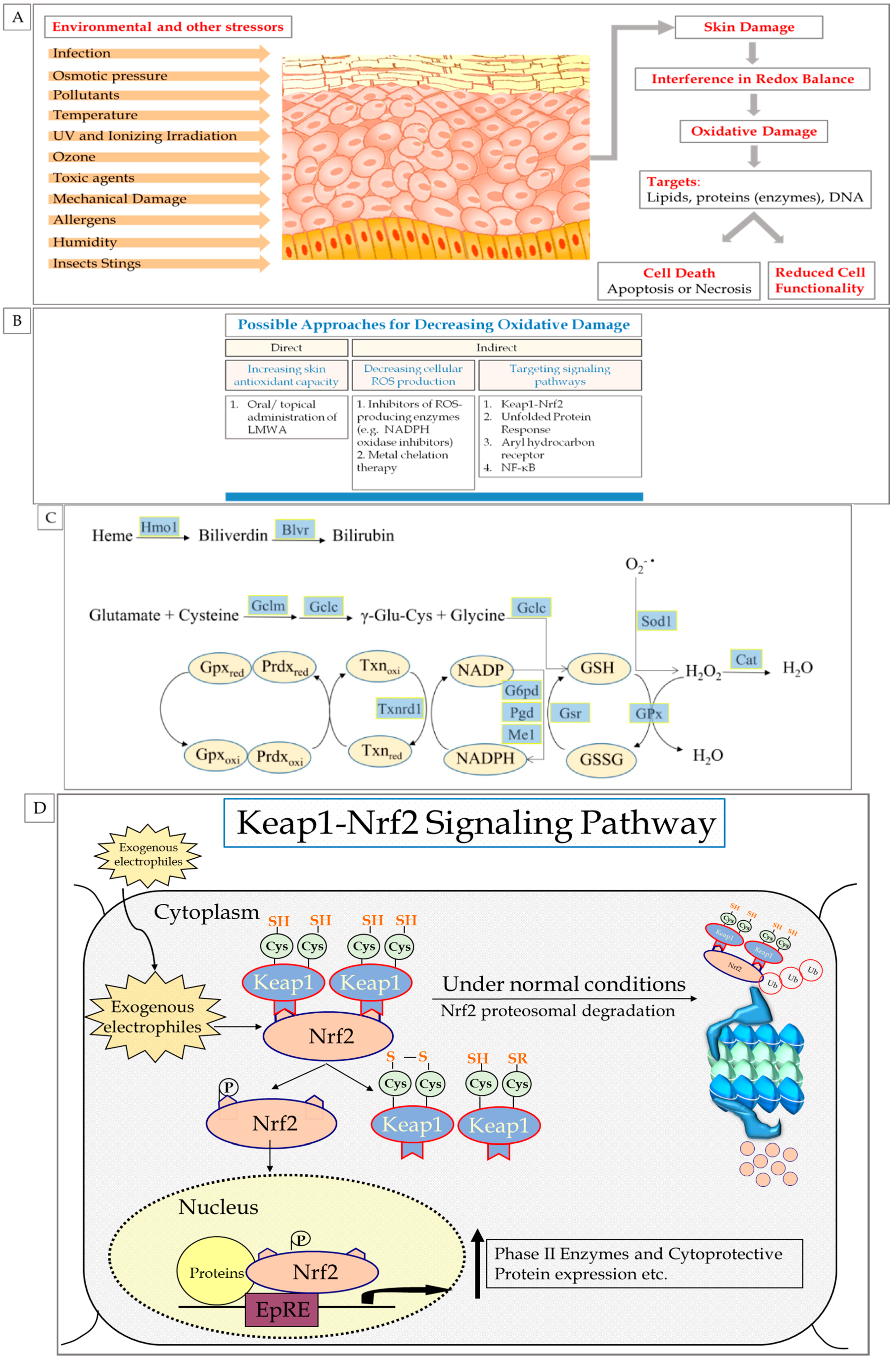
2. Oxidation Processes in Skin

2.1. Reactive Oxygen Species (ROS) and Oxidative Stress in Skin

Oxidative stress can be defined as a disturbance in the redox status of cells, tissues or organs [14]. It is caused by an imbalance between the amount of oxidants that our cells are exposed to and antioxidants which are required to retain the balance, in favor of the oxidants [14]. An oxidant, or pro-oxidant in biological systems, can be defined as a chemical substance capable of accepting electrons [14]. Pro-oxidants are generally referred to as ROS, which have a relatively short half-live with extremely high reaction rate constants [9,14]. ROS can be categorized into two groups of compounds: radical and non-radical [14]. The radical compounds contain at least one unpaired electron in the shells around the atomic nucleus and are capable of independent existence [14]. Examples for these compounds are nitric oxide radical (NO·), superoxide radical (O2·), hydroxyl radical (OH·), peroxyl radical (ROO·) and singlet oxygen (1O2) [14]. The non-radical compounds are highly reactive derivatives of oxygen, such as peroxynitrite (ONOO), hydrogen peroxide (H2O2), organic peroxides (ROOH), hypochlorous acid (HClO), aldehydes, ozone (O3) and oxygen (O2) [14]. Reactions between radical and non-radical ROS include the generation of new radical species and are usually chain reactions [10,14,15]. ROS have extremely high affinity to a variety of biological components, including proteins, DNA, lipids and the defending systems of the cell, which are composed of enzymes and reducing equivalents, or antioxidants [14]. Although ROS have short life spans, their high reactivity with biological components is responsible for their potential noxious effects. Therefore, uncontrolled quantities of ROS may be detrimental. However, ROS effects on the biological system depend on several key factors, which can be divided into two categories: factors that are mainly related to the ROS properties, including ROS origin, life span, membrane diffusion capabilities, concentration, steric conditions and oxidative potential; and factors that are more dependent on the specific ROS target, its physiological environment (including pH), the overall oxidative status and the occurrence of cytoprotective and detoxification mechanisms [9,14]. Obviously, there is a tight interconnectedness between these two categories. In the absence of a relevant biological target, ROS will not cause damage [14].

2.2. Skin Exposure to Oxidative Stress: Exogenous and Endogenous Sources

The skin is a major target of oxidative stress which can be developed into oxidative damage [14] if not blocked. Being an organ of the integumentary system, the skin has multiple roles in maintaining homeostasis and therefore performs several vital functions—regulating temperature, regulating sensation, retaining body fluids, and eliminating waste. Skin also has an endocrine function; synthesizing vitamin D and storing it along with glucose, water and fat molecules. Highly metabolic activity also characterizes skin, besides its detoxification of xenobiotic and immunological utilities. The skin barrier is essential for humans to survive in an external environment [16]. It provides the first barrier against various stressors challenging homeostasis (chemically and/or physically) and, as such, is confronted with more severe situations than any other organ [17] since it is exposed to a variety of damaging species such as superoxide radicals, hydrogen peroxide, hypochlorous acid, hydroxyl radicals, nitric oxide radicals and singlet oxygen [8]. These metabolites can also be used to produce more severe reactive species [8]. Origins of these ROS are the external environment, the skin itself and various internal sources [2,18].
External sources include air pollutants (e.g., car exhaust, industrial sources, and cigarette smoke), natural deleterious gases (e.g., ozone), ionizing and non-ionizing irradiation, food preservatives, xenobiotics, invasion of pathogenic bacteria and viruses, and various exogenous chemicals such as dust, allergens and toxin [8]. Occasionally, these factors have a synergistic activity in inducing ROS [19]. Physical damage resulting from high temperatures, humidity and mechanical damage also contribute to skin oxidative challenges [8]. Severe psychological stress, alcohol intake, poor nutrition and high calorie consumption may cause skin damage as well [20]. ROS are also produced endogenously, often as a result of normal aerobic metabolism. Oxidative phosphorylation in the mitochondria synthesizes ATP and generates ROS, mainly O2· and H2O2 [20,21]. There are thirteen potential sites of mitochondrial ROS production along the electron transfer chain and the rate of mitochondrial respiration often determines the rate of ROS generation [20,22]. Indeed, it was reported that mitochondria are the major source of oxidative stress [22]. Additional examples for endogenous sources generating ROS are the enzyme nitric oxide synthase and xanthine oxidase, which generate nitric oxide radicals and superoxide radicals, respectively, and the uncoupling of nitric oxide synthase and cytochrome P450 [23,24,25,26]. Bacterial cell invasion may also contribute to the production of endogenous ROS upon neutrophils’ activation (e.g., interaction with bacteria cells) in which neutrophils experience a respiratory burst resulting in the release of ROS efflux. Additional factors contributing to ROS production in skin are proteinases and cationic proteins [8]. Another example for an enzyme producing ROS is the enzyme NADPH oxidase, which was found to be responsible for the oxidative burst and is now considered to play a role in almost all tissue types [27]. Other endogenous ROS production routes include pathological processes such as ischemic and post-ischemic conditions and disease states including psoriasis, cancer and inflammation [8].

2.3. ROS: Beneficial and Detrimental Consequences to Skin

A continuous excessive efflux of ROS may cause deleterious damage to many skin-cell constituents such as lipids, DNA and proteins leading to reduced cell functionality and cell death [8] (Figure 1A). ROS can initiate peroxidation processes in enzymatic and non-enzymetic and/or structural proteins resulting in their conformational changes, unfolding, inactivation and degradation [8]. Tryptophan, histidine, tyrosine, lysine, methionine and cysteine residues can be oxidized during oxidative stress, resulting in additional reactive species such as endo- and hydro-peroxides, which might further cause protein cross-linking [28]. Alterations to dermal extracellular matrix proteins, collagen or elastin, might cause changes leading to abnormal mechanical properties of the skin [9,28]. Moreover, the accumulation of carbonyl proteins (carbonyl groups which are produced on protein side chains upon oxidation) has been observed in various disease conditions including psoriasis [29].
Figure 1. (A) Consequences of skin exposure to stressors; (B) Possible approaches for decreasing oxidative damage; (C) Schematic diagram representing the cytoprotective activities of the antioxidants enzymes regulated by the Keap1–Nrf2 pathway, adapted from [30]; (D) Schematic representation for the Keap1–Nrf2 pathway.
Cosmetics 03 00001 g001
The occurrence of lipid peroxidation process via ROS is of high significance. Phospholipids are major structural components of cellular membrane enabling proper functioning of the cell. Peroxidized lipids can disturb the integrity of the cellular membrane, decreasing membrane fluidity and increasing membrane leakiness to undesired substances [8]. Moreover, membrane-bound cellular receptor activity may be altered, resulting in the activation of various intracellular kinases such as the mitogen activated protein kinases (MAPK) and extracellular signal-related kinase (ERK) which are also capable of independent ROS activation. Transcription factor complexes such as AP-1 (Activation protein-1) and nuclear factor-κB (NF-κB) are then formed [8,9,28,29,31]. Lipid-membrane peroxidation might also decrease membrane signaling efficiency affecting aberrant cell proliferation responses [10]. Further reactive aldehyde species such as natural products of lipid peroxidation, including malondialdehyde, 4-hydroxynonenal, hexanal and other saturated and α,β-unsaturated aldehydes and ketones can be mutagenic and carcinogenic due to their high reactivity with DNA bases [32]. Moreover, histidine and lysine are vulnerable to modifications by aldehydes resulting from lipid peroxidation and might trigger an autoimmunity response [28]. Lipid peroxidation has been reported to be correlated with several skin inflammatory diseases including psoriasis, acne and contact dermatitis [33].
Further ROS targets include DNA and RNA. ROS (or ROS outcomes such as lipid peroxidation products) can cause base loss, base modification and single and double DNA breakage events [8]. In the case of damage to the RNA, which is more susceptible to ROS damage than DNA due to the lack of protection by both histones and cellular compartments, the result is abnormal proteins and deviant enzyme function [8,28]. In the case of DNA damage, deleterious processes in cell function occurs, which are correlated with cancer and other pathological disorders [8].
ROS pivotal mediators in skin cells are pro-inflammatory cytokines (e.g., interleukin-1α, interleukin-6) that can operate in a synergistic or additive manner and have pleiotropic activities [34]. A possible outcome of these cytokines is inflamed skin which might lead to skin infiltration by activated neutrophils that produce additional reactive species, establishing a vicious cycle [35]. The activation of various important transcription factors affected by ROS, such as AP-1 (Activation protein-1), nuclear factor-κB (NF-κB) and several signaling cascades including ERK, JNK, and p38 MAPK pathways, were found to be involved in cell growth, differentiation and degradation of skin collagen and elastic fibers contributing to the formation of wrinkles and loss of skin resilience [36].
Although ROS were demonstrated to be linked to a variety of skin diseases including skin inflammatory diseases and skin cancers, they also appear to play a crucial and essential role in the cellular existence, growth and development [8,9]. ROS are key molecules in the maintenance of skin homeostasis, possessing many functions including influence on important signaling pathways in the context of immune defense, wound healing and apoptosis [8,9]. Moreover there is a correlation between the different functions of ROS and their subcellular source, location and half-life within the cell [37]. One of the major endogenous ROS signaling molecules is hydrogen peroxide (H2O2) which is capable of regulating several transcription factors involved in cell replication, metabolism, apoptosis, and necrosis through oxidation of redox-sensitive cysteine residues [38]. The effects of ROS in biological systems, and in particular on skin, are diverse and offer a feedback control; for example, ROS can affect protein functionality either indirectly at the level of protein expression or stability, or directly at the level of posttranslational modifications [11]. Moreover, paradoxically, ROS-signaling results in enzymatic cellular antioxidant activity, as will be discussed in Section 5.

3. Endogenous Cellular Defense Systems in the Skin

The endogenous cellular defense system was developed throughout the evolutionary process as a response to a variety of stressors. This complex system has several major contributors including the immune system, the inflammatory response, cellular metabolism, and DNA repair and redox regulation systems. All of these cellular systems, operating at the molecular level, are required to maintain skin homeostasis and reduce the peril of skin pathological conditions from developing. The interconnectedness between these cellular systems allows appropriate functioning of skin cellular processes and responses to exogenous and/or endogenous stressors [39].
A major contributor to the defense system against oxidative stress is the DNA repair system which is capable of identifying damaged DNA, removing it and integrating a suitable base [8]. The skin is equipped with a DNA repair system such as the Base Excision Repair and Nucleotide Excision Repair [36]. Mitochondrial DNA is more vulnerable than nucleus DNA to oxidative stress due to a limited and less efficient DNA repair system [40]. Activity of Nucleotide Excision Repair system has been enhanced to be increased in keratinocytes exposed to low-dose UVB [41]. Dysfunctionality of these repair systems might lead to severe skin disease with phenotype of premature aging [42]. Abnormal Nucleotide Excision Repair system is the cause of 11 human diseases, among them Xeroderma pigmentosum, which leads to segmental progeria, an increase in sun sensitivity and an increased incidence of sun-induced skin cancer [42].
Another mechanism with which skin is equipped is the chelation of transition metal. Redox active transition metals are involved in ROS generation in skin. For example, an excess of free iron can catalyze ROS formation via Haber–Weiss reaction [8,11]. Chelation of redox active metals maintains metals’ redox state and prevents them from generating ROS [8,11]. Physical defense is an additional mechanism by which skin protects itself from ROS; α-tocopherol present in skin layers can stabilize cell membranes by interacting with the fatty acid chain leading to enhanced protection of skin from ROS damage [8,43].

3.1. Antioxidant Defense System

The most important defense system against oxidative stress is the antioxidant defense system [8]. This comprehensive system provides the skin with a protective antioxidant barrier and is well interlinked between its various components [44,45,46]. Most of the system's components were found distributed in skin layers, with higher concentrations in the epidermis than the dermis, correlating with ROS exposure of skin layers [47]. The main contributors of the antioxidant defense system are discussed below.

3.1.1. Low Molecular Weight Antioxidants (LMWA)

The Low Molecular Weight Antioxidants (LMWA) is a group of small, membrane-permeable, hydrophilic or lipophilic compounds having a variety of activities leading to the prevention of oxidative damage [14,48]. This group can be classified into two subsets; the first subset can be described by its ability of preventing oxidative damage by direct interaction with ROS leading to their neutralization (e.g., scavenging) [2,8,14]. This ability is obviously due to LMWA chemical characteristics, including their capability of donating electron(s) to reactive species [2,8,14]. However, the ROS scavenging ability of LMWA is limited to ROS microenvironment conditions; the LMWA needs to be in close proximity to ROS location and, moreover, its concentration must be high enough to provide efficient scavenging activity [2,14]. The second subset of LMWA can be characterized by its ability of preventing oxidative damage indirectly [2,8,14]. The mechanism of activity of these LMWA is related to their ability to chelate redox active transition metals and prevent ROS generation through Haber–Weiss reaction [8,49]. The two subsets of LMWA operate synergistically [14] and, as mentioned above, a tight connection is observed between the groups; ROS scavenging results in the conversion of the scavenger to an un-reactive radical which can be further reduced or oxidized by an additional scavenger with or without enzyme involvement [14]. An example for such a cycle is the scavenger ascorbic acid, its scavenging activity results in its formation into an ascorbyl radical which can be further reduced to ascorbic acid with the aid of glutathione.
Some LMWA can be synthesized within the cells or produced as cellular waste-products, and their concentration is regulated by the cells [14]. No gene encoded for LMWA has been reported [50]. Examples of LMWA synthesized by the cells or generated as waste-products are histidine dipeptides [14,51,52], glutathione [14,15,53], carnosine [54], lipoic acid [14,55] uric acid [14,56] and bilirubin [14,57]. However, cellular endogenous capacity is very limited and only a small portion of LMWA are derived from endogenous sources [14]. In fact, the majority of LMWA are derived from dietary sources [14]. This introduces a complexity; around the world, there is a great diversity in food-ingredient consumption for different populations. Moreover, oral consumption does not guarantee a beneficial effect on the skin since the antioxidant must cross the intestinal barrier, be metabolized and only then be distributed to the skin in its active form [58,59]. In addition, there are some crucial factors involved in the absorption process influencing LMWA concentration in skin, including transfer proteins, physical and chemical properties of specific LMWA and competition and/or interaction with other LMWA [58]. As discussed above, since ROS are highly reactive, have a short life span and are continuously produced, their scavenging in skin is of high importance and cannot be achieved by LMWA alone, as their levels found in skin are too few (measured in picomoles/mg skin) [60]. This emphasizes the importance of skin antioxidant enzymes discussed in Section 3.1.2 below.

3.1.2. Antioxidant Enzymes

The antioxidant enzymes are members of phase II enzymes. These enzymes are proteins responsible for the maintenance of cellular redox balance. This group includes the familiar antioxidant enzymes (superoxide dismutase, catalase, peroxidase, NAD(P)H dehydrogenase [quinone], heme oxygenase 1, gluthathione reductase, etc.) and, in contrast to LMWA, they are not consumed offering an efficient protection against oxidative damage [14]. This group of enzymes operate in a few dimensions; catalysis of ROS-scavenging reaction which is characterized by high affinity and reaction rates that depend on the pH of the specific activity site [11,14,59], recycling and regeneration of several LMWA such as vitamins A, C and E, and supporting and producing a suitable cellular environment for antioxidant enzymes [14,61]. ROS-scavenging reactions are mediated by specific enzymes, for example, superoxide anions dismutate to molecular oxygen, and hydrogen peroxide is catalyzed by superoxide dismutase [62]. Hydrogen peroxide can be further detoxified by catalase [63] or glutathione peroxidase. Glutathione peroxidase activity results in the formation of a glutathione disulfide by-product, which can be further regenerated by glutathione reductase to glutathione [62,64]. In addition, g-glutamylcysteinyl synthase, a rate-limiting enzyme in glutathione cellular synthesis, enhances activity in order to compensate for reduced glutathione level [62,64].
Heme oxygenase-1 (HO-1) is an inducible antioxidant enzyme, which catalyzes the degradation of heme into carbon monoxide, iron, and biliverdin which can be further converted to bilirubin, a non-enzymatic antioxidant [65,66,67]. Exhibiting a wide spectrum of cytoprotective effects in a variety of skin models including inflammatory and UV-induced oxidative damage, HO-1 induction was suggested as a marker diagnosis of oxidative stress related conditions [68,69].
Recycling LMWA antioxidants and producing an appropriate cellular environment for proper functioning of skin cells is obviously of high significance. This can be exemplified by the following groups of enzymes: glucose-6-phosphate dehydrogenase; 6-gluconate phosphate dehydrogenase; isocitrate dehydrogenases; malic enzymes; and transhydrogenase. These enzymes are responsible for the generation of NADPH from NADP+, which is necessary for regeneration of glutathione and for the maintenance of a balanced ratio of an oxidant and antioxidant environment [70,71]. An additional example is xanthine dehydrogenase which produces uric acid [72,73]. Antioxidant enzymes activities are summarized in Figure 1C.
Compelling evidence indicates that these enzymes are of high importance maintaining skin redox balance homeostasis and protecting the epidermis against oxidative damage-related conditions and diseases, including skin erythema, inflammation, wound healing and skin cancers [14,69,74,75]. It was reported that patients suffering from actinic keratoses and basal cell carcinoma (BCCs), common non-melanoma skin cancer, had reduced levels of antioxidant enzymes in plasma or serum [76]. In addition, the superoxide dismutase (SODs), CuZnSOD and MnSOD, are decreased in human non-melanoma skin cancers [77]. This diverse family of enzymes are also recognized as primary defense mechanisms against many degenerative and chronic disease conditions [61].
Additional proteins offering anti-oxidative activity to epidermal cells are the small proline-rich proteins (SPRRs). These proteins are important components of the cornified envelope cross-linked to loricrin during cornified envelope formation [78]. Having multiple cysteine residues, SPRRs demonstrate the capability of quenching ROS by forming intramolecular disulfide bonds [79]. It was reported that, during the process of aging, the cornified envelope is intensely altered [79]. However, elevated levels of SPRRs were found, which possibly indicate the presence of a compensation mechanism of the aged cornified envelope [79]. The discovery of SPRRs’ anti-oxidative capabilities emphasizes the tight link between the cornified envelope function as an epidermal barrier against the environment and its anti-oxidative line of defense, suggesting an efficient evolutionary process. This also provides a rational explanation for the changes in the skin barrier function stratum corneum lipid metabolism or protein components of the corneocytes which are involved in the development of various skin diseases and pathologies [80].
The anti-oxidative properties of the skin are very sophisticated, demonstrating diversity, complexity and feedback mechanisms in counteracting oxidative stress and maintaining skin redox balance. This is illustrated by the distribution of the anti-oxidant defense systems in the skin where antioxidant agents are distributed in the skin determined mainly (but not solely) by the lipophilic/hydrophilic ratio of the compounds [9]. The arrangement of antioxidants also correlates well with ROS skin layer exposure; the dermis being less equipped with antioxidant agents compared to the epidermis due to epidermis prior protection [9,42,47]. Vitamin C, vitamin E, glutathione, squalene, coenzyme q10, uric acid and ubiquinol were detected in the stratum corneum [9,42]. However, there is a decrease from the outer layers of the epidermis towards the deeper layers. This can be explained by a gradual oxidation process during real life environment exposure which can have additive or even synergistic effects [9,42]. Indeed, the physiologic keratin 10 oxidation gradient was reported in the stratum corneum with three-fold-higher protein carbonyl concentrations in the upper stratum corneum than in the deeper layers [42,81]. The presence and anti-oxidative capabilities of SPRRs was suggested as a compensation mechanism by which the upper layers of the stratum corneum combat oxidant exposure [42]. The deeper epidermal layers contain vitamin E, and antioxidant enzymes including catalase, superoxide dismutase, and glutathione peroxidase [9]. The dermis includes mostly hydrophilic LMWA such as vitamin C, uric acid, glutathione and antioxidant enzymes [9].

4. Can Skin Redox Balance Be Efficiently Affected by Exogenous Intervention?

Skin redox state is a complex playground regulating and integrating diverse cellular signaling pathways including metabolism, the immune system, cell survival and apoptosis [8,9]. Obviously, ROS play a key role in maintaining redox balance homeostasis, and, therefore, are highly important. The activity of ROS is not limited to cellular signaling but also involves posttranslational modifications such as cysteine residues in key signaling proteins, affecting their functionality [11,82]. It is worth noting that both oxidative stress and reductive stress can have detrimental effects on biological systems mediated by opposed mechanisms of activity (e.g., damage to cellular components and clearance of ROS such as H2O2 preventing it affecting signaling pathways respectively) [14]. Moreover, a new concept emerging, “redox biology paradigm”, states that antioxidants modulate the complex networks controlling diverse pathways in cell signaling, therefore playing a regulating role instead of protection function [82]. For example, vitamin E can control the level of reactive lipid species instead of scavenging ROS [82]. However, under specific conditions related to the exogenous environment stressors or to endogenous status of the skin, ROS are produced in excessive quantities, thus resulting in the defeat of the antioxidant defense system that may lead to the development of various severe skin diseases, including psoriasis, erythema, edema, hyperplasia, skin aging, contact dermatitis, atopic dermatitis and carcinogenesis [10,33,80,83,84]. Therefore, strategies focusing on reducing and preventing ROS-mediated damage have been extensively studied in a variety of research models [10]. The specific guidelines for providing potential relief in oxidative-damage can be categorized into two approaches. The first approach supports the reduction of the cellular ROS production while the second one refers to the antioxidant defense system and includes the increase in the enzymatic antioxidant system or an increase in skin antioxidant capacity via exogenous pharmacological intervention in an oral route, or preferably a topical route [11] (Figure 1B).
Alternatives for reducing cellular ROS production utilize specific ROS-producing enzyme inhibitors such as NADPH oxidase inhibitors [85,86]. A variety of NADPH oxidase inhibitors have been developed showing contradictory results, possibly due to different experimental conditions, and it remains undetermined as to whether such an approach is beneficial [11,85,86,87,88]. An additional suggested alternative is chelation therapy [11]. According to this therapy, suitable metal chelators may reduce metal-mediated ROS generation [11,89,90,91,92,93,94,95]. Examples of such chelation agents are polyphenol from natural compounds such as green tea (epigallocatechin-3-gallate) and curcumin [89,96]. A synergism of a metal chelator agent with an additional drug substance could probably be expected.
An increase in tissue antioxidant enzymes, such as superoxide dismutase in different forms was extensively investigated. Most of the elevated levels of superoxide dismutase were achieved by means of exogenous injection or gene therapy [11]. Chemically induced contact dermatitis in transgenic mice overexpressing superoxide dismutase was relieved with a reduction in ROS levels and pro-inflammatory cytokines observed [11,97,98]. In addition, psoriasis patients expressed superoxide dismutase to a lesser extent than healthy volunteers [98]. The results obtained from these studies demonstrated an important role of superoxide dismutase in skin inflammation, supporting a future therapeutic approach.
An increase of skin antioxidant capacity combating ROS can be achieved via the exogenous oral or topical route. The oral route must include antioxidants’ passage in the intestinal barrier resulting in their presence in blood circulation and their distribution in different tissues and organs [58]. This process might be limiting, influencing antioxidant potential effects due to low bioavailability [58]. Obviously, the mechanisms involved leading to a specific antioxidant bioavailability differ between lipid-soluble or lipophilic antioxidants (e.g., vitamin E) and water-soluble antioxidants (e.g., vitamin C). Water-soluble antioxidants arrive to the small intestine following oral consumption, usually in glycosylated form and are then hydrolyzed with the aid of endogenous enzymes [58,99,100]. In the case of an inefficient hydrolysis process, the molecule is transferred into the gastrointestinal tract where the local microflora provide the necessary hydrolysis enzymes [58]. At this stage, an antioxidant or antioxidant-metabolite is absorbed [58]. Lipophilic or lipid-soluble antioxidants are re-organized into oil droplets following oral consumption which are transferred into the small intestine, where they are transformed into mixed micelles [58,101,102]. Following micelle solubilization, antioxidants are uptaken by enterocytes by a passive mechanism and a protein-carrier-mediated transport involving lipid transporters [103,104,105]. The antioxidants are then re-packed into new structures of oil-droplets, which, through exocytosis, are transferred into the lymphatic system and enter the blood circulation [58,106]. In the case of inefficient micelle solubilization, lipid-soluble or lipophilic LMWA reach the gastrointestinal tract and are metabolized by local microflora in the colon or eliminated [58]. Oral administration of antioxidants allows their distribution to the entire skin [58]. Moreover, there is a re-load effect of antioxidants in skin; the blood constitutively delivers antioxidants to different tissues including the skin [58]. However, antioxidants need to be in their active form in order to provide their beneficial effect, and more importantly, sufficient quantities of active antioxidants must be in close proximity to the specific ROS biological target site in the skin. Still, there is compelling evidence supporting oral consumption of antioxidants either in the diet or as an additional nutritional supplement. Examples of a few antioxidants and their combinations consumed orally are described in the following. Oral administration of grape seed proanthocyanidins was shown to reduce UV-mediated skin photoaging and to decrease melanin synthesis [106]. Moreover, in mice administrated with grape seed proanthocyanidins UV-induced tumor incidence, growth, and size, as well as metastatic pulmonary nodules were inhibited [107]. In highly metastasis-specific human A375 and Hs294t melanoma cell lines, grape seed proanthocyanidins were demonstrated to be an extremely significant inhibitior of cell migration [108]. Drinking green tea has also shown to reduce UV-mediated skin tumor incidence, burden and size in mice papillomas [109,110]. Epigallocatechin-3-gallate, green tea’s main flavonoid, has demonstrated anticarcinogenic effects in mice [110,111].
Oral therapy of provitamin A and β-carotene decreased UV-induced erythema formation in clinical studies demonstrating dose-and time-dependence [112,113,114]. β-carotene has also demonstrated an ability to decrease mitochondrial mutagenesis in skin fibroblast following UV exposure [115].
Tomato carotene, lycopene, has demonstrated enhanced defense against UV-mediated damage in human skin when administrated orally. Moreover, it was used to reduce skin toxicity secondary to chemoradiation in patients with breast cancer [110,116,117,118]. However, skin photoprotective effects determined by erythema, were only observed following at least 10 weeks of oral supplementation of several carotene including lycopene [60,112,114,119,120,121,122,123]. Vitamin C and vitamin E have provided photoprotection against UVB-induced damage including reduced sunburn reaction and thymine dimers in human skin [124,125]. However, combinations of vitamin C and vitamin E resulted in a more significant photoprotection compared to the use of vitamin C or vitamin E on their own [120]. A possible straightforward way to strengthen skin anti-oxidative capabilities could be through topical antioxidant therapy.

4.1. Is Topical Application of LMWA Useful?

Topical application of LMWA can be efficient in mitigating oxidative stress and its consequences. This route of administration provides a local effect and avoids systemic circulation. Moreover, since topical application offers a possible alternative to oral delivery it avoids gastro-intestinal incompatibilities and allows a more efficient utilization of short biological half-life drugs possessing a narrow therapeutic window. Clearly, physiological and pharmacological response and patient compliance is expected to improve. However, there are several important factors that need to be considered. First, antioxidants must remain in a stable form allowing its activity in skin [58]. For example, topical application of vitamin C has met with difficulties due to a low stability in the presence of oxygen [126]. Moreover, the antioxidant needs to penetrate sufficiently into the skin layers in order to provide a proper defense against ROS attack [58]. Stability and penetration issues can be resolved using a suitable topical delivery system. There is a wide spectrum of topical delivery systems with diverse structures and dimensions offering a variety of advantageous and pharmacokinetic profiles. Still, penetration also depends on environmental factors including temperature, pH, skin hydration level, etc. [58].
Topical application of epigallocatechin-3-gallate on humans has demonstrated reduced erythema and formation of sunburnt cells dose-dependently [127,128]. Moreover, reduced oxidative stress, pyrimidine dimer formation, and inflammatory leukocytes were observed upon topical application of epigallocatechin-3-gallate in human studies [129]. UVB-mediated production of prostaglandin metabolites, including PGE2, PGF2α and PGD2, which are key players in inflammatory disorders, and, in proliferative skin diseases, was inhibited following epigallocatechin-3-gallate topical application [130]. UVB-induced skin tumor development significantly decreased following topical application of epigallocatechin-3-gallate in SKH-1 hairless mice [131]. Topical application of epigallocatechin-3-gallate prior to UVB exposure significantly reduced UVB-induced infiltration of inflammatory leukocyte and myeloperoxidase activity [130]. Epigallocatechin-3-gallate topically applied to mouse skin in non-melanoma skin cancers has shown an inhibitory effect of UVB-induced photocarcinogenesis and immunosuppression [132].
Topical application of grape seed proanthocyanidins resulted in a reduction of mutant p53-positive epidermal cells and prevented Langerhans cells from decreasing following cell sunburn [133]. In recent years, attention has been drawn to the engineering of mitochondria-targeted antioxidants such as MitoQ and Tiron [134,135]. The rational of using mitochondria targeted-antioxidants is based on antioxidant ability to cross the mitochondrial phospholipid bilayer, accumulate there, and detoxify ROS [134]. MitoQ is a derivative of ubiquinone conjugated to triphenylphosphonium, a lipophilic cation that enables this molecule to enter and accumulate within the mitochondria according to the electrochemical gradient [134]. Tiron is a biocompatible antioxidant, also capable of chelating metals such as iron [134]. Both Tiron and MitoQ were demonstrated to provide protection against UVA- and H2O2-induced damage in dermal fibroblast, suggesting new therapeutic strategies comprising a combination of antioxidants compounds with metal chelator [134,135].
Vitamin C administrated topically demonstrated an increase in collagen synthesis and a decrease in MMP (collagenase) expression [136]. A double-blind clinical study revealed that topical application of vitamin C on the forearm and neck resulted in significant improvements in skin hydration, tactile roughness, pigmentation and keratosis [137]. Topical vitamin E inhibited thymine dimer formation and mutagenesis [138,139]. It was also shown that vitamin E application decreased lipid peroxidation in vivo [140]. Topical combinations of vitamin C and E provided photoprotection in human skin more significantly compared to a single application of vitamin C or E [141].
Although exogenous intervention of LMWA is of great availability, their effects topically applied or orally consumed or supplemented remain controversial [11]. Long-term effects of such an intervention are yet to be addressed. In addition, the underlying mechanism of activity besides directly quenching ROS, involving cellular signaling pathways may have dramatic and more significant effects.

4.2. Can Activation of Cytoprotective Signaling Pathways in Skin Be Effective?

Numerous experimental sets of data from basic research, animal studies and controlled clinical studies have revealed that there is insufficient evidence to support the use of exogenous intervention of antioxidants in many chronic diseases [142,143,144,145,146,147,148,149,150,151], and, moreover, it has become evident that LMWA can be deleterious [152,153,154,155]. These observations are supported by findings using advanced mass spectrometry techniques. In various pathologies, the relative levels of protein modifications by ROS are low, and the cells and tissues are still well equipped with antioxidants [82,156,157]. An additional discovery suggesting a more complex involvement than high levels of ROS influencing and contributing to the development of oxidative stress-related pathologies is regarding the levels of ROS needed for interfering with the biological system redox balance. These levels are in orders of magnitude higher than the levels that can ever be formed in biology in cases of disease or health [82]. This leads to the new emerging concept of “redox biology paradigm” emerging, which states that antioxidants possess a role of modulating the complex networks controlling diverse pathways in cell signaling, having a regulatory role instead of a protective one [82]. It is only logical that the main target of a certain therapy should be one or more pathways that can control the pathology. This, of course, is highly complex; however, if this hypothesis is correct, the relief resulting from this therapy will be relatively rapid and will influence many aspects in the pathological state. It is also expected that exogenous intervention of targeting signaling pathways (Figure 1B), the active molecule dose will be significantly lower than the LMWA molecule, since such an exogenous intervention of LMWA must break down the redox insulation before an effect can be detected [82].
Electrophiles are compounds having diverse chemical structures, containing electron- deficient carbon centers, and, therefore, are attracted to and react with an electron rich center [158]. These molecules are involved in many complicated types of cellular functions including, chemoprevention [158,159,160,161], stress responses [158,162,163], hormesis [158,164,165] and electrophilic counterattack [158,166,167,168,169,170]. These important compounds are produced in the cell via both enzymatic and non-enzymatic lipid peroxidation and are also consumed in the diet [82]. There are few signaling pathways that are regulated by electrophiles; mainly stress signaling pathways including the Keap1–Nrf2 pathway, Heat Shock Response (HSR), and Unfolded Protein Response (UPR) [82]. Other signaling pathways are tightly linked with the bioenergetic status of the cell [82]. Additional signaling pathways that may be involved are signaling pathways that have cross-talk with the directly-regulated electrophile pathways (mentioned above) including the aryl hydrocarbon receptor (AhR), NF-κB and Notch pathways possessing cell-fate responses [171].
Therefore, electrophile therapeutics was suggested as a potential attractive and intelligent approach in the drug development field [82]. However, a few issues need to be taken into consideration. Consistent with other therapies, electrophile therapy efficiency may vary in human populations suggesting the need for a personalized therapy [82]. The fact that electrophiles are capable of modifying reactive cysteines and reacting with cysteine moieties via Michael addition [82,172] in a reversible and irreversible manner depending on the nucleophile present, might amplify their potential activity. A reservoir of electrophiles might accumulate in the target site resulting in long-term effects [82]. In addition, the electrophile effect (e.g., cell signaling effect) might also result in a potential improved effect [82]. However, this potential therapy obviously needs comprehensive research and clinical experience revealing and clarifying the wide spectrum of effects that results from signaling pathway activation. Toxicity issues might also be raised, since induced genes resulting from Keap1–Nrf2 activation differs among different electrophiles demonstrating the “cysteine code” [82,173,174,175].

5. The Role of Nrf2 in Skin Redox Balance

The induction of phase II antioxidant enzymes is regulated by the Keap1–Nrf2 pathway [176,177,178] (Figure 1D). The transcription factor nuclear factor (erythroid-derived 2)-like 2,NF-E2-related factor (Nrf2) is a key molecule controlling the expression of diverse groups of genes, including the familiar antioxidant enzymes (Figure 1C), transporters, large numbers of genes that control distinct processes such as immune and inflammatory responses, tissue remodeling and fibrosis, carcinogenesis and metastasis, and even cognitive dysfunction and addictive behavior [176,177,178]. Therefore, the potential rational strategy of maintaining human skin redox balance by modulating Nrf2 is of great interest. Nrf2 is crucial to skin in various states; it plays a role in skin homeostasis, renovation and a variety of skin diseases [12]. Nrf2 activation can affect skin nucleophilic tone, which was described as the cellular, tissue, organ, or even organism level of protection against electrophiles (e.g., ROS) by nucleophiles demonstrating para-hormesis effect [179]. Nrf2’s role in skin was originally discovered by the identification that Nrf2 is a target gene of keratinocyte growth factor (KGF) which is a cytoprotective growth factor for epithelial cells [180]. Injured skin highly expressed KGF, and, therefore, it was proposed that Nrf2 might similarly be expressed and upregulated in keratinocytes following injury [181]. Indeed, the up-regulation of Nrf2 expression in the epidermis of mouse wounds compared to normal epidermis was confirmed [181]. According to Beyer et al., endogenous Nrf2 regulates inflammation in wounded skin. In addition, it was also demonstrated that Nrf2 has a key role in the repair of an epidermal barrier defect [12]. It was shown that the lack of loricrin, one of the main components of the cornified envelope, can be compensated by the activation of Nrf2 and upregulation of Sprr2d and Sprr2h genes [182]. Moreover, it offers protection from chemically-induced skin carcinogenesis in a cell autonomous manner and not indirectly via stromal cells [181]. The activation of Nrf2 by several compounds was demonstrated to be beneficial in the prevention of skin carcinogenesis in a variety of animal studies [12]. In addition, Nrf2 activation was also investigated as a promising treatment strategy for allergic skin inflammation (e.g., allergic contact dermatitis), atopic dermatitis, psoriasis, epidermal blistering disease (e.g., Hailey–Hailey disease) and vitiligo vulgaris [12]. Nrf2 offers protection against UV-induced cytotoxicity, and, moreover, it can protect from photoaging, providing relief from aging symptoms such as wrinkle formation, loss of skin flexibility, epidermal thickening and matrix deposition [12,183].

6. Nrf2 Pathway

6.1. Nrf2 Mechanism of Action

Itoh et al. were the pioneers who first described the mechanism of Nrf2 activation [177,184]. Nrf2 is normally kept in the cytoplasm via interaction with two cysteine-rich proteins called Keap1 (Kelch-like ECH associated protein 1) [177,184,185,186,187,188,189,190] (Figure 1C). Keap1 is an actin cytoskeleton binding protein capable of mediating Nrf2 degradation via the ubiquitin proteasome pathway resulting in Nrf2 half-life of approximately 10–20 min [184,191,192,193,194]. The cysteine 273,151 and 288 residues in Keap1 are of high significance in stress-sensing activity [195,196]. Electrophiles are capable of recognizing and modifying these Keap1 cysteines, abrogating and suppressing Keap1-mediated proteasomal degradation of Nrf2, thereby causing Nrf2 stabilization, translocation and accumulation in the nucleus [197]. Nrf2 accumulation in the nucleus enables its hetrodimerization with additional proteins such as small Maf proteins and binding to the antioxidant-response element (ARE) also named as the electrophile-response element (EpRE) in the regulatory sequences of its target genes. This results in the coordinated transcriptional activation of large networks of genes encoding enzymes and detoxifying proteins, including the antioxidant enzymes, anti-inflammatory cytoprotective proteins, etc. [198,199,200,201,202].
There is conflicting evidence regarding the exact mechanism of action by which the Nrf2 transcription factor can be activated. The most common proposed models are the hinge and latch mechanism [188,203] and the Cul3 dissociation mechanism [204], both of which describe the reduction of ubiquitination and proteasomal degradation of Nrf2 [205,206,207]. According to the hinge and latch model, Keap1 homodimer binds to Nrf2 in two binding sites within the Neh2 domain of Nrf2 (termed ETGE and DLG motifs) possessing high (ETGE) and low (DLG) affinity, enabling an optimal setting of lysine residues for ubiquitin conjugation [208,209,210]. Interruption to the DLG motif relocates Nrf2, prevents its degradation and increases its nuclear accumulation [205]. The Cul3 dissociation mechanism, claims that the Keap1–Cul3 complex is interrupted causing Nrf2 ubiquitination termination and increasing its availability for nuclear translocation and accumulation [205]. While cysteine 273 and 288 residues are involved according to the hinge and latch model, and are able to form an intermolecular disulfide bond in the Keap1 protein, upon direct electrophile oxidation or covalent binding [195,196], cysteine 151 residue is involved according to the Cul3 dissociation model [205]. Differences in the stress response mediated by the Keap1-Nrf2 pathway using various electrophile concentrations and different electrophiles were reported demonstrating the “cysteine code” [175,211,212,213]. In addition, there are many kinases and phosphatases modulating the stress response, contributing to the complexity of the pathway [202,214,215,216,217,218,219].
An additional optional mechanism of activity suggested for Nrf2 activation is inhibition of Keap1–Nrf2 interaction by blocking the Keap1 domain, instead of metabolic activation by reactive electrophiles [205]. Pathway activation and regulation can also be achieved by methylation/demethylation of Cytosine-phosphate-guanine islands (CpGs) in the promoter regions, acetylation/deacetylation and methylation/demethylation of histones, or targeting of mRNAs by miRNAs [220]. Moreover, certain non-coding RNAs are also capable of influencing Nrf2 [220].

6.2. Nrf2-Activating Agents

The classic Nrf2-activating agents are electrophilic compounds which, according to both the hinge and latch model and the Cul3 dissociation model, are capable of interacting with Keap1 cysteine residues. The chemistry underlying Keap1 modification is Michael addition, which is the reductive addition of a nucleophile (e.g., Keap1 cysteine) to an α,β-unsaturated carbonyl compound (e.g., antioxidant) [179]. There are at least ten groups of both natural and synthetic compounds capable of activating the Keap1–Nrf2 pathway, including oxidizable diphenils, phenylenediamines, quiones, Michael reaction acceptors, isothiocyanates and sulfoxythiocarbamates, thiocarbamates, dithiolethiones, polyenes, hydroperoxides, trivalent arsenicals, heavy metals and dimercaptans [221,222].
It is worth noting that a pro-electrophile compound is also capable of activating the Keap1–Nrf2 pathway [158]. A pro-electrophile compound is an electrophile that becomes active following oxidation [158]. Thus, oxidative damage that needs to be combated converts the pro-electrophile, which is relatively innocuous, to an active electrophile [158]. This approach possesses a definitive advantage for human drug development over other electrophiles, because pro-drugs should manifest fewer clinical side effects [158].
Additional Nrf2-activating agents are non-electrophilic compounds exerting their effects also without resulting in oxidative damage [205]. These compounds are capable of activating the Keap1–Nrf2 pathway by directly inhibiting the interaction between Nrf2 and Keap1 inducing Nrf2-regulated genes [205]. This approach is very promising; however, these compounds demonstrated activity only in vitro and in cell culture and have not yet demonstrated efficacy in animal studies, thus further research is needed [205].
Some examples of Nrf2 activators include sulforaphane, curcumin, epigallocatechin-3-gallate, resveratrol, garlic, oganosulfur compounds, lycopene, carnosol [223,224,225], nitroxides [226] and dimethyl fumarate [227].

7. Enhanced Protection against Oxidative Damage by Nrf2-Activating Agents Using Delivery Systems

7.1. The Need for Transient and Controlled Nrf2 Activation

Eliminating oxidative stress by Keap1–Nrf2 activation offers the possibility of modulating the expression levels of hundreds of gene products that can affect oxidative stress and the related pathophysiological states, instead of attempting to restore oxidative balance by the administration of relatively small amounts of an antioxidant enzyme or cytoprotective protein [176]. In fact, Nrf2-target genes account for more than 1% of the human genome including genes responsible for antioxidant and anti-inflammatory response, drug metabolizing, and primary metabolism and bioenergetics [228]. Therefore, activation of this pathway may result in a sustained and amplified reaction providing suitable relief for a variety of pathological states [158], as well as the possibility of preventing skin damage and even malignant transformation under stress conditions [12]. However, constitutive prolonged and enhanced activation of Nrf2 in the epidermis is deleterious due to alterations in the epidermal lipid barrier, inflammation and induced keratinocyte hyperproliferation [229]. Moreover, long-term activation of this pathway can potentially trigger pro-tumorigenic activities [12,230]. By contrast, transient activation of Nrf2 in normal skin protects against UVB- and toxin-induced skin cancer [12]. Furthermore, wide spectrum studies have revealed Nrf2’s necessity for skin maintenance of homeostasis, repair activity and diseases and pathological states [12]. This is supported by the numerous and extensive efforts to find new Nrf2-activating compounds as potential drugs for various skin disorders [231].
Thus, orchestrating Nrf2-target genes by a controlled and transient activation of the Nrf2 pathway in skin, using nanotechnology-based dermal delivery systems, may present a novel strategy for prevention and treatment in various skin pathologies and under stress conditions. This pharmacological approach offers a dose-responsive amplitude on skin and targets networks of genes, resulting in the leverage of dermal cure.

7.2. Is the Usage of Topical Delivery Systems Suitable?

Three major layers comprise human skin, the epidermis, dermis and the underlying subcutaneous fat layer (hypodermis). Skin is one of the main organs in the human body which is suitable for topical delivery systems and topical application usually intended for a localized activity in skin layer(s). However, sub-therapeutic concentrations may reach the systemic circulation without causing side effects. There are several challenges that need to be addressed regarding this route of administration. The main challenge is dermal penetration which is regulated and limited by the stratum corneum, the uppermost layer of the skin [232]. The stratum corneum is responsible for the epidermal drug transport (by passive diffusion) and drug distribution in other epidermal layers including the stratum granulosum, stratum spinosum and the stratum basale. Transport across the stratum corneum follows Fick’s Law of diffusion, according to which the drug’s flux depends on the drug oil/water partition coefficient, its concentration in the delivery system, thickness of the stratum corneum, and on the surface area of the exposed skin. Additional challenges that the dermal delivery system needs to overcome are local side effects (e.g., irritation, allergic reaction, and erythema), Nrf2-activating agent physiochemical properties such as low aqueous solubility and high log P, and chemical characteristics such as instability and photosensitivity [233]. The use of a topical delivery system in the case of activating the Keap1–Nrf2 pathway may present advantages such as avoidance from hepatic first-pass metabolism, accessibility to the skin site of action, prevention of naive cells from compound exposure and obviously patient compliance [233]. The avoidance of systemic circulation of the Nrf2-activating agent may be of specific importance since it was reported that Nrf2 activation may have unfavorable systemic metabolic effects. An additional comprehensive understanding needs to be achieved regarding the way Nrf2-activating agents interact with normal and diseased cells [82]. Applying a topical dermal delivery system containing Nrf2-activating agents holds an additional rational; it was demonstrated that there is a gradient of Nrf2 expression and as a result higher ROS protection in higher epidermal layers than lower ones [12,234,235]. However, superbasal keratinocyte protection from ROS is crucial for maintaining skin integrity and homeostasis [12,235]. Topical application of Nrf2-activating agents hopefully will supply an adequate solution resulting in activation of the Nrf2 pathway by exogenous and specific pharmacological intervention.

7.3. Desired Features of an Nrf2-Activator Delivery System

Passive percutaneous penetration enhancement technologies, based on nano-colloidal delivery systems are capable of improving drug solubility, permeability and stability, achieving therapeutic concentrations and enhancing drug flux [236]. These advantages may become significant in the context of activating the Keap1–Nrf2 pathway. Using a nanotechnology-based delivery system may result in deeper penetration of the Nrf2 activating agent (e.g., electrophile or pro-electrophile compound) into the cells’ cytoplasm enabling the liberation of Nrf2 from Keap1 protein and translocation into the nucleus. However, in order to achieve this desired effect, the delivery system needs to fulfill not only the standard stringent requirements of a delivery system such as biocompatibility, drug solubility, increased penetration and minimum systemic absorption. It also requires electrophile or pro-electrophile compounds that the delivery system can encapsulate. Electrophiles, by definition, are attracted to or react with an electron rich center and therefore are highly reactive. As mentioned above, this can result in the induction of various biological pathways, including activation of the Keap1–Nrf2 pathway. However, in order to support their biological activities, electrophiles needs to reach the cells’ cytoplasm, still possessing their electrophilic properties. To achieve this goal, the electrophile first needs to be encapsulated into the delivery system without interfering with the structure of the nanotechnology-based delivery system, enabling the advantages offered by the nanometric delivery system. Second, the delivery system needs to maintain the electrophile redox state over time. Third, it needs to enhance electrophile penetration into skin layers, releasing the electrophile and allowing its vital activity. Fourth, the combined activity of the electrophile in the delivery system needs to address toxicity issues.
These requirements are universal to any encapsulated Nrf2-activating agent, thus the design of such a novel platform that activates the Keap1-Nrf2 pathway specifically and efficiently, and induces protective effects with a broad therapeutic window, is of high importance. This novel platform could use the available dermal delivery systems.
Obviously, the use of nanotechnology-based delivery systems such as miscellaneous systems, vesicular systems and nanoparticulate systems and their combinations have many advantages in increasing treatment efficiency and assuring the proper dermatologist’s choice for a specific disease state and severity (Figure 2). For example, while hydrophilic vehicles are suitable for oily to normal skin; lipophilic vehicles tend to be more suitable for dry skin conditions [237]. Together with the high variability of unique electrophiles and pro-electrophile compounds capable of activating the Keap1–Nrf2 pathway, this approach may pave the way for a variety of suitable pharmacological interventions in various skin conditions and their relief.
Figure 2. Available nanotechnology-based Nrf2-activator delivery systems.
Cosmetics 03 00001 g002

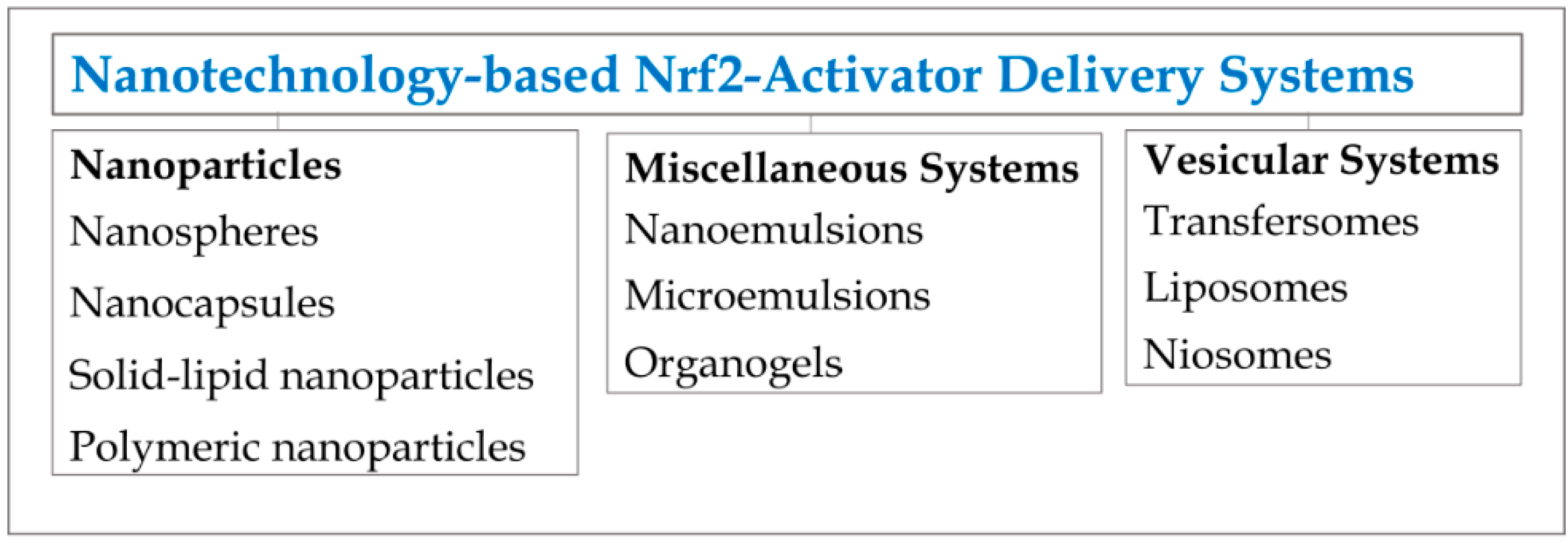
Examples of Nrf2-Activator Delivery Systems

The use of specifically-designed Nrf2-activator dermal delivery systems is an emerging concept which was recently introduced demonstrating the feasibility and advantage of this approach [238]. To date, there are delivery systems that encapsulate Nrf2 activators; however, there is a lack of specifically designed dermal Nrf2 activators in an encapsulated form. The most recent study demonstrated that encapsulated Nrf2 activators into a dermal delivery system activated the Keap1–Nrf2 pathway more efficiently than the free Nrf2 activators [238]. Moreover, an Nrf2-activator dermal delivery system has led to a significant reduction in UVB-induced cytotoxicity in human skin organ culture [238]. In another study, resveratrol, an additional Nrf2 activator was encapsulated into a novel fusogenic liposome in order to enhance its delivery and caused a rapid activation of the Keap1–Nrf2 pathway in aged cerebromicrovascular endothelial cells [239]. This study provided additional proof for the strategy of prevention and controlling oxidative stress-related pathophysiological conditions in aging [239]. An additional example providing justification for the development of an Nrf2-activator delivery system and expanding its potential application was executed with luteolin loaded phytosomes resulting in the sensitization of MDA-MB-231 cells to doxorubicin [240].
This targeted therapy possesses a key to potentially overcome a variety of skin pathologies and states. However, additional precise experimental data should be collected in order to reach a comprehensive understanding. Moreover, clinical efficacy remains to be demonstrated. An additional aspect that needs to be addressed is the influence and suitability of the dermal delivery system on the Keap1–Nrf2 pathway and specific skin conditions. This also includes the nano-structural characterization of the dermal delivery system.

8. Concluding Remarks

The prospective use of a dermal Nrf2-activator delivery system offers tremendous opportunities for controlling a variety of skin pathologies and disorders. As this review demonstrates, targeting cellular signaling pathways, specifically the Keap1–Nrf2 pathway in skin, may be a leading means for an efficient personalized treatment. This innovative approach combines the two distinct disciplines of cell biology and drug delivery platforms. On the one hand, many studies have focused on the activation of Nrf2 activators in skin for health, disease and repair [12], without further considering the delivery of these compounds to skin. On the other hand, many dermal delivery systems have been formulated and examined, without addressing the biological outcomes. Obtaining a deep knowledge regarding the integration of these two important disciplines need to be systematically supported by a wide spectrum of experimental data. Understanding the molecular mechanism for each skin pathology or state represents the first cornerstone in skin cures. Obviously, under certain circumstances, Nrf2 inhibitors, rather than Nrf2 activators, need to be considered as the preferred therapy (e.g., at specific stage of cancer evolvement). Utilizing the tools of nanotechnology provides a unified platform into which specific Nrf2 activators can be incorporated. Moreover, this targeted therapy enables loading and delivering multiple Nrf2 activators/inhibitors to obtain synergistic therapy. This approach still presents a few challenges that should be addressed, including the selection of a proper dermal delivery system tailored for individual skin biochemical needs or pathology.

Acknowledgments

Ron Kohen is affiliated with the Bloom Center of Pharmacy. He is the incumbent of the Richard and Jean Zarbin Chair in Medical Studies at the Hebrew University of Jerusalem. This work was funded by the David and Ines Myers Fund of Cleveland, OH, USA.

Author Contributions

Maya Ben-Yehuda Greenwald wrote this review; Shmuel Ben-Sasson, Havazelet Bianco-Peled and Ron Kohen revised this review.

Conflicts of Interest

The authors declare no conflict of interest.

References

  1. Trouba, K.J.; Hamadeh, H.K.; Amin, R.P.; Germolec, D.R. Oxidative stress and its role in skin disease. Antioxid. Redox Signal. 2002, 4, 665–673. [Google Scholar] [CrossRef] [PubMed]
  2. Kohen, R.; Gati, I. Skin low molecular weight antioxidants and their role in aging and in oxidative stress. Toxicology 2000, 148, 149–157. [Google Scholar] [CrossRef]
  3. Droge, W. Free radicals in the physiological control of cell function. Physiol. Rev. 2002, 82, 47–95. [Google Scholar] [CrossRef] [PubMed]
  4. Allen, R.G.; Tresini, M. Oxidative stress and gene regulation. Free Radic. Biol. Med. 2000, 28, 463–499. [Google Scholar] [CrossRef]
  5. Simon, H.U.; Haj-Yehia, A.; Levi-Schaffer, F. Role of reactive oxygen species (ROS) in apoptosis induction. Apoptosis 2000, 5, 415–418. [Google Scholar] [CrossRef] [PubMed]
  6. Roebuck, K.A. Oxidant stress regulation of IL-8 and ICAM-1 gene expression: Differential activation and binding of the transcription factors AP-1 and NF-kappaB (Review). Int. J. Mol. Med. 1999, 4, 223–253. [Google Scholar] [CrossRef] [PubMed]
  7. Lavrovsky, Y.; Chatterjee, B.; Clark, R.A.; Roy, A.K. Role of redox-regulated transcription factors in inflammation, aging and age-related diseases. Exp. Gerontol. 2000, 35, 521–532. [Google Scholar] [CrossRef]
  8. Kohen, R. Skin antioxidants: Their role in aging and in oxidative stress-new approaches for their evaluation. Biomed. Pharmacother. 1999, 53, 181–192. [Google Scholar] [CrossRef]
  9. Wölfle, U.; Seelinger, G.; Bauer, G.; Meinke, M.C.; Lademann, J.; Schempp, C.M. Reactive molecule species and antioxidative mechanisms in normal skin and skin aging. Skin Pharmacol. Physiol. 2014, 27, 316–332. [Google Scholar] [CrossRef] [PubMed]
  10. Bickers, D.R.; Athar, M. Oxidative stress in the pathogenesis of skin disease. J. Investig. Dermatol. 2006, 126, 2565–2575. [Google Scholar] [CrossRef] [PubMed]
  11. Wagener, F.A.; Carels, C.E.; Lundvig, D. Targeting the redox balance in inflammatory skin conditions. Int. J. Mol. Sci. 2013, 14, 9126–9167. [Google Scholar] [PubMed]
  12. Schäfer, M.; Werner, S. NRF2—A regulator of keratinocyte redox signaling. Free Radic. Biol. Med. 2015, 88, 243–252. [Google Scholar] [CrossRef] [PubMed]
  13. Talalay, P.; Fahey, J.W.; Healy, Z.R.; Wehage, S.L.; Benedict, A.L.; Min, C.; Dinkova-Kostova, A.T. Sulforaphane mobilizes cellular defenses that protect skin against damage by UV radiation. Proc. Natl. Acad. Sci. USA 2007, 104, 17500–17505. [Google Scholar] [CrossRef] [PubMed]
  14. Kohen, R.; Nyska, A. Oxidation of biological systems: Oxidative stress phenomena, antioxidants, redox reactions, and methods of their quantification. Toxicol. Pathol. 2002, 30, 620–650. [Google Scholar] [PubMed]
  15. Halliwell, B.; Gutteridge, J.M.C. Free Radicals in Biology and Medicine, 3rd ed.; Oxford University Press: Oxford, UK, 1999; Volume 3. [Google Scholar]
  16. Blanpain, C.D.; Fuchs, E. Epidermal homeostasis: A balancing act of stem cells in the skin. Nat. Rev. Mol. Cell Biol. 2009, 10, 207–217. [Google Scholar]
  17. Rinnerthaler, M.; Streubel, M.K.; Bischof, J.; Richter, K. Skin aging, gene expression and calcium. Exp. Gerontol. 2015, 68, 59–65. [Google Scholar] [CrossRef] [PubMed]
  18. Schallreuter, K.U.; Wood, J.M. Free radical reduction in the human epidermis. Free Radic. Biol. Med. 1989, 6, 519–532. [Google Scholar] [CrossRef]
  19. Krutmann, J.; Schruder, P.; Morita, A. Molekulare mechanismen der hautalterung durch UV-strahlung und andere exogene noxen. In Hautalterung; Springer: Berlin, Germany, 2008; pp. 23–36. (In German) [Google Scholar]
  20. Poljšak, B.; Dahmane, R. Free radicals and extrinsic skin aging. Dermatol. Res. Pract. 2012, 2012, 135206. [Google Scholar] [CrossRef] [PubMed]
  21. Richter, C.; Gogvadze, V.; Laffranchi, R.; Schlapbach, R.; Schweizer, M.; Suter, M.; Walter, P.; Yaffee, M. Oxidants in mitochondria: From physiology to diseases. Biochim. Biophys. Acta Mol. Basis Dis. 1995, 1271, 67–74. [Google Scholar] [CrossRef]
  22. Kandola, K.; Bowman, A.; Birch-Machin, M.A. Oxidative stress—A key emerging impact factor in health, ageing, lifestyle and aesthetics. Int. J. Cosmet. Sci. 2015, 37 (Suppl. 2), 1–8. [Google Scholar] [CrossRef]
  23. Deliconstantinos, G.; Villiotou, V. Gas phase oxidants of cigarette smoke increase nitric oxide synthase and xanthine oxidase activities of rabbit brain synaptosomes. Neurochem. Res. 2000, 25, 769–774. [Google Scholar] [CrossRef] [PubMed]
  24. Nakai, K.; Kadiiska, M.B.; Jiang, J.J.; Stadler, K.; Mason, R.P. Free radical production requires both inducible nitric oxide synthase and xanthine oxidase in LPS-treated skin. Proc. Natl. Acad. Sci. USA 2006, 103, 4616–4621. [Google Scholar] [CrossRef] [PubMed]
  25. Portugal-Cohen, M.; Kohen, R. Exposure of human keratinocytes to ischemia, hyperglycemia and their combination induces oxidative stress via the enzymes inducible nitric oxide synthase and xanthine oxidase. J. Dermatol. Sci. 2009, 55, 82–90. [Google Scholar] [CrossRef] [PubMed]
  26. Portugal-Cohen, M.; Numa, R.; Yaka, R.; Kohen, R. Cocaine induces oxidative damage to skin via xanthine oxidase and nitric oxide synthase. J. Dermatol. Sci. 2010, 58, 105–112. [Google Scholar] [CrossRef] [PubMed]
  27. Bedard, K.; Krause, K.-H. The NOX family of ROS-generating NADPH oxidases: Physiology and pathophysiology. Physiol. Rev. 2007, 87, 245–313. [Google Scholar] [CrossRef] [PubMed]
  28. Kammeyer, A.; Luiten, R.M. Oxidation events and skin aging. Ageing Res. Rev. 2015, 21, 16–29. [Google Scholar] [CrossRef] [PubMed]
  29. Dalle-Donne, I.; Rossi, R.; Giustarini, D.; Milzani, A.; Colombo, R. Protein carbonyl groups as biomarkers of oxidative stress. Clin. Chim. Acta 2003, 329, 23–38. [Google Scholar] [CrossRef]
  30. Kensler, T.W.; Wakabayashi, N.; Biswal, S. Cell survival responses to environmental stresses via the Keap1-Nrf2-ARE pathway. Annu. Rev. Pharmacol. Toxicol. 2007, 47, 89–116. [Google Scholar] [CrossRef] [PubMed]
  31. Halliwell, B. Reactive species and antioxidants. Redox biology is a fundamental theme of aerobic life. Plant Physiol. 2006, 141, 312–322. [Google Scholar] [CrossRef] [PubMed]
  32. Marnett, L.J. Lipid peroxidation-DNA damage by malondialdehyde. Mutat. Res. 1999, 424, 83–95. [Google Scholar] [CrossRef]
  33. Briganti, S.; Picardo, M. Antioxidant activity, lipid peroxidation and skin diseases. What’s new. J. Eur. Acad. Dermatol. Venereol. 2003, 17, 663–669. [Google Scholar] [CrossRef] [PubMed]
  34. Decker, J.M. 11th Hour: Introduction to Immunology; John Wiley & Sons: Hoboken, NJ, USA, 2009. [Google Scholar]
  35. Pillai, S.; Oresajo, C.; Hayward, J. Ultraviolet radiation and skin aging: Roles of reactive oxygen species, inflammation and protease activation, and strategies for prevention of inflammation-induced matrix degradation—A review. Int. J. Cosmet. Sci. 2005, 27, 17–34. [Google Scholar] [CrossRef] [PubMed]
  36. Wlaschek, M.; Tantcheva-Poor, I.; Naderi, L.; Ma, W.; Schneider, L.A.; Razi-Wolf, Z.; Schuller, J.; Scharffetter-Kochanek, K. Solar UV irradiation and dermal photoaging. J. Photochem. Photobiol. B Biol. 2001, 63, 41–51. [Google Scholar] [CrossRef]
  37. Gough, D.R.; Cotter, T.G. Hydrogen peroxide: A Jekyll and Hyde signalling molecule. Cell Death Dis. 2011, 2, e213. [Google Scholar] [CrossRef] [PubMed]
  38. Rhee, S.G. Cell signaling. H2O2, a necessary evil for cell signaling. Science 2006, 312, 1882–1883. [Google Scholar] [CrossRef] [PubMed]
  39. Drew, J.E. Cellular defense system gene expression profiling of human whole blood: Opportunities to predict health benefits in response to diet. Adv. Nutr. Int. Rev. J. 2012, 3, 499–505. [Google Scholar] [CrossRef] [PubMed]
  40. Birch-Machin, M.A.; Russell, E.V.; Latimer, J.A. Mitochondrial DNA damage as a biomarker for ultraviolet radiation exposure and oxidative stress. Br. J. Dermatol. 2013, 169 (Suppl. 2), 9–14. [Google Scholar] [CrossRef] [PubMed]
  41. Maeda, T.; Chua, P.P.S.; Chong, M.T.; Sim, A.B.T.; Nikaido, O.; Tron, V.A. Nucleotide excision repair genes are upregulated by low-dose artificial ultraviolet B: Evidence of a photoprotective SOS response? J. Investig. Dermatol. 2001, 117, 1490–1497. [Google Scholar] [CrossRef] [PubMed]
  42. Rinnerthaler, M.; Bischof, J.; Streubel, M.K.; Trost, A.; Richter, K. Oxidative Stress in Aging Human Skin. Biomolecules 2015, 5, 545–589. [Google Scholar] [CrossRef] [PubMed]
  43. Flora, S.J.S. Structural, chemical and biological aspects of antioxidants for strategies against metal and metalloid exposure. Oxid. Med. Cell. Longev. 2009, 2, 191–206. [Google Scholar] [CrossRef] [PubMed]
  44. Packer, J.E.; Slater, T.; Willson, R.L. Direct observation of a free radical interaction between vitamin E and vitamin C. Nature 1979, 278, 737–738. [Google Scholar] [CrossRef] [PubMed]
  45. Martensson, J.; Meister, A.; Mrtensson, J. Glutathione deficiency decreases tissue ascorbate levels in newborn rats: Ascorbate spares glutathione and protects. Proc. Natl. Acad. Sci. USA 1991, 88, 4656–4660. [Google Scholar] [CrossRef] [PubMed]
  46. Lopez-Torres, M.; Thiele, J.J.; Shindo, Y.; Han, D.; Packer, L. Topical application of alpha-tocopherol modulates the antioxidant network and diminishes ultraviolet-induced oxidative damage in murine skin. Br. J. Dermatol. 1998, 138, 207–215. [Google Scholar] [CrossRef] [PubMed]
  47. Vermeij, W.P.; Alia, A.; Backendorf, C. ROS quenching potential of the epidermal cornified cell envelope. J. Investig. Dermatol. 2011, 131, 1435–1441. [Google Scholar] [CrossRef] [PubMed]
  48. Stojiljković, D.; Pavlović, D.I.; Arsić, I. Oxidative Stress, Skin Aging and Antioxidant Therapy/Oksidacioni Stres, Starenje Kooe I Antioksidaciona Terapija. Acta Fac. Med. Naissensis 2014, 31, 207–217. [Google Scholar]
  49. Rigo, A.; Stevanato, R.; Finazzi-Agro, A.; Rotilio, G. An attempt to evaluate the rate of the haber-weiss reaction by using OH radical scavengers. FEBS Lett. 1977, 80, 130–132. [Google Scholar] [CrossRef]
  50. Yang, H.; Wang, X.; Liu, X.; Wu, J.; Liu, C.; Gong, W.; Zhao, Z.; Hong, J.; Lin, D.; Wang, Y.; et al. Antioxidant peptidomics reveals novel skin antioxidant system. Mol. Cell. Proteomics 2009, 8, 571–583. [Google Scholar] [CrossRef] [PubMed]
  51. Weinraub, D.; Levy, P.; Faraggi, M. Chemical properties of water-soluble porphyrins. 5. Reactions of some manganese (III) porphyrins with the superoxide and other reducing radicals. Int. J. Radiat. Biol. Relat. Stud. Phys. Chem. Med. 1986, 50, 649–658. [Google Scholar] [CrossRef] [PubMed]
  52. Boldyrev, A.A. Does carnosine possess direct antioxidant activity? Int. J. Biochem. 1993, 25, 1101–1107. [Google Scholar] [CrossRef]
  53. Pompella, A.; Visvikis, A.; Paolicchi, A.; de Tata, V.; Casini, A.F. The changing faces of glutathione, a cellular protagonist. Biochem. Pharmacol. 2003, 66, 1499–1503. [Google Scholar] [CrossRef]
  54. Kohen, R.; Yamamoto, Y.; Cundy, K.C.; Ames, B.N. Antioxidant activity of carnosine, homocarnosine, and anserine present in muscle and brain. Proc. Natl. Acad. Sci. USA 1988, 85, 3175–3179. [Google Scholar] [CrossRef] [PubMed]
  55. Packer, L.; Kraemer, K.; Rimbach, G. Molecular aspects of lipoic acid in the prevention of diabetes complications. Nutrition 2001, 17, 888–895. [Google Scholar] [CrossRef]
  56. Ames, B.N.; Shigenaga, M.K.; Hagen, T.M. Oxidants, antioxidants, and the degenerative diseases of aging. Proc. Natl. Acad. Sci. USA 1993, 90, 7915–7922. [Google Scholar] [CrossRef] [PubMed]
  57. Stocker, R.; Yamamoto, Y.; McDonagh, A.F.; Glazer, A.N.; Ames, B.N. Bilirubin is an antioxidant of possible physiological importance. Science 1987, 235, 1043–1046. [Google Scholar] [CrossRef] [PubMed]
  58. Myriam, M.; Sabatier, M.; Steiling, H.; Williamson, G. Skin bioavailability of dietary vitamin E, carotenoids, polyphenols, vitamin C, zinc and selenium. Br. J. Nutr. 2006, 96, 227–238. [Google Scholar] [CrossRef]
  59. Ratnam, D.V.; Ankola, D.D.; Bhardwaj, V.; Sahana, D.K.; Kumar, M.N. Role of antioxidants in prophylaxis and therapy: A pharmaceutical perspective. J. Control. Release 2006, 113, 189–207. [Google Scholar] [CrossRef] [PubMed]
  60. Fernández-García, E. Skin protection against UV light by dietary antioxidants. Food Funct. 2014, 5, 1994–2003. [Google Scholar] [CrossRef] [PubMed]
  61. Boddupalli, S.; Mein, J.R.; James, D.R.; Lakkanna, S. Induction of phase 2 antioxidant enzymes by broccoli sulforaphane: Perspectives in maintaining the antioxidant activity of vitamins A, C, and E. Front. Genet. 2012, 3. [Google Scholar] [CrossRef] [PubMed]
  62. Auf dem Keller, U.; Kumin, A.; Braun, S.; Werner, S. Reactive oxygen species and their detoxification in healing skin wounds. J. Investig. Dermatol. Symp. Proc. 2006, 11, 106–111. [Google Scholar] [CrossRef] [PubMed]
  63. Shindo, Y.; Witt, E.; Packer, L. Antioxidant defense mechanisms in murine epidermis and dermis and their responses to ultraviolet light. J. Investig. Dermatol. 1993, 100, 260–265. [Google Scholar] [CrossRef] [PubMed]
  64. Schafer, F.Q.; Buettner, G.R. Redox environment of the cell as viewed through the redox state of the glutathione disulfide/glutathione couple. Free Radic. Biol. Med. 2001, 30, 1191–1212. [Google Scholar] [CrossRef]
  65. Haines, D.D.; Lekli, I.; Teissier, P.; Bak, I.; Tosaki, A. Role of haeme oxygenase-1 in resolution of oxidative stress-related pathologies: Focus on cardiovascular, lung, neurological and kidney disorders. Acta Physiol. 2012, 204, 487–501. [Google Scholar] [CrossRef] [PubMed]
  66. Maines, M.D. Heme oxygenase: Function, multiplicity, regulatory mechanisms, and clinical applications. FASEB J. 1988, 2, 2557–2568. [Google Scholar] [PubMed]
  67. Tenhunen, R.; Marver, H.S.; Schmid, R. The enzymatic conversion of heme to bilirubin by microsomal heme oxygenase. Proc. Natl. Acad. Sci. USA 1968, 61, 748. [Google Scholar] [CrossRef] [PubMed]
  68. Tyrrell, R.M. Solar ultraviolet A radiation: An oxidizing skin carcinogen that activates heme oxygenase-1. Antioxid. Redox Signal. 2004, 6, 835–840. [Google Scholar] [CrossRef] [PubMed]
  69. Saw, C.L.; Yang, A.Y.; Huang, M.-T.; Liu, Y.; Lee, J.H.; Khor, T.O.; Su, Z.-Y.; Shu, L.; Lu, Y.; Conney, A.H.; et al. Nrf2 null enhances UVB-induced skin inflammation and extracellular matrix damages. Cell Biosci. 2014, 4. [Google Scholar] [CrossRef] [PubMed]
  70. Chance, B.; Sies, H.; Boveris, A. Hydroperoxide metabolism in mammalian organs. Physiol. Rev. 1979, 59, 527–605. [Google Scholar] [PubMed]
  71. Ying, W. NAD+/NADH and NADP+/NADPH in cellular functions and cell death: Regulation and biological consequences. Antioxid. Redox Signal. 2008, 10, 179–206. [Google Scholar] [CrossRef] [PubMed]
  72. Ames, B.N.; Cathcart, R.; Schwiers, E.; Hochstein, P. Uric acid provides an antioxidant defense in humans against oxidant- and radical-caused aging and cancer: A hypothesis. Proc. Natl. Acad. Sci. USA 1981, 78, 6858–6862. [Google Scholar] [CrossRef] [PubMed]
  73. Gul, M.; Kutay, F.Z.; Temocin, S.; Hanninen, O. Cellular and clinical implications of glutathione. Indian J. Exp. Biol. 2000, 38, 625–634. [Google Scholar] [PubMed]
  74. Peres, P.S.; Terra, V.A.; Guarnier, F.A.; Cecchini, R.; Cecchini, A.L. Photoaging and chronological aging profile: Understanding oxidation of the skin. J. Photochem. Photobiol. B Biol. 2011, 103, 93–97. [Google Scholar] [CrossRef] [PubMed]
  75. Chun, K.-S.; Kundu, J.; Kundu, J.K.; Surh, Y.-J. Targeting Nrf2-Keap1 signaling for chemoprevention of skin carcinogenesis with bioactive phytochemicals. Toxicol. Lett. 2014, 229, 73–84. [Google Scholar] [CrossRef] [PubMed]
  76. Engin, A. Glutathione content of human skin carcinomas. Arch. Dermatol. Res. 1976, 257, 53–55. [Google Scholar] [CrossRef] [PubMed]
  77. Kobayashi, T.; Matsumoto, M.; Iizuka, H.; Suzuki, K.; Taniguchi, N. Superoxide dismutase in psoriasis, squamous cell carcinoma and basal cell epithelioma: An immunohistochemical study. Br. J. Dermatol. 1991, 124, 555–559. [Google Scholar] [CrossRef] [PubMed]
  78. Patel, S.; Kartasova, T.; Segre, J.A. Mouse Sprr locus: A tandem array of coordinately regulated genes. Mamm. Genome 2003, 14, 140–148. [Google Scholar] [CrossRef] [PubMed]
  79. Rinnerthaler, M.; Duschl, J.; Steinbacher, P.; Salzmann, M.; Bischof, J.; Schuller, M.; Wimmer, H.; Peer, T.; Bauer, J.W.; Richter, K. Age-related changes in the composition of the cornified envelope in human skin. Exp. Dermatol. 2013, 22, 329–335. [Google Scholar] [CrossRef] [PubMed]
  80. Baroni, A.; Buommino, E.; de Gregorio, V.; Ruocco, E.; Ruocco, V.; Wolf, R. Structure and function of the epidermis related to barrier properties. Clin. Dermatol. 2012, 30, 257–262. [Google Scholar] [CrossRef] [PubMed]
  81. Thiele, J.J.; Hsieh, S.N.; Briviba, K.; Sies, H. Protein oxidation in human stratum corneum: Susceptibility of keratins to oxidation in vitro and presence of a keratin oxidation gradient in vivo. J. Investig. Dermatol. 1999, 113, 335–339. [Google Scholar] [CrossRef] [PubMed]
  82. Levonen, A.-L.; Hill, B.G.; Kansanen, E.; Zhang, J.; Darley-Usmar, V.M. Redox regulation of antioxidants, autophagy, and the response to stress: Implications for electrophile therapeutics. Free Radic. Biol. Med. 2014, 71, 196–207. [Google Scholar] [CrossRef] [PubMed]
  83. Black, H.S. ROS: A step closer to elucidating their role in the etiology of light-induced skin disorders. J. Investig. Dermatol. 2004, 122, xiii–xiv. [Google Scholar] [CrossRef] [PubMed]
  84. Valacchi, G.; Sticozzi, C.; Pecorelli, A.; Cervellati, F.; Cervellati, C.; Maioli, E. Cutaneous responses to environmental stressors. Ann. N. Y. Acad. Sci. 2012, 1271, 75–81. [Google Scholar] [CrossRef] [PubMed]
  85. Lambeth, J.D.; Krause, K.-H.; Clark, R.A. NOX enzymes as novel targets for drug development. Semin. Immunopathol. 2008, 30, 339–363. [Google Scholar] [CrossRef] [PubMed]
  86. Jaquet, V.; Scapozza, L.; Clark, R.A.; Krause, K.-H.; Lambeth, J.D. Small-molecule NOX inhibitors: ROS-generating NADPH oxidases as therapeutic targets. Antioxid. Redox Signal. 2009, 11, 2535–2552. [Google Scholar] [CrossRef] [PubMed]
  87. Zhang, W.-J.; Wei, H.; Frei, B. Genetic deficiency of NADPH oxidase does not diminish, but rather enhances, LPS-induced acute inflammatory responses in vivo. Free Radic. Biol. Med. 2009, 46, 791–798. [Google Scholar] [CrossRef] [PubMed]
  88. Marriott, H.M.; Jackson, L.E.; Wilkinson, T.S.; Simpson, A.J.; Mitchell, T.J.; Buttle, D.J.; Cross, S.S.; Ince, P.G.; Hellewell, P.G.; Whyte, M.K.B.; et al. Reactive oxygen species regulate neutrophil recruitment and survival in pneumococcal pneumonia. Am. J. Respir. Crit. Care Med. 2008, 177, 887–895. [Google Scholar] [CrossRef] [PubMed]
  89. Hegde, M.L.; Bharathi, P.; Suram, A.; Venugopal, C.; Jagannathan, R.; Poddar, P.; Srinivas, P.; Sambamurti, K.; Rao, K.J.; Scancar, J.; et al. Challenges associated with metal chelation therapy in Alzheimer’s disease. J. Alzheimers Dis. 2009, 17, 457–468. [Google Scholar] [PubMed]
  90. Blennow, K.; de Leon, M.J.; Zetterberg, H. Alzheimer’s disease. Lancet 2006, 377, 1019–1031. [Google Scholar] [CrossRef]
  91. Dong, J.; Atwood, C.S.; Anderson, V.E.; Siedlak, S.L.; Smith, M.A.; Perry, G.; Carey, P.R. Metal binding and oxidation of amyloid-β within isolated senile plaque cores: Raman microscopic evidence. Biochemistry 2003, 42, 2768–2773. [Google Scholar] [CrossRef] [PubMed]
  92. Finefrock, A.E.; Bush, A.I.; Doraiswamy, P.M. Current status of metals as therapeutic targets in Alzheimer’s disease. J. Am. Geriatr. Soc. 2003, 51, 1143–1148. [Google Scholar] [CrossRef] [PubMed]
  93. Khan, A.; Ashcroft, A.E.; Korchazhkina, O.V.; Exley, C. Metal-mediated formation of fibrillar ABri amyloid. J. Inorg. Biochem. 2004, 98, 2006–2010. [Google Scholar] [CrossRef] [PubMed]
  94. Valko, M.; Morris, H.; Cronin, M.T.D. Metals, toxicity and oxidative stress. Curr. Med. Chem. 2005, 12, 1161–1208. [Google Scholar] [CrossRef] [PubMed]
  95. Ricchelli, F.; Drago, D.; Filippi, B.; Tognon, G.; Zatta, P. Aluminum-triggered structural modifications and aggregation of β-amyloids. Cell. Mol. Life Sci. 2005, 62, 1724–1733. [Google Scholar] [CrossRef] [PubMed]
  96. Mandel, S.; Amit, T.; Bar-Am, O.; Youdim, M.B.H. Iron dysregulation in Alzheimer’s disease: Multimodal brain permeable iron chelating drugs, possessing neuroprotective-neurorescue and amyloid precursor protein-processing regulatory activities as therapeutic agents. Prog. Neurobiol. 2007, 82, 348–360. [Google Scholar] [CrossRef] [PubMed]
  97. Ha, H.-Y.; Kim, Y.; Ryoo, Z.Y.; Kim, T.-Y. Inhibition of the TPA-induced cutaneous inflammation and hyperplasia by EC-SOD. Biochem. Biophys. Res. Commun. 2006, 348, 450–458. [Google Scholar] [CrossRef] [PubMed]
  98. Kim, Y.; Kim, B.H.; Lee, H.; Jeon, B.; Lee, Y.S.; Kwon, M.-J.; Kim, T.-Y. Regulation of skin inflammation and angiogenesis by EC-SOD via HIF-1α and NF-κB pathways. Free Radic. Biol. Med. 2011, 51, 1985–1995. [Google Scholar] [CrossRef] [PubMed]
  99. Day, A.J.; Canada, F.J.; Diaz, J.C.; Kroon, P.A.; McLauchlan, R.; Faulds, C.B.; Plumb, G.W.; Morgan, M.R.A.; Williamson, G. Dietary flavonoid and isoflavone glycosides are hydrolysed by the lactase site of lactase phlorizin hydrolase. FEBS Lett. 2000, 468, 166–170. [Google Scholar] [CrossRef]
  100. Nemeth, K.; Plumb, G.W.; Berrin, J.-G.; Juge, N.; Jacob, R.; Naim, H.Y.; Williamson, G.; Swallow, D.M.; Kroon, P.A. Deglycosylation by small intestinal epithelial cell β-glucosidases is a critical step in the absorption and metabolism of dietary flavonoid glycosides in humans. Eur. J. Nutr. 2003, 42, 29–42. [Google Scholar] [CrossRef] [PubMed]
  101. Hollander, D.; Rim, E.; Muralidhara, K.S. Mechanism and site of small intestinal absorption of alpha-tocopherol in the rat. Gastroenterology 1975, 68, 1492–1499. [Google Scholar] [PubMed]
  102. Hollander, D.; Ruble, P.E. Beta-carotene intestinal absorption: Bile, fatty acid, pH, and flow rate effects on transport. Am. J. Physiol. 1978, 235, G686–G691. [Google Scholar]
  103. During, A.; Hussain, M.M.; Morel, D.W.; Harrison, E.H. Carotenoid uptake and secretion by CaCo-2 cells: β-carotene isomer selectivity and carotenoid interactions. J. Lipid Res. 2002, 43, 1086–1095. [Google Scholar] [CrossRef] [PubMed]
  104. During, A.; Dawson, H.D.; Harrison, E.H. Carotenoid transport is decreased and expression of the lipid transporters SR-BI, NPC1L1, and ABCA1 is downregulated in Caco-2 cells treated with ezetimibe. J. Nutr. 2005, 135, 2305–2312. [Google Scholar] [PubMed]
  105. Reboul, E.; Abou, L.; Mikail, C.L.; Ghiringhelli, O.; Andre, M.; Portugal, H.; Jourdheuil-Rahmani, D.; Amiot, M.; Lairon, D.; Borel, P. Lutein transport by Caco-2 TC-7 cells occurs partly by a facilitated process involving the scavenger receptor class B type I (SR-BI). Biochem. J. 2005, 387, 455–461. [Google Scholar] [CrossRef] [PubMed]
  106. Cho, H.-S.; Kwak, D.H.; Choi, I.S.; Park, H.K.; Kang, S.J.; Yoo, H.S.; Lee, M.S.; Oh, K.-W.; Hong, J.T. Inhibitory effect of proanthocyanidin on ultraviolet B irradiation-induced melanogenesis. J. Toxicol. Environ. Health A 2009, 72, 1475–1483. [Google Scholar] [CrossRef] [PubMed]
  107. Mittal, A.; Elmets, C.A.; Katiyar, S.K. Dietary feeding of proanthocyanidins from grape seeds prevents photocarcinogenesis in SKH-1 hairless mice: Relationship to decreased fat and lipid peroxidation. Carcinogenesis 2003, 24, 1379–1388. [Google Scholar] [CrossRef] [PubMed]
  108. Vaid, M.; Singh, T.; Katiyar, S.K. Grape seed proanthocyanidins inhibit melanoma cell invasiveness by reduction of PGE2 synthesis and reversal of epithelial-to-mesenchymal transition. PLoS ONE 2011, 6, e21539. [Google Scholar] [CrossRef] [PubMed]
  109. Wang, Z.Y.; Huang, M.-T.; Ho, C.-T.; Chang, R.; Ma, W.; Ferraro, T.; Reuhl, K.R.; Yang, C.S.; Conney, A.H. Inhibitory effect of green tea on the growth of established skin papillomas in mice. Cancer Res. 1992, 52, 6657–6665. [Google Scholar] [PubMed]
  110. Tong, L.X.; Young, L.C. Nutrition: The future of melanoma prevention? J. Am. Acad. Dermatol. 2014, 71, 151–160. [Google Scholar] [CrossRef] [PubMed]
  111. Vayalil, P.K.; Mittal, A.; Hara, Y.; Elmets, C.A.; Katiyar, S.K. Green tea polyphenols prevent ultraviolet light-induced oxidative damage and matrix metalloproteinases expression in mouse skin. J. Investig. Dermatol. 2004, 122, 1480–1487. [Google Scholar] [CrossRef] [PubMed]
  112. Stahl, W.; Heinrich, U.; Jungmann, H.; Sies, H.; Tronnier, H. Carotenoids and carotenoids plus vitamin E protect against ultraviolet light-induced erythema in humans. Am. J. Clin. Nutr. 2000, 71, 795–798. [Google Scholar] [PubMed]
  113. Lee, J.; Jiang, S.; Levine, N.; Watson, R.R. Carotenoid supplementation reduces erythema in human skin after simulated solar radiation exposure. Proc. Soc. Exp. Biol. Med. 2000, 223, 170–174. [Google Scholar] [CrossRef] [PubMed]
  114. Heinrich, U.; Gartner, C.; Wiebusch, M.; Eichler, O.; Sies, H.; Tronnier, H.; Stahl, W. Supplementation with β-carotene or a similar amount of mixed carotenoids protects humans from UV-induced erythema. J. Nutr. 2003, 133, 98–101. [Google Scholar] [PubMed]
  115. Eicker, J.R.; Kurten, V.; Wild, S.; Riss, G.; Goralczyk, R.; Krutmann, J.; Berneburg, M. Betacarotene supplementation protects from photoaging-associated mitochondrial DNA mutation. Photochem. Photobiol. Sci. 2003, 2, 655–659. [Google Scholar] [CrossRef] [PubMed]
  116. Asgari, M.M.; Maruti, S.S.; Kushi, L.H.; White, E. A cohort study of vitamin D intake and melanoma risk. J. Investig. Dermatol. 2009, 129, 1675–1680. [Google Scholar] [CrossRef] [PubMed]
  117. Di Franco, R.; Calvanese, M.; Murino, P.; Manzo, R.; Guida, C.; di Gennaro, D.; Anania, C.; Ravo, V. Skin toxicity from external beam radiation therapy in breast cancer patients: Protective effects of Resveratrol, Lycopene, Vitamin C and anthocianin (Ixor®). Radiat. Oncol. 2012, 7, 1–6. [Google Scholar] [CrossRef] [PubMed]
  118. USDA. USDA-NCC Carotenoid Database for U.S. Foods—1998. Available online: http://healthybusiness.co.za/blog/wp-content/uploads/USDA_carotenoids-in-foods.pdf (accessed on 9 August 2013).
  119. Mathews-Roth, M.M.; Pathak, M.A.; Parrish, J.; Fitzpatrick, T.B.; Kass, E.H.; Toda, K.; Clemens, W. A clinical trial of the effects of oral beta-carotene on the responses of human skin to solar radiation. J. Investig. Dermatol. 1972, 59, 349–353. [Google Scholar] [CrossRef] [PubMed]
  120. Gollnick, H.P.M.; Hopfenmller, W.; Hemmes, C.; Chun, S.C.; Schmid, C.; Sundermeier, K.; Biesalski, H.K. Systemic beta carotene plus topical UV-sunscreen are an optimal protection against harmful effects of natural UV-sunlight: Results of the Berlin-Eilath study. Eur. J. Dermatol. 1996, 6, 200–205. [Google Scholar]
  121. Stahl, W.; Heinrich, U.; Wiseman, S.; Eichler, O.; Sies, H.; Tronnier, H. Dietary tomato paste protects against ultraviolet light-induced erythema in humans. J. Nutr. 2001, 131, 1449–1451. [Google Scholar] [PubMed]
  122. Wolf, C.; Steiner, A.; Hunigsmann, H. Do oral carotenoids protect human skin against ultraviolet erythema, psoralen phototoxicity, and ultraviolet-induced DNA damage? J. Investig. Dermatol. 1988, 90, 55–57. [Google Scholar] [CrossRef] [PubMed]
  123. Garmyn, M.; Ribaya-Mercado, J.D.; Russel, R.M.; Bhawan, J.; Gilchrest, B.A. Effect of beta-carotene supplementation on the human sunburn reaction. Exp. Dermatol. 1995, 4, 104–111. [Google Scholar] [CrossRef] [PubMed]
  124. Placzek, M.; Gaube, S.; Kerkmann, U.; Gilbertz, K.-P.; Herzinger, T.; Haen, E.; Przybilla, B. Ultraviolet B-induced DNA damage in human epidermis is modified by the antioxidants ascorbic acid and d-α-tocopherol. J. Investig. Dermatol. 2005, 124, 304–307. [Google Scholar] [CrossRef] [PubMed]
  125. Eberlein-Konig, B.; Ring, J. Relevance of vitamins C and E in cutaneous photoprotection. J. Cosmet. Dermatol. 2005, 4, 4–9. [Google Scholar] [CrossRef] [PubMed]
  126. Makrantonaki, E.; Zouboulis, C.C. Skin alterations and diseases in advanced age. Drug Discov. Today Dis. Mech. 2008, 5, e153–e162. [Google Scholar] [CrossRef]
  127. Elmets, C.A.; Singh, D.; Tubesing, K.; Matsui, M.; Katiyar, S.; Mukhtar, H. Cutaneous photoprotection from ultraviolet injury by green tea polyphenols. J. Am. Acad. Dermatol. 2001, 44, 425–432. [Google Scholar] [CrossRef] [PubMed]
  128. Bickers, D.R.; Athar, M. Novel approaches to chemoprevention of skin cancer. J. Dermatol. 2000, 27, 691–695. [Google Scholar] [CrossRef] [PubMed]
  129. Katiyar, S.K. Skin photoprotection by green tea: Antioxidant and immunomodulatory effects. Curr. Drug Targets Immune Endocr. Metabol. Disord. 2003, 3, 234–242. [Google Scholar] [CrossRef] [PubMed]
  130. Katiyar, S.K.; Matsui, M.S.; Elmets, C.A.; Mukhtar, H. Polyphenolic antioxidant (−)-epigallocatechin-3-gallate from green tea reduces UVB-induced inflammatory responses and infiltration of leukocytes in human skin. Photochem. Photobiol. 1999, 69, 148–153. [Google Scholar] [CrossRef]
  131. Mittal, A.; Piyathilake, C.; Hara, Y.; Katiyar, S.K. Exceptionally high protection of photocarcinogenesis by topical application of (−)-epigallocatechin-3-gallate in hydrophilic cream in SKH-1 Hairless Mouse Model: Relationship to inhibition of UVB-induced global DNA hypomethylation. Neoplasia 2003, 5, 555–565. [Google Scholar] [CrossRef]
  132. Gensler, H.L.; Timmermann, B.N.; Valcic, S.; Wachter, G.A.; Dorr, R.; Dvorakova, K.; Alberts, D.S. Prevention of photocarcinogenesis by topical administration of pure epigallocatechin gallate isolated from green tea. 1996, 26, 325–335. [Google Scholar] [CrossRef] [PubMed]
  133. Yuan, X.-Y.; Liu, W.; Hao, J.-C.; Gu, W.-J.; Zhao, Y.-S. Topical grape seed proanthocyandin extract reduces sunburn cells and mutant p53 positive epidermal cell formation, and prevents depletion of Langerhans cells in an acute sunburn model. Photomed. Laser Surg. 2012, 30, 20–25. [Google Scholar] [CrossRef] [PubMed]
  134. Oyewole, A.O.; Wilmot, M.-C.; Fowler, M.; Birch-Machin, M.A. Comparing the effects of mitochondrial targeted and localized antioxidants with cellular antioxidants in human skin cells exposed to UVA and hydrogen peroxide. FASEB J. 2014, 28, 485–494. [Google Scholar] [CrossRef] [PubMed]
  135. Oyewole, A.O.; Birch-Machin, M.A. Mitochondria-targeted antioxidants. FASEB J. 2014, 29, 4766–4771. [Google Scholar] [CrossRef] [PubMed]
  136. Varani, J.; Spearman, D.; Perone, P.; Fligiel, S.E.G.; Datta, S.C.; Wang, Z.Q.; Shao, Y.; Kang, S.; Fisher, G.J.; Voorhees, J.J. Inhibition of type I procollagen synthesis by damaged collagen in photoaged skin and by collagenase-degraded collagen in vitro. Am. J. Pathol. 2001, 158, 931–942. [Google Scholar] [CrossRef]
  137. Humbert, P.G.; Haftek, M.; Creidi, P.; Lapiere, C.; Nusgens, B.; Richard, A.; Schmitt, D.; Rougier, A.; Zahouani, H. Topical ascorbic acid on photoaged skin. Clinical, topographical and ultrastructural evaluation: Double-blind study vs. placebo. Exp. Dermatol. 2003, 12, 237–244. [Google Scholar] [CrossRef] [PubMed]
  138. Ricciarelli, R.; Maroni, P.; Özer, N.; Zingg, J.-M.; Azzi, A. Age-dependent increase of collagenase expression can be reduced by α-tocopherol via protein kinase C inhibition. Free Radic. Biol. Med. 1999, 27, 729–737. [Google Scholar] [CrossRef]
  139. Chen, W.; Barthelman, M.; Martinez, J.; Alberts, D.; Gensler, H.L. Inhibition of cyclobutane pyrimidine dimer formation in epidermal p53 gene of UV-irradiated mice by alpha-tocopherol. Nutr. Cancer 1997, 29, 205–211. [Google Scholar] [CrossRef] [PubMed]
  140. Zussman, J.; Ahdout, J.; Kim, J. Vitamins and photoaging: Do scientific data support their use? J. Am. Acad. Dermatol. 2010, 63, 507–525. [Google Scholar] [CrossRef] [PubMed]
  141. Murray, J.C.; Burch, J.A.; Streilein, R.D.; Iannacchione, M.A.; Hall, R.P.; Pinnell, S.R. A topical antioxidant solution containing vitamins C and E stabilized by ferulic acid provides protection for human skin against damage caused by ultraviolet irradiation. J. Am. Acad. Dermatol. 2008, 59, 418–425. [Google Scholar] [CrossRef] [PubMed]
  142. Montuschi, P.; Barnes, P.J.; Roberts, L.J. Isoprostanes: Markers and mediators of oxidative stress. FASEB J. 2004, 18, 1791–1800. [Google Scholar] [CrossRef] [PubMed]
  143. Traber, M.G.; Stevens, J.F. Vitamins C and E: Beneficial effects from a mechanistic perspective. Free Radic. Biol. Med. 2011, 51, 1000–1013. [Google Scholar] [CrossRef] [PubMed]
  144. Traber, M.G. Does vitamin E decrease heart attack risk? Summary and implications with respect to dietary recommendations. J. Nutr. 2001, 131, 395S–397S. [Google Scholar] [PubMed]
  145. Morrow, J.D. Quantification of isoprostanes as indices of oxidant stress and the risk of atherosclerosis in humans. Arterioscler. Thromb. Vasc. Biol. 2005, 25, 279–286. [Google Scholar] [CrossRef] [PubMed]
  146. Roberts, L.J.; Oates, J.A.; Linton, M.F.; Fazio, S.; Meador, B.P.; Gross, M.D.; Shyr, Y.; Morrow, J.D. The relationship between dose of vitamin E and suppression of oxidative stress in humans. Free Radic. Biol. Med. 2007, 43, 1388–1393. [Google Scholar] [CrossRef] [PubMed]
  147. Huang, H.-Y.; Caballero, B.; Chang, S.; Alberg, A.J.; Semba, R.D.; Schneyer, C.R.; Wilson, R.F.; Cheng, T.Y.; Vassy, J.; Prokopowicz, G.; et al. The efficacy and safety of multivitamin and mineral supplement use to prevent cancer and chronic Disease in adults: A systematic review for a national institutes of health state-of-the-science conference. Ann. Intern. Med. 2006, 145, 372–385. [Google Scholar] [CrossRef] [PubMed]
  148. Gey, K.F. Prospects for the prevention of free radical disease, regarding cancer and cardiovascular disease. Br. Med. Bull. 1993, 49, 679–699. [Google Scholar] [PubMed]
  149. Wright, M.E.; Lawson, K.A.; Weinstein, S.J.; Pietinen, P.; Taylor, P.R.; Virtamo, J.; Albanes, D. Higher baseline serum concentrations of vitamin E are associated with lower total and cause-specific mortality in the Alpha-Tocopherol, Beta-Carotene Cancer Prevention Study. Am. J. Clin. Nutr. 2006, 84, 1200–1207. [Google Scholar] [PubMed]
  150. Marchioli, R.; Levantesi, G.; Macchia, A.; Marfisi, R.M.; Nicolosi, G.L.; Tavazzi, L.; Tognoni, G.; Valagussa, F.; Investigators, G.I.P. Vitamin E increases the risk of developing heart failure after myocardial infarction: Results from the GISSI-Prevenzione trial. J. Cardiovasc. Med. 2006, 7, 347–350. [Google Scholar] [CrossRef] [PubMed]
  151. Traber, M.G.; Frei, B.; Beckman, J.S. Vitamin E revisited: Do new data validate benefits for chronic disease prevention? Curr. Opin. Lipidol. 2008, 19, 30–38. [Google Scholar] [CrossRef] [PubMed]
  152. Bjelakovic, G.; Nikolova, D.; Gluud, L.L.; Simonetti, R.G.; Gluud, C. Antioxidant supplements for prevention of mortality in healthy participants and patients with various diseases. Cochrane Database Syst. Rev. 2012, 3. [Google Scholar] [CrossRef]
  153. Miller, E.R.; Pastor-Barriuso, R.; Dalal, D.; Riemersma, R.A.; Appel, L.J.; Guallar, E. Meta-analysis: High-dosage vitamin E supplementation may increase all-cause mortality. Ann. Intern. Med. 2005, 142, 37–46. [Google Scholar] [CrossRef] [PubMed]
  154. Zhu, Z. Window Period for Oxidative Stress Attenuating Intervention (WPOS Theory). Am. J. Biomed. Sci. 2009, 1, 250–259. [Google Scholar] [CrossRef]
  155. Myung, S.K.; Ju, W.; Cho, B.; Oh, S.W.; Park, S.M.; Koo, B.K.; Park, B.J. Efficacy of vitamin and antioxidant supplements in prevention of cardiovascular disease: Systematic review and meta-analysis of randomised controlled trials. BMJ 2013, 346, f10. [Google Scholar] [CrossRef] [PubMed]
  156. Stocker, R.; Keaney, J.F. Role of oxidative modifications in atherosclerosis. Physiol. Rev. 2004, 84, 1381–1478. [Google Scholar] [CrossRef] [PubMed]
  157. Terentis, A.C.; Thomas, S.R.; Burr, J.A.; Liebler, D.C.; Stocker, R. Vitamin E oxidation in human atherosclerotic lesions. Circ. Res. 2002, 90, 333–339. [Google Scholar] [CrossRef] [PubMed]
  158. Satoh, T.; McKercher, S.R.; Lipton, S.A. Nrf2/ARE-mediated antioxidant actions of pro-electrophilic drugs. Free Radic. Biol. Med. 2013, 65, 645–657. [Google Scholar] [CrossRef] [PubMed]
  159. Talalay, P. Chemoprotection against cancer by induction of phase 2 enzymes. Biofactors 2000, 12, 5–12. [Google Scholar] [CrossRef] [PubMed]
  160. Hayes, J.D.; McMahon, M.; Chowdhry, S.; Dinkova-Kostova, A.T. Cancer chemoprevention mechanisms mediated through the Keap1-Nrf2 pathway. Antioxid. Redox Signal. 2010, 13, 1713–1748. [Google Scholar] [CrossRef] [PubMed]
  161. Itoh, K.; Tong, K.I.; Yamamoto, M. Molecular mechanism activating Nrf2-Keap1 pathway in regulation of adaptive response to electrophiles. Free Radic. Biol. Med. 2004, 36, 1208–1213. [Google Scholar] [CrossRef] [PubMed]
  162. Taguchi, K.; Motohashi, H.; Yamamoto, M. Molecular mechanisms of the Keap1-Nrf2 pathway in stress response and cancer evolution. Genes Cells 2011, 16, 123–140. [Google Scholar] [CrossRef] [PubMed]
  163. Calabrese, V.; Cornelius, C.; Dinkova-Kostova, A.T.; Calabrese, E.J.; Mattson, M.P. Cellular stress responses, the hormesis paradigm, and vitagenes: Novel targets for therapeutic intervention in neurodegenerative disorders. Antioxid. Redox Signal. 2010, 13, 1763–1811. [Google Scholar] [CrossRef] [PubMed]
  164. Mattson, M.P. Hormesis defined. Ageing Res. Rev. 2008, 7, 1–7. [Google Scholar] [CrossRef] [PubMed]
  165. Birringer, M. Hormetics: Dietary triggers of an adaptive stress response. Pharm. Res. 2011, 28, 2680–2694. [Google Scholar] [CrossRef] [PubMed]
  166. Satoh, T.; Okamoto, S.I.; Cui, J.; Watanabe, Y.; Furuta, K.; Suzuki, M.; Tohyama, K.; Lipton, S.A. Activation of the Keap1/Nrf2 pathway for neuroprotection by electrophillic phase II inducers. Proc. Natl. Acad. Sci. USA 2006, 103, 768–773. [Google Scholar] [CrossRef] [PubMed]
  167. Satoh, T.; Lipton, S.A. Redox regulation of neuronal survival mediated by electrophilic compounds. Trends Neurosci. 2007, 30, 37–45. [Google Scholar] [CrossRef] [PubMed]
  168. Satoh, T.; Kosaka, K.; Itoh, K.; Kobayashi, A.; Yamamoto, M.; Shimojo, Y.; Kitajima, C.; Cui, J.; Kamins, J.; Okamoto, S.I.; et al. Carnosic acid, a catechol-type electrophilic compound, protects neurons both in vitro and in vivo through activation of the Keap1/Nrf2 pathway via S-alkylation of targeted cysteines on Keap1. J. Neurochem. 2008, 104, 1116–1131. [Google Scholar] [CrossRef] [PubMed]
  169. Satoh, T.; Rezaie, T.; Seki, M.; Sunico, C.R.; Tabuchi, T.; Kitagawa, T.; Yanagitai, M.; Senzaki, M.; Kosegawa, C.; Taira, H.; et al. Dual neuroprotective pathways of a pro-electrophilic compound via HSF-1-activated heat-shock proteins and Nrf2-activated phase 2 antioxidant response enzymes. J. Neurochem. 2011, 119, 569–578. [Google Scholar] [CrossRef] [PubMed]
  170. Satoh, T.; Akhtar, M.W.; Lipton, S.A. Combating oxidative/nitrosative stress with electrophilic counterattack strategies. In Oxidative Stress and Redox Regulation; Springer: Berlin, Germany, 2013; pp. 277–307. [Google Scholar]
  171. Wakabayashi, N.; Slocum, S.L.; Skoko, J.J.; Shin, S.; Kensler, T.W. When NRF2 talks, who’s listening? Antioxid. Redox Signal. 2010, 13, 1649–1663. [Google Scholar] [CrossRef] [PubMed]
  172. Cooper, C.E.; Patel, R.P.; Brookes, P.S.; Darley-Usmar, V.M. Nanotransducers in cellular redox signaling: Modification of thiols by reactive oxygen and nitrogen species. Trends Biochem. Sci. 2002, 27, 489–492. [Google Scholar] [CrossRef]
  173. Higdon, A.N.; Landar, A.; Barnes, S.; Darley-Usmar, V.M. The electrophile responsive proteome: Integrating proteomics and lipidomics with cellular function. Antioxid. Redox Signal. 2012, 17, 1580–1589. [Google Scholar] [CrossRef] [PubMed]
  174. Stamatakis, K.; Perez-Sala, D. Prostanoids with cyclopentenone structure as tools for the characterization of electrophilic lipid-protein interactomes. Ann. NY Acad. Sci. 2006, 1091, 548–570. [Google Scholar] [CrossRef] [PubMed]
  175. Suzuki, T.; Motohashi, H.; Yamamoto, M. Toward clinical application of the Keap1-Nrf2 pathway. Trends Pharmacol. Sci. 2013, 34, 340–346. [Google Scholar] [CrossRef] [PubMed]
  176. Hybertson, B.M.; Gao, B.; Bose, S.K.; McCord, J.M. Oxidative stress in health and disease: The therapeutic potential of Nrf2 activation. Mol. Asp. Med. 2011, 32, 234–246. [Google Scholar] [CrossRef] [PubMed]
  177. Itoh, K.; Chiba, T.; Takahashi, S.; Ishii, T.; Igarashi, K.; Katoh, Y.; Oyake, T.; Hayashi, N.; Satoh, K.; Hatayama, I.; et al. An Nrf2/small Maf heterodimer mediates the induction of phase II detoxifying enzyme genes through antioxidant response elements. Biochem. Biophys. Res. Commun. 1997, 236, 313–322. [Google Scholar] [CrossRef] [PubMed]
  178. McMahon, M.; Itoh, K.; Yamamoto, M.; Chanas, S.A.; Henderson, C.J.; McLellan, L.I.; Wolf, C.R.; Cavin, C.; Hayes, J.D. The Cap‘n’Collar basic leucine zipper transcription factor Nrf2 (NF-E2 p45-related factor 2) controls both constitutive and inducible expression of intestinal detoxification and glutathione biosynthetic enzymes. Cancer Res. 2001, 61, 3299–3307. [Google Scholar] [PubMed]
  179. Forman, H.J.; Davies, K.J.A.; Ursini, F. How do nutritional antioxidants really work: Nucleophilic tone and para-hormesis versus free radical scavenging in vivo. Free Radic. Biol. Med. 2014, 66, 24–35. [Google Scholar] [CrossRef] [PubMed]
  180. Braun, S.; Hanselmann, C.; Gassmann, M.G.; auf dem Keller, U.; Born-Berclaz, C.; Chan, K.; Kan, Y.W.; Werner, S. Nrf2 transcription factor, a novel target of keratinocyte growth factor action which regulates gene expression and inflammation in the healing skin wound. Mol. Cell. Biol. 2002, 22, 5492–5505. [Google Scholar] [CrossRef] [PubMed]
  181. Beyer, T.A.; Auf dem Keller, U.; Braun, S.; Schafer, M.; Werner, S. Roles and mechanisms of action of the Nrf2 transcription factor in skin morphogenesis, wound repair and skin cancer. Cell Death Differ. 2007, 14, 1250–1254. [Google Scholar] [CrossRef] [PubMed]
  182. Huebner, A.J.; Dai, D.; Morasso, M.; Schmidt, E.E.; Schafer, M.; Werner, S.; Roop, D.R. Amniotic fluid activates the Nrf2/keap1 pathway to repair an epidermal barrier defect in utero. Dev. Cell 2012, 23, 1238–1246. [Google Scholar] [CrossRef] [PubMed]
  183. Hirota, A.; Kawachi, Y.; Yamamoto, M.; Koga, T.; Hamada, K.; Otsuka, F. Acceleration of UVB-induced photoageing in Nrf2 gene-deficient mice. Exp. Dermatol. 2011, 20, 664–668. [Google Scholar] [CrossRef] [PubMed]
  184. Itoh, K.; Wakabayashi, N.; Katoh, Y.; Ishii, T.; Igarashi, K.; Engel, J.D.; Yamamoto, M. Keap1 represses nuclear activation of antioxidant responsive elements by Nrf2 through binding to the amino-terminal Neh2 domain. Genes Dev. 1999, 13, 76–86. [Google Scholar] [CrossRef] [PubMed]
  185. Dhakshinamoorthy, S.; Jaiswal, A.K. Functional characterization and role of INrf2 in antioxidant response element-mediated expression and antioxidant induction of NAD(P)H: Quinone oxidoreductasel gene. Oncogene 2001, 20, 3906–3917. [Google Scholar] [CrossRef] [PubMed]
  186. Moi, P.; Chan, K.; Asunis, I.; Cao, A.; Kan, Y.W. Isolation of NF-E2-related factor 2 (Nrf2), a NF-E2-like basic leucine zipper transcriptional activator that binds to the tandem NF-E2/AP1 repeat of the beta-globin locus control region. Proc. Natl. Acad. Sci. USA 1994, 91, 9926–9930. [Google Scholar] [CrossRef] [PubMed]
  187. Rushmore, T.H.; Morton, M.R.; Pickett, C.B. The antioxidant responsive element. Activation by oxidative stress and identification of the DNA consensus sequence required for functional activity. J. Biol. Chem. 1991, 266, 11632–11639. [Google Scholar] [PubMed]
  188. Tong, K.I.; Kobayashi, A.; Katsuoka, F.; Yamamoto, M. Two-site substrate recognition model for the Keap1-Nrf2 system: A hinge and latch mechanism. Biol. Chem. 2006, 387, 1311–1320. [Google Scholar] [CrossRef] [PubMed]
  189. Wakabayashi, N.; Itoh, K.; Wakabayashi, J.; Motohashi, H.; Noda, S.; Takahashi, S.; Imakado, S.; Kotsuji, T.; Otsuka, F.; Roop, D.R.; et al. Keap1-null mutation leads to postnatal lethality due to constitutive Nrf2 activation. Nat. Genet. 2003, 35, 238–245. [Google Scholar] [CrossRef] [PubMed]
  190. Suzuki, T.; Maher, J.; Yamamoto, M. Select heterozygous Keap1 mutations have a dominant-negative effect on wild-type Keap1 in vivo. Cancer Res. 2011, 71, 1700–1709. [Google Scholar] [CrossRef] [PubMed]
  191. Itoh, K.; Wakabayashi, N.; Katoh, Y.; Ishii, T.; O’Connor, T.; Yamamoto, M. Keap1 regulates both cytoplasmic-nuclear shuttling and degradation of Nrf2 in response to electrophiles. Genes Cells 2003, 8, 379–391. [Google Scholar] [CrossRef] [PubMed]
  192. Sekhar, K.R.; Yan, X.X.; Freeman, M.L. Nrf2 degradation by the ubiquitin proteasome pathway is inhibited by KIAA0132, the human homolog to INrf2. Oncogene 2002, 21, 6829–6834. [Google Scholar] [CrossRef] [PubMed]
  193. McMahon, M.; Itoh, K.; Yamamoto, M.; Hayes, J.D. Keap1-dependent proteasomal degradation of transcription factor Nrf2 contributes to the negative regulation of antioxidant response element-driven gene expression. J. Biol. Chem. 2003, 278, 21592–21600. [Google Scholar] [CrossRef] [PubMed]
  194. Kobayashi, A.; Kang, M.I.; Okawa, H.; Ohtsuji, M.; Zenke, Y.; Chiba, T.; Igarashi, K.; Yamamoto, M. Oxidative stress sensor Keap1 functions as an adaptor for Cul3-based E3 ligase to regulate proteasomal degradation of Nrf2. Mol. Cell. Biol. 2004, 24, 7130–7139. [Google Scholar] [CrossRef] [PubMed]
  195. He, X.; Ma, Q. Critical cysteine residues of Kelch-like ECH-associated protein 1 in arsenic sensing and suppression of nuclear factor erythroid 2-related factor 2. J. Pharmacol. Exp. Ther. 2010, 332, 66–75. [Google Scholar] [CrossRef] [PubMed]
  196. Yamamoto, T.; Suzuki, T.; Kobayashi, A.; Wakabayashi, J.; Maher, J.; Motohashi, H.; Yamamoto, M. Physiological significance of reactive cysteine residues of Keap1 in determining Nrf2 activity. Mol. Cell. Biol. 2008, 28, 2758–2770. [Google Scholar] [CrossRef] [PubMed]
  197. Nguyen, T.; Yang, C.S.; Pickett, C.B. The pathways and molecular mechanisms regulating Nrf2 activation in response to chemical stress. Free Radic. Biol. Med. 2004, 37, 433–441. [Google Scholar] [CrossRef] [PubMed]
  198. Tkachev, V.O.; Menshchikova, E.B.; Zenkov, N.K. Mechanism of the Nrf2/Keap1/ARE signaling system. Biochemistry (Moscow) 2011, 76, 407–422. [Google Scholar] [CrossRef]
  199. Giudice, A.; Arra, C.; Turco, M.C. Review of molecular mechanisms involved in the activation of the Nrf2-ARE signaling pathway by chemopreventive agents. In Transcription Factors; Springer: Berlin, Germany, 2010; pp. 37–74. [Google Scholar]
  200. Hur, W.; Gray, N.S. Small molecule modulators of antioxidant response pathway. Curr. Opin. Chem. Biol. 2011, 15, 162–173. [Google Scholar] [CrossRef] [PubMed]
  201. Sykiotis, G.P.; Bohmann, D. Stress-activated cap-n-collar transcription factors in aging and human disease. Sci. Signal. 2010, 3. [Google Scholar] [CrossRef] [PubMed]
  202. Sun, Z.; Huang, Z.; Zhang, D.D. Phosphorylation of Nrf2 at multiple sites by MAP kinases has a limited contribution in modulating the Nrf2-dependent antioxidant response. PLoS ONE 2009, 4, e6588. [Google Scholar] [CrossRef] [PubMed]
  203. Tong, K.I.; Padmanabhan, B.; Kobayashi, A.; Shang, C.; Hirotsu, Y.; Yokoyama, S.; Yamamoto, M. Different electrostatic potentials define ETGE and DLG motifs as hinge and latch in oxidative stress response. Mol. Cell. Biol. 2007, 27, 7511–7521. [Google Scholar] [CrossRef] [PubMed]
  204. Eggler, A.L.; Liu, G.; Pezzuto, J.M.; van Breemen, R.B.; Mesecar, A.D. Modifying specific cysteines of the electrophile-sensing human Keap1 protein is insufficient to disrupt binding to the Nrf2 domain Neh2. Proc. Natl. Acad. Sci. USA 2005, 102, 10070–10075. [Google Scholar] [CrossRef] [PubMed]
  205. Richardson, B.G.; Jain, A.D.; Speltz, T.E.; Moore, T.W. Non-electrophilic modulators of the canonical Keap1/Nrf2 pathway. Bioorg. Med. Chem. Lett. 2015, 25, 2261–2268. [Google Scholar] [CrossRef] [PubMed]
  206. Fukutomi, T.; Takagi, K.; Mizushima, T.; Ohuchi, N.; Yamamoto, M. Kinetic, thermodynamic, and structural characterizations of the association between Nrf2-DLGex degron and Keap1. Mol. Cell. Biol. 2014, 34, 832–846. [Google Scholar] [CrossRef] [PubMed]
  207. Baird, L.; Llores, D.; Swift, S.; Dinkova-Kostova, A.T. Regulatory flexibility in the Nrf2-mediated stress response is conferred by conformational cycling of the Keap1-Nrf2 protein complex. Proc. Natl. Acad. Sci. USA 2013, 110, 15259–15264. [Google Scholar] [CrossRef] [PubMed]
  208. Suzuki, T.; Yamamoto, M. Molecular basis of the Keap1-Nrf2 system. Free Radic. Biol. Med. 2015, 88, 93–100. [Google Scholar] [CrossRef] [PubMed]
  209. McMahon, M.; Thomas, N.; Itoh, K.; Yamamoto, M.; Hayes, J.D. Dimerization of substrate adaptors can facilitate cullin-mediated ubiquitylation of proteins by a “tethering” mechanism: A two-site interaction model for the Nrf2-Keap1 complex. J. Biol. Chem. 2006, 281, 24756–24768. [Google Scholar] [CrossRef] [PubMed]
  210. Tong, K.I.; Katoh, Y.; Kusunoki, H.; Itoh, K.; Tanaka, T.; Yamamoto, M. Keap1 recruits Neh2 through binding to ETGE and DLG motifs: Characterization of the two-site molecular recognition model. Mol. Cell. Biol. 2006, 26, 2887–2900. [Google Scholar] [CrossRef] [PubMed]
  211. Kansanen, E.; Kivela, A.M.; Levonen, A.L. Regulation of Nrf2-dependent gene expression by 15-deoxy-Δ12,14-prostaglandin J2. Free Radic. Biol. Med. 2009, 47, 1310–1317. [Google Scholar] [CrossRef] [PubMed]
  212. Levonen, A.; Landar, A.; Ramachandran, A.; Ceaser, E.; Dickinson, D.; Zanoni, G.; Morrow, J.; Darley-Usmar, V. Cellular mechanisms of redox cell signalling: Role of cysteine modification in controlling antioxidant defences in response to electrophilic lipid oxidation products. Biochem. J. 2004, 378, 373–382. [Google Scholar] [CrossRef] [PubMed]
  213. Kobayashi, M.; Li, L.; Iwamoto, N.; Nakajima-Takagi, Y.; Kaneko, H.; Nakayama, Y.; Eguchi, M.; Wada, Y.; Kumagai, Y.; Yamamoto, M. The antioxidant defense system Keap1-Nrf2 comprises a multiple sensing mechanism for responding to a wide range of chemical compounds. Mol. Cell. Biol. 2009, 29, 493–502. [Google Scholar] [CrossRef] [PubMed]
  214. Hayes, J.D.; McMahon, M. NRF2 and KEAP1 mutations: Permanent activation of an adaptive response in cancer. Trends Biochem. Sci. 2009, 34, 176–188. [Google Scholar] [CrossRef] [PubMed]
  215. Sun, Z.; Chin, Y.E.; Zhang, D.D. Acetylation of Nrf2 by p300/CBP augments promoter-specific DNA binding of Nrf2 during the antioxidant response. Mol. Cell. Biol. 2009, 29, 2658–2672. [Google Scholar] [CrossRef] [PubMed]
  216. McCubrey, J.A.; LaHair, M.M.; Franklin, R.A. Reactive oxygen species-induced activation of the MAP kinase signaling pathways. Antioxid. Redox Signal. 2006, 8, 1775–1789. [Google Scholar] [CrossRef] [PubMed]
  217. Owuor, E.D.; Kong, A.N.T. Antioxidants and oxidants regulated signal transduction pathways. Biochem. Pharmacol. 2002, 64, 765–770. [Google Scholar] [CrossRef]
  218. Numazawa, S.; Ishikawa, M.; Yoshida, A.; Tanaka, S.; Yoshida, T. Atypical protein kinase C mediates activation of NF-E2-related factor 2 in response to oxidative stress. Am. J. Physiol. Cell Physiol. 2003, 285, C334–C342. [Google Scholar] [CrossRef] [PubMed]
  219. Bloom, D.A.; Jaiswal, A.K. Phosphorylation of Nrf2 at Ser40 by protein kinase C in response to antioxidants leads to the release of Nrf2 from INrf2, but is not required for Nrf2 stabilization/accumulation in the nucleus and transcriptional activation of antioxidant response element-mediated NAD(P)H: Quinone oxidoreductase-1 gene expression. J. Biol. Chem. 2003, 278, 44675–44682. [Google Scholar] [PubMed]
  220. Guo, Y.; Yu, S.; Zhang, C.; Kong, A.N.T. Epigenetic regulation of Keap1-Nrf2 signaling. Free Radic. Biol. Med. 2015, 88, 337–349. [Google Scholar] [CrossRef] [PubMed]
  221. Bryan, H.K.; Olayanju, A.; Goldring, C.E.; Park, B.K. The Nrf2 cell defence pathway: Keap1-dependent and -independent mechanisms of regulation. Biochem. Pharmacol. 2013, 85, 705–717. [Google Scholar] [CrossRef] [PubMed]
  222. Prestera, T.; Holtzclaw, W.D.; Zhang, Y.; Talalay, P. Chemical and molecular regulation of enzymes that detoxify carcinogens. Proc. Natl. Acad. Sci. 1993, 90, 2965–2969. [Google Scholar] [CrossRef] [PubMed]
  223. Jeong, W.S.; Jun, M.; Kong, A.N.T. Nrf2: A potential molecular target for cancer chemoprevention by natural compounds. Antioxid. Redox Signal. 2006, 8, 99–106. [Google Scholar] [CrossRef] [PubMed]
  224. Lau, A.; Wang, X.J.; Zhao, F.; Villeneuve, N.F.; Wu, T.; Jiang, T.; Sun, Z.; White, E.; Zhang, D.D. A noncanonical mechanism of Nrf2 activation by autophagy deficiency: Direct interaction between Keap1 and p62. Mol. Cell. Biol. 2010, 30, 3275–3285. [Google Scholar] [CrossRef] [PubMed]
  225. Kensler, T.W.; Wakabayashi, N. Nrf2: Friend or foe for chemoprevention? Carcinogenesis 2010, 31, 90–99. [Google Scholar] [CrossRef] [PubMed]
  226. Ben Yehuda Greenwald, M.; Anzi, S.; Ben Sasson, S.; Bianco-Peled, H.; Kohen, R. Can nitroxides evoke the Keap1-Nrf2-ARE pathway in skin? Free Radic. Biol. Med. 2014, 77, 258–269. [Google Scholar] [CrossRef] [PubMed]
  227. Brennan, M.S.; Matos, M.F.; Li, B.; Hronowski, X.; Gao, B.; Juhasz, P.; Rhodes, K.J.; Scannevin, R.H. Dimethyl fumarate and monoethyl fumarate exhibit differential effects on KEAP1, NRF2 activation, and glutathione depletion in vitro. PLoS ONE 2015, 10, e0120254. [Google Scholar]
  228. Holmstrom, K.M.; Baird, L.; Zhang, Y.; Hargreaves, I.; Chalasani, A.; Land, J.M.; Stanyer, L.; Yamamoto, M.; Dinkova-Kostova, A.T.; Abramov, A.Y. Nrf2 impacts cellular bioenergetics by controlling substrate availability for mitochondrial respiration. Biol. Open 2013, 2, 761–770. [Google Scholar] [CrossRef] [PubMed]
  229. Schafer, M.; Farwanah, H.; Willrodt, A.H.; Huebner, A.J.; Sandhoff, K.; Roop, D.; Hohl, D.; Bloch, W.; Werner, S. Nrf2 links epidermal barrier function with antioxidant defense. EMBO Mol. Med. 2012, 4, 364–379. [Google Scholar] [CrossRef] [PubMed]
  230. Lau, A.; Villeneuve, N.F.; Sun, Z.; Wong, P.K.; Zhang, D.D. Dual roles of Nrf2 in cancer. Pharm. Res. 2008, 58, 262–270. [Google Scholar] [CrossRef] [PubMed]
  231. Lieder, F.; Reisen, F.; Geppert, T.; Sollberger, G.; Beer, H.D.; auf dem Keller, U.; Schafer, M.; Detmar, M.; Schneider, G.; Werner, S. Identification of UV-protective activators of nuclear factor erythroid-derived 2-related factor 2 (Nrf2) by combining a chemical library screen with computer-based virtual screening. J. Biol. Chem. 2012, 287, 33001–33013. [Google Scholar] [CrossRef] [PubMed] [Green Version]
  232. Barry, B.W. Novel mechanisms and devices to enable successful transdermal drug delivery. Eur. J. Pharm. Sci. 2001, 14, 101–114. [Google Scholar] [CrossRef]
  233. Raza, K.; Kumar, M.; Kumar, P.; Malik, R.; Sharma, G.; Kaur, M.; Katare, O.P. Topical delivery of aceclofenac: Challenges and promises of novel drug delivery systems. BioMed Res. Int. 2014, 2014, 406731. [Google Scholar] [CrossRef] [PubMed]
  234. Lee, Y.; Shin, J.M.; Jang, S.; Choi, D.K.; Seo, M.S.; Kim, H.R.; Sohn, K.C.; Im, M.; Seo, Y.J.; Lee, J.H.; et al. Role of nuclear factor E2-related factor 2 (Nrf2) in epidermal differentiation. Arch. Dermatol. Res. 2014, 306, 677–682. [Google Scholar] [CrossRef] [PubMed]
  235. Schafer, M.; Dutsch, S.; auf dem Keller, U.; Werner, S. Nrf2: A central regulator of UV protection in the epidermis. Cell Cycle 2010, 9, 2917–2918. [Google Scholar] [CrossRef] [PubMed]
  236. Gašperlin, M.; Gosenca, M. Main approaches for delivering antioxidant vitamins through the skin to prevent skin ageing. Expert Opin. Drug Deliv. 2011, 8, 905–919. [Google Scholar] [CrossRef] [PubMed]
  237. Weiss, S.C. Conventional topical delivery systems. Dermatol. Ther. 2011, 24, 471–476. [Google Scholar] [CrossRef] [PubMed]
  238. Ben Yehuda Greenwald, M.; Frusic-Zlotkin, M.; Soroka, Y.; Sasson, S.B.; Bianco-Peled, H.; Bitton, R.; Kohen, R. Nitroxide delivery system for Nrf2 activation and skin protection. Eur. J. Pharm. Biopharm. 2015, 94, 123–134. [Google Scholar] [CrossRef] [PubMed]
  239. Csiszar, A.; Csiszar, A.; Pinto, J.T.; Gautam, T.; Kleusch, C.; Hoffmann, B.; Tucsek, Z.; Toth, P.; Sonntag, W.E.; Ungvari, Z. Resveratrol encapsulated in novel fusogenic liposomes activates Nrf2 and attenuates oxidative stress in cerebromicrovascular endothelial cells from aged rats. J. Gerontol. A Biol. Sci. Med. Sci. 2014, 70, 303–313. [Google Scholar] [CrossRef] [PubMed]
  240. Sabzichi, M.; Hamishehkar, H.; Ramezani, F.; Sharifi, S.; Tabasinezhad, M.; Pirouzpanah, M.; Ghanbari, P.; Samadi, N. Luteolin-loaded phytosomes sensitize human breast carcinoma MDA-MB 231 cells to doxorubicin by suppressing Nrf2 mediated signalling. Asian Pac. J. Cancer Prev. 2014, 15, 5311–5316. [Google Scholar] [CrossRef] [PubMed]

