Review

Safety Evaluation of Cosmetic Ingredients: In Vitro Opportunities for the Identification of Contact Allergens

1
Laboratory of Toxicology, Dipartimento di Scienze Farmacologiche e Biomolecolari (DiSFeB), Università degli Studi di Milano, Via G. Balzaretti, 9, 20133 Milan, Italy
2
3Rs Management and Consulting ApS, Kongens Lyngby 2800, Denmark
*
Author to whom correspondence should be addressed.
Cosmetics 2014, 1(1), 61-74; https://doi.org/10.3390/cosmetics1010061
Submission received: 5 February 2014 / Revised: 6 March 2014 / Accepted: 11 March 2014 / Published: 20 March 2014
(This article belongs to the Special Issue What Do You Know about Cosmetics?)

Abstract

:
Irritant and allergic contact dermatitis are undesired side effects in the development of drugs and cosmetics as well as after contact with environmental or industrial chemicals. Over the last decades, a great deal of progress has been made in the development of alternative In vitro test to assess these issues. Driven by the 7th Amendment to the European Cosmetic Directive, the EU policy on chemicals (the registration, evaluation, authorization and restriction of chemicals (REACH) system), the update of the European legislation on the protection of animals used in research, and emerging visions and strategies for predicting toxicity, in vitro methods are likely to play a major role in the near future. On 12 December 2013, the European Union Reference Laboratory for Alternatives to Animal Testing (EURL ECVAM, part of the European Commission Joint Research Centre) published its Recommendation on the Direct Peptide Reactivity Assay (DPRA) for skin sensitization, capable of distinguishing sensitizers from non-sensitizers. Other assays (i.e., KeratinoSens™ assay) will follow shortly. While a number of methods are at various stages of development and use, currently it is not possible to rank chemicals for their sensitizing potency, an issue that is important for a full safety assessment. It is expected that a predictive method to totally replace animal testing will be in the form of a test battery comprising molecular, cell-based, and/or computational methods, the so-called “Integrated Approaches to Testing and Assessment”. This review aims to discuss the state-of-the-art in the field of in vitro assessment of contact sensitizers.

Graphical Abstract

1. Introduction

The immune system has evolved to protect the host against invasive microorganisms such as bacteria and viruses, and against malignant cells. It can be targeted by many chemicals, including environmental contaminants and drugs, with potentially adverse effects on the hostʼs health. Therefore, any dysregulation(s) of the homeostasis of the immune cells must be considered as a hazard, which should be carefully evaluated during the risk assessment phase [1].
Immunotoxicology represents an important aspect in the safety evaluation of chemicals, as they can lead to:
  • Inappropriate immunostimulation, which may result in immune-mediated diseases, including hypersensitivity reactions and autoimmune diseases. In industrialized countries, hypersensitivity reactions represent the most frequently reported immunotoxic effects of chemicals, with prevalence in the general population of 15%–20% [2]. Contact allergy is, therefore, a common and important environmental and occupational health hazard.
  • Decreased immunocompetence (immunosuppression due to secondary immunodeficiencies), which may lead in repeated, more severe, prolonged infections or activation of opportunistic infections, and in the development of virus-related malignancies [3,4,5,6].
In susceptible individuals, xenobiotics may initiate, facilitate or exacerbate pathological immune processes, resulting in autoimmunity, allergy and cancer. Different mechanisms can lead to immunotoxicity: chemicals can kill immune cells, resulting in bone marrow toxicity and immunosuppression; chemicals can interfere with general or immune specific signaling pathways, resulting in changes in the expression of surface markers, cytokine production, cell differentiation and activation [7]. Small molecular weight chemicals (<1000 Da) can bind to proteins, forming complete antigens or exposing cryptic self-proteins, leading to allergy or autoimmune disorders. Immunosuppressive chemicals can breakdown central tolerance of autoreactive B or T cells. This, especially during in utero or early life exposure, has been related later in life to autoimmunity and abnormal hypersensitivity [8].

2. Current in Vivo Models to Assess Hypersensitivity

Chemical allergy is of considerable importance to the toxicologist, who has the responsibility of identifying and characterizing the skin and respiratory sensitization potential of chemicals, and estimating the risk they pose to human health.
A skin sensitizer is a chemical able to cause an allergic contact dermatitis in susceptible individuals. Skin sensitization needs to be assessed within the framework of existing and forthcoming legislation for all chemicals, and it is a key endpoint for cosmetic ingredients. Allergic contact dermatitis (ACD) is a preventable disease. A reduction of ACD can be achieved by: correct detection of skin sensitizers; characterization of potency; understanding of human skin exposure; and application of adequate risk assessment and management strategies [2].
The evaluation of the contact sensitization potential of chemicals is currently done using the local lymph node assay (LLNA), as described in the Organization for Economic Co-Operation and Development (OECD) guideline 429 [9]. The LLNA is a murine model developed to evaluate the skin sensitization potential of chemicals [10]. The LLNA is an alternative method to traditional Guinea pig assays (i.e., Guinea pig maximization test and the Buehler test), as in comparison, it provides important animal welfare benefits, including elimination of potential pain and distress, and 33%–60% fewer animals. The LLNA respond to two of the three Rs, namely refinement and reduction. In addition, in the recently adopted reduced LLNA (updated OECD 429), a further reduction (40%) in the number of animals can be achieved. The reduced LLNA, however, can only be used for negative classification and not if dose-response data are required. If a positive/equivocal response is obtained, additional testing may be needed.
As alternative to the traditional LLNA, which requires the use of 3H-thymidine or 125I-iodiodeoxyuridine to assess lymph node proliferation, the LLNA:DA and the LLNA:BrdU-ELISA have been recently adopted by the OECD (TG442A, LLNA:DA and TG442B, LLNA:BrdU-ELISA). The nonradioactive LLNA methods measure cell proliferation by assessing the level of ATP by a bioluminescence assay, or by the incorporation of BrdU followed by ELISA. Both assays have the same limitations of the traditional LLNA (i.e., false negative findings with certain metals, false positive findings with certain skin irritants, such as some surfactants or solubility issue of the test substance, as for certain medical devices), but allow for broad use, with reduced hazard for the environment and laboratory workers.
In addition, the LLNA has proven very useful in assessing the skin sensitizing potency of chemicals, based on the estimation of the concentration of chemical required to induce a stimulation index of three relative to concurrent vehicle-treated controls (i.e., EC3 value). Low EC3 values correlated well with sensitizers known to be potent in man, whereas high EC3 values were usually associated with weakly human sensitizers [11]. It has been accepted, that an EC3 value of >2% leads to a GHS (Globally Harmonised System of Classification and Labelling of Chemicals) classification as sub-category 1B, indicating weaker sensitization; all others are classified as category 1A, indicating stronger sensitization. Even if respiratory allergens are positive in the LLNA, this categorization cannot be applied to respiratory sensitizers [12].
The methods described above represent the animal models currently used for regulatory purposes. In the context of in vitro methods, it is important to remember that regulators will only accept alternatives to animal tests in toxicology, if they will allow them to classify and label chemicals in the same way as the current tests. In vitro toxicity tests can be accepted for regulatory purposes only after a successful experimental validation study.
In the guiding principles for the use of available tools in the context of skin sensitization safety assessment for cosmetic ingredients, the Scientific Committee for Consumer Safety (SCCS) recommends a weight of evidence (WoE) approach, which in most cases make use of LLNA data (SCCS/1294/10; SCCS/1416/11). A stepwise approach for both hazard identification and risk assessment is suggested for a decision regarding the risk of skin sensitization under the conditions of use [13]. Future WoE-based approaches will depend on the use, optimization and new development of non-animal tools and their integration into appropriate assessment/testing strategies as below highlight.

3. In Vitro Assessment of Immunotoxicity

In the screening of new chemicals, it should be very important for ethical, safety and economic reasons to have methods to identify immunotoxic compounds without the use of animals. There is a pressing need for alternative non-animal methods to reduce and ultimately replace animal tests for this endpoint as also required by some European regulations (i.e., Cosmetics Regulation and REACH (registration, evaluation, authorization and restriction of chemicals)). In particular, adopted in 2007, the European chemicals policy, commonly known as REACH, demands the safety assessment of thousands of marketed chemical substances. As stated in the first article of the REACH regulation: “This Regulation should also promote the development of alternative methods for the assessment of hazards of substances.” Furthermore, in compliance with the 7th Amendment to Directive 76/768/EEC, animal testing and marketing of cosmetic ingredients and their products within Europe are fully forbidden since March 2013. These clear-cut regulatory developments collectively illustrate the need for alternative methods.
A variety of public and private projects are currently dedicated to the development and application of alternative testing methods, which will also have a direct impact on immunotoxicity testing [14]. For example, within Tox21, the project launched in 2007 by the U.S. National Research Council and commissioned by the EPA, and within ToxCast [15], the evaluation of in vitro effects of chemicals on cells such as human endothelial cells, bronchial epithelial cells, keratinocytes, fibroblasts, peripheral blood mononuclear cells is going provide information also on their potential immunotoxic effects [16].
As validated animal models exist, hypersensitivity and immunosuppression are considered the primary focus for developing in vitro methods, with the clear notion that in vitro assays to detect developmental immunotoxicity, immunostimulation and autoimmunity are also highly needed [17].
In the last decades, an incredible progress has been made in the development of alternative methods to assess immunotoxicity. These advances are such that these methods can be, at least, used for the pre-screening and hazard identification of direct immunotoxicants [18].
Following the in vitro possibilities to identify skin sensitizers are reported to provide a perspective on the efforts made in the field of in vitro immunotoxicology.

In Vitro Assessment of Contact Sensitizers

Several in vivo methods exist that have been proven to be very accurate in terms of predictive identification and potency classification of sensitizers [19]. The challenge is now to obtain the same quality of information using in silico or in vitro methods. Four goals have been identified for a full replacement of skin sensitization animal data [20]:
  • Hazard identification: prediction of potential sensitizer (yes/no answer);
  • Classification and labeling (i.e., GHS, EU-CLP (European Union regulation-Classification, Labelling and Packaging)): besides yes/no answer, some potency determination is required;
  • Hazard characterization: prediction of potency of the sensitizer, i.e., non-sensitizer, weak, moderate, strong, extreme (dose-response information);
  • Risk assessment: accurate evaluation of relative skin sensitizing potency to support effective risk assessment.
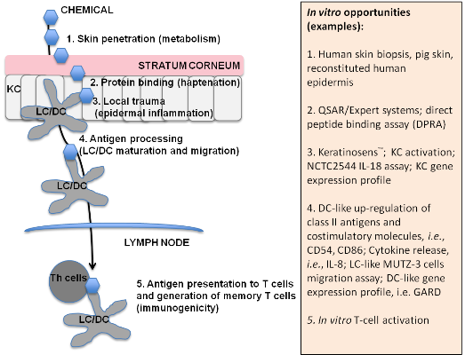
Chemical allergens are low molecular weight compounds (<1000 Dalton) too small to be seen by our immune system (hapten), therefore, they must bind to self-macromolecules to form a complete antigen. The complex formation is related to electrophilic reactivity and hydrophobicity of the allergen. Chemical allergens can be divided in three classes: (1) haptens; (2) pro-haptens, which require metabolic activation; and (3) pre-haptens, which spontaneously oxidize to form haptens. The metabolic competence of the in vitro system must be therefore carefully evaluated, as the lack of it may lead to false negative results.
ACD is a delayed type hypersensitivity reaction caused mainly by reactive T helper 1 and interferon (IFN)-γ producing CD8+ T cells (Tc1), which requires previous sensitization by the same chemicals [21,22]. In order for a chemical to induce skin sensitization, several key steps must be taken. Briefly, following skin absorption and the formation of the complete antigen, the development of ACD then requires the activation of innate immune cells, including keratinocyes (KC) required for maturation and migration of dendritic cells (DC), and DC, required for the activation of T cells. The acquisition of specific immune response will then take place at the level of draining lymph nodes, where DC migrate and stimulate the activation of hapten-specific responsive T-cells and the generation of Tc1 effector cells.
In Table 1, these key passages and the in vitro opportunities are reported. Some of these tests have been or are currently in prevalidation or have been submitted for consideration. In particular, the direct peptide reactivity assay (DPRA), which measures depletion of a cysteine- and a lysine-containing peptide in the presence of the test chemical [23], and the KeratinoSens™ assay [24], which is based on a luciferase reporter gene under the control of an anti-oxidant response element of the human AKR1C2 gene stably inserted into HaCaT keratinocytes, have successfully passed the prevalidation phase at EURL/ECVAM (European Union Reference Laboratory for alternatives to animal testing) , and recommendations have been published [25]. Soon, the human cell line activation assay (h-CLAT) [26], addressing CD86 and CD54 upregulation in THP-1 cells, will be available as well. The documents available online summarize the assayʼs mechanistic relevance, the performance as well as the applicability and limitations and makes recommendations for further work.
A number of assays for the in vitro identification of contact and respiratory sensitization have been developed within the integrated European Framework Program 6 Project Sens-it-iv, (LSHB-CT-2005-018681; 2005–2011) [27]. The Sens-it-iv Toolbox is the major deliverable of this project. In Table 2, the most advanced and promising assays developed within, or with contribution of Sens-it-iv are reported. Many of the tests listed in Table 2 have been submitted to the EURL-ECVAM for prevalidation.
One unifying characteristic of chemical allergens is the reactivity with proteins for the effective induction of skin sensitization. Most of chemical allergens are electrophilic and react with nucleophilic amino acids. One potential alternative approach to skin sensitization hazard identification is the use of (Quantitative) structure activity relationships ((Q)SARs) coupled with appropriate documentation and performance characteristics. A number of (Q)SARs and expert systems have been developed and are described in the literature, i.e., OECD (Organization for Economic Cooperation and Development) toolbox, ToxTree, TOPKAT, Derek, TOPS-MODE, etc. None of the systems appear to perform sufficiently well to act as a standalone tool for hazard identification, but they may be very useful within a structured decision support system as part of a safety assessment strategy [28]. Goebel et al. [13] offer a nice overview of published reactivity-based QSAR models and their use in non-animal safety assessment of skin sensitization.
Concerning chemical reactivity, in the DPRA, generally, non-allergens and weak allergens demonstrated minimal to low peptide reactivity, whereas moderate to extremely potent allergens displayed moderate to high peptide reactivity. Classifying minimal reactivity as non-sensitizers and low, moderate, and high reactivity as sensitizers, a prediction accuracy of 89% was reported [23].
Besides its barrier function, the skin has been recognized as an immunologically active tissue. KC may convert nonspecific exogenous stimuli into the production of cytokines, adhesion molecules and chemotactic factors [29]. After KC, Langerhans cells (LC) represent the second most prominent cell type in the epidermis (2%–5% of the epidermal population). LC are the principal antigen-presenting cells (APC) in the skin [30]. Due to their anatomical location and their significant role in the development of ACD, the use of both of these cell types to evaluate sensitizing potency in vitro is amply justifiable. In addition to chemical processing, LC activation and migration requires the binding of cytokines produced by KC as a result of initial chemical exposure. Furthermore, the irritant capacity of allergens might present an additional risk factor so that irritant allergens may be stronger allergens than non-irritant ones [31].
Starting from the in vivo observation that in mice, IL-1α expression by KC was selectively up-regulated after application of contact sensitizers but not tolerogen or irritant [32], similar results were reproduced in vitro using the murine KC cell line HEL30 [33]. Other authors [34] obtained similar results and, furthermore observed that the rank of potency was similar to the ranking established using the LLNA. Similarly, using human KC it has been demonstrated that allergens, but not irritants or tolerogens, induced IL-12 [35,36]. Among other cytokines produced by KC, IL-18 has been demonstrated to favor TH1 type immune responses by enhancing the secretion of pro-inflammatory mediators such as TNFα, IL-8, and IFNγ, and to play a key proximal role in the induction of allergic contact dermatitis. The increase in intracellular IL-18 content was used to discriminate contact allergens from low molecular weight respiratory allergens and irritants [37]. At non cytotoxic concentrations (cell viability >80% as assessed by MTT (3-(4,5-dimethylthiazol-2-yl)-2,5-diphenyltetrazolium bromide) reduction assay), all contact sensitizers tested induced a dose-dependent increase in IL-18, whereas both irritants and respiratory allergens failed, indicating that cell-associated IL-18 may provide an in vitro tool for identification and discrimination of contact vs. respiratory allergens and/or irritants [38,39,40]. A total of 33 chemicals were tested, with an overall accuracy of 89.7%.
In line with this, we recently published a multicenter study, involving different laboratories located in Europe and in the US, on the possibility to use the release of IL-18 in epidermal equivalent 3D models (EE) for the in vitro identification and ranking potency of contact allergens [41]. The assay provides a single test for the identification and classification of skin sensitizing chemicals, including chemicals with low water solubility or stability, which are problematic to use in traditional cell culture systems. A protocol was developed using different 3D-epidermal models including in house VUMC (VU University Medical Centre) model, epiCS® (previously EST1000TM), MatTek EpiDermTM and SkinEthicTM RHE. Following topical exposure for 24 h to 17 contact allergens and 13 non-sensitizers a robust increase in IL-18 release was observed only after exposure to contact allergens. A putative prediction model has been proposed from data obtained from two laboratories yielding 95% accuracy. Correlating the in vitro EE sensitizer potency data, which assesses the chemical concentration which results in 50% cytotoxicity (EE-EC50) with human and animal data showed a superior correlation with human DSA05 (μg/cm2) data (Spearman correlation r = 0.8500; p = 0.0061) compared to LLNA data (Spearman correlation r = 0.5968; p = 0.0542), where DSA05 represents the induction dose per skin area that produces a positive response in 5% of the tested population. Also a good correlation was observed for release of IL-18 (SI-2) into culture supernatants with human DSA05 data (Spearman correlation r = 0.8333; p = 0.0154). The advantage of the proposed assay would be the possibility to identify in the same test the allergenic potential of a chemical (by the amount of IL-18 release) and its potency (by its irritant potential). The in vitro reconstructed 3D epidermis model allows a chemical exposure that mimics human exposure (topical application), and overcomes all drawbacks of traditional submerged culture. Furthermore, 3D organotypic epidermis models possess in vivo-like barrier properties and metabolizing capabilities, providing significant benefits over monolayer culture models for the assessment of contact sensitization potential. This easily transferable human in vitro assay appears to be very promising, but additional testing of a larger chemical set is required to fully evaluate the usefulness of this assay and to establish a definitive prediction model.
Besides the EE model, it is important to note that other in vitro methods can also be used for potency classification [26,42]. A first indication of potency may come, for example, from the concentration required to induce a threshold of positive response (CD86 ≥ 150) in the h-CLAT system. A good correlation (r = 0.839, p < 0.01) was indeed found between the h-CLAT thresholds and LLNA EC3 values [42], if a chemical has a Minimum Induction Threshold >10 it should be ranked as strong sensitizer, if <10 as a weak.
The key passage 4 in chemical-induced skin sensitization involves DC maturation and migration. Upon antigen capture, the DC undergoes a maturation process leading to the up-regulation of co-stimulatory molecules (CD86, CD80, and CD40), MHC Class II molecules and the CD83 protein [43]. The establishment of human in vitro models of DC had offered the possibility to demonstrate that haptens were able to directly activate cultured DC [44,45,46,47]. A recent ECVAM workshop has reviewed the state of the art of the use of DC and human myeloid cell lines for the predictive identification of skin sensitization hazard [47]. Among the several endpoints investigated in different experimental models, CD86, IL-8, and p38 MAP kinase appear to be the most promising and robust biomarkers described to date in DC based assays [18,48,49].
Activated dendritic cells then migrate to the T-lymphocyte regions of lymphoid organs, where they lose antigen-processing activity and become potent immunostimulatory cells. Triggering of T cell responses to chemicals is the key event (key passage 5) that decides whether initial sensitization that results in transient inflammation in the target organ will lead to manifest disease upon subsequent contact with the chemical. Since T cells are the pathogenic effector in chemical-induced allergic contact dermatitis, respiratory allergy and adverse drug reaction, T cell-based in vitro assay are important to identify contact and respiratory allergens. In this context, the T cell priming assay represents a promising tool to analyze the human naïve T cell repertoire [50].
Although it should not be ruled out a priori that skin sensitization testing may, in the future, be addressed by one single method only, all methods currently under evaluation or evaluated for skin sensitization at EURL-ECVAM are not intended as a stand-alone methods, but for use within integrated approaches in combination with other information to predict the skin sensitization potential of chemicals and, where possible, the potency assessment of skin sensitizers. It is expected that in vitro data will be integrated via statistical correlation into a testing strategy along with the peptide reactivity data, bioavailability data, and some informed rating of structural alerts in order to establish an acceptable exposure level [51]. This will need to build experience in how to apply/integrate non-animal data to different exposure scenario for risk assessment decision-making. A range of integrated approaches to testing and assessment solutions may also be foreseen to cover different regulatory goals (i.e., hazard identification, classification, and potency assessment). The testing strategy will largely depend on information at hand, and it is expected to be chemical-specific [52].
It has been anticipated that it will take at least another 7–9 years for the full replacement of the in vivo animal models currently used to assess sensitization [20,52]. Nevertheless, these in vitro methods can already be used for hazard identification, i.e., to discriminate between sensitizers and non-sensitizers, and in the near future, potency classification for labeling is a realistic possibility.
Table 1. Key passages in chemical-induced skin sensitization and in vitro opportunities.
Key passage numberIn vitro opportunitiesReferences
1. Skin penetration and access to viable epidermisHuman skin biopsis, pig skin, reconstituted human epidermis[53,54]
2. Binding to macromolecules (haptenation)QSAR/Expert systems; Peptide binding assay (DPRA); allergen-peptide/protein interaction assay[23,28,54,55]
3. Local trauma: epidermal inflammation (danger signals)Keratinosens™; KC activation; NCTC2544 IL-18 assay; KC gene expression profile [24,33,34,35,36,37,38,39,40,56,57]
4. Antigen processing: dendritic cells maturation and migrationDC-like up-regulation of class II antigens and costimulatory molecules, i.e., CD54, CD86; Cytokine release, i.e., IL-8; LC-like MUTZ-3 cells migration assay; DC-like gene expression profile[26,41,42,43,44,45,46,47,58,59,60,61,62,63,64]
5. Antigen presentation to TH cells and memory T-cell generation (immunogenicity)In vitro T-cell activation[50,65,66]
Table 2. The Sens-it-iv Toolbox for skin sensitization.
Target cellsName of the assayN° of chemicalsTSFSOP
KeratinocytesNCTC2544 IL-18 assay33yesyes
Human Epidermal Equivalent30yesyes
Dendritic cellsGARD49yesyes
Maturation #1 (CD86, CD54, IL-8)Stopped--
Maturation # 2 (DotScan)20yesyes
Migration12yesyes
T cellsPrimary T-cell stimulation6yesE
OthersNeutrophils-THP-1 metabolization Proteomic marker profile12yesE
N°, number of chemicals tested; TSF, test submission form to EURL-ECVAM; SOP, standard operation procedure; E, evaluation of more compounds by the lead laboratory.

4. Conclusions

The growing political and practical pressure against the use of traditional animal tests to characterize chemical toxicity is the main driving force for the development of alternative methods for the screening and prioritization of toxicants, including immunotoxicity. Significant progress has been made in the last decade, and several alternative in vitro assays to detect contact hypersensitivity have the potential to reduce animal use and possibly testing cost.
However, for a full replacement of contact sensitization animal testing the following gaps still need to be filled:
  • Applicability domains: solubility, stability, activation, cost, use of serum, etc.
  • Lack of bio-availability information: real exposure at cellular level?
  • Better understanding the mechanisms defining potency
    Pathway analysis and marker signature identification
    Quantitative relation between marker signatures and potency of a chemical
    Quantitative and qualitative relation with T cell responses
  • Best Integrated Testing Strategy/ies need to be identified
There is a clear need for continued investment in the development of methods and approaches that will allow the correct identification in vitro of potential contact allergens and their potency. Intensive international and inter-laboratory cooperation and coordination will be necessary to reach this goal.
The different in vitro methods developed have inevitably some limitations as above outlined. The choice of the test will depend largely on the laboratory skills and resources, on eventual constrains dictated by internal policies, such as impossibility to use primary human cells or tissues or to use radioactive materials.
Even if it is true that continuous cell lines are not the physiological equivalents of primary cells, they can be valid surrogates, avoiding the use of living donors, the eventual shortage of donors and the unavoidable donor to donor variability. Some of the above mentioned methods are also ideal for high-throughput screening, which is critical for a rapid screening and prioritization of toxicants for further animal investigations.
As the underlying mechanisms of immunotoxicity are complex, see the involvement of several cells, chemical needs to comply several properties before an immunotoxic effect will be manifest. At present, a battery of tests is considered a more realistic approach to detect chemical-induced immunotoxicity.
The conclusion is that there is still much to be achieved, but the journey has been fascinating and there have been some very important developments. The hope is that continued investment in research will make in vitro immunotoxicity testing a reality in the twenty-first century.

Conflicts of Interest

The authors declare no conflict of interest.

References

  1. Germolec, D.E. Sensitivity and predictivity in immunotoxicity testing: Immune endpoints and disease resistance. Toxicol. Lett. 2004, 149, 109–114. [Google Scholar] [CrossRef]
  2. Peiser, M.; Tralau, T.; Heidler, J.; Api, A.M.; Arts, J.H.E.; Basketter, D.A.; English, J.; Diepgen, T.L.; Fuhlbrigge, C.; Gaspari, A.A.; et al. Allergic contact dermatitis: Epidemiology, molecular mechanisms, in vitro methods and regulatory aspects. Cell. Mol. Life Sci. 2012, 69, 763–781. [Google Scholar] [CrossRef]
  3. Sia, I.G.; Paya, C.V. Infectious complications following renal transplantation. Surg. Clin. N. Am. 1998, 78, 95–112. [Google Scholar] [CrossRef]
  4. Klein, N.C.; Go, C.H.; Cunha, B.A. Infections associated with steroid use. Infect. Dis. Clin. N. Am. 2001, 15, 423–432. [Google Scholar] [CrossRef]
  5. Sleijffers, A.; Garssen, J.; van Loveren, H. Ultraviolet radiation, resistance to infectious diseases, and vaccination responses. Methods 2002, 28, 111–121. [Google Scholar]
  6. Via, T.; Descotes, J. Immunosuppressive drugs and cancer. Toxicology 2003, 185, 229–240. [Google Scholar] [CrossRef]
  7. Esser, C.; Jux, B. Small chemicals, bioactivation, and the immune system—A fragile balance of i-tox and benefits? Che. Biodivers. 2009, 6, 2138–2143. [Google Scholar]
  8. Dietert, R.R. Role of developmental immunotoxicity and immune dysfunction in chronic disease and cancer. Reprod. Toxicol. 2011, 31, 319–326. [Google Scholar] [CrossRef]
  9. Organization for Economic Co-Operation and Development (OECD). Skin Sensitisation: Local Lymph Node Assay. In OECD Guidelines for the Testing of Chemicals; No. 429; OECD: Paris, France, 2010. [Google Scholar]
  10. Kimber, I.; Weisenberger, C. A murine local lymph node assay for the identification of contact allergens. Assay development and results of an initial validation study. Arch. Toxicol. 1989, 63, 274–282. [Google Scholar] [CrossRef]
  11. Basketter, D.A.; Balikie, L.; Dearman, R.J.; Kimber, I.; Ryan, C.A.; Gerberick, G.F.; Harvey, P.; Evans, P.; White, I.R.; Rycroft, R.J.G. Use of the local lymph node assay for the estimation of relative contact allergenic potency. Contact Dermat. 2000, 42, 344–348. [Google Scholar]
  12. Basketter, D.A.; Kimber, I. Assessing the potency of respiratory allergens: Uncertainties and challenges. Reg. Toxicol. Pharmacol. 2011, 61, 365–373. [Google Scholar] [CrossRef]
  13. Goebel, C.; Aeby, P.; Ade, N.; Alépée, N.; Aptula, A.; Araki, D.; Dufour, E.; Gilmour, N.; Hibatallah, J.; Keller, D.; et al. Guiding principles for the implementation of non-animal safety assessment approaches for cosmetics: Skin sensitisation. Reg. Toxicol. Pharmacol. 2012, 63, 40–52. [Google Scholar] [CrossRef]
  14. Luebke, R. Immunotoxicity screening and prioritization in the twenty-first Century. Toxicol. Pathol. 2012, 40, 294–299. [Google Scholar] [CrossRef]
  15. United Environmental Protection Agency. Available online: http://www.epa.gov/ncct/toxcas (accessed on 14 March 2014).
  16. Houck, K.A.; Dix, D.J.; Judson, R.S.; Kavlock, R.J.; Yang, J.; Berg, E.L. Profiling bioactivity of the ToxCast chemical library using BioMAP primary human cell systems. J. Biomol. Screen 2009, 14, 1054–1066. [Google Scholar] [CrossRef]
  17. Hartung, T.; Corsini, E. Immunotoxicology: Challenges in the 21st century and in vitro opportunities. ALTEX 2013, 30, 411–426. [Google Scholar] [CrossRef]
  18. Galbiati, V.; Mitjans, M.; Corsini, E. Present and future of in vitro immunotoxicology in drug development. J. Immunotoxicol. 2010, 7, 255–267. [Google Scholar] [CrossRef]
  19. Kimber, I.; Basketter, D.A.; Gerberick, G.F.; Ryan, C.A.; Dearman, R.J. Chemical allergy: Translating biology into hazard characterization. Toxicol. Sci. 2011, 120, S238–S268. [Google Scholar] [CrossRef]
  20. Aeby, P.; Ashikaga, T.; Bessou-Touya, S.; Schepky, A.; Gerberick, F.; Kern, P.; Marrec-Fairley, M.; Maxwell, G.; Ovigne, J.-M.; Sakaguchi, H.; et al. Identifying and characterizing chemical skin sensitizers without animal testing: Colipaʼs research and method development program. Toxicol. in Vitro 2010, 24, 1465–1473. [Google Scholar]
  21. McFadden, J.P.; Puangpet, P.; Basketter, D.A.; Dearman, R.J. Why does allergic contact dermatitis exist? Br. J. Dermatol. 2013, 168, 692–699. [Google Scholar] [CrossRef]
  22. Martin, S.F. Allergic contact dermatitis: Xenoinflammation of the skin. Curr. Opin. Immunol. 2012, 24, 720–729. [Google Scholar] [CrossRef]
  23. Gerberick, G.F.; Vassallo, J.D.; Foertsch, L.M.; Price, B.B.; Chaney, J.G.; Lepoittevin, J.-P. Quantification of chemical peptide reactivity for screening contact allergens: A classification tree model approach. Toxicol. Sci. 2007, 97, 417–427. [Google Scholar] [CrossRef]
  24. Emter, R.; Ellis, G.; Natsch, A. Performance of a novel keratinocyte-based reporter cell line to screen skin sensitizers in vitro. Toxicol. Appl. Pharmacol. 2010, 245, 281–290. [Google Scholar]
  25. Joint Research Centre. Available online: http://ihcp.jrc.ec.europa.eu/our_labs/eurl-ecvam/eurl-ecvam-recommendations (accessed on 14 March 2014).
  26. Sakaguchi, H.; Ashikaga, T.; Miyazawa, M.; Kosaka, N.; Ito, Y.; Yoneyama, K.; Sono, S.; Itagaki, H.; Toyoda, H.; Suzuki, H. The relationship between CD86/CD54 expression and THP-1 cell viability in an in vitro skin sensitization test—Human cell line activation test (h-CLAT). Cell Biol. Toxicol. 2009, 25, 109–126. [Google Scholar] [CrossRef]
  27. Roggen, E.L. Application of the acquired knowledge and implementation of the Sens-it-iv toolbox for identification and classification of skin and respiratory sensitizers. Toxicol. in Vitro 2013, 27, 1122–1126. [Google Scholar]
  28. Gerberick, F.; Aleksic, M.; Basketter, D.; Casati, S.; Karlberg, A.-T.; Kern, P.; Kimber, L.; Lepoittevin, J.P.; Natsch, A.; Ovigne, J.M.; et al. Chemical reactivity measurement and the predictive identification of skin sensitizers. Altern. Lab. Anim. 2008, 36, 215–242. [Google Scholar]
  29. Barker, J.N.W.N.; Griffiths, C.E.M.; Nickoloff, B.J.; Mitra, R.S.; Dixit, V.M. Keratinocytes as initiators of inflammation. Lancet 1991, 337, 211–214. [Google Scholar] [CrossRef]
  30. Katz, S.I.; Tamaki, K.; Sachs, D.H. Epidermal Langerhans cells are derived from cells originating in bone marrow. Nature 1979, 282, 324–326. [Google Scholar] [CrossRef]
  31. Bonneville, M.; Chavagnac, C.; Vocanson, M.; Rozieres, A.; Benetiere, J.; Pernet, I.; Denis, A.; Nicolas, J.-F.; Hennino, A. Skin contact irritation conditions the development and severity of allergic contact dermatitis. J. Invest. Dermatol. 2007, 127, 1430–1435. [Google Scholar]
  32. Enk, A.H.; Katz, S.I. Early molecular events in the induction phase of contact sensitivity. Proc. Natl. Acad. Sci. USA 1992, 89, 1398–1402. [Google Scholar] [CrossRef]
  33. Corsini, E.; Primavera, A.; Marinovich, M.; Galli, C.L. Selective induction of cell-associated IL-1α in murine keratinocytes by chemical allergens. Toxicology 1998, 129, 193–200. [Google Scholar] [CrossRef]
  34. Van Och, F.M.; van Loveren, H.; van Wolfswinkel, J.C.; Machielsen, A.J.C.; Vandebriel, R.J. Assessment of potency of allergenic activity of low molecular weight compounds based on IL-1α and IL-18 production by a murine and human keratinocyte cell line. Toxicology 2005, 210, 95–109. [Google Scholar] [CrossRef]
  35. Muller, G.; Saloga, J.; Germann, T.; Bellinghausen, I.; Mohamadzadeh, M.; Knop, J.; Enk, A.H. Identification and induction of human keratinocyte-derived IL-12. J. Clin. Invest. 1994, 94, 1799–1805. [Google Scholar] [CrossRef]
  36. Corsini, E.; Limiroli, E.; Marinovich, M.; Galli, C.L. Selective induction of IL-12 by chemical allergens in reconstituted human epidermis. Altern. Lab. Anim. 1999, 27, 261–269. [Google Scholar]
  37. Corsini, E.; Mitjans, M.; Galbiati, V.; Lucchi, L.; Galli, C.L.; Marinovich, M. Use of IL-18 production in a human keratinocyte cell line to discriminate contact sensitizers from irritants and low molecular weight respiratory allergens. Toxicol. in Vitro 2009, 23, 789–796. [Google Scholar] [CrossRef]
  38. Galbiati, V.; Mitjans, M.; Lucchi, L.; Viviani, B.; Galli, C.L.; Marinovich, M.; Corsini, E. Further development of the NCTC 2544 IL-18 assay to identify in vitro contact allergens. Toxicol. in Vitro 2011, 25, 724–732. [Google Scholar] [CrossRef]
  39. Corsini, E.; Galbiati, V.; Mitjans, M.; Galli, C.L.; Marinovich, M. NCTC 2544 and IL-18 production: A tool for the identification of contact allergens. Toxicol. in Vitro 2014, 28, 13–17. [Google Scholar] [CrossRef]
  40. Gibbs, S.; Corsini, E.; Spiekstra, S.W.; Galbiati, V.; Fuchs, H.W.; DeGeorge, G.; Troese, M.; Patrick, H.; Deng, W.; Roggen, E. An epidermal equivalent assay for identification and ranking potency of contact sensitizers. Toxicol. Appl. Pharmacol. 2013, 272, 529–541. [Google Scholar] [CrossRef]
  41. Mitjans, M.; Viviani, B.; Lucchi, L.; Galli, C.L.; Marinovich, M.; Corsini, E. Role of p38 MAPK in the selective release of IL-8 induced by chemical allergen in naive THP-1 cells. Toxicol. in Vitro 2008, 22, 386–395. [Google Scholar] [CrossRef]
  42. Caux, C.; Massacrier, C.; Vanbervliet, B.; Dubois, B.; van Kooten, C.; Durand, I.; Banchereau, J. Activation of human dendritic cells through CD40 cross-linking. J. Exp. Med. 1994, 180, 1263–1272. [Google Scholar] [CrossRef]
  43. Aiba, S.; Terunuma, A.; Manome, H.; Tagami, H. Dendritic cells differently respond to haptens and irritants by their production of cytokines and expression of co-stimulatory molecules. Eur. J. Immunol. 1997, 27, 3031–3038. [Google Scholar] [CrossRef]
  44. Degwert, J.S.; Hoppe, U.; Kligman, L.H. In vitro model for contact sensitization: I. Stimulatory capacities of human blood-derived dendritic cells and their phenotypical alterations in the presence of contact sensitizers. Toxicol. in Vitro 1997, 11, 613–618. [Google Scholar] [CrossRef]
  45. Rougier, N.; Redziniak, G.; Mougin, D.; Schmitt, D.; Vincent, C. In vitro evaluation of the sensitization potential of weak contact allergens using Langerhans-like dendritic cells and autologous T-cells. Toxicology 2000, 145, 73–82. [Google Scholar] [CrossRef]
  46. De Smedt, A.C.A.; van den Heuvel, R.L.; Zwi Berneman, N.; Schoeters, G.E.R. Modulation of phenotype, cytokine production and stimulatory function of CD34+-derived DC by NiCl2 and SDS. Toxicol. in Vitro 2001, 15, 319–325. [Google Scholar] [CrossRef]
  47. Weigt, H.; Muhlradt, P.F.; Larbig, M.; Krug, N.; Braun, A. The toll-like receptor-2/6 agonist macrophage-activating lipopeptide-2 cooperates with IFN-γ to reverse the Th2 skew in an in vitro allergy model. J. Immunol. 2004, 172, 6080–6086. [Google Scholar]
  48. Casati, S.; Aeby, P.; Basketter, D.A.; Cavani, A.; Gennari, A.; Gerberick, G.F.; Griem, P.; Hartung, T.; Kimber, I.; Lepoittevin, J.-P.; et al. Dendritic cells as a tool for the predictive identification of skin sensitisation hazard. Altern. Lab. Anim. 2005, 33, 47–62. [Google Scholar]
  49. Dos Santos, G.G.; Reinders, J.; Ouwehand, K.; Rustemeyer, T.; Scheper, R.J.; Gibbs, S. Progress on the development of human in vitro dendritic cell based assays for assessment of the sensitizing potential of a compound. Toxicol. Appl. Pharmacol. 2009, 236, 372–382. [Google Scholar] [CrossRef]
  50. Martin, S.F.; Esser, P.R.; Schmucker, S.; Dietz, L.; Naisbitt, D.J.; Park, B.K.; Vocanson, M.; Nicolas, J.-F.; Keller, M.; Pichler, W.J.; et al. T-cell recognition of chemicals, protein allergens and drugs: Towards the development of in vitro assays. Cell. Mol. Life Sci. 2010, 67, 4171–4184. [Google Scholar] [CrossRef]
  51. Jaworska, J.; Harol, A.; Kern, P.S.; Gerberick, G.F. Integrating non-animal test information into an adaptive testing strategy—Skin sensitization proof of concept case. Altex 2011, 28, 211–225. [Google Scholar] [CrossRef]
  52. Adler, S.; Basketter, D.; Creton, S.; Pelkonen, O.; van Benthem, J.; Zuang, V.; Andersen, K.E.; Angers-Loustau, A.; Aptula, A.; Bal-Price, A.; et al. Alternative (non-animal) methods for cosmetics testing: Current status and future prospects-2010. Arch. Toxicol. 2011, 85, 367–485. [Google Scholar] [CrossRef]
  53. Organization for Economic Co-operation and Development (OECD). Skin Absorption: In Vitro Method. In OECD Guidelines for the Testing of Chemicals; No. 428; OECD: Paris, France, 2004. [Google Scholar]
  54. Basketter, D.A.; Pease, C.; Kasting, G.; Kimber, I.; Casati, S.; Cronin, M.; Diembeck, W.; Gerberick, F.; Hadgraft, J.; Hartung, T.; et al. Skin sensitisation and epidermal disposition: The relevance of epidermal disposition for sensitisation hazard identification and risk assessment. Altern. Lab. Anim. 2007, 35, 137–154. [Google Scholar]
  55. Patlewicz, G.; Aptula, A.O.; Uriarte, E.; Roberts, D.W.; Kern, P.S.; Gerberick, G.F.; Kimber, I.; Dearman, R.J.; Ryan, C.A.; Basketter, D.A. An evaluation of selected global (Q)SARs/expert systems for the prediction of skin sensitization potential. SAR QSAR Environ. Res. 2007, 18, 515–541. [Google Scholar] [CrossRef]
  56. Dos Santos, G.G.; Spiekstra, S.W.; Sampat-Sardjoepersad, S.C.; Reinders, J.; Scheper, R.J.; Gibbs, S. A potential In vitro epidermal equivalent assay to determine sensitizer potency. Toxicol. in Vitro 2011, 25, 347–357. [Google Scholar] [CrossRef]
  57. Vandebriel, R.J.; Pennings, J.L.; Baken, K.A.; Pronk, T.E.; Boorsma, A.; Gottschalk, R.; van Loveren, H. Keratinocyte gene expression profiles discriminate sensitizing and irritating compounds. Toxicol. Sci. 2010, 117, 81–89. [Google Scholar] [CrossRef]
  58. Dietz, L.; Kinzebach, S.; Ohnesorge, S.; Franke, B.; Goette, I.; Koenig-Gressel, D.; Thierse, H.-J. Proteomic allergen-peptide/protein interaction assay for the identification of human skin sensitizers. Toxicol. in Vitro 2013, 27, 1157–1162. [Google Scholar]
  59. Ashikaga, T.; Hoya, M.; Itagaki, H.; Katsumura, Y.; Aiba, S. Evaluation of CD86 expression and MHC calss II molecule internalization in THP-1 human monocytic cells as predictive endpoints for contact sensitizers. Toxicol. in Vitro 2002, 16, 711–716. [Google Scholar] [CrossRef]
  60. Azam, P.; Peiffer, J.L.; Chamousset, D.; Tissier, M.-H.; Bonnet, P.-A.; Vian, L.; Fabre, I.; Ourlin, J.-C. The cytokine-dependent MUTZ-3 cell line as an in vitro model for the screening of contact sensitizers. Toxicol. Appl. Pharmacol. 2006, 212, 14–23. [Google Scholar] [CrossRef]
  61. Arrighi, J.F.; Rebsamen, M.; Rousset, F.; Kindler, V.; Hauser, C. A critical role for p38 mitogen-activated protein kinase in the maturation of human blood-derived dendritic cells induced by lipopolysaccharide, TNF-α, and contact sensitizers. J. Immunol. 2001, 166, 3837–3845. [Google Scholar]
  62. Yoshida, Y.; Skaguchi, H.; Ito, Y.; Okuda, M.; Suzuki, H. Evaluation of the skin sensitization potential of chemicals using expression of co-stimulatory molecules, CD54 and CD86, on the naive THP-1 cell line. Toxicol. in Vitro 2003, 17, 221–228. [Google Scholar] [CrossRef]
  63. Rees, B.; Spiekstra, S.W.; Carfi, M.; Ouwehand, K.; Williams, C.A.; Corsini, E.; McLeod, J.D.; Gibbs, S. Inter-laboratory study of the in vitro dendritic cell migration assay for identification of contact allergens. Toxicol. in Vitro 2011, 25, 2124–2134. [Google Scholar]
  64. De Fraissinette, A.B.; Cordier, A.; Ulrich, P. Modulation of the activity of human monocyte-derived dendritic cells by chemical haptens, a metal allergen, and a staphylococcal superantigen. Toxicol. Sci. 1999, 52, 189–198. [Google Scholar]
  65. Gorbachev, A.V.; Fairchild, R.L. Induction and regulation of T-cell priming for contact hypersensitivity. Crit. Rev. Immunol. 2001, 21, 451–472. [Google Scholar]
  66. Rustemeyer, T.; de Ligter, S.; von Blomberg, B.M.; Frosch, P.J.; Scheper, R.J. Human T-lymphocyte priming in vitro by haptenated autologous dendritic cells. Clin. Exp. Immunol. 1999, 117, 209–216. [Google Scholar]

