Article

Physicochemical and Biological Activity Analysis of Low-Density Polyethylene Substrate Modified by Multi-Layer Coatings Based on DLC Structures, Obtained Using RF CVD Method

1
Faculty of Materials Science and Ceramics, AGH University of Science and Technology, A. Mickiewicza Av. 30, 30 059 Kraków, Poland
2
Department of Technologies and Materials, Faculty of Mechanical Engineering, Technical University in Kosice, Masiarska 74, 040 01 Kosice, Slovakia
3
Institute of Materials Science and Engineering, Łódz University of Technology, Stefanowskiego Str. 1/15, 90 924 Łódz, Poland
4
Faculty of Chemistry, Jagiellonian University, Gronostajowa 2, 30 387 Kraków, Poland
*
Author to whom correspondence should be addressed.
Coatings 2018, 8(4), 135; https://doi.org/10.3390/coatings8040135
Submission received: 6 March 2018 / Revised: 31 March 2018 / Accepted: 5 April 2018 / Published: 10 April 2018
(This article belongs to the Special Issue Functional Ceramic Coatings)

Abstract

:
In this paper, the surface properties and selected mechanical and biological properties of various multi-layer systems based on diamond-like carbon structure deposited on low-density polyethylene (LDPE) substrate were studied. Plasma etching and layers deposition (incl. DLC, N-DLC, Si-DLC) were carried out using the RF CVD (radio frequency chemical vapor deposition) method. In particular, polyethylene with deposited N-DLC and DLC layers in one process was characterized by a surface hardness ca. seven times (up to ca. 2.3 GPa) higher than the unmodified substrate. Additionally, its surface roughness was determined to be almost two times higher than the respective plasma-untreated polymer. It is noteworthy that plasma-modified LDPE showed no significant cytotoxicity in vitro. Thus, based on the current research results, it is concluded that a multilayer system (based on DLC coatings) obtained using plasma treatment of the LDPE surface can be proposed as a prospective solution for improving mechanical properties while maintaining biocompatibility.

1. Introduction

Polymers such as polyethylene (PE) are the most widely used materials for medical applications due to their properties (i.e., high flexibility, low density, high chemical resistance, biocompatibility) [1,2]. However, its medical application often requires a surface modification and enhancement of the surface properties (i.e., low surface hardness). Therefore, special surface treatments must be applied to improve the physicochemical properties. This can be achieved using thin layer technology, including oxygen and nitrogen plasma discharge [3], laser irradiation [4,5], deposition of anti-wear and/or functional coatings (i.e., diamond-like carbon (DLC)) [6,7,8] and immobilization of biopolymers (e.g., chitosan and its derivatives) [9,10]. Since plasma treatment results in the generation of high-energy species such as radicals, ions, or molecules in an excited electronic state, this enables surface reactions to take place and leads to surface activation and modification [11]. Such plasma techniques can transform PE into a valuable material for medical applications due to the modification of the surface without any interference in the substrate interior [12].
In the case of biomedical applications, the pre-treated processes (i.e., plasma etching) have a huge impact on the coating properties, biocompatibility, and enhancement of cell attachment [13]. Plasma treatment is the most versatile surface modification technique and involves electron-induced excitations, ionization, and dissociation to facilitate the production of tailored surface properties such as wettability, roughness, and many more chemical, physical, and biological properties of the polymer surface. For enhanced adhesion, the surface free energy of the polymer should be larger than that of the substrate it will be bonded with. This is why surface modification of polyethylene is required to increase the surface free energy, which corresponds to a decrease of contact angle. In addition, it is very beneficial for many applications to obtain protective and gas barrier coatings (based on DLC structure) as well as obtaining the most biocompatible surfaces (based on DLC and chitosan structures) on the polymeric substrate [7,9,14,15,16]. DLC layers are characterized by high hardness (up to 30 GPa) and a high Young’s modulus, but usually also high internal stresses (up to 7 GPa). These properties are related to the presence of a sp3 C fraction in the structure [17]. However, the stresses can be reduced by incorporating other atoms into the structure (i.e., Si, O, N, F [18,19,20] or metals [21,22]). It is worth mentioning that the reduction in stresses is often associated with a reduction in hardness and elastic modulus of the layers [6,23].
In this paper, the RF CVD (radio frequency chemical vapor deposition) method was used to modify the physicochemical parameters of LDPE (low-density polyethylene) substrate. The experimental part consisted of different approaches to studying various multi-layer systems (i.e., DLC, N-DLC layers, or Si-DLC layers). Every time, the polymer substrate was first treated and functionalized by plasma etching using Ar+ ions. Precise characterization before and after surface modification was performed, presented, and discussed. The surface properties of the modified PE were determined by scanning electron microscopy (SEM) with chemical composition analysis (EDX). Atomic structure and topography were examined by infrared spectroscopy (Fourier transform infrared-attenuated total reflectance (FTIR-ATR)) and atomic force microscopy (AFM), respectively. The nanoindentation method was applied to assess hardness and Young’s modulus profiles. Cytotoxicity in vitro against the MG-63 cell line was evaluated by Alamar Blue assay. Additionally, the influence of surface modification on wettability and surface free energy of modified polyethylene substrate was also examined.

2. Materials and Methods

2.1. Sample Preparation and Surface Treatment

The material used in this study was low-density polyethylene prepared in the form of regular samples (width/length/height—10 mm/7 mm/4 mm). The average chemical composition of this material was 99.5 at.% carbon and 0.5 at.% oxygen (according to EDS analysis, EDAX Genesis, EDAX Inc., Mahwah, NJ, USA) and provided by Sigma-Aldrich (Karlsruhe, Germany). The oxygen content determined on the LDPE surface is probably the result of the adsorption of this element on the surface in air atmosphere. Before coating deposition on PE substrates, the samples were chemically purified in isopropanol and subjected to Ar+ ion pre-treatment (etching process) in plasma conditions. The process of PE surface modification was performed in a RF CVD reactor (Elettrorava S.p.A., Turin, Italy), which generated plasma using radio-frequency discharge (13.56 MHz) under low-pressure conditions. The distance between the electrodes (cathode–anode) was 20 mm. All plasma processes (treatment and layers deposition) on the PE substrate were performed at room temperature (297 K) due to the low thermal resistance (melting point 383 K) of polyethylene. The treatment in the RF reactor of the polyethylene surface (ion etching) was conducted for all samples in order to prepare the substrates for further modification processes and surface purification from adsorbed gases (e.g., O2). This stage of processing was conducted in Ar flow conditions (75 cm3/min) under a pressure of 53 Pa and plasma density of 0.08 W/cm2 for 10 min. The experiments consisted of four independent series of PE surface modification, including the deposition of DLC, N-DLC, and Si-DLC coatings, as well as multilayer systems. These processes were carried out in accordance with the technical parameters presented in Table 1.
In addition, the unmodified substrate (PE_0 series) was also tested to compare the obtained test results. The obtained samples were characterized using techniques adequate for material engineering as well as the evaluation of biological activity.

2.2. Surface Characterization and Mechanical Study

The surface microstructure and chemical composition of unmodified and coated polyethylene was examined using scanning electron microscopy (NOVA NANO SEM 200, FEI, Hillsboro, OR, USA) with energy dispersive X-ray spectroscopy analysis (EDX). In the case of the detection of light elements (i.e., C, N, and O), an accelerating voltage of 5 eV was applied. In addition, the thickness of the obtained layers was examined based on the cross-section of the tested samples. Furthermore, the topography of tested samples was investigated using atomic force microscopy (AFM, Bruker, Santa Barbara, CA, USA) equipped with peak force tapping mode with a MultiMode 8 (Bruker, Santa Barbara, CA, USA) microscope with a Sb-doped silicon tip of 8 nm diameter. The chemical structure of the polymer surface was examined using FTIR-ATR (Fourier transform infrared) spectroscopy on a Bio-Rad FTS60 V device (Digilab Division, Cambridge, MA, USA). The spectra were measured within 400–4000 cm−1, 275 scans, and 4 cm−1 resolution.
Contact angle measurements were conducted using the sessile drop technique performed on a DSA10Mk2 (Kruss, Hamburg, Germany) analyzer. Wettability and surface energy measurements were made using ultra-high-quality water (UHQ—water produced with the use of UHQ-PS, Elga, Buckinghamshire, UK) and diiodomethane (Aldrich, Taufkichen, Germany) droplets with a volume of 0.2 μL. For each tested sample, five independent measurements were carried out on the surface.
Indentation technique with G200 Nano Indenter® (MTS, Oak Ridge, TN, USA) equipped with Berkovich-type diamond tip was used to assess the mechanical properties of the surface. Profiles of hardness and modulus of elasticity were acquired by the continuous stiffness measurement (CSM) method, where the maximum penetration depth was set to 2 µm.

2.3. Cytotoxicity

Human osteoblast-like MG-63 cell lines (ATCC: CRL-1427) were cultured in DMEM (Dulbecco's Modified Eagle’s Medium) medium (Immuniq, Żory, Poland) without phenol red with the addition of 10% fetal bovine serum (FBS) and 1% streptomycin/penicillin (Gibco-BRL, Life Technologies, Karlsruhe, Germany) in standard cell conditions (37 °C, 5% CO2). Cytotoxicity assay was conducted for both pristine polyethylene and modified samples. The samples were sterilized overnight in ethanol vapor. Then, the samples were placed in a sterile 24-well culture plate and a cell suspension with a density of 50 × 104 cells/0.2 mL was added. Cells without the tested samples were treated as a reference sample. Treated and untreated cells were incubated for 72 h. Every 24 h, the medium was changed for a fresh one. After that time, cytotoxicity was determined by Alamar Blue assay (Sigma-Aldrich, Bornem, Belgium) according to the well-described protocol in [24]. In brief, cells were washed with phosphate-buffered saline (PBS) and incubated with resazurin sodium salt solution (25 µM in PBS) for 4 h at 37 °C in the dark. The fluorescence caused by the cellular metabolic activity was measured at 605 nm (excitation wavelength 560 nm) with a multimode microplate reader (Infinite 200M PRO NanoQuant, Tecan, Männedorf, Switzerland). Cytotoxicity was expressed as a percentage of viable cells after treatment with coated polyethylene samples in reference to untreated cells (control). The experiment was repeated three times. Cell morphology was examined using a fluorescence microscope (Olympus IX51, Olympus, Tokyo, Japan) with an excitation filter of 470/20 nm. After staining with acridine orange (viable cells) and propidium iodide (dead cells), cells were observed for morphological changes. At least five viewing fields containing ca. 100 cells each were analyzed. Photographs of CT26 cells after treatment with the tested alloys were taken using an inverted microscope equipped with a reflected fluorescence system (Olympus IX51, Olympus, Tokyo, Japan).

3. Results

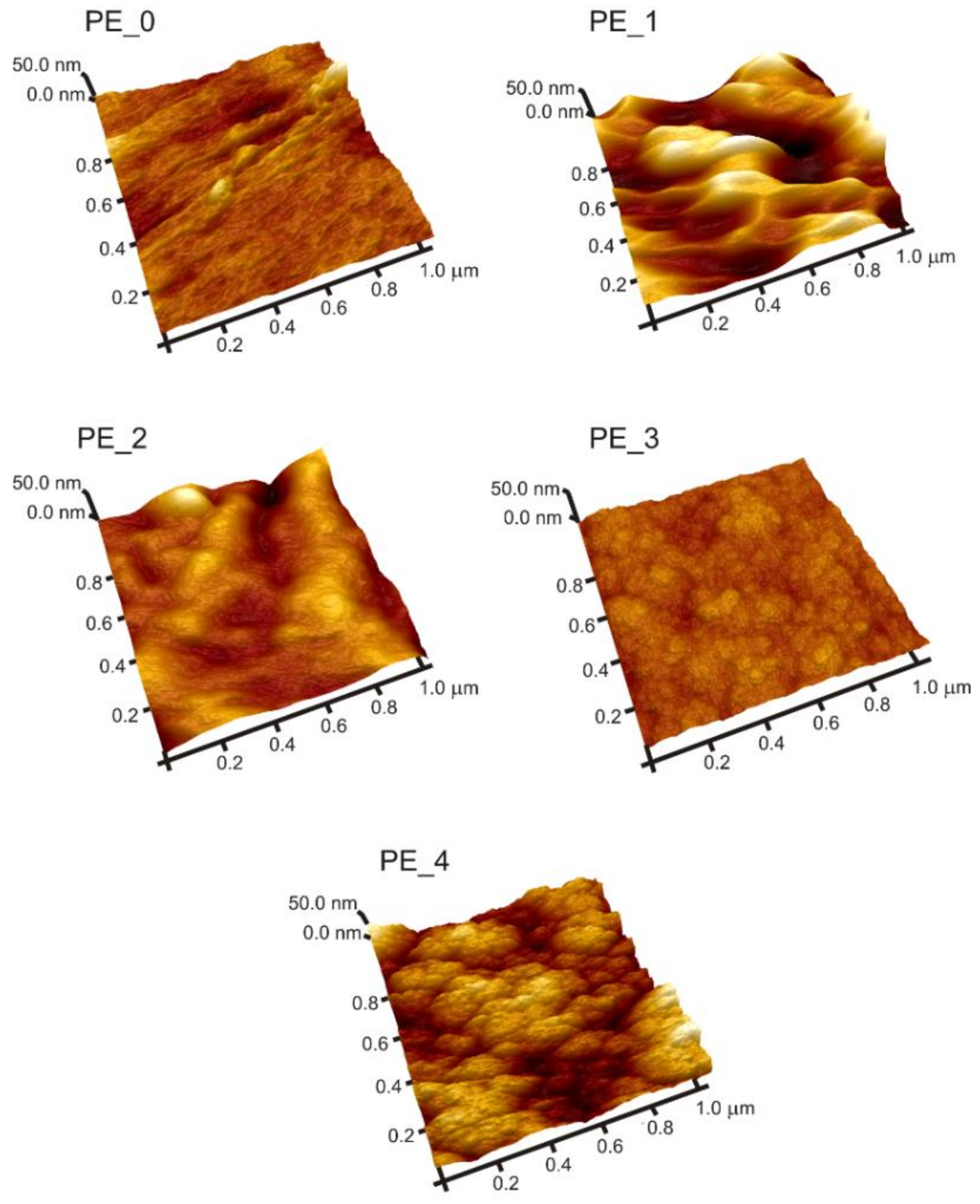
3.1. Morphological Analysis

SEM and AFM techniques were used to obtain detailed information about morphological and topographical changes of polyethylene induced by plasma treatment and deposition of DLC-based coatings. It is worth noting that air and oxygen plasma treatments are more aggressive than argon plasma. Even though the ions of these gases are reactive and aggressive in contact with the surface layer of the polymeric substrate [25], these atmospheres are also used to clean the surface prior to the coating deposition. For instance, Rohrbeck et al. [26] applied an oxygen plasma cleaning process (10 min, 200 W), and after such treatment the initially smooth polymer surface turned out to be considerably rougher, and trenches and holes were more pronounced. However, in our work, the etching process with application of argon (less-reactive gas than oxygen and air) was carried out under the plasma power of 8 W, and in these conditions no significant negative influence of temperature on LDPE was observed. SEM analysis (Figure 1) revealed that each surface modification resulted in the formation of continuous and homogenous structures on the surface, without any cracks. Only in the case of modifications with DLC layer deposition (PE_1 series), could a more diverse microstructure be observed, with visible heterogeneities in the micro scale.
The new formed structures showed more details with atomic force microscopy, at the nanometric scale (Figure 2).
Analysis of AFM images of modified substrates showed granular-like structures, which in the case of Si-DLC coatings were composed of agglomerated clusters (see Figure 2, sample PE_4).
A similar effect was observed Catena et al. when DLC layers were deposited on polyethylene [1]. In this respect, it is worth mentioning that plasma treatment of PE surfaces (with coating deposition) caused an increase in the surface area of tested samples, which is also beneficial for cell adhesion. The characteristic bulges (observed in sample PE_3 and PE_4) are similar to those presented by Catena et al. [27], caused by intrinsic stress release phenomena. More details concerning the surface roughness values of all samples, their chemical composition, and layer thicknesses are presented in Table 2.
It is important that determined roughness values (Ra) for samples after coatings deposition were similar in the case of one-layer modifications (ca. 24–30 nm) as well as for the two-layer ones (ca. 13–16 nm). These values were two to over three times higher than the value of this parameter for the unmodified polyethylene (ca. 9 nm). On one hand, the plasma treatment contributed to the increase in surface roughness of the PE substrate, and on the other hand it influenced the surface structure of the modified samples. The increases of surface roughness value after plasma processes are in good agreement with results obtained by Novotná et al. [28].
The chemical composition of tested samples (series PE_1–PE_4 in Table 2) confirms that the obtained coatings consisted of C, N, and Si elements, depending on the chemical composition of the gas mixture during plasma processes in the RF reactor. In the case of PE_2 series (with the N-DLC coating), nitrogen was incorporated into the structure to ca. 8 at.%, while Si atoms (for the PE_4 series) in the Si-DLC structure to ca. 27 at.%. It is worth noting that the addition of N and Si atoms to the diamond-like carbon structure caused a decrease in the value of internal stresses inside the obtained coatings as well as their hardness, which was also observed in other works [29,30,31]. However, the presence of silicon above (ca. 16 at.%) positively affected the anti-bacterial properties of the DLC coatings as well, which was also confirmed by Bociaga et al. [19]. The chemical composition studies of the tested samples revealed the presence of oxygen atoms in the structure, up to ca. 3 at.% in the case of the PE_1, PE_2, and PE_3 series.
The growth of oxygen concentration after plasma treatments was strongly affected by the creation of polar oxygen groups, which was also concluded by Novotná et al. [28]. In the case of PE_4, the thickness of Si-DLC layer was above 1 µm and the N-DLC layer was out of range of EDS analysis. This can be explained by the absence of nitrogen in the average content (at.%). In the case of PE_1, PE_2 and PE_3 series oxygen appeared in EDX analysis (up to ca. 3 at.%), possibly as the result of the adsorption of this element after the coating deposition process at ambient air conditions. In the case of the series with a Si-DLC layer (PE_4), the content of oxygen atoms was much higher (ca. 20 at.%), which can be associated with a large silicon content in the DLC structure, and therefore increased compliance for the incorporation of oxygen into the top surface of the modified PE substrate. The presence of oxygen was attributed to the surface oxidation. This process was also observed by Batory et al. [32]. The confirmation of this fact was by the presence of the Si-O atomic groups in the IR spectra as well as the highest range of surface hardness for obtained Si-DLC layers (the tested PE_4 sample, vide infra Figure 3 and see Section 3.4). This is mainly due to the very high binding energy for Si–O (ca. 532 eV), compared to the value for C–H (ca. 338.5 kJ/mol) and Si–H (ca. 298.7 kJ/mol) [33]. Additionally, Si–H bonds were less stable than C–H bonds, which also confirms the incorporation of oxygen into the Si-DLC structure.

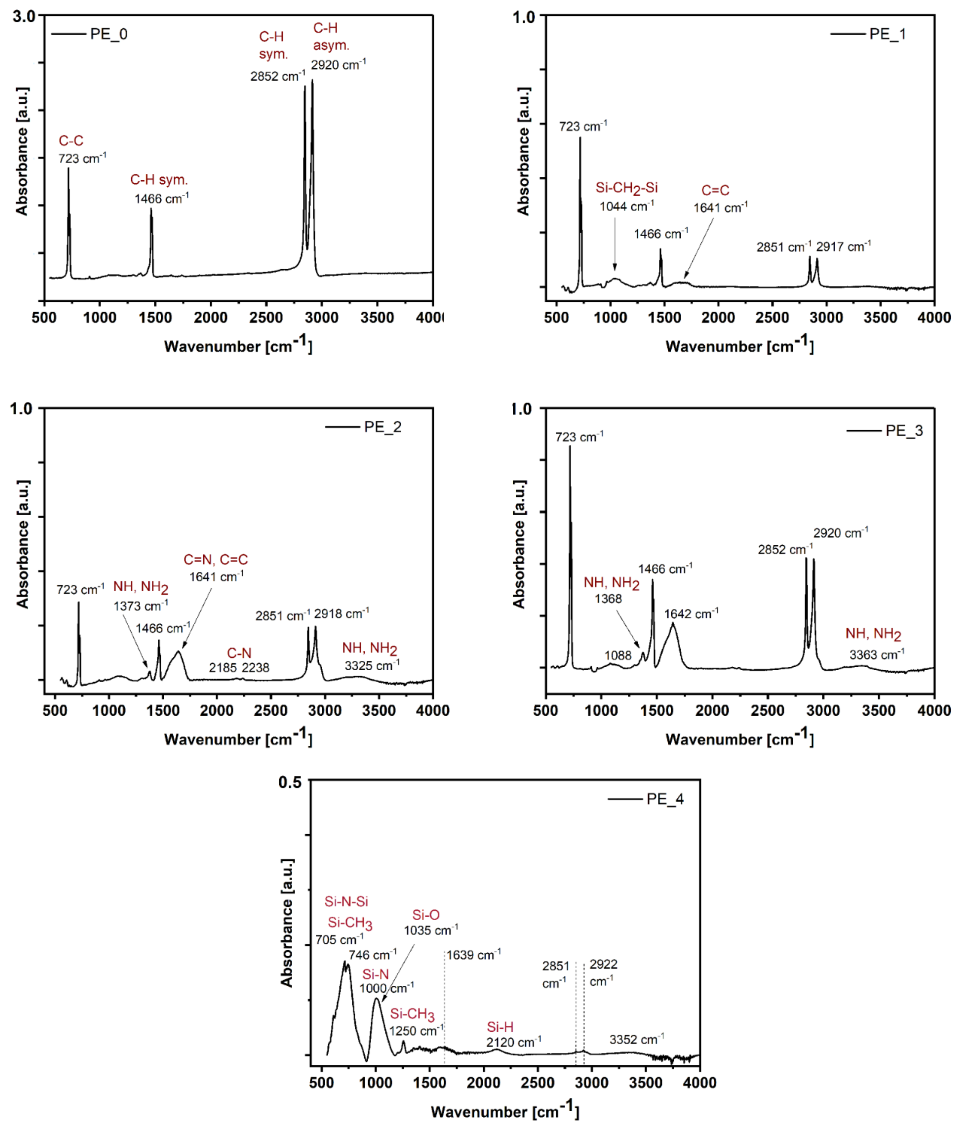
3.2. FTIR-ATR Analysis

The modification of the PE surface led to significant changes in atomic structure, which was confirmed by the FTIR-ATR method. Obtained results are shown in Figure 3.
The IR spectra of unmodified PE (PE_0 series) showed two large peaks at 2920 and 2852 cm−1, which correspond to C–H asymmetric and symmetric stretching vibrations in the CH2 group, respectively. Two smaller absorption peaks at 1466 and 723 cm−1 can be identified as C–H symmetric and C–C bonds. It can be clearly concluded that this spectra is characteristic for unmodified polyethylene [2]. Deposition of DLC coating (see Figure 3—PE_1) noticeably changed the IR spectra of pure PE, the peaks assigned to C–H and C–C vibrations decreased, and the reordered spectra are typical for DLC structures. This modification also caused the appearance of a spectral line at 1641 cm−1 that was assigned to vibrations in C=C groups.
In the case of the next modification (PE_2 series), due to the obtained N-DLC layer, the new spectra lines were centered at 3325 cm−1 (assigned to NH and NH2 groups, in the energy range 3300–3400 cm−1) [34,35] and additionally confirmed by a peak at ca. 1373 cm−1 [36]. Furthermore, the relatively wide peak (in comparison to spectra for PE_1 series, centered at 1641 cm−1) was also attributed to C=N bonds vibrations [37,38], while two weak spectral lines (for 2185 and 2238 cm−1) were attributed to stretching vibrations in C–N groups [39].
In the case of PE substrate modification with the deposition of two layers (N-DLC/DLC, PE_3 series), the highest intensity (about two times higher than the IR spectra for PE_2 series) of spectral lines was assigned to C–H, C=C, and C–C vibration groups.
The last surface modification of polyethylene substrate (after deposition of N-DLC/Si-DLC layers, PE_4 series) resulted in significant changes to the obtained IR spectra. The spectra were dominated by various atomic groups containing Si atoms, including Si–H (2120 cm1) [40], Si–CH3 (1250 cm1) [41], Si–N (750–1050 cm1) [36], and Si–CH2–Si (1090–1020 cm1) vibrations [42]. In addition, in the range of 600 cm1 to 850 cm1, many vibration modes, Si–C stretching, Si–N–Si asymmetrical stretching, CH3–Si rocking-stretching, and Si–H bending [42,43] can be noticed. The high value of oxygen content in this case (ca. 21 at.%, based on EDS analysis) is probably associated with vibrations in the Si–O in Si–O–Si groups, which was assigned to the 1035 cm1 spectral line [41]. It is noteworthy that in the case of modification with Si-DLC layers, the high dissociation energy of the Si–O bonding (798 kJ/mol) [44] resulted in a significant increase in the mechanical resistance of the modified surface.

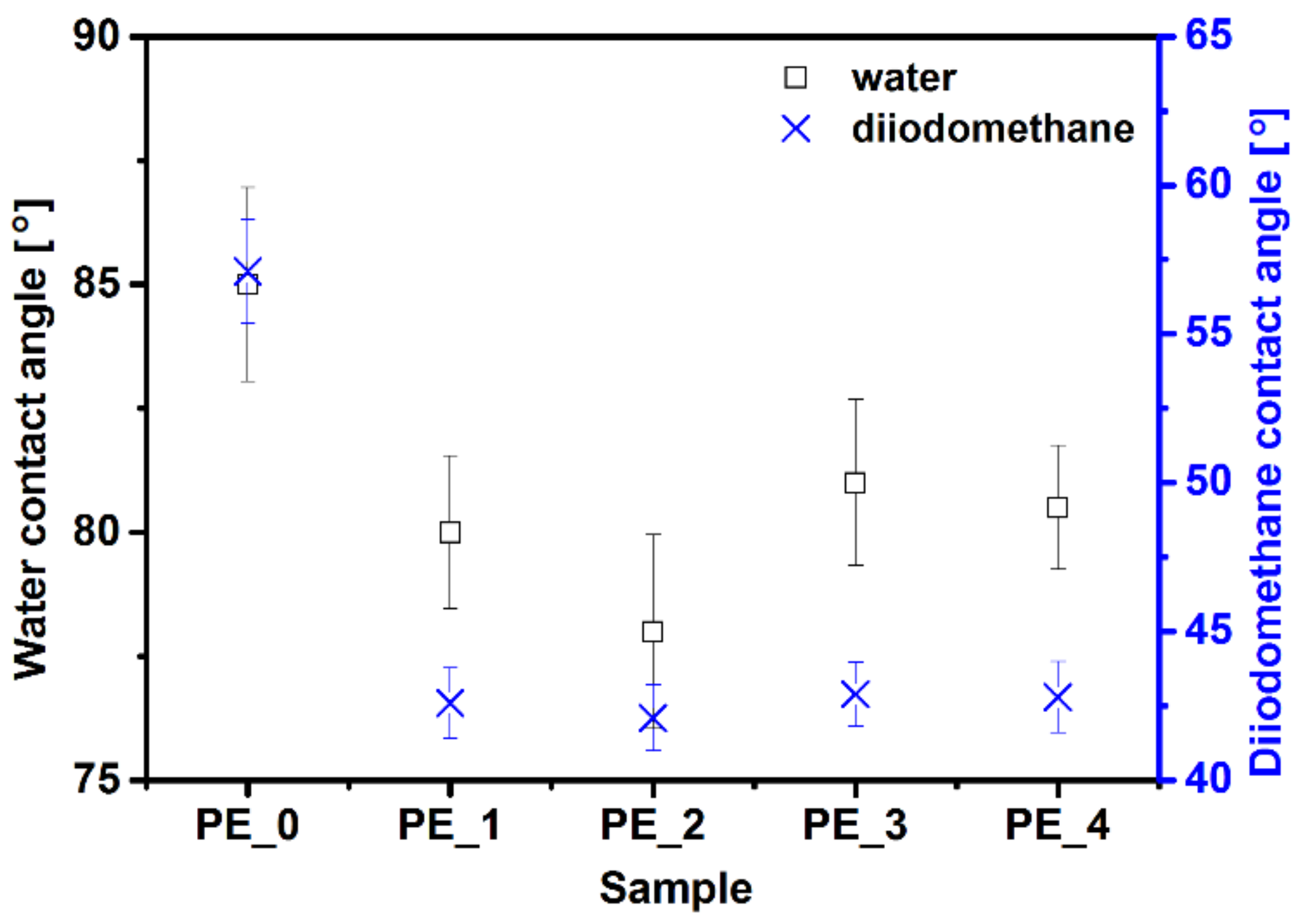
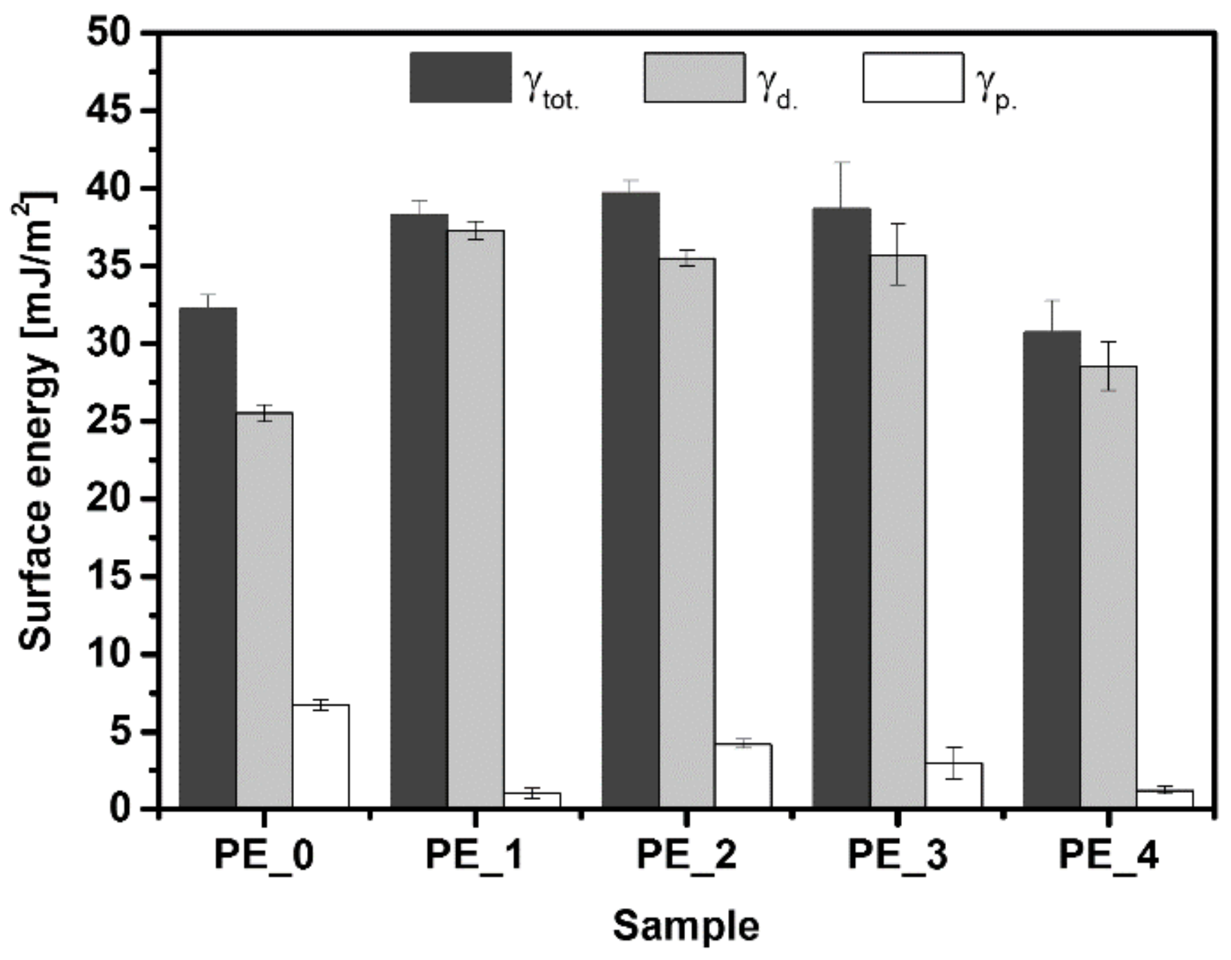
3.3. Contact Angle and Surface Energy Analysis

Unmodified polyethylene is a low-energy hydrophobic material which must be modified in order to be useful in biomedical applications. Wettability is one of the most important surface parameters for biomedical applications, because hydrophilic material with higher surface energy favors cell adhesion and biocompatibility [4]. The contact angle value of untreated polyethylene (PE_0 series) was determined to be high (ca. 85°) for two measuring liquids (water and diiodomethane), see Figure 4.
A significant decrease in contact angle values after the deposition of DLC-based structures was observed for all tested series, while the contact angle for diiodomethane was lower than for water. A similar effect after the deposition of two different diamond-like carbon structures (flexible-DLC and robust-DLC) was described by Catena et al. [1]. This shows that in most cases plasmochemical treatment causes the contact angle to decrease, which was discussed broadly in many papers [2,10,45,46]. Polyethylene with N-DLC coating (PE_2 series) is probably the best for biomedical applications, because it exhibits low and comparable water and diiodomethane contact angles.
Figure 5 shows results of surface free energy obtained for the tested samples, including polar and dispersive components of SFE. The influence of plasma treatment for LDPE on surface free energy was also described by Pandiyaraj et al. [47,48].
The authors concluded that usually in such processes oxygen flow results in an increase in the polar component (by incorporation of polar functional groups), without significantly changing the dispersive component. In our case, the performed experiments demonstrated that after deposition of DLC-based structures, the dispersive component increased, while the polar component decreased in relation to unmodified PE (PE_0 series). For example, in the case of modification with undoped DLC coatings (PE_1 series), the dispersive and polar components of surface free energy increased up to 37.5 mJ/m2d.) and decreased to 1.0 mJ/m2p.), respectively. Despite the fact that the total surface energy of all modified samples increased considerably, the best results (the highest γtot. value and the lowest contact angle value) were obtained for the PE_2 series. This leads to the conclusion that the deposition of DLC layers (mainly N-DLC) can improve biocompatibility by increasing the surface energy of the substrate.

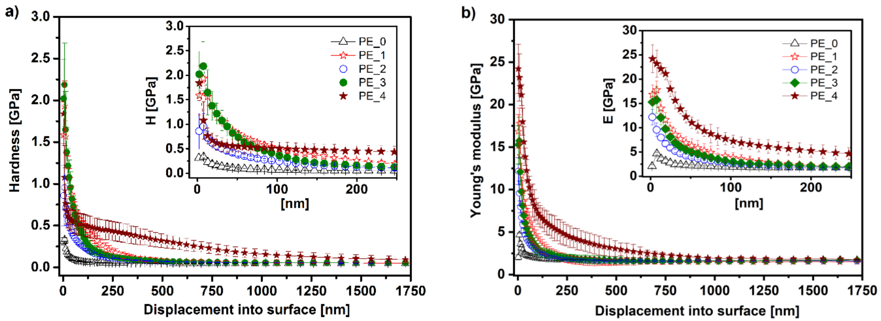
3.4. Mechanical Analysis

Surface modification of the LDPE surface under plasmochemical conditions improves its mechanical properties. The hardness and Young’s modulus profiles in relation to displacement into the surface are shown in Figure 6.
Deposition of DLC-based coatings generally improves hardness (by up to nine times), especially at a distance of 600 nm from the surface (Figure 6a). The unmodified polyethylene (sample PE_0) was characterized by increased hardness only up to about 50 nm displacement into the surface (hardness of 0.3 GPa) and then stabilized at a value of ca. 0.1 GPa. In the case of the DLC layer obtained on the PE substrate (PE_1 series), we observed a hardness increase of up to ca. 2 GPa. The DLC structure doped with N atoms (N-DLC coating, PE_2 series) was characterized by lower hardness than the previous one (PE_1), the surface hardness achieved a value of ca. 1 GPa, and the strengthening remained at ca. 550 nm distance from the surface. The PE_3 series, corresponding to N-DLC/DLC multilayer, exhibited the highest surface hardness of up to 2.3 GPa, at a similar distance from the surface as in the case of the PE_2 series (ca. 600 nm). As a result, the addition of N to the structure of DLC caused a decrease in layer hardness compared to undoped DLC coatings. A comparable relationship was also observed for the addition of Si, but only at a distance of about 50 nm from the surface. Similar dependencies were observed by Ruijun et al. [49] and Wang et al. [50]. For the tested samples, the highest strengthening range (up to ca. 1750 nm from the surface) was shown in the PE_4 series, after deposition of the N-DLC/Si-DLC coating. In this case, the hardness increased to ca. 1.8 GPa.
Similar relationships were observed for the Young’s modulus of tested samples (Figure 6b). Unmodified polyethylene reached a maximum value of 5 GPa near the surface, while in the interior it was about 2 GPa, which is a typical value for polyethylene. First modifications (DLC, N-DLC, and DLC/N-DLC corresponding to PE_1–PE_3) caused significant alterations in the Young’s modulus of the samples (18 GPa for PE_1, 12 GPa for PE_2, and 16 GPa for PE_3), but these modifications increased these values only up to about 200 nm displacement into the surface. Again, the best mechanical properties were exhibited by the PE_4 series (N-DLC/Si-DLC modification), with a maximum Young’s modulus value of 25 GPa on the surface. Increased E modulus in relation to unmodified PE remained escalated for about 1000 nm. This value is substantial, because it is the closest result to bone stiffness, which can be related to the enhanced biocompatibility required for implants.

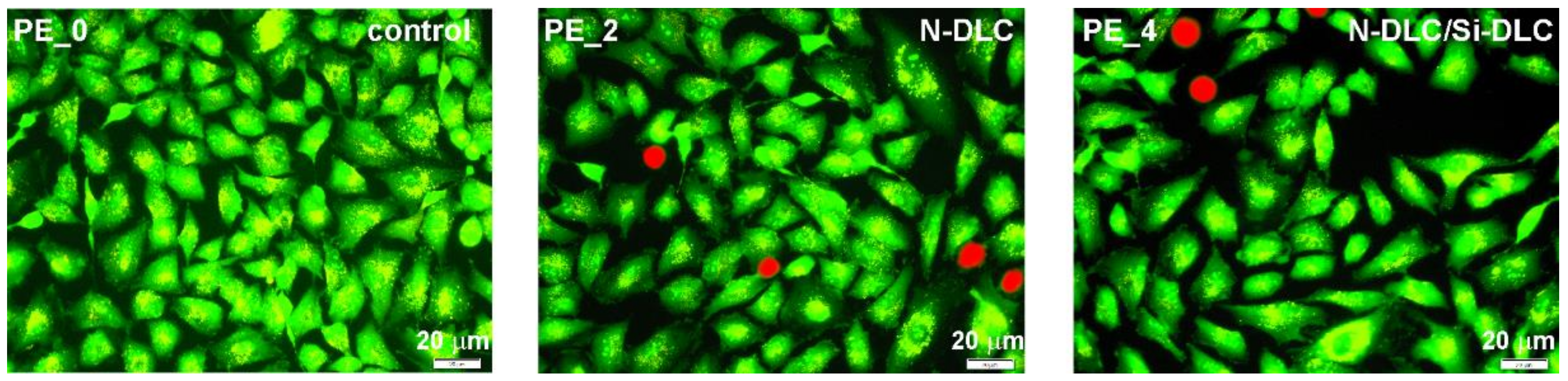
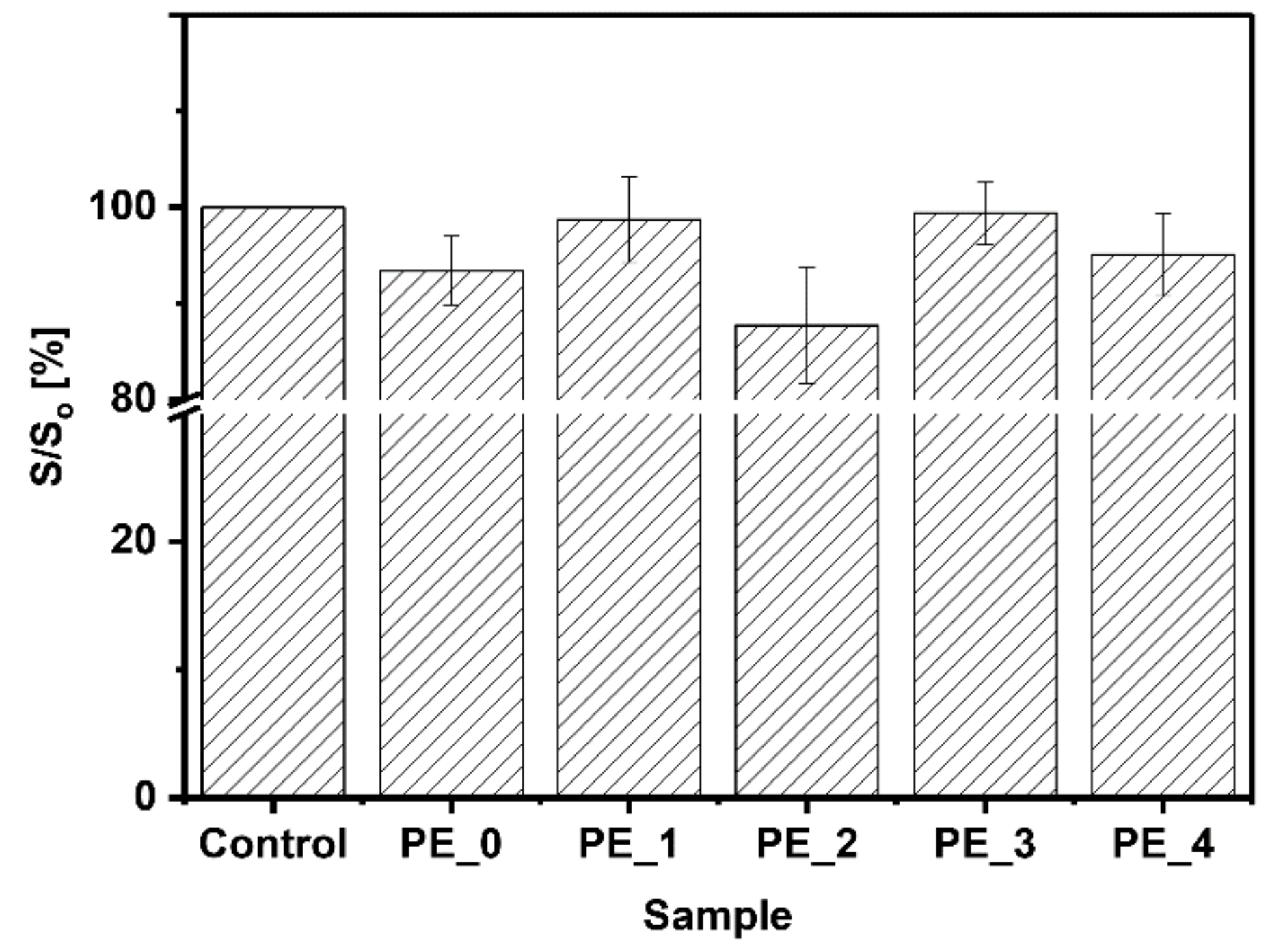
3.5. Cytotoxicity Assay

In vitro cytotoxicity was evaluated on human osteoblast-like MG-63 cells after 72 h. No significant changes in cell viability were observed in all tested series. As shown in Figure 7, the decrease in cell survival after treatment for all tested samples was assessed to be not higher than 12%.
The most significant cytotoxicity (ca. 12%) showed only LDPE substrate after deposition of N-DLC layer on the top (series PE_2). This elevated cytotoxicity can be explained by a high content of N (ca. 8 at.%) in the modified surface.
Additionally, detailed analysis based on cellular morphology observation under fluorescent microscopy (Figure 8) did not reveal any significant changes in mitochondrial shape and size, and no apoptotic bodies were formed after this long incubation time.
Additionally, PE_3 modification (with the deposition of N-DLC/DLC layers) resulted in comparable biocompatibility to the control (untreated MG-63 cells) with significant improvement of mechanical properties (vide supra Figure 6).
The proposed DLC, N-DLC, or Si-DLC coatings on polymeric substrate were less cytotoxic than studied by us previously (DLC layers doped with N and Si atoms and deposited on titanium alloy). The latter coatings influenced the viability of the treated MG-63 cells, decreasing the cell surviving fraction by even up ca. 29% (modification C; after nitriding process and Si/N-DLC deposition) [51]. It can be concluded that the application of N-DLC or Si-DLC resulted in lower cytotoxic effect than the addition of N and Si atoms to the DLC structure during coating deposition.

4. Conclusions

On the basis of the performed experiments, it can be concluded that the modification of the LDPE in plasma conditions obtaining multi-layer coatings based on DLC structures resulted in profitable changes of physicochemical and biological properties for their medical applications. In particular, it allowed for a required hardness gradient on the modified surface to be obtained, and as a result made the polymer surface harder and less elastic. It was shown that all modifications in which DLC structures (doped and undoped with Si or N atoms) were obtained improved the wettability of the polymer surface, which is an important property in osseointegration processes. Importantly, in the case of N-DLC/DLC (PE_3 series) and DLC/Si-DLC (PE_4 series) multi-layers deposition on the LDPE surface, a significant improvement in hardness was observed—up to ca. 2.3 and 2.5 GPa, respectively. In addition, a high content of Si (ca. 27 at.%) in the top layer of PE_4 series enabled the incorporation of oxygen in the DLC structure after the modification process at ambient air conditions. All obtained surface modifications resulted in considerable increase in the mechanical properties of the tested surface by improving the PE substrate strengthening at a distance of ca. 1.7 µm from the surface. It is noteworthy that for all tested series no significant changes in cell viability in vitro were observed.

Acknowledgments

This work has been supported by the statutory research of Department of Physical Chemistry and Modeling of Faculty of Materials Science and Ceramics, AGH-UST (subject number 11.11.160.768).

Author Contributions

Karol Kyzioł conceived the article concept, designed and performed the experiments, and wrote the paper; Julia Oczkowska analyzed the data and prepared part of the manuscript; Daniel Kottfer, Marek Klich, Łukasz Kaczmarek and Zbigniew Grzesik analyzed the data, participated in discussion; Agnieszka Kyzioł designed and performed biological experiments and analyzed the obtained results.

Conflicts of Interest

The authors declare no conflict of interest.

References

  1. Catena, A.; Agnello, S.; Rösken, L.M.; Bergen, H.; Recktenwald, E.; Bernsmann, F.; Busch, H.; Cannas, M.; Gelardi, F.M.; Hahn, B.; et al. Characteristics of industrially manufactured amorphous hydrogenated carbon (a-C:H) depositions on high-density polyethylene. Carbon N. Y. 2016, 96, 661–671. [Google Scholar] [CrossRef]
  2. De Geyter, N.; Morent, R.; Leys, C. Surface characterization of plasma-modified polyethylene by contact angle experiments and ATR-FTIR spectroscopy. Surf. Interface Anal. 2008, 40, 608–611. [Google Scholar] [CrossRef]
  3. Junkar, I.; Vesel, A.; Cvelbar, U.; Mozetič, M.; Strnad, S. Influence of oxygen and nitrogen plasma treatment on polyethylene terephthalate (PET) polymers. Vacuum 2009, 84, 83–85. [Google Scholar] [CrossRef]
  4. Slepicka, P.; Kasalkova, N.S.; Siegel, J.; Kolska, Z.; Bacakova, L.; Svorcik, V. Nano-structured and functionalized surfaces for cytocompatibility improvement and bactericidal action. Biotechnol. Adv. 2015, 33, 1120–1129. [Google Scholar] [CrossRef] [PubMed]
  5. Riveiro, A.; Soto, R.; Del Val, J.; Comesaña, R.; Boutinguiza, M.; Quintero, F.; Lusquiños, F.; Pou, J. Laser surface modification of ultra-high-molecular-weight polyethylene (UHMWPE) for biomedical applications. Appl. Surf. Sci. 2014, 302, 236–242. [Google Scholar] [CrossRef]
  6. Love, C.A.; Cook, R.B.; Harvey, T.J.; Dearnley, P.A.; Wood, R.J.K. Diamond like carbon coatings for potential application in biological implants—A review. Tribol. Int. 2013, 63, 141–150. [Google Scholar] [CrossRef]
  7. Fischer, C.B.; Rohrbeck, M.; Wehner, S.; Richter, M.; Schmeißer, D. Interlayer formation of diamond-like carbon coatings on industrial polyethylene: Thickness dependent surface characterization by SEM, AFM and NEXAFS. Appl. Surf. Sci. 2013, 271, 381–389. [Google Scholar] [CrossRef]
  8. Choudhury, D.; Morita, T.; Sawae, Y.; Lackner, J.M.; Towler, M.; Krupka, I. A Novel functional layered diamond-like carbon coating for orthopedics applications. Diam. Relat. Mater. 2015, 61, 56–69. [Google Scholar] [CrossRef]
  9. Xin, Z.; Hou, J.; Ding, J.; Yang, Z.; Yan, S.; Liu, C. Surface functionalization of polyethylene via covalent immobilization of O-stearoyl-chitosan. Appl. Surf. Sci. 2013, 279, 424–431. [Google Scholar] [CrossRef]
  10. Pandiyaraj, K.N.; Ferraria, A.M.; Rego, A.M.B.D.; Deshmukh, R.R.; Su, P.; Halleluyah, J.M.; Halim, A.S. Low-pressure plasma enhanced immobilization of chitosan on low-density polyethylene for bio-medical applications. Appl. Surf. Sci. 2015, 328, 1–12. [Google Scholar] [CrossRef]
  11. Holmes, C.; Tabrizian, M. Surface Functionalization of Biomaterials; Elsevier Inc.: New York, NY, USA, 2014; pp. 187–206. [Google Scholar]
  12. Švorčík, V.; Kotál, V.; Slepička, P.; Macková, A.; Novotná, M.; Hnatowicz, V. Modification of surface properties of polyethylene by Ar plasma discharge. Nucl. Instrum. Methods Phys. Res. Sect. B Beam Interact. Mater. Atoms. 2006, 244, 365–372. [Google Scholar] [CrossRef]
  13. Chung, T.W.; Liu, D.Z.; Wang, S.Y.; Wang, S.S. Enhancement of the growth of human endothelial cells by surface roughness at nanometer scale. Biomaterials 2003, 24, 4655–4661. [Google Scholar] [CrossRef]
  14. Ray, S.C.; Mukherjee, D.; Sarma, S.; Bhattacharya, G.; Mathur, A.; Roy, S.S.; McLaughlin, J.A. Functional diamond like carbon (DLC) coatings on polymer for improved gas barrier performance. Diam. Relat. Mater. 2017, 80, 59–63. [Google Scholar] [CrossRef]
  15. Dufils, J.; Faverjon, F.; Héau, C.; Donnet, C.; Benayoun, S.; Valette, S. Evaluation of a variety of a-C:H coatings on PEEK for biomedical implants. Surf. Coat. Technol. 2017, 313, 96–106. [Google Scholar] [CrossRef]
  16. Ashtijoo, P.; Bhattacherjee, S.; Sutarto, R.; Hu, Y.; Yang, Q. Fabrication and characterization of adherent diamond-like carbon based thin films on polyethylene terephthalate by end hall ion beam deposition. Surf. Coat. Technol. 2016, 308, 90–97. [Google Scholar] [CrossRef]
  17. Grill, A. Diamond-like carbon: State of the art. Diam. Relat. Mater. 1999, 8, 428–434. [Google Scholar] [CrossRef]
  18. Zhang, T.F.; Wan, Z.X.; Ding, J.C.; Zhang, S.; Wang, Q.M.; Kim, K.H. Microstructure and high-temperature tribological properties of Si-doped hydrogenated diamond-like carbon films. Appl. Surf. Sci. 2018, 435, 963–973. [Google Scholar] [CrossRef]
  19. Bociaga, D.; Kaminska, M.; Sobczyk-Guzenda, A.; Jastrzebski, K.; Swiatek, L.; Olejnik, A. Surface properties and biological behaviour of Si-DLC coatings fabricated by a multi-target DC-RF magnetron sputtering method for medical applications. Diam. Relat. Mater. 2016, 67, 41–50. [Google Scholar] [CrossRef]
  20. Batory, D.; Jedrzejczak, A.; Kaczorowski, W.; Szymanski, W.; Kolodziejczyk, L.; Clapa, M.; Niedzielski, P. Influence of the process parameters on the characteristics of silicon-incorporated a-C:H:SiOx coatings. Surf. Coat. Technol. 2015, 271, 112–118. [Google Scholar] [CrossRef]
  21. Bociaga, D.; Jakubowski, W.; Komorowski, P.; Sobczyk-Guzenda, A.; Jędrzejczak, A.; Batory, D.; Olejnik, A. Surface characterization and biological evaluation of silver-incorporated DLC coatings fabricated by hybrid RF PACVD/MS method. Mater. Sci. Eng. C 2016, 63, 462–474. [Google Scholar] [CrossRef] [PubMed]
  22. Usman, A.; Rafique, M.S.; Khaleeq-ur-Rahman, M.; Siraj, K.; Anjuma, S.; Latifa, H.; Khan, T.M.; Mehmood, M. Growth and characterization of Ni:DLC composite films using pulsed laser deposition technique. Mater. Chem. Phys. 2011, 126, 649–654. [Google Scholar] [CrossRef]
  23. Dean, J.; Aldrich-Smith, G.; Clyne, T.W. Use of nanoindentation to measure residual stresses in surface layers. Acta Mater. 2011, 59, 2749–2761. [Google Scholar] [CrossRef]
  24. Vega-Avila, E.; Pugsley, M.K. An overview of colorimetric assay methods used to assess survival or proliferation of mammalian cells. Proc. West. Pharmacol. Soc. 2011, 54, 10–14. [Google Scholar] [CrossRef] [PubMed]
  25. Couto, E.; Tan, I.H.; Demarquette, N.; Caraschi, J.C.; Leão, A. Oxygen plasma treatment of sisal fibers and polypropylene: Effects on mechanical properties of composites. Polym. Eng. Sci. 2002, 42, 790–797. [Google Scholar] [CrossRef]
  26. Rohrbeck, M.; Körsten, S.; Fischer, C.B.; Wehner, S.; Kessler, B. Diamond-like carbon coating of a pure bioplastic foil. Thin Solid Films 2013, 545, 558–563. [Google Scholar] [CrossRef]
  27. Catena, A.; Agnello, S.; Cannas, M.; Gelardi, F.M.; Wehner, S.; Fischer, C.B. Evolution of the sp2 content and revealed multilayer growth of amorphous hydrogenated carbon (a-C:H) films on selected thermoplastic materials. Carbon 2017, 117, 351–359. [Google Scholar] [CrossRef]
  28. Novotná, Z.; Rimpelová, S.; Ju, P.; Veselý, M.; Kolská, Z.; Václav, Š. The interplay of plasma treatment and gold coating and ultra-high molecular weight polyethylene: On the cytocompatibility. Mater. Sci. Eng. C 2017, 71, 125–131. [Google Scholar] [CrossRef] [PubMed]
  29. Chouquet, C.; Gerbaud, G.; Bardet, M.; Barrat, S.; Billard, A.; Sanchette, F.; Ducros, C. Structural and mechanical properties of a-C:H and Si doped a-C:H thin films grown by LF-PECVD. Surf. Coat. Technol. 2010, 204, 1339–1346. [Google Scholar] [CrossRef]
  30. Charitidis, C.A. Nanomechanical and nanotribological properties of carbon-based thin films: A review. Int. J. Refract. Met. Hard Mater. 2010, 28, 51–70. [Google Scholar] [CrossRef]
  31. Ray, S.C.; Pong, W.F.; Papakonstantinou, P. Iron, nitrogen and silicon doped diamond like carbon (DLC) thin films: A comparative study. Thin Solid Films 2016, 610, 42–47. [Google Scholar] [CrossRef]
  32. Batory, D.; Jedrzejczak, A.; Szymanski, W.; Niedzielski, P.; Fijalkowski, M.; Louda, P.; Kotela, I.; Hromadka, M.; Musil, J. Mechanical characterization of a-C:H:SiOx coatings synthesized using radio-frequency plasma-assisted chemical vapor deposition method. Thin Solid Films 2015, 590, 299–305. [Google Scholar] [CrossRef]
  33. Ong, S.E.; Zhang, S.; Du, H.; Sun, D. Relationship between bonding structure and mechanical properties of amorphous carbon containing silicon. Diam. Relat. Mater. 2007, 16, 1628–1635. [Google Scholar] [CrossRef]
  34. De Graaf, A.; Dinescu, G.; Longueville, J.L.; Van de Sanden, M.C.M.; Schram, D.C.; Dekempeneer, E.H.A.; Van Ijzendoorn, L.J. Amorphous hydrogenated carbon nitride films deposited via an expanding thermal plasma at high growth rates. Thin Solid Films 1998, 333, 29–34. [Google Scholar] [CrossRef]
  35. Kyzioł, K.; Koper, K.; Kaczmarek, Ł.; Grzesik, Z. Plasmochemical modification of aluminum-zinc alloys using NH3-Ar atmosphere with anti-wear coatings deposition. Mater. Chem. Phys. 2017, 189, 198–206. [Google Scholar] [CrossRef]
  36. Giorgis, F.; Pirri, C.F.; Tresso, E. Structural properties of a-Si1−xNx: H films grown by plasma enhanced chemical vapour deposition by SiH4 + NH3 + H2 gas mixtures. Thin Solid Films 1997, 307, 298–305. [Google Scholar] [CrossRef]
  37. Szörényi, T.; Fuchs, C.; Fogarassy, E.; Hommet, J.; Le Normand, F. Chemical analysis of pulsed laser deposited a-CNx films by comparative infrared and X-ray photoelectron spectroscopies. Surf. Coat. Technol. 2000, 125, 308–312. [Google Scholar] [CrossRef]
  38. Jonas, S.; Januś, M.; Jaglarz, J.; Kyzioł, K. Formation of SixNy(H) and C:N:H layers by plasma-assisted chemical vapor deposition method. Thin Solid Films 2016, 600, 162–168. [Google Scholar] [CrossRef]
  39. Wang, Y.H.; Moitreyee, M.R.; Kumar, R.; Shen, L.; Zeng, K.Y.; Chai, J.W.; Pan, J.S. A comparative study of low dielectric constant barrier layer, etch stop and hardmask films of hydrogenated amorphous Si-(C, O, N). Thin Solid Films 2004, 460, 211–216. [Google Scholar] [CrossRef]
  40. Vassallo, E.; Cremona, A.; Ghezzi, F.; Dellera, F.; Laguardia, L.; Ambrosone, G.; Coscia, U. Structural and optical properties of amorphous hydrogenated silicon carbonitride films produced by PECVD. Appl. Surf. Sci. 2006, 252, 7993–8000. [Google Scholar] [CrossRef]
  41. Kafrouni, W.; Rouessac, V.; Julbe, A.; Durand, J. Synthesis of PECVD a-SiCxNy: H membranes as molecular sieves for small gas separation. J. Memb. Sci. 2009, 329, 130–137. [Google Scholar] [CrossRef]
  42. Kim, M.T.; Lee, J. Characterization of amorphous SiC:H films deposited from hexamethyldisilazane. Thin Solid Films 1997, 303, 173–179. [Google Scholar] [CrossRef]
  43. Kyzioł, K.; Koper, K.; Środa, M.; Klich, M.; Kaczmarek, Ł. Influence of gas mixture during N+ ion modification under plasma conditions on surface structure and mechanical properties of Al–Zn alloys. Surf. Coat. Technol. 2015, 278, 30–37. [Google Scholar] [CrossRef]
  44. Lanigan, J.L.; Wang, C.; Morina, A.; Neville, A. Repressing oxidative wear within Si doped DLCs. Tribol. Int. 2016, 93, 651–659. [Google Scholar] [CrossRef]
  45. Popelka, A.; Kronek, J.; Novák, I.; Kleinová, A.; Mičušík, M.; Špírková, M.; Omastová, M. Surface modification of low-density polyethylene with poly(2-ethyl-2-oxazoline) using a low-pressure plasma treatment. Vacuum 2014, 100, 53–56. [Google Scholar] [CrossRef]
  46. Pandiyaraj, K.N.; RamKumar, M.C.; Arun Kumar, A.; Padmanabhan, P.V.A.; Deshmukh, R.R.; Bendavid, A.; Su, P.; Sachdev, A.; Gopinat, P. Cold atmospheric pressure (CAP) plasma assisted tailoring of LDPE film surfaces for enhancement of adhesive and cytocompatible properties: Influence of operating parameters. Vacuum 2016, 130, 34–47. [Google Scholar] [CrossRef]
  47. Pandiyaraj, K.N.; Arun Kumar, A.; RamKumar, M.C.; Deshmukh, R.R.; Bendavid, A.; Su, P.; Kumar, S.U.; Gopinath, P. Effect of cold atmospheric pressure plasma gas composition on the surface and cyto-compatible properties of low density polyethylene (LDPE) films. Curr. Appl. Phys. 2016, 16, 784–792. [Google Scholar] [CrossRef]
  48. Pandiyaraj, K.N.; Deshmukh, R.R.; Ruzybayev, I.; Shah, S.I.; Su, P.; Halleluyah, M., Jr.; Halim, A.S. Influence of non-thermal plasma forming gases on improvement of surface properties of low density polyethylene (LDPE). Appl. Surf. Sci. 2014, 307, 109–119. [Google Scholar] [CrossRef]
  49. Ruijun, Z.; Hongtao, M. Nano-mechanical properties and nano-tribological behaviors of nitrogen-doped diamond-like carbon (DLC) coatings. J. Mater. Sci. 2006, 41, 1705–1709. [Google Scholar] [CrossRef]
  50. Wang, J.; Pu, J.; Zhang, G.; Wang, L. Tailoring the structure and property of silicon-doped diamond-like carbon films by controlling the silicon content. Surf. Coat. Technol. 2013, 235, 326–332. [Google Scholar] [CrossRef]
  51. Kyzioł, K.; Kaczmarek, Ł.; Brzezinka, G.; Kyzioł, A. Structure, characterization and cytotoxicity study on plasma surface modified Ti-6Al-4V and γ-TiAl alloys. Chem. Eng. J. 2014, 240, 516–526. [Google Scholar] [CrossRef]
Figure 1. SEM images of unmodified polyethylene (PE) (PE_0 series) and selected modified PE surface after plasma processes: (PE_1) DLC deposition; (PE_2) N-DLC deposition; (PE_3) N-DLC and DLC deposition; (PE_4) N-DLC and Si-DLC deposition.
Coatings 08 00135 g001
Figure 2. Atomic force microscopy (AFM) images of unmodified PE (PE_0 series) and modified polyethylene surface modification: (PE_1) DLC deposition; (PE_2) N-DLC deposition; (PE_3) N-DLC and DLC deposition; (PE_4) N-DLC and Si-DLC deposition.
Coatings 08 00135 g002
Figure 3. FTIR-ATR spectra of unmodified (PE_0) and surface-modified low-density polyethylene: (PE_1) DLC deposition; (PE_2) N-DLC deposition; (PE_3) N-DLC and DLC deposition; (PE_4) N-DLC and Si-DLC deposition.
Coatings 08 00135 g003
Figure 4. Contact angle values for water (black) and diiodomethane (blue) of PE surface before and after plasma modification.
Coatings 08 00135 g004
Figure 5. Surface free energy (γtot.—total surface energy, γd.—dispersive surface energy, γp.—polar surface energy) of unmodified and modified polyethylene, calculated using two different measuring liquids.
Coatings 08 00135 g005
Figure 6. (a) Hardness and (b) Young’s modulus profiles of tested samples.
Coatings 08 00135 g006
Figure 7. Cytotoxicity test results represented as survivability of cells (in %).
Coatings 08 00135 g007
Figure 8. Live (green) and dead (red) human osteoblast-like MG-63 cells after 72 h of treatment. Cells were stained with acridine orange and propidium iodide, indicating viable and dead necrotic or late apoptotic cells, respectively.
Coatings 08 00135 g008
Table 1. Technical parameters applied in surface modification of LDPE (low-density polyethylene) substrates using the RF CVD (radio frequency chemical vapor deposition) method.
SeriesProcessGas MixtureParameters
GasFlow (sccm)ρPrf. (W/cm2)p (Pa)t (min)
PE_1DLC depositionAr/H2/CH450/80/80.804060
PE_2N-DLC depositionAr/N2/CH475/84/100.604060
PE_3N-DLC depositionAr/N2/CH475/84/100.604030
DLC depositionAr/H2/CH450/80/80.804030
PE_4N-DLC depositionAr/N2/CH475/84/100.604030
Si-DLC depositionAr/CH4/SiH475/8/80.505330
Note: ρPrf.—plasma density; p—pressure in the chamber; t—process time.
Table 2. Layer thickness (d), surface roughness (Ra), and chemical composition of unmodified (PE_0 series) and modified polyethylene with obtained coatings.
Sampled (µm)Ra (nm)Chemical Composition (at.%)
CONSi
PE_09 ± 299.5 ± 0.10.5 ± 0.1
PE_10.96 ± 0.0624 ± 1096.9 ± 0.13.1 ± 0.1
PE_20.77 ± 0.0230 ± 489.7 ± 0.12.2 ± 0.18.1 ± 0.1
PE_30.83 ± 0.0416 ± 295.0 ± 0.12.7 ± 0.12.3 ± 0.1
PE_41.95 ± 0.09 *13 ± 351.3 ± 0.121.5 ± 0.10.0 **27.2 ± 0.1
Ra: arithmetic average roughness (nm), measured using AFM; *: the thickness of Si-DLC and N-DLC layers was ca. 1.55 µm and ca. 0.40 µm, respectively; **: N-DLC layer was out of range of EDS analysis (thickness of Si-DLC above 1 µm).

