
Deep Brain Stimulation in Huntington’s Disease—Preliminary Evidence on Pathophysiology, Efficacy and Safety

Abstract
:1. Introduction
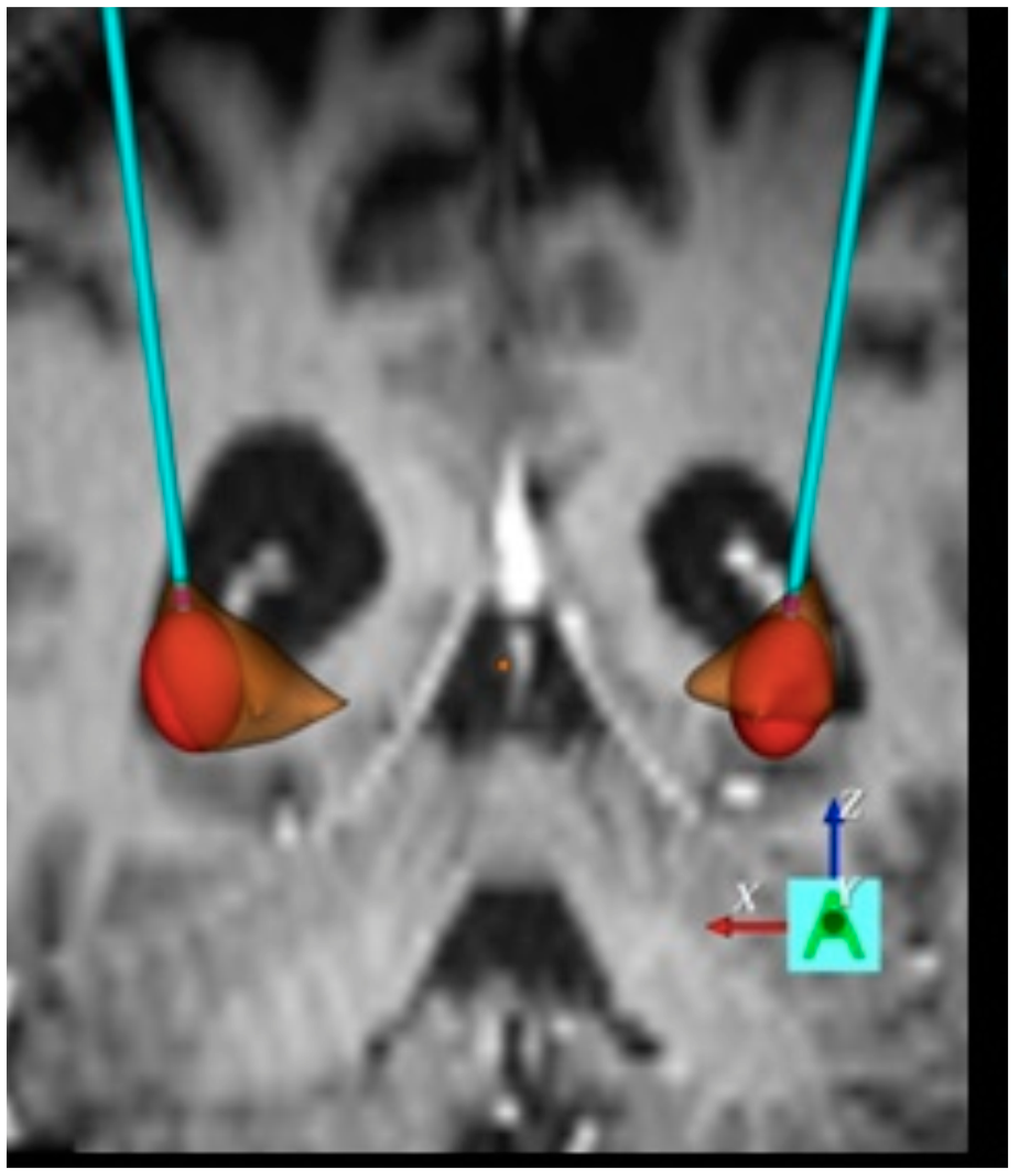
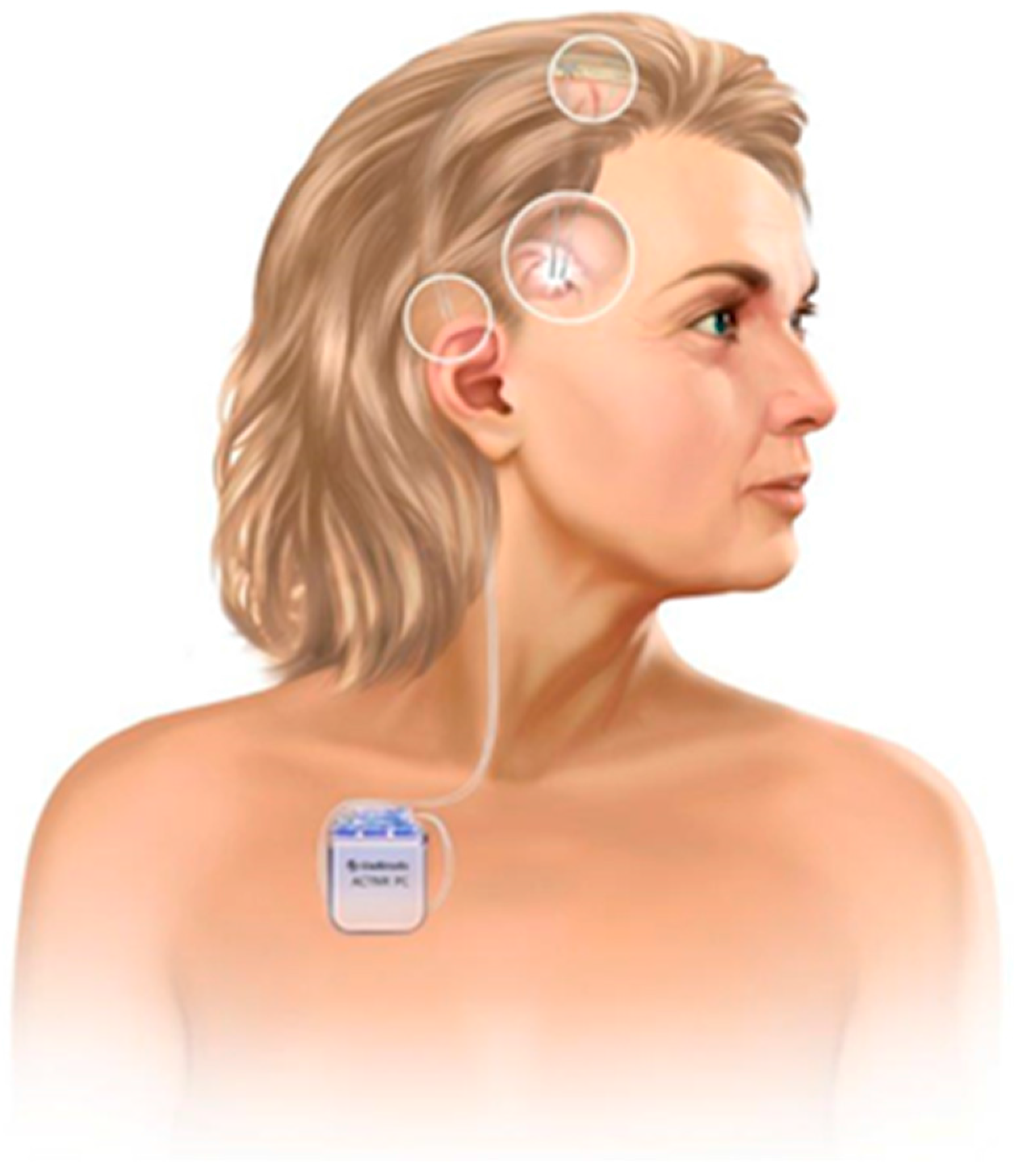
2. Evolution of Deep Brain Stimulation for HD
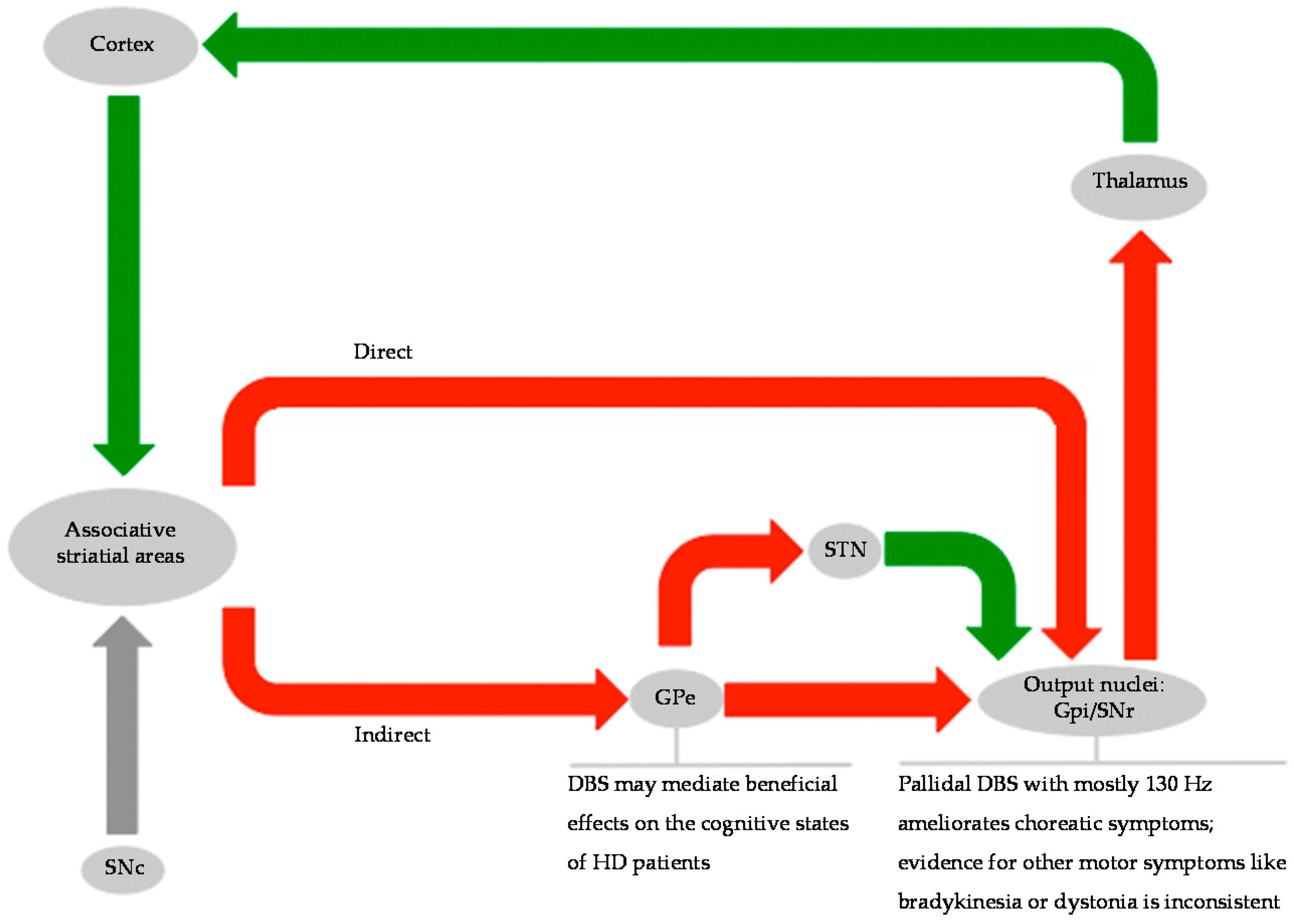
3. Invasive Assessment of the Basal Ganglia Network in HD
4. Clinical Implications of DBS in HD
4.1. Clinical Implications of DBS on Hyperkinetic and Hypokinetic Symptoms
4.2. Clinical Implications of DBS on Non-Motor-Functions
5. Safety of DBS in HD
6. Outlook
- (1)
- No significant atrophy;
- (2)
- Mild cortical atrophy as common in neurodegenerative disorders;
- (3)
- Severe cortical atrophy and additional atrophy periventricular and of the target basal ganglia structures.
7. Conclusions
Acknowledgments
Author Contributions
Conflicts of Interest
References
- Hartmann, C.J.; Groiss, S.J.; Vesper, J.; Schnitzler, A.; Wojtecki, L. Brain stimulation in Huntington’s disease. Neurodegener. Dis. Manag. 2016, 6, 223–236. [Google Scholar] [CrossRef] [PubMed]
- Walker, F.O. Huntington’s disease. Semin. Neurol. 2007, 27, 143–150. [Google Scholar] [CrossRef] [PubMed]
- Mitchell, I.J.; Cooper, A.J.; Griffiths, M.R. The selective vulnerability of striatopallidal neurons. Prog. Neurobiol. 1999, 59, 691–719. [Google Scholar] [CrossRef]
- Albin, R.L.; Reiner, A.; Anderson, K.D.; Penney, J.B.; Young, A.B. Striatal and nigral neuron subpopulations in rigid huntington’s disease: Implications for the functional anatomy of chorea and rigidity-akinesia. Ann. Neurol. 1990, 27, 357–365. [Google Scholar] [CrossRef] [PubMed]
- Wichmann, T.; DeLong, M.R. Functional and pathophysiological models of the basal ganglia. Curr. Opin. Neurobiol. 1996, 6, 751–758. [Google Scholar] [CrossRef]
- Louis, E.D.; Anderson, K.E.; Moskowitz, C.; Thorne, D.Z.; Marder, K. Dystonia-predominant adult-onset huntington disease: Association between motor phenotype and age of onset in adults. Arch. Neurol. 2000, 57, 1326–1330. [Google Scholar] [CrossRef] [PubMed]
- Thompson, P.D.; Berardelli, A.; Rothwell, J.C.; Day, B.L.; Dick, J.P.; Benecke, R.; Marsden, C.D. The coexistence of bradykinesia and chorea in huntington’s disease and its implications for theories of basal ganglia control of movement. Brain J. Neurol. 1988, 111, 223–244. [Google Scholar] [CrossRef]
- Spiegel, E.A.; Wycis, H.T.; Marks, M.; Lee, A.J. Stereotaxic apparatus for operations on the human brain. Science 1947, 106, 349–350. [Google Scholar] [CrossRef] [PubMed]
- Gildenberg, P.L. History repeats itself. Stereotact. Funct. Neurosurg. 2003, 80, 61–75. [Google Scholar] [CrossRef] [PubMed]
- Benabid, A.L.; Pollak, P.; Louveau, A.; Henry, S.; de Rougemont, J. Combined (thalamotomy and stimulation) stereotactic surgery of the VIM thalamic nucleus for bilateral parkinson disease. Appl. Neurophysiol. 1987, 50, 344–346. [Google Scholar] [CrossRef] [PubMed]
- Delgado, J.M.; Hamlin, H.; Chapman, W.P. Technique of intracranial electrode implacement for recording and stimulation and its possible therapeutic value in psychotic patients. Confin. Neurol. 1952, 12, 315–319. [Google Scholar] [CrossRef] [PubMed]
- Delgado, J.M.; Mark, V.; Sweet, W.; Ervin, F.; Weiss, G.; Bach, Y.R.G.; Hagiwara, R. Intracerebral radio stimulation and recording in completely free patients. J. Nerv. Ment. Dis. 1968, 147, 329–340. [Google Scholar] [CrossRef] [PubMed]
- Sem-Jacobsen, C.W. Depth-electrographic observations in psychotic patients: A system related to emotion and behavior. Acta Psychiatr. Scand. 1959, 34, 412–416. [Google Scholar] [CrossRef]
- Alberts, W.W.; Wright, E.W., Jr.; Levin, G.; Feinstein, B.; Mueller, M. Threshold stimulation of the lateral thalamus and globus pallidus in the waking human. Electroencephalogr. Clin. Neurophysiol. 1961, 13, 68–74. [Google Scholar] [CrossRef]
- Sem-Jacobsen, C.W. Depth-electrographic observations related to Parkinson’s disease. Recording and electrical stimulation in the area around the third ventricle. J. Neurosurg. 1966, 24, S388–S402. [Google Scholar]
- Mundinger, F. New stereotactic treatment of spasmodic torticollis with a brain stimulation system (author’s transl.). Med. Klin. 1977, 72, 1982–1986. [Google Scholar]
- Miocinovic, S.; Somayajula, S.; Chitnis, S.; Vitek, J.L. History, applications, and mechanisms of deep brain stimulation. JAMA Neurol. 2013, 70, 163–171. [Google Scholar] [CrossRef] [PubMed]
- Herrington, T.M.; Cheng, J.J.; Eskandar, E.N. Mechanisms of deep brain stimulation. J. Neurophysiol. 2016, 115, 19–38. [Google Scholar] [CrossRef] [PubMed]
- Benabid, A.L.; Pollak, P.; Gervason, C.; Hoffmann, D.; Gao, D.M.; Hommel, M.; Perret, J.E.; de Rougemont, J. Long-term suppression of tremor by chronic stimulation of the ventral intermediate thalamic nucleus. Lancet 1991, 337, 403–406. [Google Scholar] [CrossRef]
- Grill, W.M.; Snyder, A.N.; Miocinovic, S. Deep brain stimulation creates an informational lesion of the stimulated nucleus. Neuroreport 2004, 15, 1137–1140. [Google Scholar] [CrossRef] [PubMed]
- Krack, P.; Pollak, P.; Limousin, P.; Hoffmann, D.; Benazzouz, A.; Le Bas, J.F.; Koudsie, A.; Benabid, A.L. Opposite motor effects of pallidal stimulation in Parkinson’s disease. Ann. Neurol. 1998, 43, 180–192. [Google Scholar] [CrossRef] [PubMed]
- Kumar, R.; Lang, A.E.; Rodriguez-Oroz, M.C.; Lozano, A.M.; Limousin, P.; Pollak, P.; Benabid, A.L.; Guridi, J.; Ramos, E.; van der Linden, C.; et al. Deep brain stimulation of the globus pallidus pars interna in advanced Parkinson’s disease. Neurology 2000, 55, S34–S39. [Google Scholar] [PubMed]
- Volkmann, J.; Mueller, J.; Deuschl, G.; Kuhn, A.A.; Krauss, J.K.; Poewe, W.; Timmermann, L.; Falk, D.; Kupsch, A.; Kivi, A.; et al. Pallidal neurostimulation in patients with medication-refractory cervical dystonia: A randomised, sham-controlled trial. Lancet Neurol. 2014, 13, 875–884. [Google Scholar] [CrossRef]
- Vidailhet, M.; Jutras, M.F.; Grabli, D.; Roze, E. Deep brain stimulation for dystonia. J. Neurol. Neurosurg. Psychiatry 2013, 84, 1029–1042. [Google Scholar] [CrossRef] [PubMed]
- Damier, P.; Thobois, S.; Witjas, T.; Cuny, E.; Derost, P.; Raoul, S.; Mertens, P.; Peragut, J.C.; Lemaire, J.J.; Burbaud, P.; et al. Bilateral deep brain stimulation of the globus pallidus to treat tardive dyskinesia. Arch. Gen. Psychiatry 2007, 64, 170–176. [Google Scholar] [CrossRef] [PubMed]
- Trottenberg, T.; Paul, G.; Meissner, W.; Maier-Hauff, K.; Taschner, C.; Kupsch, A. Pallidal and thalamic neurostimulation in severe tardive dystonia. J. Neurol. Neurosurg. Psychiatry 2001, 70, 557–559. [Google Scholar] [CrossRef] [PubMed]
- Timmermann, L.; Pauls, K.A.; Wieland, K.; Jech, R.; Kurlemann, G.; Sharma, N.; Gill, S.S.; Haenggeli, C.A.; Hayflick, S.J.; Hogarth, P.; et al. Dystonia in neurodegeneration with brain iron accumulation: Outcome of bilateral pallidal stimulation. Brain 2010, 133, 701–712. [Google Scholar] [CrossRef] [PubMed]
- Miquel, M.; Spampinato, U.; Latxague, C.; Aviles-Olmos, I.; Bader, B.; Bertram, K.; Bhatia, K.; Burbaud, P.; Burghaus, L.; Cho, J.W.; et al. Short and long term outcome of bilateral pallidal stimulation in chorea-acanthocytosis. PLoS ONE 2013, 8, e79241. [Google Scholar] [CrossRef] [PubMed]
- Guehl, D.; Cuny, E.; Tison, F.; Benazzouz, A.; Bardinet, E.; Sibon, Y.; Ghorayeb, I.; Yelnick, J.; Rougier, A.; Bioulac, B.; et al. Deep brain pallidal stimulation for movement disorders in neuroacanthocytosis. Neurology 2007, 68, 160–161. [Google Scholar] [CrossRef] [PubMed]
- Krauss, J.K.; Loher, T.J.; Weigel, R.; Capelle, H.H.; Weber, S.; Burgunder, J.M. Chronic stimulation of the globus pallidus internus for treatment of non-dYT1 generalized dystonia and choreoathetosis: 2-year follow up. J. Neurosurg. 2003, 98, 785–792. [Google Scholar] [CrossRef] [PubMed]
- Gill, S.; Curran, A.; Tripp, J.; Melarickas, L.; Hurran, C.; Stanley, O. Hyperkinetic movement disorder in an 11-year-old child treated with bilateral pallidal stimulators. Dev. Med. Child Neurol. 2001, 43, 350–353. [Google Scholar] [CrossRef] [PubMed]
- Vidailhet, M.; Yelnik, J.; Lagrange, C.; Fraix, V.; Grabli, D.; Thobois, S.; Burbaud, P.; Welter, M.L.; Xie-Brustolin, J.; Braga, M.C.; et al. Bilateral pallidal deep brain stimulation for the treatment of patients with dystonia-choreoathetosis cerebral palsy: A prospective pilot study. Lancet Neurol. 2009, 8, 709–717. [Google Scholar] [CrossRef]
- Spiegel, E.A.; Wycis, H.T. Thalamotomy and pallidotomy for treatment of choreic movements. Acta Neurochir. 1952, 2, 417–422. [Google Scholar] [CrossRef] [PubMed]
- Cubo, E.; Shannon, K.M.; Penn, R.D.; Kroin, J.S. Internal globus pallidotomy in dystonia secondary to Huntington’s disease. Mov. Disord. 2000, 15, 1248–1251. [Google Scholar] [CrossRef]
- Tobin, A.J.; Signer, E.R. Huntington’s disease: The challenge for cell biologists. Trends Cell Biol. 2000, 10, 531–536. [Google Scholar] [CrossRef]
- Alexander, G.E.; DeLong, M.R.; Strick, P.L. Parallel organization of functionally segregated circuits linking basal ganglia and cortex. Annu. Rev. Neurosci. 1986, 9, 357–381. [Google Scholar] [CrossRef] [PubMed]
- Alexander, G.E.; Crutcher, M.D.; DeLong, M.R. Basal ganglia-thalamocortical circuits: Parallel substrates for motor, oculomotor, “prefrontal” and “limbic” functions. Prog. Brain Res. 1990, 85, 119–146. [Google Scholar] [PubMed]
- Smith, Y.; Bevan, M.D.; Shink, E.; Bolam, J.P. Microcircuitry of the direct and indirect pathways of the basal ganglia. Neuroscience 1998, 86, 353–387. [Google Scholar] [PubMed]
- Reiner, A.; Albin, R.L.; Anderson, K.D.; D’Amato, C.J.; Penney, J.B.; Young, A.B. Differential loss of striatal projection neurons in huntington disease. Proc. Natl. Acad. Sci. USA 1988, 85, 5733–5737. [Google Scholar] [CrossRef] [PubMed]
- Albin, R.L.; Reiner, A.; Anderson, K.D.; Dure, L.S.; Handelin, B.; Balfour, R.; Whetsell, W.O., Jr.; Penney, J.B.; Young, A.B. Preferential loss of striato-external pallidal projection neurons in presymptomatic huntington’s disease. Ann. Neurol. 1992, 31, 425–430. [Google Scholar] [CrossRef] [PubMed]
- Albin, R.L.; Young, A.B.; Penney, J.B. The functional anatomy of basal ganglia disorders. Trends Neurosci. 1989, 12, 366–375. [Google Scholar] [CrossRef]
- Penney, J.B., Jr.; Young, A.B. Striatal inhomogeneities and basal ganglia function. Mov. Disord. 1986, 1, 3–15. [Google Scholar] [CrossRef] [PubMed]
- Georgiou, N.; Bradshaw, J.L.; Phillips, J.G.; Bradshaw, J.A.; Chiu, E. The Simon effect and attention deficits in Gilles de la Tourette’s syndrome and Huntington’s disease. Brain J. Neurol. 1995, 118, 1305–1318. [Google Scholar] [CrossRef]
- Litvan, I.; Paulsen, J.S.; Mega, M.S.; Cummings, J.L. Neuropsychiatric assessment of patients with hyperkinetic and hypokinetic movement disorders. Arch. Neurol. 1998, 55, 1313–1319. [Google Scholar] [CrossRef] [PubMed]
- Raymond, L.A.; Andre, V.M.; Cepeda, C.; Gladding, C.M.; Milnerwood, A.J.; Levine, M.S. Pathophysiology of Huntington’s disease: Time-dependent alterations in synaptic and receptor function. Neuroscience 2011, 198, 252–273. [Google Scholar] [CrossRef] [PubMed]
- Joel, D. Open interconnected model of basal ganglia-thalamocortical circuitry and its relevance to the clinical syndrome of Huntington’s disease. Mov. Disord. 2001, 16, 407–423. [Google Scholar] [CrossRef] [PubMed]
- Gross, R.E.; Krack, P.; Rodriguez-Oroz, M.C.; Rezai, A.R.; Benabid, A.L. Electrophysiological mapping for the implantation of deep brain stimulators for Parkinson’s disease and tremor. Mov. Disord. 2006, 21, S259–S283. [Google Scholar] [CrossRef] [PubMed]
- Starr, P.A.; Rau, G.M.; Davis, V.; Marks, W.J., Jr.; Ostrem, J.L.; Simmons, D.; Lindsey, N.; Turner, R.S. Spontaneous pallidal neuronal activity in human dystonia: Comparison with Parkinson’s disease and normal macaque. J. Neurophysiol. 2005, 93, 3165–3176. [Google Scholar] [CrossRef] [PubMed]
- Tang, J.K.; Moro, E.; Lozano, A.M.; Lang, A.E.; Hutchison, W.D.; Mahant, N.; Dostrovsky, J.O. Firing rates of pallidal neurons are similar in Huntington’s and Parkinson’s disease patients. Exp. Brain Res. 2005, 166, 230–236. [Google Scholar] [CrossRef] [PubMed]
- Starr, P.A.; Kang, G.A.; Heath, S.; Shimamoto, S.; Turner, R.S. Pallidal neuronal discharge in Huntington’s disease: Support for selective loss of striatal cells originating the indirect pathway. Exp. Neurol. 2008, 211, 227–233. [Google Scholar] [CrossRef] [PubMed]
- Delorme, C.; Rogers, A.; Lau, B.; Francisque, H.; Welter, M.L.; Fernandez Vidal, S.; Yelnik, J.; Durr, A.; Grabli, D.; Karachi, C. Deep brain stimulation of the internal pallidum in Huntington’s disease patients: Clinical outcome and neuronal firing patterns. J. Neurol. 2016, 263, 290–298. [Google Scholar] [CrossRef] [PubMed]
- Groiss, S.J.; Elben, S.; Reck, C.; Voges, J.; Wojtecki, L.; Schnitzler, A. Local field potential oscillations of the globus pallidus in Huntington’s disease. Mov. Disord. 2011, 26, 2577–2578. [Google Scholar] [CrossRef] [PubMed]
- Wojtecki, L.; Groiss, S.J.; Ferrea, S.; Elben, S.; Hartmann, C.J.; Dunnett, S.B.; Rosser, A.; Saft, C.; Sudmeyer, M.; Ohmann, C.; et al. A prospective pilot trial for pallidal deep brain stimulation in Huntington’s disease. Front. Neurol. 2015, 6, 177. [Google Scholar] [CrossRef] [PubMed]
- Gonzalez, V.; Cif, L.; Biolsi, B.; Garcia-Ptacek, S.; Seychelles, A.; Sanrey, E.; Descours, I.; Coubes, C.; de Moura, A.M.; Corlobe, A.; et al. Deep brain stimulation for Huntington’s disease: Long-term results of a prospective open-label study. J. Neurosurg. 2014, 121, 114–122. [Google Scholar] [CrossRef] [PubMed]
- Moro, E.; Lang, A.E.; Strafella, A.P.; Poon, Y.Y.; Arango, P.M.; Dagher, A.; Hutchison, W.D.; Lozano, A.M. Bilateral globus pallidus stimulation for Huntington’s disease. Ann. Neurol. 2004, 56, 290–294. [Google Scholar] [CrossRef] [PubMed]
- Fawcett, A.P.; Moro, E.; Lang, A.E.; Lozano, A.M.; Hutchison, W.D. Pallidal deep brain stimulation influences both reflexive and voluntary saccades in Huntington’s disease. Mov. Disord. 2005, 20, 371–377. [Google Scholar] [CrossRef] [PubMed]
- Hebb, M.O.; Garcia, R.; Gaudet, P.; Mendez, I.M. Bilateral stimulation of the globus pallidus internus to treat choreathetosis in Huntington’s disease: Technical case report. Neurosurgery 2006, 58, E383. [Google Scholar] [CrossRef] [PubMed]
- Fasano, A.; Mazzone, P.; Piano, C.; Quaranta, D.; Soleti, F.; Bentivoglio, A.R. GPi-DBS in Huntington’s disease: Results on motor function and cognition in a 72-year-old case. Mov. Disord. 2008, 23, 1289–1292. [Google Scholar] [CrossRef] [PubMed]
- Biolsi, B.; Cif, L.; Fertit, H.E.; Robles, S.G.; Coubes, P. Long-term follow-up of Huntington disease treated by bilateral deep brain stimulation of the internal globus pallidus. J. Neurosurg. 2008, 109, 130–132. [Google Scholar] [CrossRef] [PubMed]
- Garcia-Ruiz, P.J.; Ayerbe, J.; del Val, J.; Herranz, A. Deep brain stimulation in disabling involuntary vocalization associated with Huntington’s disease. Parkinsonism Relat. Disord. 2012, 18, 803–804. [Google Scholar] [CrossRef] [PubMed]
- Spielberger, S.; Hotter, A.; Wolf, E.; Eisner, W.; Muller, J.; Poewe, W.; Seppi, K. Deep brain stimulation in Huntington’s disease: A 4-year follow-up case report. Mov. Disord. 2012, 27, 806–807. [Google Scholar] [CrossRef] [PubMed]
- Huys, D.; Bartsch, C.; Poppe, P.; Lenartz, D.; Huff, W.; Prutting, J.; Timmermann, L.; Klosterkotter, J.; Maarouf, M.; Rommel, T.; et al. Management and outcome of pallidal deep brain stimulation in severe Huntington’s disease. Fortschr. Neurol. Psychiatr. 2013, 81, 202–205. [Google Scholar] [CrossRef] [PubMed]
- Velez-Lago, F.M.; Thompson, A.; Oyama, G.; Hardwick, A.; Sporrer, J.M.; Zeilman, P.; Foote, K.D.; Bowers, D.; Ward, H.E.; Sanchez-Ramos, J.; et al. Differential and better response to deep brain stimulation of chorea compared to dystonia in Huntington’s disease. Stereotact. Funct. Neurosurg. 2013, 91, 129–133. [Google Scholar] [CrossRef] [PubMed]
- Cislaghi, G.; Capiluppi, E.; Saleh, C.; Romano, L.; Servello, D.; Mariani, C.; Porto, M. Bilateral globus pallidus stimulation in westphal variant of Huntington disease. Neuromodul. J. Int. Neuromodul. Soc. 2014, 17, 502–505. [Google Scholar] [CrossRef] [PubMed]
- Gruber, D.; Kuhn, A.A.; Schoenecker, T.; Kopp, U.A.; Kivi, A.; Huebl, J.; Lobsien, E.; Mueller, B.; Schneider, G.H.; Kupsch, A. Quadruple deep brain stimulation in Huntington’s disease, targeting pallidum and subthalamic nucleus: Case report and review of the literature. J. Neural 2014, 121, 1303–1312. [Google Scholar] [CrossRef] [PubMed]
- Loutfi, G.; Linder, J.; Hariz, G.-M.; Hariz, M.; Blomstedt, P. Pallidal deep brain stimulation in the treatment of Huntington’s chorea. Brain Disord. Ther. 2014, 3. [Google Scholar] [CrossRef]
- Kang, G.A.; Heath, S.; Rothlind, J.; Starr, P.A. Long-term follow-up of pallidal deep brain stimulation in two cases of Huntington’s disease. J. Neurol. Neurosurg. Psychiatry 2011, 82, 272–277. [Google Scholar] [CrossRef] [PubMed]
- Zittel, S.; Moll, C.K.; Gulberti, A.; Tadic, V.; Rasche, D.; Baumer, T.; Fellbrich, A.; Bruggemann, N.; Engel, A.K.; Tronnier, V.; et al. Pallidal deep brain stimulation in Huntington’s disease. Parkinsonism Relat. Disord. 2015, 21, 1105–1108. [Google Scholar] [CrossRef] [PubMed]
- Amtage, F.; Feuerstein, T.J.; Meier, S.; Prokop, T.; Piroth, T.; Pinsker, M.O. Hypokinesia upon pallidal deep brain stimulation of dystonia: Support of a gabaergic mechanism. Front. Neurol. 2013, 4, 198. [Google Scholar] [CrossRef] [PubMed]
- Berman, B.D.; Starr, P.A.; Marks, W.J., Jr.; Ostrem, J.L. Induction of bradykinesia with pallidal deep brain stimulation in patients with cranial-cervical dystonia. Stereotact. Funct. Neurosurg. 2009, 87, 37–44. [Google Scholar] [CrossRef] [PubMed]
- Schrader, C.; Capelle, H.H.; Kinfe, T.M.; Blahak, C.; Bazner, H.; Lutjens, G.; Dressler, D.; Krauss, J.K. GPi-DBS may induce a hypokinetic gait disorder with freezing of gait in patients with dystonia. Neurology 2011, 77, 483–488. [Google Scholar] [CrossRef] [PubMed]
- Lopez-Sendon Moreno, J.L.; Garcia-Caldentey, J.; Regidor, I.; del Alamo, M.; Garcia de Yebenes, J. A 5-year follow-up of deep brain stimulation in Huntington’s disease. Parkinsonism Relat. Disord. 2014, 20, 260–261. [Google Scholar] [CrossRef] [PubMed]
- Paulsen, J.S. Cognitive impairment in huntington disease: Diagnosis and treatment. Curr. Neurol. Neurosci. Rep. 2011, 5, 474–483. [Google Scholar] [CrossRef] [PubMed]
- Beglinger, L.J.; O’Rourke, J.J.; Wang, C.; Langbehn, D.R.; Duff, K.; Paulsen, J.S. Earliest functional declines in Huntington disease. Psychiatry Res. 2010, 178, 414–418. [Google Scholar] [CrossRef] [PubMed]
- Smith, M.A.; Brandt, J.; Shadmehr, R. Motor disorder in Huntington’s disease begins as a dysfunction in error feedback control. Nature 2000, 403, 544–549. [Google Scholar] [CrossRef] [PubMed]
- Beste, C.; Saft, C.; Andrich, J.; Gold, R.; Falkenstein, M. Error processing in Huntington’s disease. PLoS ONE 2006, 1, e86. [Google Scholar] [CrossRef] [PubMed]
- Ayalon, L.; Doron, R.; Weiner, I.; Joel, D. Amelioration of behavioral deficits in a rat model of Huntington’s disease by an excitotoxic lesion to the globus pallidus. Exp. Neurol. 2004, 186, 46–58. [Google Scholar] [CrossRef]
- Temel, Y.; Cao, C.; Vlamings, R.; Blokland, A.; Ozen, H.; Steinbusch, H.W.; Michelsen, K.A.; von Horsten, S.; Schmitz, C.; Visser-Vandewalle, V. Motor and cognitive improvement by deep brain stimulation in a transgenic rat model of Huntington’s disease. Neurosci. Lett. 2006, 406, 138–141. [Google Scholar] [CrossRef] [PubMed]
- Ligot, N.; Krystkowiak, P.; Simonin, C.; Goldman, S.; Peigneux, P.; Van Naemen, J.; Monclus, M.; Lacroix, S.F.; Devos, D.; Dujardin, K.; et al. External globus pallidus stimulation modulates brain connectivity in Huntington’s disease. J. Cereb. Blood Flow Metab. 2011, 31, 41–46. [Google Scholar] [CrossRef] [PubMed]
- Beste, C.; Muckschel, M.; Elben, S.; C, J.H.; McIntyre, C.C.; Saft, C.; Vesper, J.; Schnitzler, A.; Wojtecki, L. Behavioral and neurophysiological evidence for the enhancement of cognitive control under dorsal pallidal deep brain stimulation in Huntington’s disease. Brain Struct. Funct. 2015, 220, 2441–2448. [Google Scholar] [CrossRef] [PubMed]
- Reich, M.M.; Steigerwald, F.; Sawalhe, A.D.; Reese, R.; Gunalan, K.; Johannes, S.; Nickl, R.; Matthies, C.; McIntyre, C.C.; Volkmann, J. Short pulse width widens the therapeutic window of subthalamic neurostimulation. Ann. Clin. Transl. Neurol. 2015, 2, 427–432. [Google Scholar] [CrossRef] [PubMed]
- Volkmann, J.; Stiegerwald, S.; Reich, M. Deep brain stimulation at short pulse width results in superior therapeutic windows for treatment of Parkinson’s disease: A randomized, controlled, double-blind neurostimulation trial (CUSTOM-DBS). In Proceedings of the 18th International Congress of Parkinson’s Disease and Movement Disorders, Stockholm, Sweden, 8–12 June 2014.
- Contarino, M.F.; Bour, L.J.; Verhagen, R.; Lourens, M.A.; de Bie, R.M.; van den Munckhof, P.; Schuurman, P.R. Directional steering: A novel approach to deep brain stimulation. Neurology 2014, 83, 1163–1169. [Google Scholar] [CrossRef] [PubMed]
- Pollo, C.; Kaelin-Lang, A.; Oertel, M.F.; Stieglitz, L.; Taub, E.; Fuhr, P.; Lozano, A.M.; Raabe, A.; Schupbach, M. Directional deep brain stimulation: An intraoperative double-blind pilot study. Brain J. Neurol. 2014, 137, 2015–2026. [Google Scholar] [CrossRef] [PubMed]
- Quinn, E.J.; Blumenfeld, Z.; Velisar, A.; Koop, M.M.; Shreve, L.A.; Trager, M.H.; Hill, B.C.; Kilbane, C.; Henderson, J.M.; Bronte-Stewart, H. Beta oscillations in freely moving Parkinson’s subjects are attenuated during deep brain stimulation. Mov. Disord. 2015, 30, 1750–1758. [Google Scholar] [CrossRef] [PubMed]
- Fenoy, A.J.; Simpson, R.K., Jr. Risks of common complications in deep brain stimulation surgery: Management and avoidance. J. Neurosurg. 2014, 120, 132–139. [Google Scholar] [CrossRef] [PubMed]


| Study | n | FUP (Months) | Age (Years) | DisDur (Years) | Chorea | Bradykinesia | Dystonia | Total Motor | Comments |
|---|---|---|---|---|---|---|---|---|---|
| Moro et al. [55] | 1 | 8 | 43 | 8 | 44% | 14% | 38% | 31% | DBS frequency of 130 Hz but not 40 Hz worsened bradykinesia. Increased regional cerebral blood flow in cortical motor regions. |
| Fawcett et al. [56] | 1 | 4 | 42 | n.a. | 56% | n.a. | 60% | 26% | Moderate improvement of speech, swallowing and gait, task-specific improvement of oculomotor function. |
| Hebb et al. [57] | 1 | 12 | 41 | 13 | 57% | n.a. | n.a. | 15% | Chorea improves with higher stimulation frequency (180 Hz), no frequency-dependent effect of bradykinesia |
| Fasano et al. [58] | 1 | 12 | 72 | 17 | 77% | 60% | 100% | n.a. | Worsening of gait, apathy, cognitive decline, functional gain minimal, turning off at 11 months did not induce chorea |
| Biolsi et al. [59] | 1 | 48 | 60 | 10 | 21% | n.a. | n.a. | 5% | Chorea reduced by 56%, when comparing DBS-on vs. DBS-off. L-Dopa-responsive worsening of bradykinesia. Cognition stable. |
| Groiss et al. [52] | 1 | 12 | 65 | n. a. | 47% | n.a. | 31% | n.a | Primary focus on local field potentials. Hypokinesia observed at 180 Hz stimulation improved from 40 Hz DBS |
| Garcia-Ruiz et al. [60] | 1 | 12 | 30 | 10 | n.a. | n.a. | n.a. | 48% | Marked improvement of vocalization. No effect of DBS on hypokinesia and rigidity. Facilitated activities of daily living. |
| Spielberger et al. [61] | 1 | 48 | 30 | 9 | 75% | 5% | 70% | −4% | Worsening of chorea with 40 Hz DBS, best results with 130 Hz DBS. Progression of bradykinesia compatible with natural course. |
| Huys et al. [62] | 1 | 12 | 40 | 3 | 16% | Improved cognition 6 months after surgery followed by a decline at 12 months (but improved results compared to baseline assessment) | |||
| Velez-Lago et al. [63] | 1 | 60 | 60 | 2 | 56% | n.a. | −40% | −98% | Chorea (69%), dystonia (40%), and overall motor score (37%) improved up to 24 months after surgery. Stable cognition. |
| Cislaghi et al. [64] | 1 | 48 | 31 | 16 | 67% | n.a. | n.a. | n.a. | Significant improvement in chorea in juvenile HD. Impairment of bradykinesia. No effect on cognitive function. |
| Gruber et al. [65] | 1 | 48 | 41 | 9 | 60% | 42% | 50% | 19% | GPI DBS-induced bradykinesia alleviated with STN DBS. Cognitive decline compatible with the natural course of HD. |
| Loutfi et al. [66] | 1 | 12 | 59 | 12 | 27% | n.a. | −40% | 12% | Stable cognition, modest improvement of verbal fluency, marked improvement of behavioral assessment. |
| Study | n | FUP (Months) | Age (Years) | DisDur (Years) | Chorea | Bradykinesia | Dystonia | Total Motor | Comments |
|---|---|---|---|---|---|---|---|---|---|
| Kang et al. [67] | 2 | 24 | 57 | 10 | 63% | −11% | n.a. | 22% | Best results on chorea with either 40 Hz (patient 1) or 130 Hz (patient 2) DBS. Cognitive decline compatible with natural disease progression. |
| 24 | 50 | 5 | 59% | 0% | 0 to 6 | 4% | |||
| Velez-Lago et al. [63] | 2 | 12 | 27 | 7 | 74% | n.a. | n.a. | 43% | Despite good effect on chorea, there was no improvement for a patient with predominant dystonia. Worsening of bradykinesia and rigidity |
| 9 | 19 | 6 | n.a. | n.a. | 20% | −24% | |||
| Gonzales et al. [54] | 7 | 36 | 78 | 5 | 65% | n.a. | n.a. | −10% | Bradykinesia worsened over time. Additionally, DBS-dependent effects could be observed. Reduction of pulse width reduced bradykinesia. Non-significant worsening of dystonia over time. Despite progressive decline of cognition, cognitive levels were not significantly worse compared to baseline |
| 36 | 39 | 8 | 69% | 14% | |||||
| 36 | 74 | 4 | 20% | −11% | |||||
| 36 | 54 | 8 | 79% | −30% | |||||
| 36 | 37 | 3 | 67% | 33% | |||||
| 12 | 30 | 3 | 70% | 40% | |||||
| 12 | 36 | 3 | 80% | −64% | |||||
| Wojtecki et al. [53] | 6 | 6 | 52 | 3 | 66% | 5% | 56% | 42% | First randomized, double-blind study up to date. First study comparing GPE and GPI DBS. Patients 4 and 5 suffered from juvenile variant of HD and therefore exclusively presented with hypokinetic-rigid symptoms and dystonia. DBS of GPE and GPI did not lead to significantly different results. Heterogeneous results concerning functional outcome. |
| 6 | 71 | 21 | 63% | −9% | 85% | 27% | |||
| 6 | 38 | 10 | 46% | −22% | 55% | 11% | |||
| 6 | 25 | 11 | n. a. | −19% | −44% | −3% | |||
| 6 | 23 | 8 | n. a. | −10% | −37% | −9% | |||
| 6 | 29 | 4 | 66% | 17% | 0% | 28% | |||
| Zittel et al. [68] | 3 | 36 | 54 | 5 | 50% | −36% | 100% | 11% | Heterogeneous results concerning DBS effects on bradykinesia and dystonia. Mini-mental status examination stable over time, while more complex tests revealed diverging results |
| 12 | 35 | 4 | 58% | 11% | −250% | 25% | |||
| 12 | 45 | 7 | 40% | 39% | −100% | 20% | |||
| Delorme et al. [51] | 3 | 30 | 56 | 10 | 15% | −100% | 0 to 1 | −18% | Greater effect size (with mean improvement of 55% of chorea and 32% for the total score), if not compared to baseline but DBS off assessment at follow-up. DBS via ventral electrode contacts was more effective than DBS via dorsal contacts |
| 24 | 24 | 3 | 67% | 0 to 4 | 0 to 6 | 20% | |||
| 12 | 50 | 10 | 29% | −33% | −140% | −2% |
© 2016 by the authors; licensee MDPI, Basel, Switzerland. This article is an open access article distributed under the terms and conditions of the Creative Commons Attribution (CC-BY) license (http://creativecommons.org/licenses/by/4.0/).
Share and Cite
Wojtecki, L.; Groiss, S.J.; Hartmann, C.J.; Elben, S.; Omlor, S.; Schnitzler, A.; Vesper, J. Deep Brain Stimulation in Huntington’s Disease—Preliminary Evidence on Pathophysiology, Efficacy and Safety. Brain Sci. 2016, 6, 38. https://doi.org/10.3390/brainsci6030038
Wojtecki L, Groiss SJ, Hartmann CJ, Elben S, Omlor S, Schnitzler A, Vesper J. Deep Brain Stimulation in Huntington’s Disease—Preliminary Evidence on Pathophysiology, Efficacy and Safety. Brain Sciences. 2016; 6(3):38. https://doi.org/10.3390/brainsci6030038
Chicago/Turabian StyleWojtecki, Lars, Stefan Jun Groiss, Christian Johannes Hartmann, Saskia Elben, Sonja Omlor, Alfons Schnitzler, and Jan Vesper. 2016. "Deep Brain Stimulation in Huntington’s Disease—Preliminary Evidence on Pathophysiology, Efficacy and Safety" Brain Sciences 6, no. 3: 38. https://doi.org/10.3390/brainsci6030038