Article

Development of a Novel Scaffold of Chitosan, Type IV Collagen and Integrin α3β1 As Alternative Scaffold for Primary Culture of Podocytes

by
Diana Ginette Zárate-Triviño
1,*,
Sara Paola Hernández-Martínez
1,
Juan José Bollain-y-Goytia-de-la-Rosa
2,
Moisés Armides Franco-Molina
1,
Eduardo Arturo Elizalde-Peña
3,
Margarita Idalia Hernández-Villegas
1,
Gloria Azucena Rangel-Ochoa
4 and
Cristina Rodríguez-Padilla
1
1
Immunology and Virology department, Faculty of Biological Science, Autonomous University of Nuevo Leon, Pedro de Alba s/n, San Nicolás de Los Garza, Nuevo León 66455, México
2
Immunology and Molecular Biology department, Academic Unit of Biological Science, Autonomous University of Zacatecas, Avenida de la Revolución Mexicana s/n, Guadalupe, Zacatecas 98600, México
3
Investigation and Postgraduate Unit, Faculty of Engineering, Autonomous University of Querétaro, Cerro de las Campanas s/n, Querétaro 76010, México
4
General Hospital “Luz Gonzalez Cosio”, Col. Ciudad Gobierno, Zacatecas 98160, México
*
Author to whom correspondence should be addressed.
Appl. Sci. 2018, 8(6), 930; https://doi.org/10.3390/app8060930
Submission received: 24 April 2018 / Revised: 21 May 2018 / Accepted: 26 May 2018 / Published: 5 June 2018
(This article belongs to the Section Applied Biosciences and Bioengineering)

Abstract

:
Loss of podocytes has been a main pathology present in renal diseases; the leak of these specialized cells increases the permeability of the glomerular basal membrane (GMB) and protein release affecting the glomeruli, the main structure of the kidney. The study of different physiopathology mechanism has been a challenge because of the short lifetime of podocytes in vitro. We obtained and characterized composites based on Chitosan (CTS), polyvinyl alcohol (PVA), type IV collagen and integrin α3β1 as a possible application in primary culture of podocytes. Podocytes were extracted from the urine of patients with Idiopathic Nephrotic Syndrome (INS). To evaluate biocompatibility, we assessed cell viability through the lactate dehydrogenase assay. Immunohistochemical staining was used to detect the expression of specific proteins from podocytes such as podocin, and podocalyxin and CD80, a marker of cellular stress. The results showed that our synthesis method promotes the copolymerization of the components in the scaffold. Due to its reactivity, the amine group of chitosan made links with type IV collagen and integrin α3β1. The swelling and degradation tests allowed us to select the material with the best mechanical properties for cellular culture. The expression of podocin and podocalyxin remains the same in the culture of podocytes on the scaffold; in contrast, CD80 expression increased. The viability of podocytes cultured on the CTS/PVA/type IV collagen/integrin α3β1 scaffold increased in comparison to the culture control.

1. Introduction

Clinical advances have identified the podocyte as the main cell involved in kidney disease, because structural alteration of podocytes such as hypertrophy, dedifferentiation, detachment, and apoptosis contribute to the development of idiopathic nephrotic syndrome (INS), minimal change disease, focal segmental glomerulosclerosis, membranous glomerulopathy, and lupus nephritis [1,2,3].
Podocytes alterations are associated with loss of podocalyxin, a protein involved in structural maintenance pedicels (podocyte foot processes) which anchor the cell to the glomerular basal membrane [2,4,5].
Currently, cell lines are used for the study of kidney disease; however, the process of immortalization affects the expression of anchor and structural proteins, hindering the understanding of the physiopathologic mechanisms [6,7]. Primary cultures of cells extracted from patients have been used to elucidate the origin of kidney-associated diseases, however, the viability of primary podocytes in vitro is around 2 or 3 weeks at most, with loss of morphology and drastic decrease of viability [8]. Thus, it is necessary to improve culture conditions and develop new techniques that prolong the half-life of primary podocytes while keeping the production of their specific proteins.
Scaffolds are support structures that simulate the physiological conditions for the recovery and repair of damaged organs and tissues, [9,10,11] and are an option for primary cell cultures [12].
Scaffolds are made of bio-compatible polymers that are either present in the physiological environment of the cell or that provide requirements for a specific cell type. One of the polymers used for scaffolds is chitosan, a nitrogenous polysaccharide obtained from the alkaline deacetylation of chitin derived from the exoskeleton of marine animals such as crabs, shrimps, lobsters, and krill [13,14]. Several studies show that chitosan stimulates cell adhesion, migration and proliferation. Chitosan interacts with other molecules through its amine groups, improving their mechanical and biological properties. Polyvinyl alcohol (PVA) a nontoxic synthetic polymer, is one of such molecules. Blends of chitosan-PVA have been studied as base for hydrogels and scaffolds with biological applications [15,16].
We found no reports of chitosan-PVA scaffolds for podocyte primary culture; furthermore, for their maintenance, podocytes require other substances such as integrin α3β1, necessary for attachment to the basal membrane [17] and IV collagen, a main component of the basal membrane.
The aim of this work is to study the performance of a chitosan/PVA/IV collagen/integrin α3β1 scaffold for primary podocytes culture, evaluating their half-life, cell adhesion, and specific protein expression, as an alternative in vitro model for the study of physio-pathological mechanisms involved in renal disease.

2. Materials and Methods

2.1. Reagents

Chitosan medium molecular weight, poly (vinyl alcohol), collagen from human placenta type IV, Roswell Park Memorial Institute medium (RPMI 1640) (Sigma Aldrich, St. Louis, MO, USA), recombinant human integrin α3β1 (R&D Systems, Minneapolis, MN, USA). MAbs-IgG1 mouse anti-podocalyxin antibody, anti-podocin antibody, anti-CD80 (B7.1) antibody, anti-IgG Rabbit antibody (FITC), anti-IgG1 mouse antibody (TR), anti-IgG goat antibody (FITC) (Santa Cruz Biotechnology, Dallas, TX, USA). Anti-IgG1 mouse antibody (Rat anti-mouse IgG1 MicroBeads), MiniMACS separator (Miltenyi Biotec, Bergisch Gladbach, Germany), fetal bovine serum (Gibco, Thermo Fisher Scientific, Waltham, MA, USA). Kit Quick-Prep™ Urinalysis, L-Lactate Assay Kit (Cayman Chemical Company, Ann Arbor, MI, USA), 24-well tissue culture polystyrene plates (Corning Costar, Sigma-Aldrich, Saint Louis, MO, USA).

2.2. Chitosan, PVA, Type IV Collagen, and Integrin α3β1 Scaffold Preparation

PVA hydrogels were prepared by fully dissolving 1.0 g of polymer powder in 100 mL of Milli-Q water, under magnetic stirring at 90 °C (solution A). Chitosan hydrogels were prepared by fully dissolving 2 g of polymer powder in 100 mL of 0.2 M CH3COOH under magnetic stirring for 24 h (solution B). Subsequently, solution A was added to solution B to obtain Chitosan/PVA with a 7:1 (v/v) mass ratio. Finally, type IV collagen and recombinant human integrin α3β1, at 7% w/v or 0.03% w/v ratio were added. The solution was poured into plastic molds and dried in an oven for 48 h at 30 °C.

2.3. Characterization for Fourier Transform Infrared Spectroscopy (FTIR)

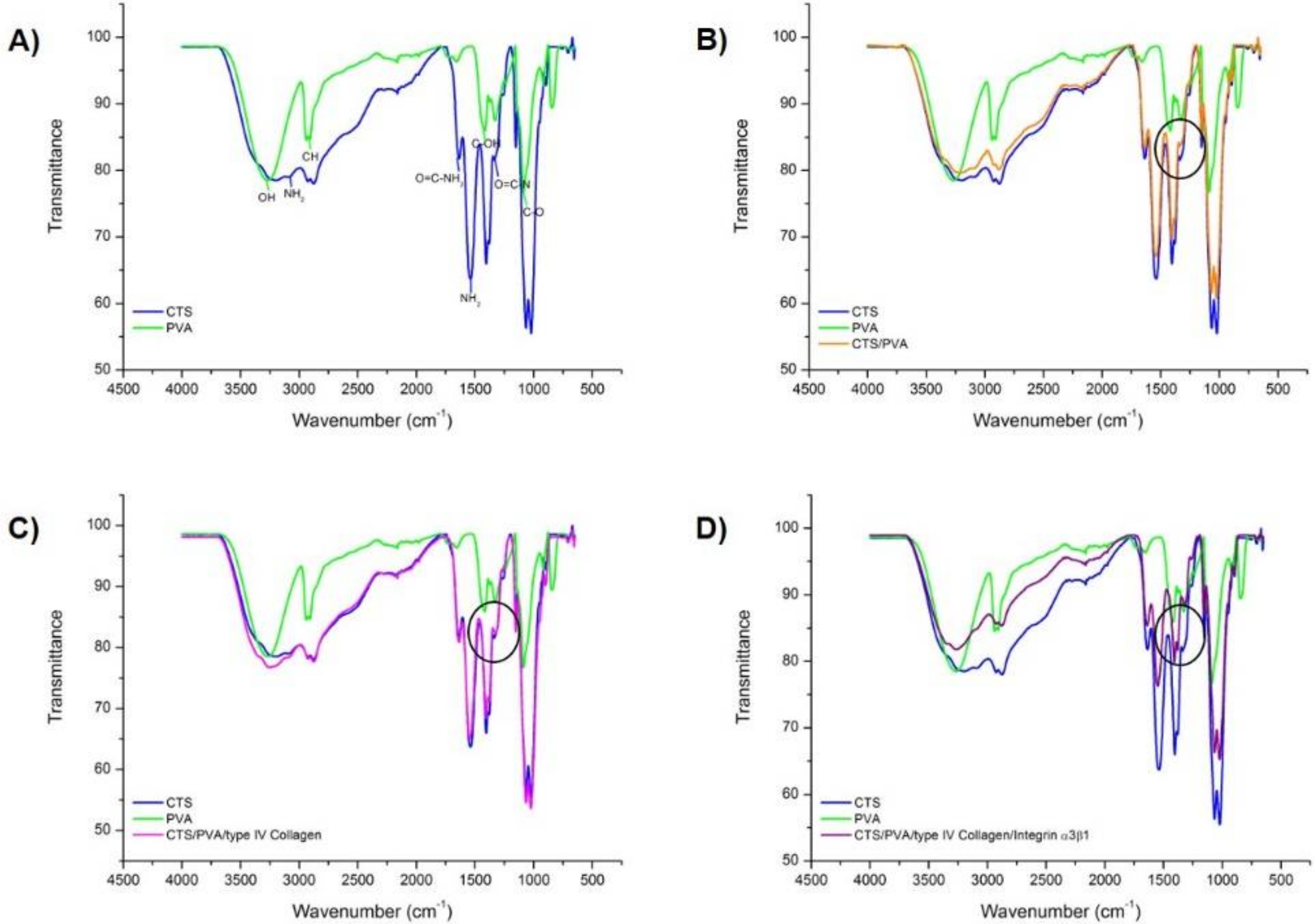
The study of the contribution of each component to the interaction was carried by means of attenuated total reflection (ATR) used in conjunction with Fourier Transform Infrared Spectroscopy (FTIR) technique. The spectra of CTS, PVA, CTS/PVA, CTS/PVA/type IV collagen and CTS/PVA/type IV collagen/integrin α3β1 samples were recorded by a Universal ATR Sampling Accessory Perkin Elmer by the accumulation of 16 scan at 4 cm−1 resolution in the wavenumber range 4500–500 cm−1. Specimens were placed onto the diamond ATR crystal using a top-plate and pressure arm. Data were analyzed with the software PERKIN ELMER spectrum.

2.4. Swelling and Degradation Tests under Physiological Mimicking Conditions

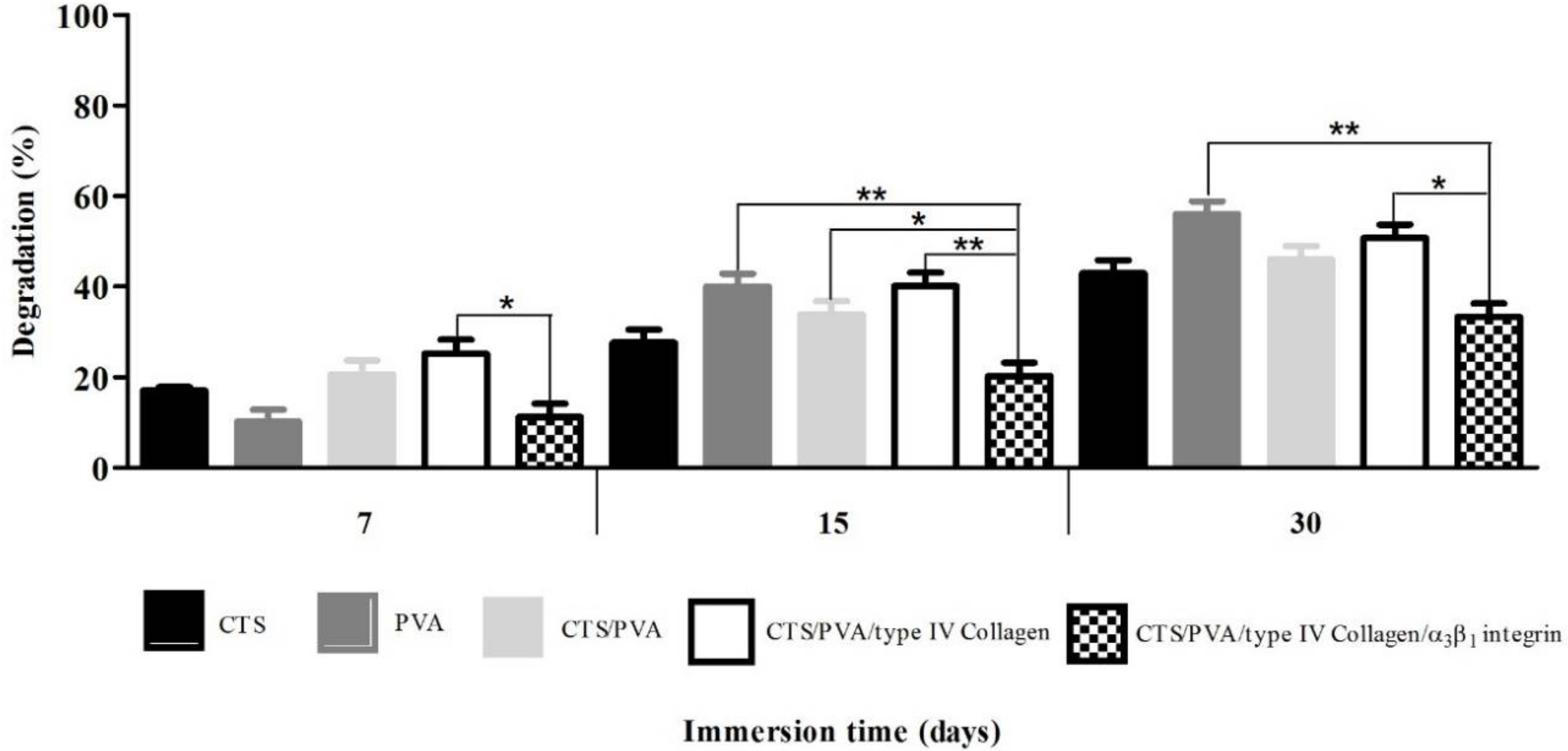
The CTS, PVA, CTS/PVA, CTS/PVA/type IV collagen and CTS/PVA/type IV collagen/integrin α3β1 scaffolds were cut into 2 × 2 cm2 pieces and soaked in RPMI 1640 medium for the swelling and degradation assay. At the end of each soaking period, the solution excess on the samples was wiped with a lint-free tissue paper.

2.4.1. Swelling Test

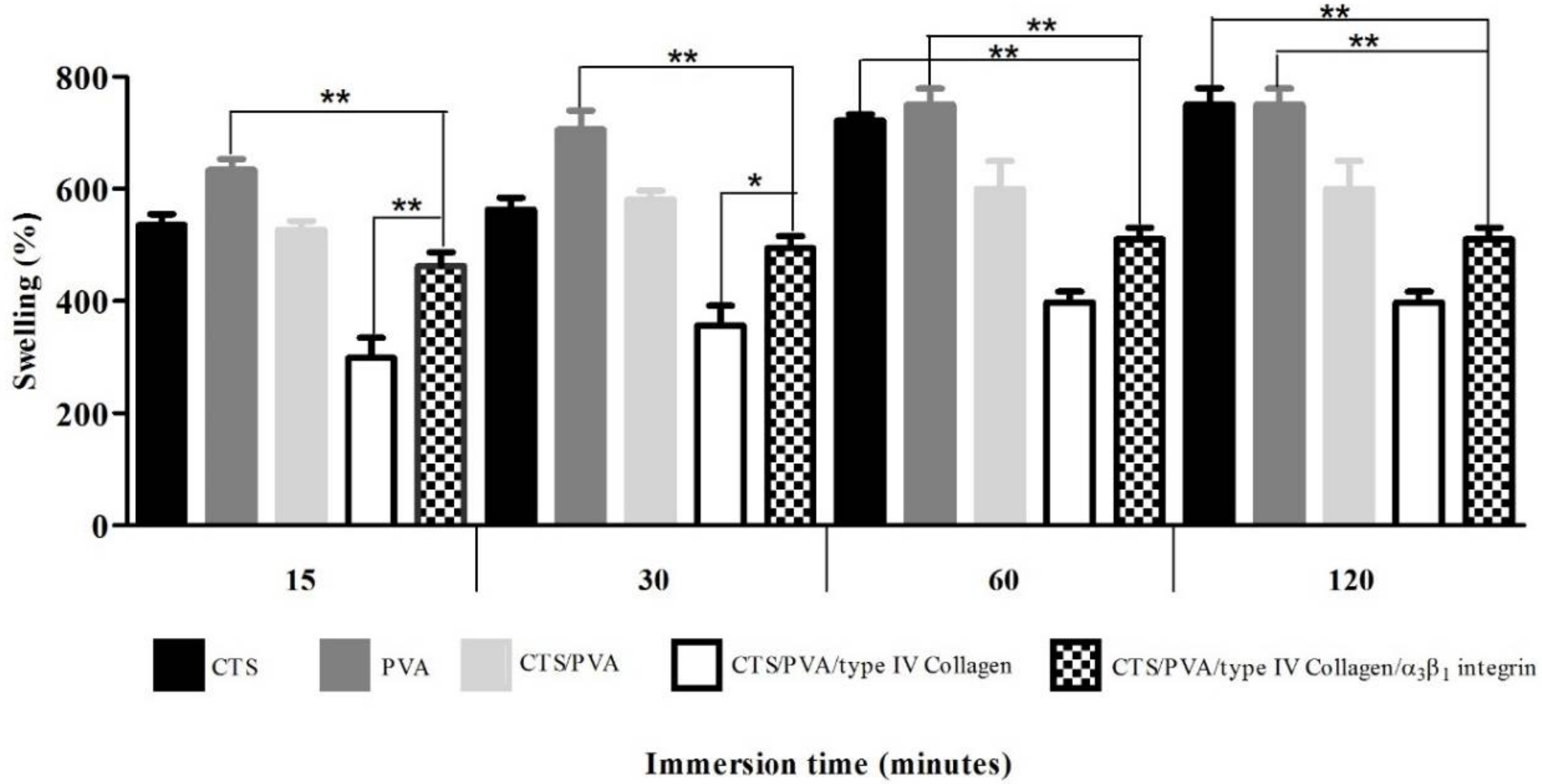
The swelling test indicates the nutrient uptake of our scaffold from the culture medium and their delivery to the cell. A rigid structure is produced by the polymer interaction, which reduces the flexibility and number of hydrophobic groups in the scaffold, affecting the swelling velocity.
For fluid uptake measurements, CTS, PVA, CTS/PVA, CTS/PVA/type IV collagen and CTS/PVA/type IV collagen/integrin α3β1 scaffold samples were weighed before being immersed in RPMI 1640 medium at 37 °C. At determinate time intervals (3, 7, 15, and 30 min), the samples were removed quickly and carefully, dried lightly to remove excess medium, and then were weighed. The degree of welling was determinate by the following Equation (1): medium absorption (S), wet weight (Wf) and dry weight (W0).
S =   W f     W 0   ÷   W 0   ×   100
Each medium absorption experiment was made by triplicate and the average value is reported.

2.4.2. Degradation Test

The degradation study determines time-dependent alterations in the physicochemical characteristics of the scaffolds, this includes polymer mass loss through solvation and depolymerization. To find out the degradation index (Di), samples were incubated in RPMI 1640 medium at 37 °C. At determinate time intervals (3, 7, 15, and 30 days), the samples were removed quickly and carefully, dried lightly to remove excess medium, and then were weighed. The degradation index was calculated based on the mass loss using the Equation (2): wet weight (Wt) and dry weight (W0).
D i   =   W 0     W t   ÷   W 0   ×   100
Each degradation experiment was made by triplicate and the average value is reported.

2.5. Biological Samples

Urine samples were obtained from twenty-two children: twelve patients with INS, 7 boys and 5 girls with an average age of 4.8 years, and ten healthy children 6 boys and 4 girls, with an average age of 7.8 years (Table 1). All patients were diagnosed according to the criteria of the International Study of Kidney Disease in Children. Informed consent was obtained from the parents of each child as necessary. This study was approved by the Ethics Committee of the Zacatecas General Hospital, Mexico.

2.6. Podocyte Isolation

Podocytes were isolated from urine and mice kidneys. The freshly urine were processed in less than 30 min and were centrifuged for 5 min at 1500 rpm. The Balb/c mice kidneys were trypsinized for 10 min at 37 °C and then centrifuged for 5 min at 1500 rpm. After decanting the supernatant, both pellets were labeled with mAbs anti-Podocalyxin antibody (Podocalyxin-like 1 (3D3) mAbs-IgG1 mouse) for a 1 h. Subsequently, the cells were magnetically labeled with the anti-IgG1 mouse antibody (Rat anti-mouse IgG1). Then, the cell suspension was loaded onto a MACS Column, which was placed in the magnetic field of a MiniMACS separator. The viability and number of podocytes obtained by positive selection was determined using the Quick-Prep TM Urinalysis kit, using the Equation (3):
  c e l l µ L   =   c e l l s   p e r   f i e l d   ×   2   ( d i l u t i o n   f a c t o r )

2.7. Podocyte Culture

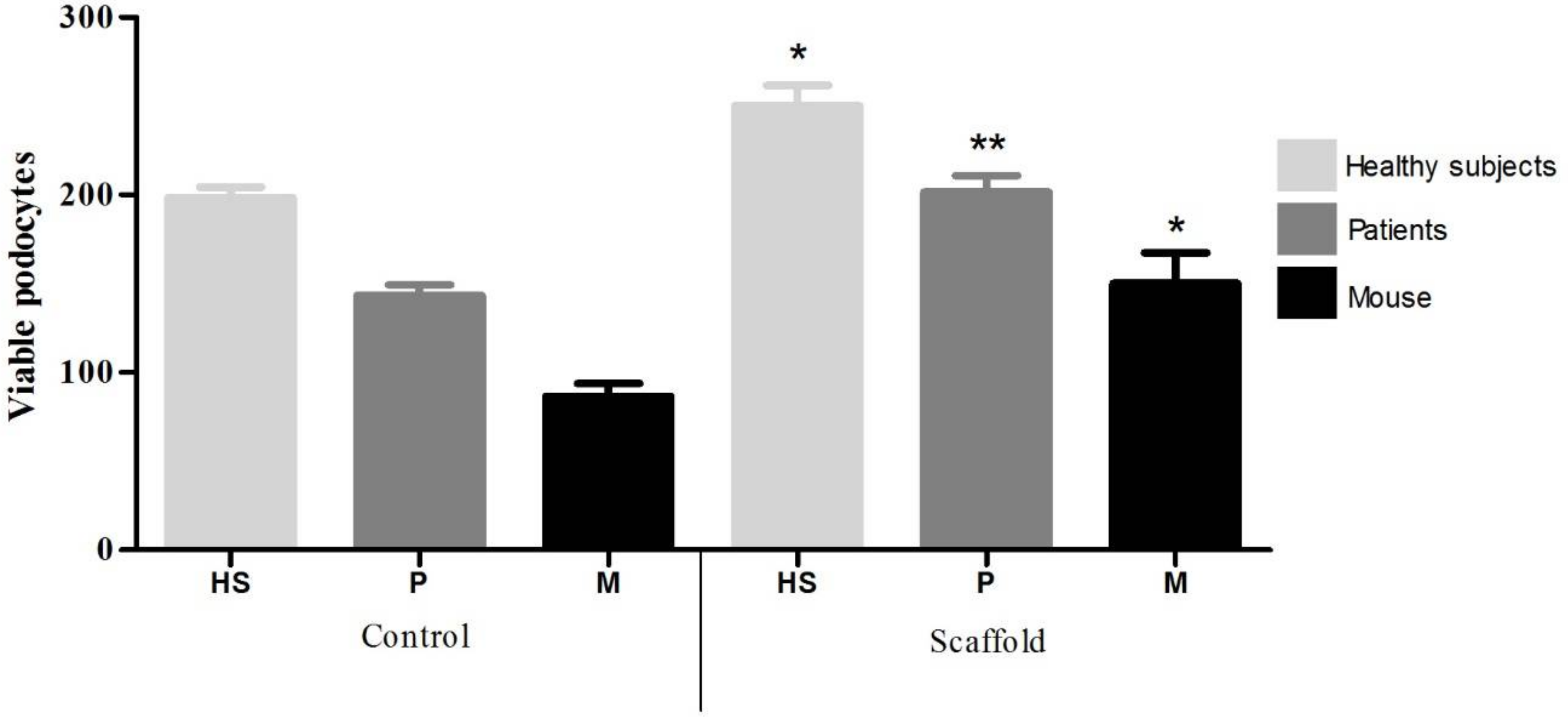
The CTS/PVA/type IV collagen/integrin α3β1 scaffold was placed in a 24-well tissue culture polystyrene plate (Corning Costar, Sigma-Aldrich, Saint Louis, MO, USA). Then, 1 mL cell suspension at a density of 1 × 10⁴ cell/mL in RPMI 1640 medium was placed on each well and maintained in a humidified atmosphere with 5% CO2 at 37 °C. As a culture control, we used a polystyrene plate (Corning Costar, Sigma-Aldrich, Saint Louis, MO, USA), where 1 mL cell suspension at a density of 1 × 10⁴ cell/mL in RPMI 1640 medium were added. After 30 days of culture, podocytes were stained with 4′,6-diamidino-2-phenylindole (DAPI) to be counted and determine the number of viable cells.

2.8. Co-Localization of Podocin, Podocalyxin, and CD80

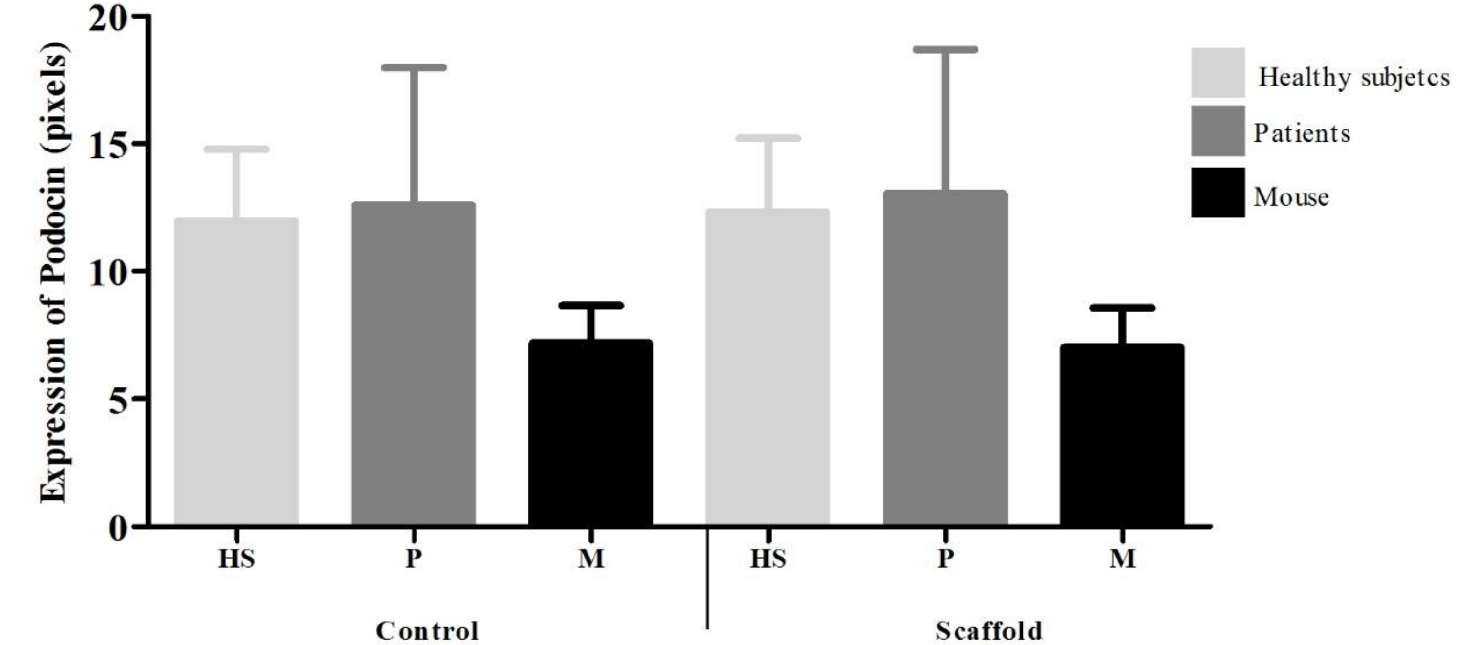
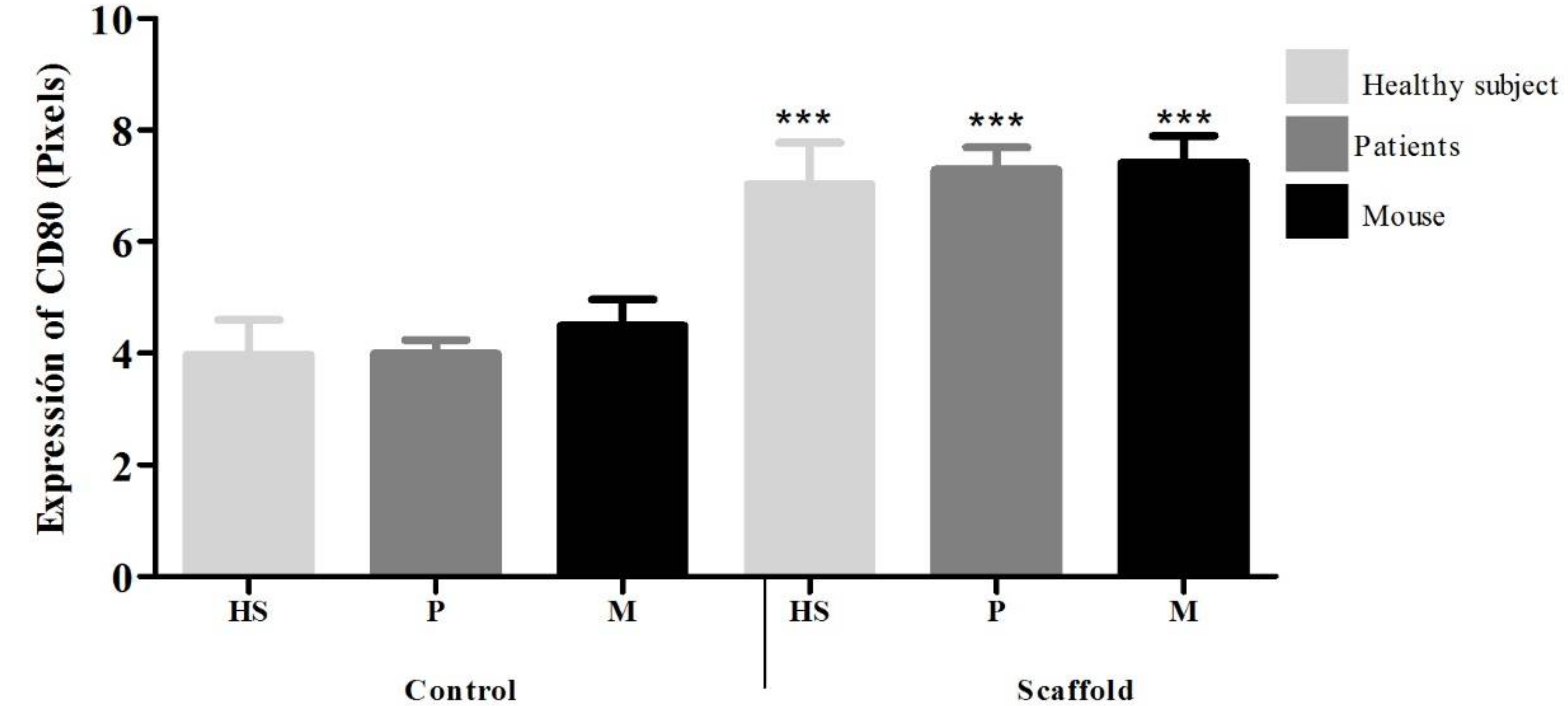
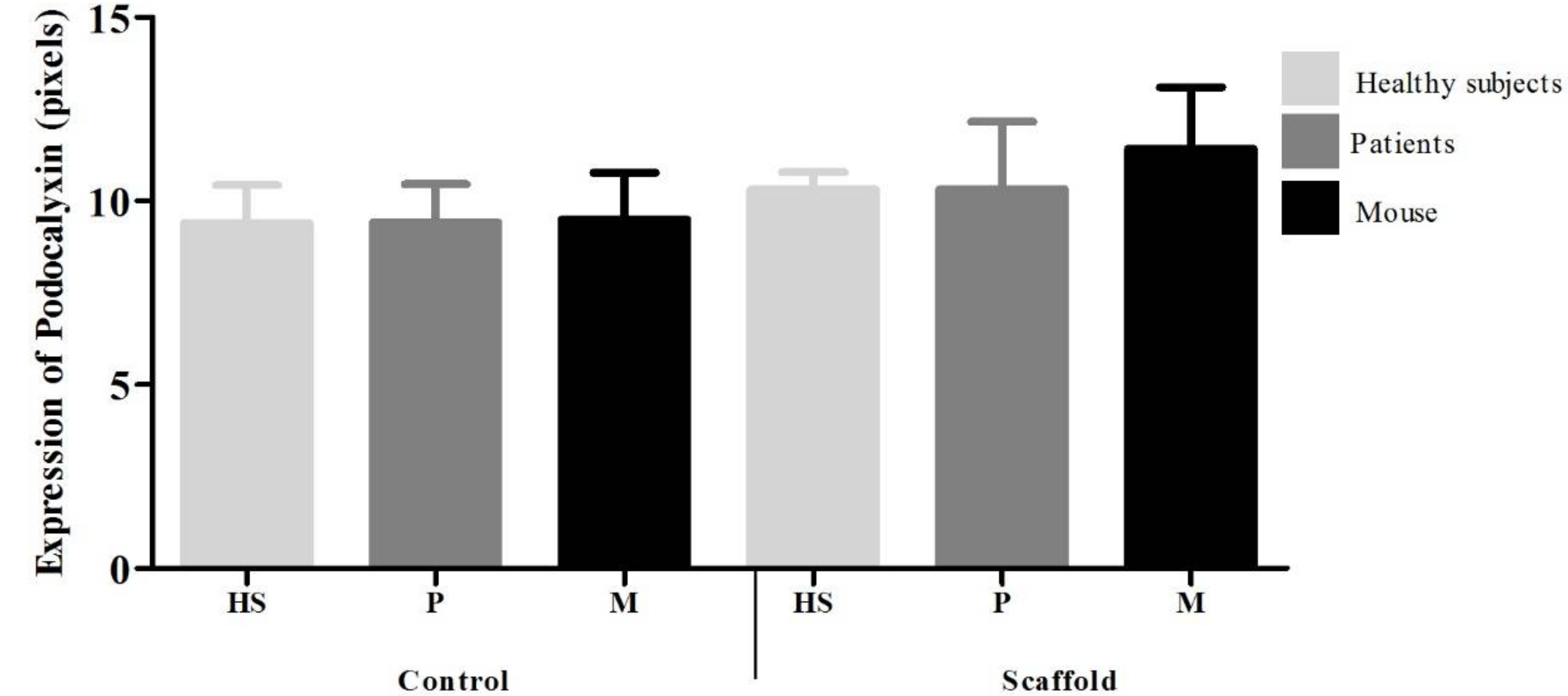
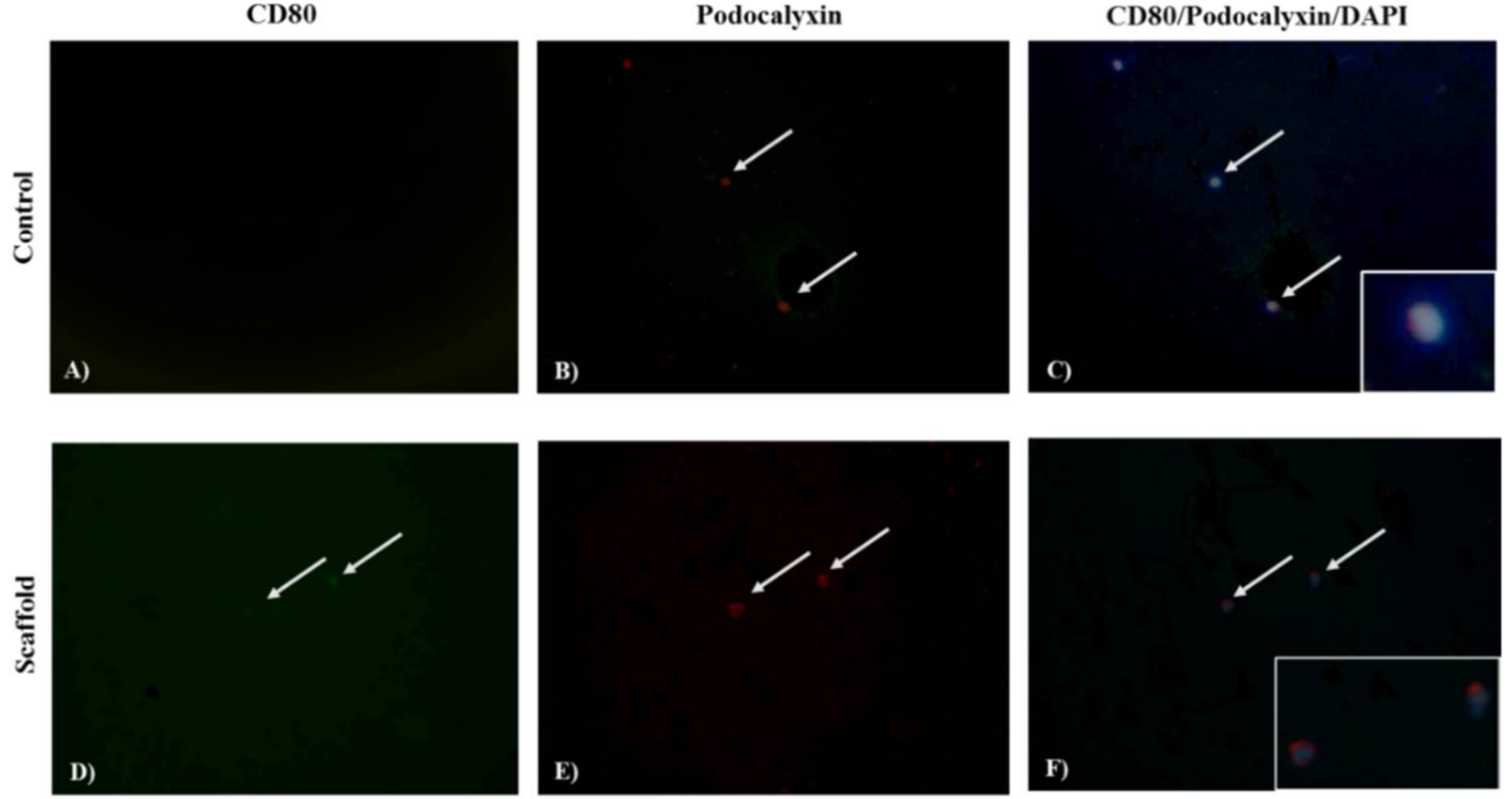
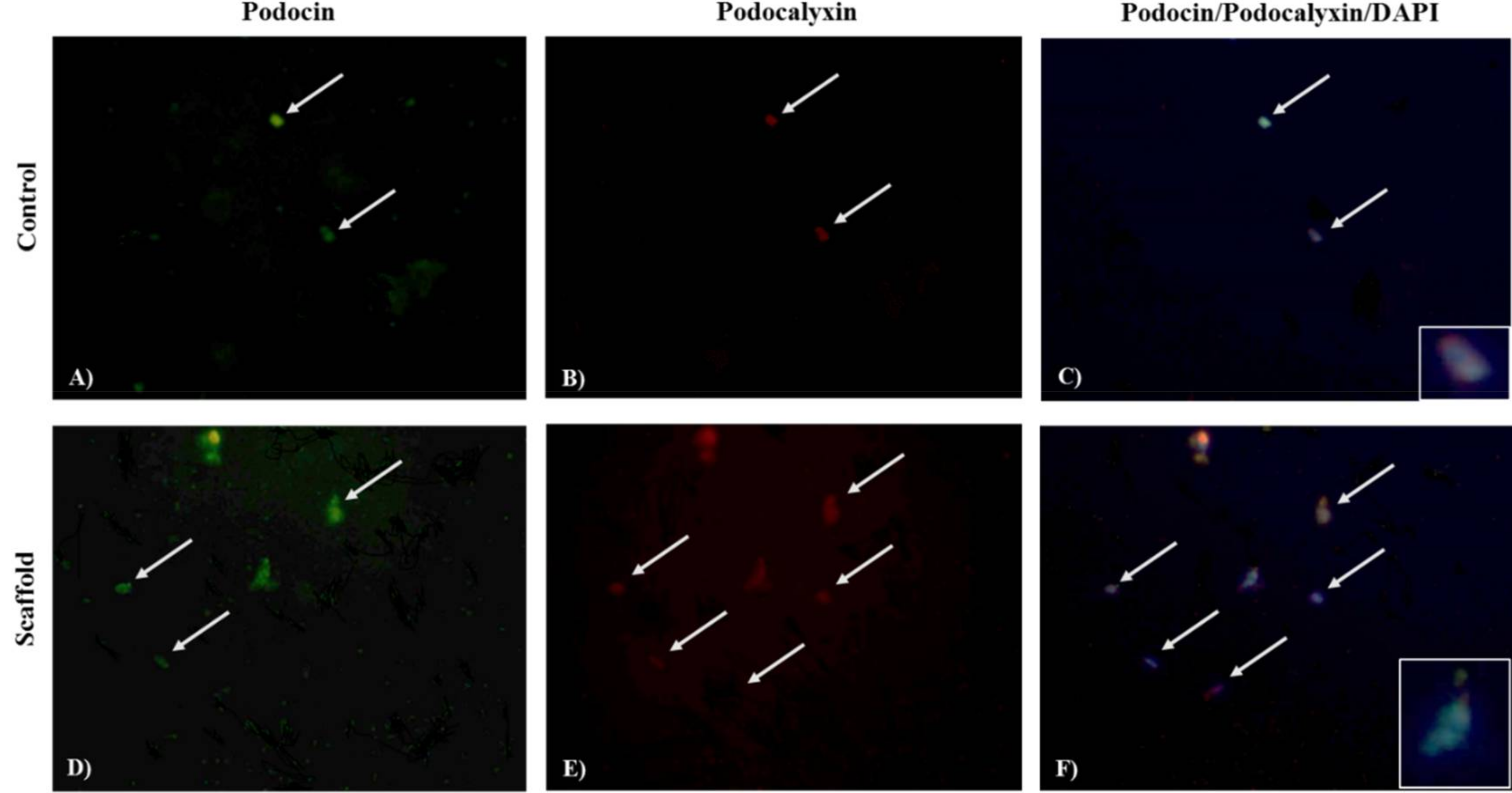
Expression of Podocin, Podocalyxin, and CD80 was identified by indirect immunofluorescent assay (IF). Podocytes were washed with PBS and fixed with 4% paraformaldehyde at 4 °C for 5 min. Then, were permeabilized with Triton 1X (Triton 0.1% with 1% sodium citrate in PBS) for 2 min, and washes three times with PBS. After, the nonspecific binding sites were blocked with 20% fetal bovine serum in PBS for 30 min and incubated for 1 h with anti-Podocin antibody (rabbit anti-human polyclonal IgG, Podocalyxin-like 1 (3D3) mAbs-IgG1 mouse or anti-CD80 (B7.1) antibody Goat anti-human polyclonal IgG diluted 1:100 in PBS. After washes with PBS, the presence of bound antibodies was detected by Goat anti-rabbit IgG FITC, Goat anti-mouse IgG1 TR, and Donkey anti-goat IgG FITC, staining. Podocytes were counterstained with 4′,6-diamidino-2-phenylindole (DAPI) for 30 min, after washing with methanol and methanol-PBS were mounted in PBS/glycerol (3/7) for be photographed at 40X in a microscope (Olympus BX-40). The images were analyzed using Image-Pro Plus software, version 7.0. (Media Cybernetics, Rockville, MD, USA) and the intensity of signal was expressed in pixels. Podocin and CD80 were identified by emitting fluorescence in green, and Podocalixin red color, with the contrast of the DAPI was determined the cellular localization. The analysis was conducted independently by two researchers.

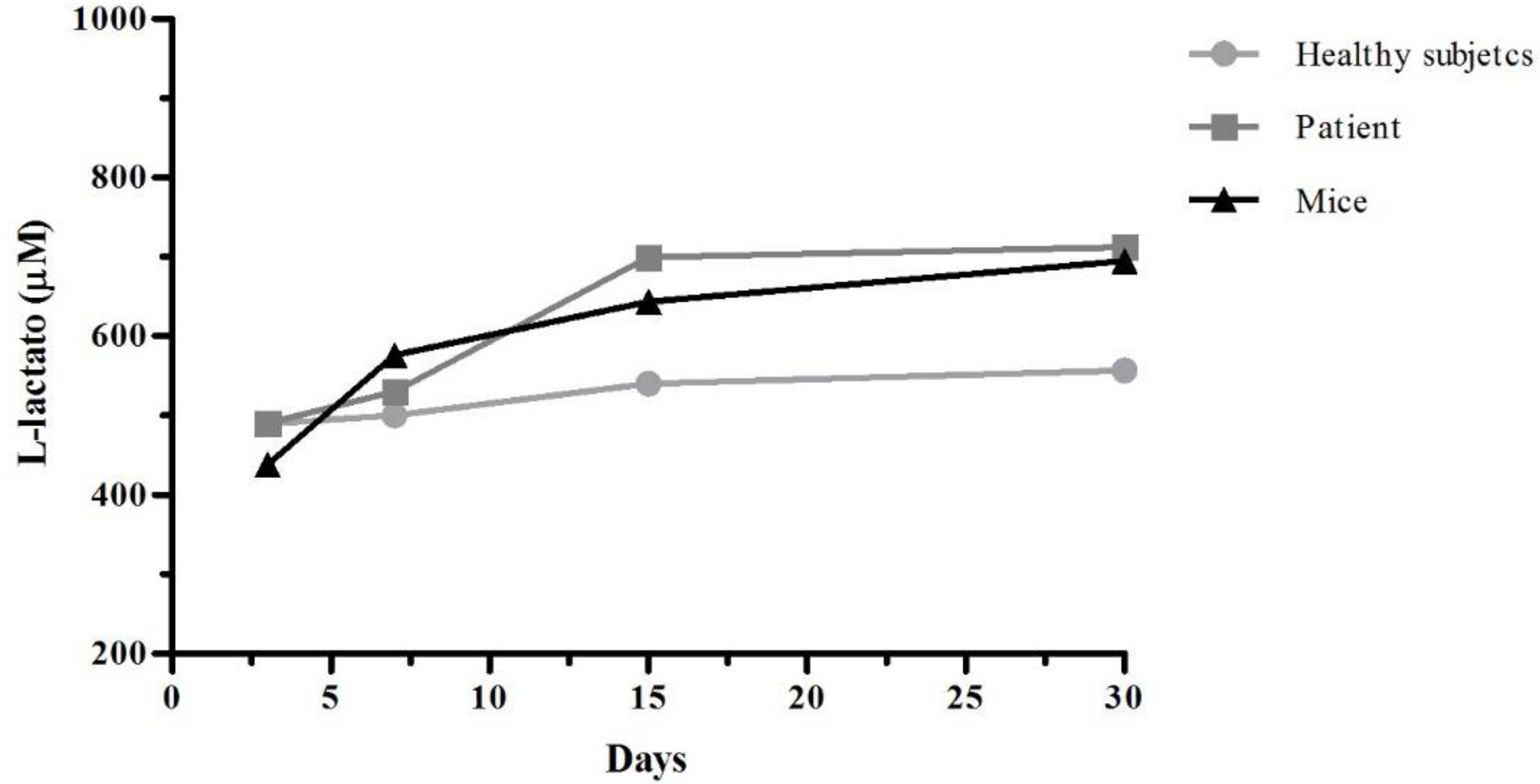
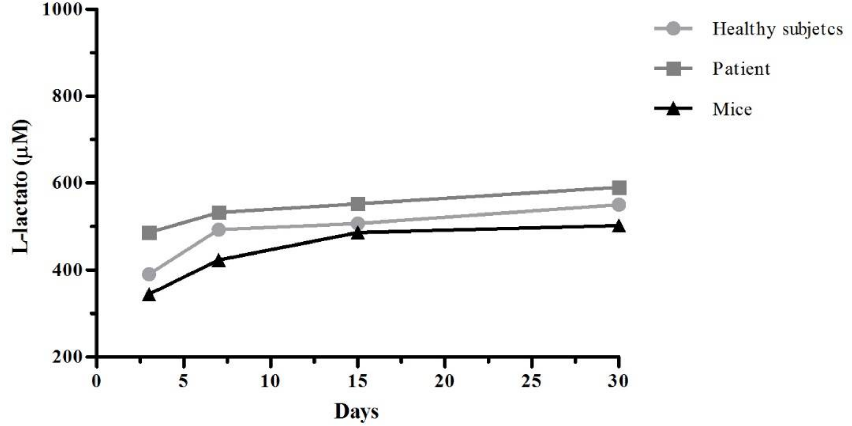
2.9. Cell Viability Assay

Lactate dehydrogenase (LDH) is a cytoplasmic enzyme that is present in the cells. When the cytoplasmic membranes suffer damage, the release of LDH is produce. The quantification of this enzyme in cell culture supernatants it can be used as an indicator of cell death. After 3, 7, 15, and 30 days of culture, the supernatants were recovered and was work according with specifications of L-Lactate Assay Kit (Cayman Chemical Company, Ann Arbor, MI, USA). In the assay, lactate dehydrogenase catalyzes the oxidation of lactate to pyruvate, along with the concomitant reduction of NAD+ to NADH. The measure of formation of NADH was done at 585 nm.

2.10. Statistical Analysis

Statistical analyses were performed using GraphPad Prism 6 program. The results are presented as the mean ± SD. Comparisons were made using one-way ANOVA followed by a Tukey test contrast with a confidence interval of 95%. * p < 0.05, ** p < 0.01 and *** p < 0.001.

3. Results

3.1. Chitosan Amine Groups Are Involved in the Crosslinking Mechanism

FTIR spectroscopy was used to compare the characteristic chemical groups of chitosan and PVA and confirm the crosslinked network formation with type IV collagen and integrin α3β1. Figure 1 shows the FTIR spectra of the CTS, PVA, CTS/PVA, CTS/PVA/type IV collagen and CTS/PVA/type IV collagen/integrin α3β1 scaffolds. Figure 1A shows the spectrum of pure chitosan. The band at 3100 cm−1 corresponds to the N-H groups; two bands at 1640 cm−1 and 1540 cm−1 were observed which has been reported as amine I (O=C–NH) and II (NH2), respectively. The broad band at 1336 cm−1 is related to the O=C–N stretching vibration in chitosan. The FTIR spectrum of PVA (Figure 1A) shows the broad band observed at 3280 cm−1 associated with the hydrogen bonds in the polymeric chains. At 2850 cm−1, the band refers to the stretching (C–H) from alkyl groups and conformation of C–OH groups between 1232 cm−1 and 1417 cm−1. The IR spectra of the CTS/PVA (Figure 1B), CTS/PVA/type IV collagen (Figure 1C) and CTS/PVA/type IV collagen/integrin α3β1 (Figure 1D) scaffolds show a reduction of intensities at 1336 cm−1, corresponding to the O=C–N stretching vibration, this suggest a crosslinking from CTS, PVA, type IV collagen and integrin α3β1.

3.2. The Percentage of Swelling Is Influenced by the Crosslinking Mechanism

Swelling experiments were conducted with CTS, PVA, CTS/PVA, CTS/PVA/type IV collagen and CTS/PVA/type IV collagen/integrin α3β1 scaffolds. The swelling behavior is shown in Figure 2, an initial rapid mass uptake in 15 and 30 min is observed for CTS, PVA and CTS/PVA, followed by mass stabilization at 60 min only for PVA and CTS/PVA scaffold, in contrast, whit CTS scaffold where the mass increased. On the other hand, CTS/PVA/ type IV collagen/integrin α3β1 scaffold has a mass uptake constantly from 15 min. These results reveal that swelling is influenced by crosslinking as CTS and PVA scaffolds show higher percentage of swelling of 575% ± 58.99 and 657.6% ± 45.42 during the time of the test, which is reduced in CTS/PVA (530% ± 31.14), CTS/PVA/type IV collagen (300% ± 35.35), and CTS/PVA/type IV collagen/integrin α3β1 (460% ± 13.06).

3.3. Type IV Collagen and Integrin α3β1 Prevent the Polymer Mass Loss

The results for degradation behavior of CTS, PVA, CTS/PVA, CTS/PVA/type IV collagen and CTS/PVA/type IV collagen/integrin α3β1 scaffolds are depicted in Figure 3. A decrease in the degradation index of scaffolds formulated with type IV collagen and integrin α3β1 (16.48 ± 3.7) was observed, in contrast with CTS (22.03 ± 7.88), PVA (26.80 ± 5.59), CTS/PVA (25.28 ± 9.48) and CTS/PVA/type IV collagen (31.94 ± 2.17). This suggest that the type IV collagen and integrin α3β1 act as chemical crosslinkers, this hypothesis is supported by the FTIR results (Figure 1) that indicate a strong interaction between type IV collagen and integrin α3β1 with amine groups at 1336 cm−1. In addition, CTS and PVA scaffolds show loss of mass due to depolymerization of chains for lack of crosslinking agents.

3.4. The CTS/PVA/type IV Collagen/Integrin α3β1 Scaffold Favors the Attachment of Podocytes

The number of podocytes extracted is depicted in Figure 4, with 8000 viable cells/mL from Balb/c mice kidney, 6000 viable cells/mL from urine of patients with INS, and 2000 viable cells/mL from urine of healthy subjects. After 30 days of culture, podocytes were stained with 4′,6-diamidino-2-phenylindole (DAPI) to be counted and determine the number of viable cells on culture control and CTS/PVA/type IV collagen/integrin α3β1 scaffold. We observed a higher number of viable podocytes between the different groups on scaffold (HS = 250 ± 20, INS = 201.7 ± 16.07 and mouse = 150 ± 30), compared to the control culture (HS = 198.3 ± 10.41, INS = 143.3 ± 10.21 and mouse = 86.67 ± 12.58) (Figure 5).

3.5. Podocytes on CTS/PVA/Type IV Collagen/Integrin α3β1 Scaffold Were Viable after 30 Days of Culture

Figure 6 and Figure 7 show the quantification of lactate dehydrogenase on supernatants from podocytes cultivated on CTS/PVA/type IV collagen/integrin α3β1 scaffold and control culture. We observed a slight increase on concentration of L-lactate in podocytes from control culture (521.8 HS, 608.1 INS and 588.2 mice) in contrast with podocytes from CTS/PVA/type IV collagen/integrin α3β1 scaffold (485.3 μM in HS, 540.2 INS, and 439 mice). There is not significant difference.

3.6. The Expression of CD80 Increased on Podocytes Cultured on CTS/PVA/Type IV Collagen/Integrin α3β1 Scaffold

Expression of CD80 by intensity pixels in podocytes on CTS/PVA/type IV collagen/integrin α3β1 scaffold (HS = 7 ± 2.23, INS = 7.3 ± 0.78 and mouse = 7.4 ± 1.43) was statistically different than podocytes on control culture (HS = 3.9 ± 1.8, INS = 3.9 ± 0.59 and mouse = 4.5 ± 1.13). (Figure 8). On the other hand, the expression of Podocin (Figure 9) on scaffold (HS = 12.34 ± 5.7, INS = 13.07 ± 9.73 and mouse = 7.02 ± 2.66) and control culture (HS = 11.96 ± 5.65, INS = 12.60 ± 9.3 and mouse = 7.19 ± 2.52) it does not have significant modification. Similar to Podocin, expression of podocalyxin (Figure 10) does not have modification, scaffold (HS = 10.31 ± 1.75, INS = 10.32 ± 5.18 and mouse = 11.42 ± 5.01) and control culture (HS = 9.4 ± 3.7, INS = 9.4 ± 2.76 and mouse = 9.49 ± 3.1), these results were obtained of IF images, expression of CD80 (Figure 11A,D), podocin (Figure 12A,D) and podocalyxin (Figure 12B,E).

4. Discussion

Keeping ex vivo podocytes alive will help to clarify the pathophysiological mechanisms involved in glomerular diseases to design new strategies for diagnosis and treatment. Because of this, the development of biocompatible materials that simulate the environmental conditions of specific cells could be useful in future studies.
Our results indicate that swelling percentages of scaffolds CTS and PVA are of the order of 600%, decreasing with the increasing crosslinking type IV collagen and integrin α3β1. These results coincide with those of Caruapoma and Santiago [18] who report that scaffolds of CTS, PVA, and CTS/PVA present swelling percentages of the order of 600%, although they used 10 kilograys (kGy) of gamma radiation to obtain the scaffolds.
Costa et al. [13] reported a decrease in the degradation percentage of scaffold CTS/PVA by increasing of glutaraldehyde (GA) as a crosslinker agent. These results and ours indicate that cross-linking of the polymer chains maintains the stability of the supports and reduce degradation; this is confirmed by the results obtained from FTIR, indicating polymeric crosslinking of type IV collagen and integrin α3β1 through the group O=C–N of Chitosan. These findings coincide with the results reported by Costa et al., where GA has higher affinity for amino groups (NH2) of Chitosan in comparison to hydroxyl groups (OH) of PVA.
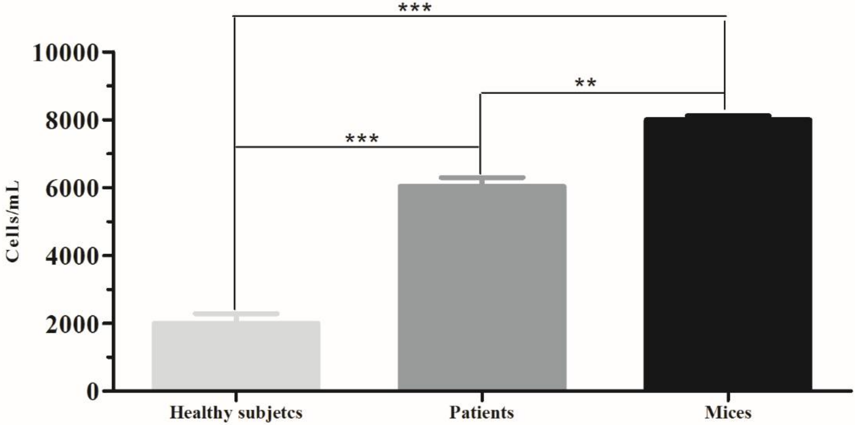
In patients with certain glomerular diseases, podocytes in urine have been detected, correlating with the loss of kidney function. Therefore, we obtained a higher number of podocytes in ISN patients (6000 cells) than HS (2000 cells). These results are consistent with those published by Petermann et al. [19], reporting the presence of viable podocytes in a urine a model of passive Heymann nephritis induced in rats (2500 cells), in contrast to controls where no presence of viable podocytes was observed. Our results are also consistent with those of Nakamura et al. [20] that detected viable podocytes (0.8–5.3 cells/mL) in the urine of 50 diabetic patients and an absence of viable podocytes in the 10 HS.
Evaluating the scaffold in culture, it was observed by IF that a higher number of podocytes adhered to the scaffolds (HS = 2, ISN = 3.5 and mouse = 2), in contrast to the control culture (HS = 1.75, ISN = 2.5 and 1.5 = mouse). These results agree with those of Chuang et al. [12] who reported greater adherence of fibroblasts taken from skin (5 cells per field) on a support of CTS/PVA and presence of microvilli, in contrast to a support of PVA where fewer cells adhered (3 cells per field) and without presence of microvilli. Expression of podocalyxin in podocytes is observed homogeneously both on the scaffold (HS = 10.3, INS = 10.3 and mouse = 11.4) and control cultures (HS = 9.4, INS = 9.4 and mouse = 9.4) however the expression of CD80 is increased in podocytes grown on the scaffold (HS = 7, INS = 7.3 and mouse = 7.4) in contrast to the control culture (HS = 3.9, INS = 3.9 and mouse = 4.5). There are reports that have observed that biomaterials promote increase in the expression of costimulatory molecules (e.g., Ex. CD80, CD86, CD40) of primary culture cells [21]. Recent data also indicate that podocyte cells, under certain circumstances, can be induced to express CD80 (B7.1) [22]. So, we believe that CTS provides an active signaling pathway that leads to the expression of this receptor in podocytes. Cytotoxicity tests using cell cultures are the first step to determine the safety of biomaterials and identifying active compounds. Our results of cell viability show a greater LDH release in culture control in contrast with scaffold culture podocytes. These results agree those of Chuang et al. [12] where CTS/PVA scaffold cultured fibroblast viability was of 70–80%, compared to 30% viability on PVA alone; however, they determined cell viability by Thiazolyl Blue Tetrazolium Bromide MTT technique; and they measured cell viability on day 4 of culture.

5. Conclusions

The material synthesized present a low percent of degradation in relation to CTS, PVA and CTS/PVA, the addition of collagen and integrin α3β1 improved the stability of the scaffold. The FTIR spectra suggest a crosslinking between the components. CTS/PVA/type IV collagen/integrin α3β1 keeping significantly the viability of podocytes in contrast with the conventional culture (polystyrene plate) after 30 days showing the expression of structural proteins. The CTS/PVA/type IV collagen/integrin α3β1 showed promising characteristics such as cellular support and can it employ in the determination of proteins involved in pathological diseases.

Author Contributions

D.G.Z.-T., S.P.H.-M., M.A.F.-M. and C.R.-P. analyzed data and revised the manuscript. D.G.Z.-T., S.P.H.-M. carried out developed and composition design of material, synthesized and characterized of materials. S.P.H.-M., J.J.B.-y.-G.-d.-l.-R., immunofluorescence assay and microscopy. M.A.F.-M. helped to obtained draft manuscript and CD80 detection, E.A.E.-P. supervised the FT-IR analysis S.P.H.-M., M.I.H.-V. cell viability, swelling and degradation test G.A.R.-O. collected samples of patients. D.G.Z.-T. designed synthesis and experiments wrote manuscript. D.G.Z.-T. and S.P.H.-M. wrote the manuscript.

Acknowledgments

The authors would like to thank to CONACYT for scholarship (No. 377122), we thank Reina Araceli Mauricio Sanchez for methodology technical assistance, Alexandra Arreola for article revision and the laboratory of virology and immunology of biological science faculty of the Universidad Autónoma de Nuevo León for technical support and funding.

Conflicts of Interest

The authors declare no conflict of interest.

References

  1. Hu, G.; Jiao, B. Mechanism of podocyte detachment: Targeting transmembrane molecules between podocytes and glomerular basement membrane. Biomed. Aging Pathol. 2013, 3, 36–42. [Google Scholar] [CrossRef]
  2. Jefferson, J.A.; Nelson, P.J.; Najafian, B.; Shankland, S.J. Podocyte Disorders: Core Curriculum 2011. Am. J. Kidney Dis. 2011, 58, 666–677. [Google Scholar] [CrossRef] [PubMed] [Green Version]
  3. Gigante, M.; Piemontese, M.; Gesualdo, L.; Iolascon, A.; Aucella, F. Molecular and Genetic Basis of Inherited Nephrotic Syndrome. Int. J. Nephrol. 2011, 2011, 792195. [Google Scholar] [CrossRef] [PubMed]
  4. Diéguez, S. Molecular bases of the glomerular filtration barrier-steroid resistant nephrotic syndrome. In Proceedings of the Nephrology Conference, Burgos, Spain, 10–25 November 2003. [Google Scholar]
  5. Neal, C. Podocytes What’s under yours? (Podocytes and foot processes and how they change in nephropathy). Front. Endocrinol. 2015, 6, 1–11. [Google Scholar] [CrossRef] [PubMed]
  6. Da Sacco, S.; Lemley, K.V.; Sedrakyan, S.; Zanusso, I.; Petrosyan, A. A Novel Source of Cultured Podocytes. PLoS ONE 2013, 8, e81812. [Google Scholar] [CrossRef] [PubMed]
  7. Java, A.; Liszewski, M.K.; Hourcade, D.E.; Zhang, F.; Atkinson, J.P. Role of complement receptor 1 (CR1; CD35) on epithelial cells: A model for understanding complement-mediated damage in the kidney. Mol. Immunol. 2015, 67, 584–595. [Google Scholar] [CrossRef] [PubMed] [Green Version]
  8. Saleem, M.A.; O’Hare, M.J.; Reiser, J.; Coward, R.J.; Inwar, C.D. A conditionally immortalized human podocytes cell line demonstrating nephrin and podocin expression. J. Am. Soc. Nephrol. 2002, 13, 630–638. [Google Scholar] [PubMed]
  9. Colorado, A.C.; Agudelo, C.A.; Moncada, M.E. Analysis of Biomaterials for use in skin tissue engineering. Biomed. Eng. 2010, 14, 11–23. [Google Scholar]
  10. Dvir, T.; Timko, B.; Kohane, D.; Langer, R. Nanotechnological strategies for engineering complex tissues. Nat. Nanotechnol. 2011, 6, 13–22. [Google Scholar] [CrossRef] [PubMed]
  11. Tamayo, A.; Akbari, M.; Annabi, N.; Paul, A.; Khademhosseini, A.; Juncker, D. Fiber-Based Tissue Engineer: Progress, Challenges and Opportunities. Biotechnol. Adv. 2013, 31, 669–687. [Google Scholar] [CrossRef] [PubMed]
  12. Chuang, W.Y.; Young, T.H.; Yao, C.H.; Chiu, W.Y. Properties of the poly(vinyl alcohol)/chitosan blend and its effects on the culture of fibroblast in vitro. Biomaterials 1999, 20, 1479–1487. [Google Scholar] [CrossRef]
  13. Costa, E.; Pereira, M.; Mansur, H. Properties and biocompatibility of chitosan films modified by blending with PVA and chemically crosslinked. J. Mater. Sci. Mater. Med. 2009, 20, 553–561. [Google Scholar]
  14. Raftery, R.; O’Brien, F.; Cryan, S. Chitosan for Gene Delivery and Orthopedic Tissue Engineering Applications. Molecules 2013, 18, 5611–5647. [Google Scholar] [CrossRef] [PubMed] [Green Version]
  15. Karimi, A.; Navidbakhsh, M.; Faghihi, S. Fabrication and Mechanical Characterization of a Polyvinyl Alcohol Sponge for Tissue Engineering Applications. Perfusion 2014, 29, 231–237. [Google Scholar] [CrossRef] [PubMed]
  16. Londoño, M.; Jaramillo, J.M.; Sabater Rr Vélez, J.M. Dielectric Properties of Poly(vinyl alcohol) Hydrogels Prepared by Freezing/thawing Technique. Rev. EIA 2012, 18, 105–114. [Google Scholar]
  17. Sawada, K.; Toyoda, M.; Kaneyama, N.; Shiraiwa, S.; Moriya, H.; Miyatake, H.; Tanaka, E.; Yamamoto, N.; Miyauchi, M.; Kimura, M.; et al. Upregulation of α3β1-Integrin in Podocytes in Early-Stage Diabetic Nephropathy. J. Diabetes Res. 2016, 2016, 9265074. [Google Scholar] [CrossRef] [PubMed]
  18. Carhuapoma, W.; Santiago, J. Characterization of chitosan-polyvinyl alcohol hidrogel obtained by gamma radation. Iberoam. J. Polym. 2005, 6, 333–346. [Google Scholar]
  19. Petermann, A.; Krofft, R.; Blonski, M.; Hiromura, K.; Vaughn, M.; Pichler, R.; Griffin, S.; Wada, T.; Pippin, J.; Durvasula, R.; et al. Podocytes that detach in experimental membranous nephropathy are viable. Kidney Int. 2003, 64, 1222–1231. [Google Scholar] [CrossRef] [PubMed]
  20. Nakamura, T.; Ushiyama, C.H.; Suzuki, S.; Hara, M.; Shimada, N.; Ebihara, I.; Koide, H. Urinary excretion of podocytes in patients with diabetic nephropathy. Nephrol. Dial. Transp. 2000, 15, 1379–1383. [Google Scholar] [CrossRef] [Green Version]
  21. Vicente Ozores, S. Polysaccharide Nanocapsule as Vaccines Adjuvant Vehicles; University of Santiago de Compostela: Santiago, Spain, 2013. [Google Scholar]
  22. Segarra, A.; Carnicer, C.; Arbós, A.; Quiles, M.; Agraz, I.; Ostos, E. Biomarkers in nephrotic syndrome: Some more steps on the long road. Nephrol. J. 2012, 32, 558–572. [Google Scholar]
Figure 1. Spectra of Fourier Transform Infrared Spectroscopy (FTIR): (A) Chitosan (CTS) and polyvinyl alcohol (PVA), (B) CTS, PVA and CTS/PVA, (C) CTS, PVA and CTS/PVA/type IV Collagen and (D) CTS, PVA and CTS/PVA/type IV Collagen/Integrin α3β1.
Applsci 08 00930 g001
Figure 2. Swelling percentage in scaffolds of CTS, PVA, CTS/PVA, CTS/PVA/type IV Collagen and CTS/PVA/type IV Collagen/integrin α3β1. * p < 0.05, ** p < 0.01.
Applsci 08 00930 g002
Figure 3. Degradation percentage in scaffolds of CTS, PVA, CTS/PVA, CTS/PVA/type IV Collagen and CTS/PVA/type IV Collagen/integrin α3β1 at 7, 15 and 30 days. * p < 0.05, ** p < 0.01.
Applsci 08 00930 g003
Figure 4. Podocytes extracted from urine of patients with Idiopathic Nephrotic Syndrome (INS), healthy subjects, and mice kidney by positive selection. Using system MACS Column and MiniMACS separator. ** p < 0.01, *** p < 0.001.
Applsci 08 00930 g004
Figure 5. Podocytes viable on CTS/PVA/type IV Collagen/integrin α3β1 scaffold. Control culture (polystyrene plate) after 30 days of culture. * p < 0.05, ** p < 0.01.
Applsci 08 00930 g005
Figure 6. Concentration of L-lactato dehydrogenase from podocyte supernatants on control culture (polystyrene plate) to 3, 7, 15 and 30 days.
Applsci 08 00930 g006
Figure 7. Concentration of L-lactato dehydrogenase from podocyte supernatants on CTS/PVA/type IV Collagen/integrin α3β1 scaffold to 3, 7, 15 and 30 days.
Applsci 08 00930 g007
Figure 8. CD80 expression immunofluorescence in podocytes extracted from urine of patients with INS, healthy subjects, and mice kidney after 30 days of culture. The CD80 expression was measured using Image-Pro Plus software, version 7.0. The intensity of signal is shown by pixel intensity. *** p < 0.001.
Applsci 08 00930 g008
Figure 9. Podocin expression immunofluorescence in podocytes extracted from urine of patients with INS, healthy subjects, and mice kidney after 30 days of culture. The Podocin expression was measured using Image-Pro Plus software, version 7.0. The intensity of signal is shown by pixel intensity.
Applsci 08 00930 g009
Figure 10. Podocalyxin expression immunofluorescence in podocytes extracted from urine of patients with INS, healthy subjects, and mice kidney after 30 days of culture. The Podocalyxin expression was measured using Image-Pro Plus software, version 7.0. The intensity of signal is shown by pixel intensity.
Applsci 08 00930 g010
Figure 11. CD80 and Podocalyxin expression in podocytes from urine of patients with INS. (AD) Immunofluorescence of CD80. (BE) Immunofluorescence of Podocalyxin used as marker of podocytes. (CF) Merge of 4′,6-diamidino-2-phenylindole (DAPI) with CD80 and Podocalyxin in cultured podocyte (magnification 40×).
Applsci 08 00930 g011
Figure 12. Podocin and Podocalyxin expression in podocytes from urine of patients with INS. (AD) Immunofluorescence of Podocin. (BE) Immunofluorescence of Podocalyxin used as marker of podocytes. (CF) Merge of DAPI with Podocin and Podocalyxin in cultured podocyte (magnification 40×).
Applsci 08 00930 g012
Table 1. Clinical and biochemical characteristic of the study population.
STUDY GROUPS
GroupsGenderAge (Years)pH UrinePTU (mg/m2/h)Cholesterol Levels (mg/dL)Triglyceride Levels (mg/dL)Serum Albumin (mg/dL)Serum Creatinine (mg/dL)GFR (mL/m)
INSM = 58.33%
F = 41.66%
4.85.759.43227.83165.583.20.327232.5
HSM = 60%
F = 40%
7.85.4------------------------------------
INS: Idiopathic Nephrotic Syndrome, HC: Healthy Subjects, PTU: Proteinuria, GFR: Glomerular Filtration Tests.

