Article

Prophylactic Effects of Polymethoxyflavone-Rich Orange Peel Oil on Nω-Nitro-L-Arginine-Induced Hypertensive Rats

1
College of Food Science, Southwest University, Chongqing 400715, China
2
Citrus Research Institute, Southwest University, Chongqing 400712, China
3
Chongqing Collaborative Innovation Center for Functional Food, Chongqing University of Education, Chongqing 400067, China
4
College of Biological and Chemical Engineering, Chongqing University of Education, Chongqing 400067, China
5
College of Plant Protections, Southwest University, Chongqing 400715, China
6
Chongqing Zhongxian Fruit Industry Bureau, Chongqing 404000, China
*
Authors to whom correspondence should be addressed.
These authors contributed equally to this work.
Appl. Sci. 2018, 8(5), 752; https://doi.org/10.3390/app8050752
Submission received: 13 March 2018 / Revised: 29 April 2018 / Accepted: 4 May 2018 / Published: 9 May 2018
(This article belongs to the Section Applied Biosciences and Bioengineering)

Abstract

:
The prophylactic effects of the polymethoxyflavones (PMFs) in long-leaf orange peel oil (OPO) were determined using an Nω-nitro-L-arginine-induced hypertensive rat model. The OPO contained eight PMF components, namely sinensetin, hexamethoxyflavone, tetramethyl-O-isoscutellarein, nobiletin, tetramethyl-O-scutellarein, heptamethoxyflavone, 5-demethylnobiletin and tangeretin. After treatment with OPO, the SP (systolic pressure) and DP (diastolic pressure) in hypertensive rats were reduced. The NO (nitric oxide) contents in serum, heart, liver and kidney of OPO-treated Nω-nitro-L-arginine (L-NNA)-induced hypertensive rats were higher than those in untreated hypertensive rats, but the MDA (malondialdehyde) contents in OPO-treated rats were lower than those of the control rats (untreated hypertensive rats). ET-1 (endothelin-1), VEGF (vascular endothelial growth factor) and E-selectin serum levels in hypertensive rats could be reduced, but the CGRP (calcium gene-related peptide) level could be increased by OPO treatment. The results of the qPCR assay showed that OPO upregulated HO-1 (heme oxygenase-1), nNOS (neuronal nitric oxide synthase) and eNOS (endothelial nitric oxide synthase) mRNA expression and downregulated ADM (adrenomedullin), RAMP2 (receptor activity modifying protein 2) and iNOS (inducible nitric oxide synthase) expression in hypertensive rats. The Western blot results also proved that OPO upregulated nNOS and eNOS protein expression and downregulated iNOS expression in hypertensive rats. Based on this study, we could conclude that OPO showed good antihypertensive effects, and the effect was concentration dependent.

Graphical Abstract

1. Introduction

The flavonoids in citrus fruits are a major class of secondary metabolites that have significant biological activities. While the exact roles of these stress response and defensive compounds in plant tissue remain unclear [1], the broad-spectrum bioactivities and functionalities of citrus flavonoids, and in particular polymethoxyflavones (PMFs), in mammalian systems have been intensely studied and well-documented in recent years. PMFs display strong anti-allergic, antioxidant, antibacterial, anti-proliferative, anti-inflammatory and anticancer activities [2,3,4,5]. They have also shown inhibitive, preventative and anti-proliferative activities against melanoma cells both in vitro and in vivo [6,7,8,9]. Epidemiological studies have indicated that ingestion of nobiletin, a citrus PMF, is associated with a reduced risk of certain types of cancer and chronic disease [10].
Members of the PMF family have a 2-phenyl-γ-pyrone skeleton, and each member differs in the number, type and positions of methoxy substituents in their core structure. Because of the hydrophobicity of methoxy groups relative to hydroxyl groups, PMFs are more lipophilic than polyhydroxylated flavonoids, such as quercetin, luteolin, scutellarein and naringenin. Therefore, the PMFs have higher membrane permeabilities and are readily absorbed into the human blood stream [11], thus increasing their bioavailability. Likewise, because of their high lipophilicity, almost all PMFs in citrus fruits occur in the peel flavedo, the peripheral surface layer that contains the peel oil, paraffin wax and other hydrophobic components.
Nitric oxide can regulate blood vessel tension and reduce blood pressure. Nitric oxide is also a key material for the prevention of cardiovascular and cerebrovascular diseases, such as hypertension [12]. Nω-nitro-L-arginine (L-NNA) can be used as an NOS inhibitor, and it can inhibit nitric oxide generation, causing high blood pressure [13]. Based on this characteristic, L-NNA is often used in animal experiments for inducing high blood pressure. NOS inhibitors can reduce endothelium-dependent relaxation induced by various vasodilatation factors [14]. A large dose of NOS inhibitor can dramatically reduce the generation of nitric oxide in vascular endothelia in a short time, which promotes the proliferation of vascular smooth muscle cells and noticeably changes the structure of vascular tissues [15]. This process can damage endothelial function and elevate arterial blood pressure, which may further limit nitric oxide synthesis in endothelial cells for maintaining high blood pressure [16]. Angiotensin II is also used for experimental hypertension models [17,18,19]. Angiotensin II can cause over-activation of the rennin-angiotensin-aldosterone system, inducing hypertension, and nitric oxide can prevent different kinds of hypertension [20].
Orange (Citrus sinensis) is the most cultivated and consumed citrus fruit around the world. The value of orange peel oil for food and beverage flavorings, perfumery and aromatherapy has long been recognized. Cold-pressed orange peel oil, which has the best quality, only accounts for approximately 0.6% of the total fruit by weight; however, it contains more than half the amount of the total polymethoxyflavones present in the whole fruit, which makes it the most bioactive fraction of the orange fruit [18]. In the present study, we used a rat model of NOS inhibitory-type hypertension caused by L-NNA to determine the preventative and alleviative effects of PMF-rich orange peel oil. The blood pressures were determined and the molecular markers in the blood and tissues of the rats analyzed to explore the underlying mechanisms of orange peel oil (OPO) activity. The NO (nitric oxide) and MDA (malonaldehyde) contents in serum, heart, liver, kidney and stomach and the ET-1 (endothelin-1), CGRP (calcium gene-related peptide) and VEGF (vascular endothelial growth factor) contents in serum were determined. The mRNA expressions of HO-1, ADM, RAMP2, nNOS, eNOS and iNOS in heart muscle were determined by the qPCR assay, and the nNOS, eNOS and iNOS protein expression was also determined. The results of this study will provide a basis for further utilization of orange peel oil and its PMFs.

2. Materials and Methods

2.1. Samples, Standards and Solvents

Fresh mature orange (Citrus sinensis) cv. Changyecheng (Long-leaf Orange) fruits were purchased from a local orchard. The long-leaf orange, a recently-developed cultivar that is being increasingly planted in China, had been selected as a mutation from a common orange seedling in Chongqing, China. This cultivar was approved by the Crop Variety Approval Committee of Chongqing in 2014 as having excellent potential for both fresh eating and juice processing.
The orange peel oil was obtained by using a cold-pressing method mechanically similar to that used in industry. The fruit flavedo layer was manually peeled and extracted using a household squeezer. The aqueous peel oil emulsion was collected and quickly centrifuged at 12,000 rpm at 4 °C for 10 min. The upper-most oil layer was then collected into large 50-mL tapered centrifuge tubes and frozen at −80 °C for two weeks to remove residual water. Anhydrous orange peel oil (OPO) was stored at 4 °C until subsequent sample preparation, chromatographic analysis and animal experiments.
HPLC-grade standards of nine polymethoxyflavones (PMFs) were used to identify the constituents. Isosinensetin, tetramethyl-O-isoscutellarein, tetramethyl-O-scutellarein, heptamethoxyflavone and 5-demethylnobiletin were purchased from Yuanye Biotech (Shanghai, China); tangeretin was purchased from ChromaDex (Irvine, CA, USA); sinensetin and nobiletin were purchased from Toronto Research Chemicals (Toronto, ON, Canada); and 3,5,6,7,3′,4′-hexamethoxyflavone was purchased from Purify Tech Co. Ltd. (Chengdu, China). Stock solutions were prepared of the individual standard at concentrations of approximately 1 mg Ml−1 in methanol and stored at −20 °C. All reference standards were analyzed by employing a Thermo Ultimate 3000 HPLC with a Thermo Quantiva triple quadrupole electrospray ionization tandem mass spectrometer; the results showed that the purities of the stock solutions were all ≥98%. The stock solution of every standard compound was diluted in series so the calibration equation could be determined.
A GC-grade D-limonene reference standard was purchased from Toronto Research Chemicals (Toronto, ON, Canada), and its purity was found to be ≥95% using an Agilent 7890B GC equipped with an MS detector.
HPLC-grade acetonitrile, methanol, hexane and tetrahydrofuran (THF) were purchased from Honeywell (Morris Plains, NJ, USA). Analytical-grade phosphoric acid was purchased from Kelong Co. (Chengdu, Sichuan, China). Water was freshly purified using a Milli-Q plus Advantage A10 system (Molsheim, France).

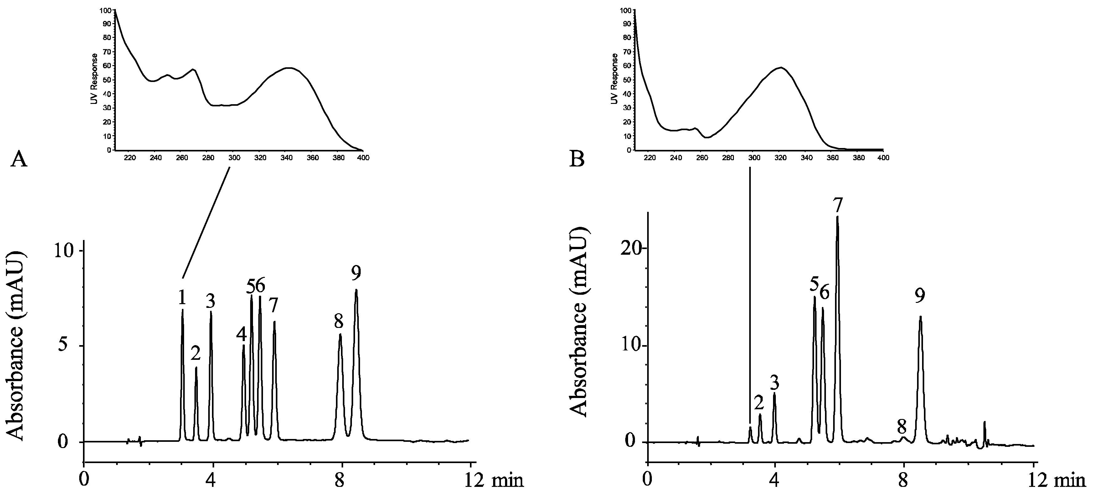
2.2. HPLC Analysis of the Polymethoxyflavones in OPO

Five microliters of orange peel oil were dissolved in 995 μL of methanol to generate the HPLC sample, and the injection volume was 2 μL. An Agilent Poroshell EC-C8 column (i.d. 4.6 × 150 mm, 2.7 μm particle) was used with an Agilent 1260 Infinity chromatography system. The system consisted of a G1311B quaternary pump, a G1329B auto sampler, a G1316A thermostated column compartment and a G1315D diode array detector (DAD). The solvent consisted of 0.05% phosphoric acid/water (A), methanol (B) and 50% THF/water (C), and the flow rate was 1 mL min−1. The solvent program consisted of the following linear gradients after a 0–5.00 min isocratic period, 52% B and 7.5% C; 5.01–7.50 min: 58–62% B and 0% C; 7.50–10.00 min: 62–100% B and 0% C; and 10.01–35.00 min: 52% B and 7.5% C to re-establish the initial conditions. The unlisted percentages of eluent were solvent A. The column temperature was maintained at 30 °C. The diode array detector was set to scan from 190–400 nm. The bandwidth and slit were set at 4 nm; the response time was 2.0 s; and the sampling frequency was 2.5 Hz. To identify the constituents, ultraviolet (UV) absorbance data and retention times of the sample peaks were compared with those of standards. The standard calibration equations were employed to quantify the identified PMF compounds.

2.3. GC-MS Analysis of D-Limonene in OPO

A microliter of OPO was dissolved in hexane to prepare 1 mL of sample solution (1000 × diluted). The qualitative and semi-quantitative determinations of OPO D-limonene were made using an Agilent model 5977A MSD mass spectrometer (MS) with a 7693A autosampler and a Model 7890B gas chromatograph (GC) equipped with a 30 m × 0.25 mm i.d. HP-5 column with a film thickness of 0.25 μm (Agilent, Palo Alto, CA, USA). The initial oven temperature was held at 35 °C for 5 min. It was then increased at 7 °C/min to 250 °C and held for 10 min. The injection port and ionizing source were both kept at 230 °C; the quadrupole was kept at 150 °C. Mass spectra were collected from m/z 30–400. Compounds were identified using the NIST14 library (NIST, Gaithersburg, MD, USA), as well as by comparing the mass spectra and retention times with those of the D-limonene reference standard.

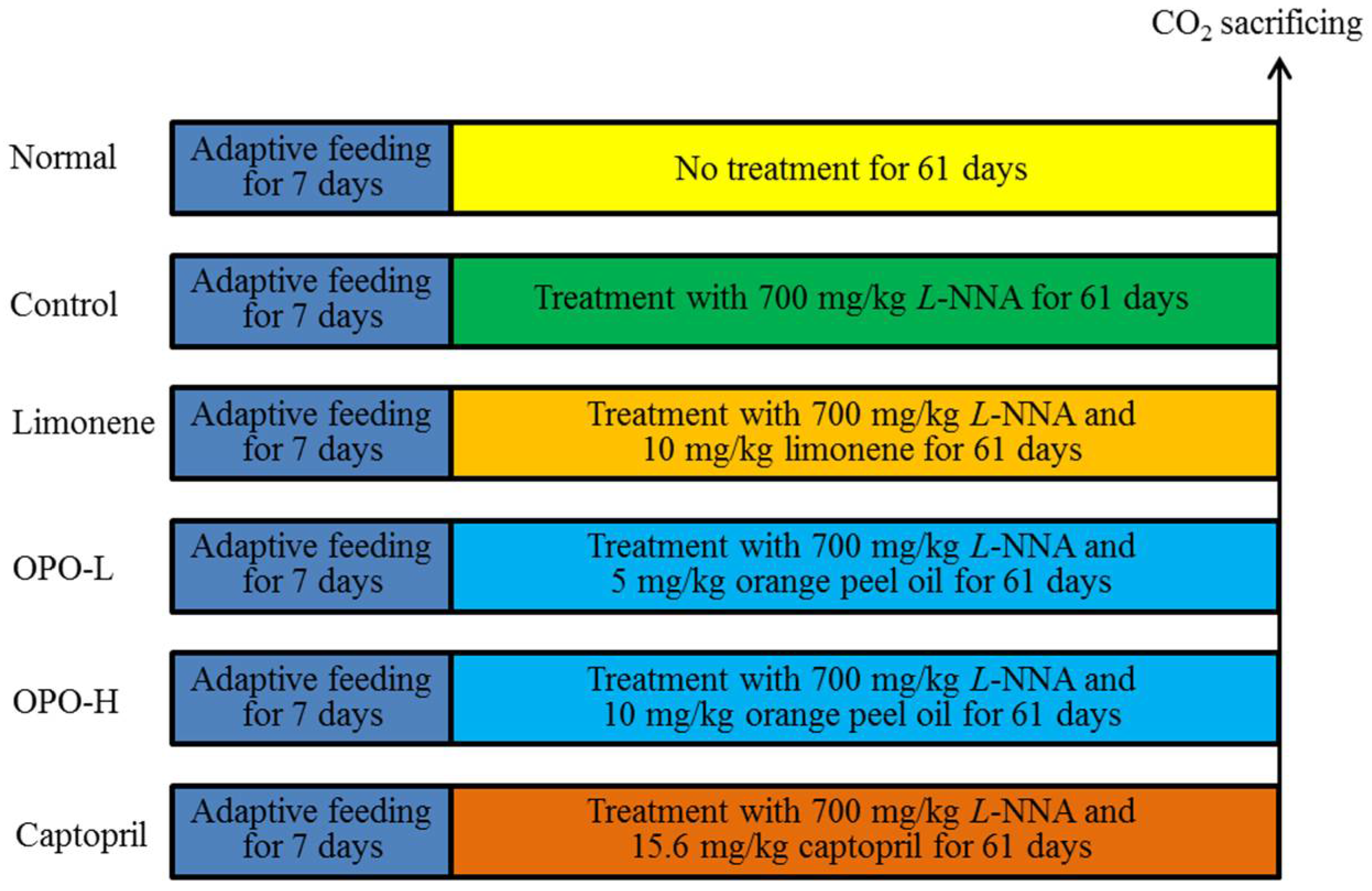
2.4. Animal Experiments

Sixty male Sprague-Dawley (SD) rats, which were 6 weeks of age and had bodyweights of 165 ± 10 g, were purchased from the Experimental Animal Center of Chongqing Medical University (Chongqing, China). The rats were fed in a pathogen-free animal house (temperature 25 ± 2 °C, relative humidity 50% ± 5%; a 12-h light/dark cycle, SCXK (Yu) 2012-0001, Chongqing, China). These rats were averagely divided into six groups for the experiment, i.e., the normal group, control group, orange peel oil-low (OPO-L) group, orange peel oil-high (OPO-H) group, limonene group and captopril group. Rats were housed two per cage, and the pair of rats both belonged to the same test group. Prior to beginning the experiments, the animals were allowed 7 days to acclimatize to the sphygmomanometric procedures.
Rats in the normal group were fed a commercial diet and had access to tap water, and they were not given any drug. The rats of other groups were treated with 700 mg/kg L-NNA (Nω-nitro-L-arginine, Shanghai Gold Wheat Biological Technology Co. Ltd., Shanghai, China) by gavage for 61 days. Every day starting at the beginning of the seventh day, 5 mL/kg and 10 mL/kg doses of orange peel oil were feed via gavage to rats in the OPO-L and OPO-H groups, respectively (Figure 1). Ten milliliters per kilogram bodyweight of food-grade limonene (Citrosuco, Matão, SP, Brazil) were fed to each rat in the limonene group, whereas 15.6 mg captopril/kg (Sigma, St. Louis, MO, USA) were fed by gavage to each rat in the captopril group [19].
On every third day, bodyweight and water-intake were recorded. After treatment with L-NNA at approximately 6 p.m., a 60 min buffer period was given to allow the possible elevation of blood pressure that was agitated by gavage to subside (a pilot study was applied to the rats, and the result showed that blood pressure calmed down normally in approximately 0.5 h; data not shown). Then, systolic and diastolic pressure were recorded by the tail-cuff method using a BP-100A auto-sphygmomanometric system (Techman Soft Inc., Chengdu, China). The operational principle and data profile are shown in Figure 1. At least four consecutive cycles (inflation/deflation) were performed on each rat, and the mean of the last four recordings among which there was a difference of less than a 10-mmHg was accepted. Then, all rats were sacrificed using CO2; the heart, liver, kidney, stomach and blood vessel tissue and blood were collected. These experiments followed a protocol approved by the Animal Ethics Committee of Chongqing Medical University (Chongqing, China).

2.5. Serum Level Assay

After the blood was taken, the blood was solidified for 1 h–2 h at 37 °C. Then, the blood was centrifuged at 3000 rpm for 10 min, the serum separated and the insoluble matter discarded. Finally, the serum levels of ET-1 (No. H093), CGRP (No. H217), VEGF (H044), E-selectin (H029), NO (A012-1) and MDA (A003-1) were determined using the corresponding kits (Nanjing Jiancheng Bioengineering Institute, Nanjing, Jiangsu, China).

2.6. Tissue Level Assay

Tissue samples (1.0 g) were removed and rinsed in saline. The blood was removed, and the tissue was broken down into 10% tissue homogenates at 10,000 r/min. The NO and MDA contents were determined using the corresponding kits (Nanjing Jiancheng Bioengineering Institute, Nanjing, Jiangsu, China).

2.7. Real-Time Quantitative PCR Assay

The colon tissues of the rats were disrupted, and then, the total RNA of the colon tissue was extracted with RNAzol. The total RNA content extracted was diluted to 1 μg/μL. A 5-μL aliquot of the diluted total RNA extracting solution was used to obtain the cDNA template according to the manufacturer’s instructions of the reverse transcription kit. A 2-μL aliquot of the cDNA template was mixed with 10 μL of SYBR Green PCR Master Mix and 1 μL of each of the upstream and downstream primers (Table 1). A total of 40 cycles of 95 °C for 15 s was performed moving to 55 °C after 60 s followed by 30 s; then 72 °C for 35 s; then 95 °C for 30 s; and finally, 55 °C for 35 s. GAPDH was used as the reference to calculate the relative expression levels of the genes according to the formula 2-ΔΔCt = ΔCt(detecting gene) − ΔCt(GAPDH) [19].

2.8. Western Blot Assay

Approximately 0.5 g of colonic tissue were used to produce 1 mL of cell lysate from each rat. Briefly, the tissues were homogenized and then centrifuged at 1200 rpm at 4 °C, and the supernatant was collected and stored at −80 °C until analysis. A protein standard curve was generated to determine the protein concentration in each sample according to the instructions of the BCA Protein Concentration Assay Kit (Thermo Fisher Scientific, Waltham, MA, USA). Approximately 150 μg of protein were mixed with twice that volume of buffer solution, and this solution was subjected to 12% SDS-PAGE (Thermo Fisher Scientific) gel electrophoresis at 140 V for 2 h. The resolved proteins were then transferred onto a PVDF membrane (Thermo Fisher Scientific) at 4 °C, 100 mA, for 1 h and then blocked using 5% skim milk at 25 °C with shaking for 1 h. After incubating overnight at 4 °C, the membranes were washed with TBST three times for 10 min each. Then, the membranes were incubated at 25 °C for 2 h with a secondary antibody (Thermo Fisher Scientific) and then washed three times with TBST for 10 min each. Finally, the grey values of the groups were compared [19].

2.9. Statistical Analysis

The data were expressed as the mean ± the standard (SD) deviation. The significant differences (p < 0.05) in the data from different groups were calculated using Duncan’s multiple range test using SAS Version 9.2 (SAS Institute Inc., Cary, NC, USA).

3. Results

3.1. Polymethoxyflavones and D-Limonene Contents in Orange Peel Oil

Cold-pressed citrus peel oil mainly consists of volatile terpenoids and non-volatile components. The terpenoids generally do not absorb UV light; therefore, they cannot be determined by HPLC-UV, but they are suitable for GC-MS analysis. In contrast, the non-volatile compounds, like polymethoxyflavones, absorb more UV light, but they have a higher boiling point; thus, they are typically separated and analyzed using HPLC with a UV detector.
The HPLC chromatogram of the non-volatile components in long-leaf orange peel oil (OPO) is shown in Figure 2. By comparing the retention time (Rt) and UV spectra of the oil sample (Figure 2B) and those of the mixed standards (Figure 2A), eight polymethoxyflavones (Compounds 2–9) were identified in the OPO. The small peak at 3.2 min (Figure 2B) was confirmed to not be isosinensetin (1) because its Rt was slightly later than the corresponding standard peak (3.0 min), and their 210–400-nm UV spectra were different, as shown above each chromatogram. The total ion current (TIC) obtained from GC-MS analysis of the volatile components in OPO is shown in Figure 3. The most abundant peak was identified as D-limonene.
The quantitative results of the PMFs and limonene are shown in Table 2. In general, D-limonene accounts for more than 95% of the total volatiles, and it is the major component of orange peel oil; the non-volatiles, which are dissolved in limonene, consist primarily of PMFs, as these compounds account for more than 91% of the total peak area. Specifically, nobiletin and heptamethoxyflavone were the most abundant PMFs in long-leaf OPO, followed by tangeretin, tetramethyl-O-scutellarein and hexamethoxyflavone. 5-Demethylnobiletin, the 5-hydroxyl derivative of nobiletin, was the least concentrated PMF in long-leaf OPO.

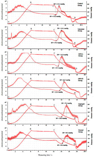
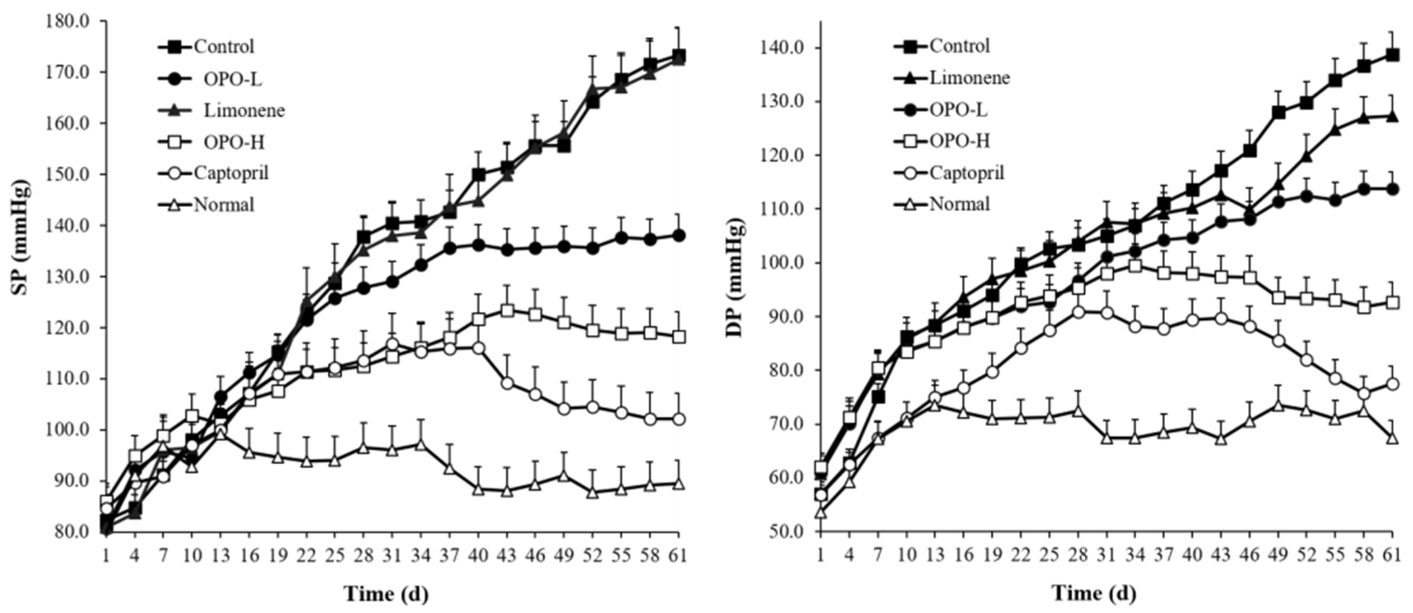
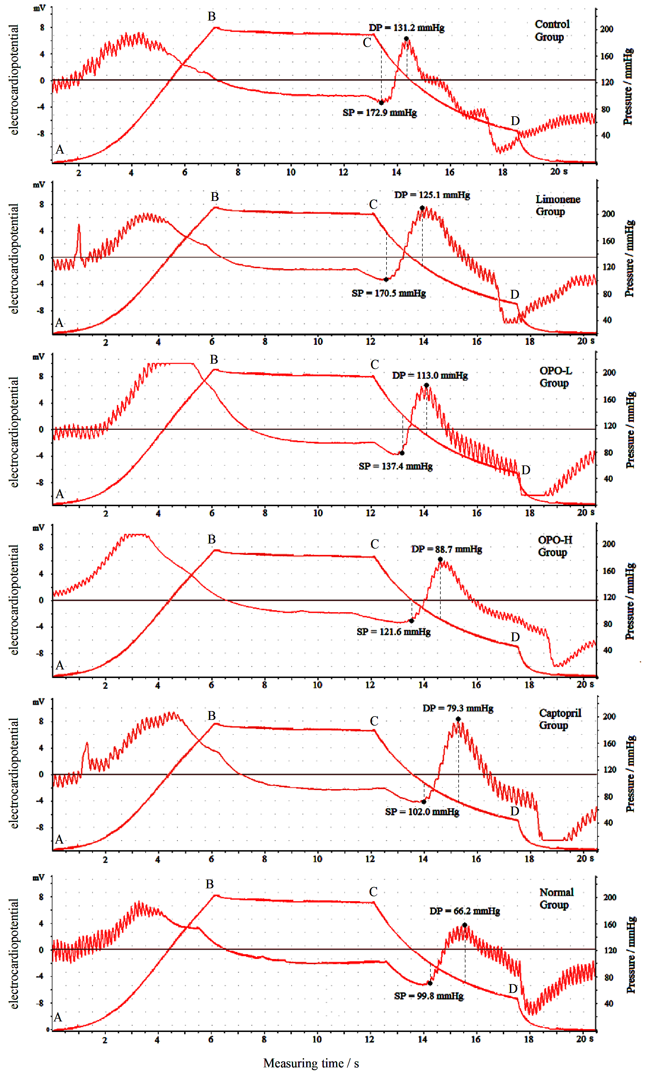
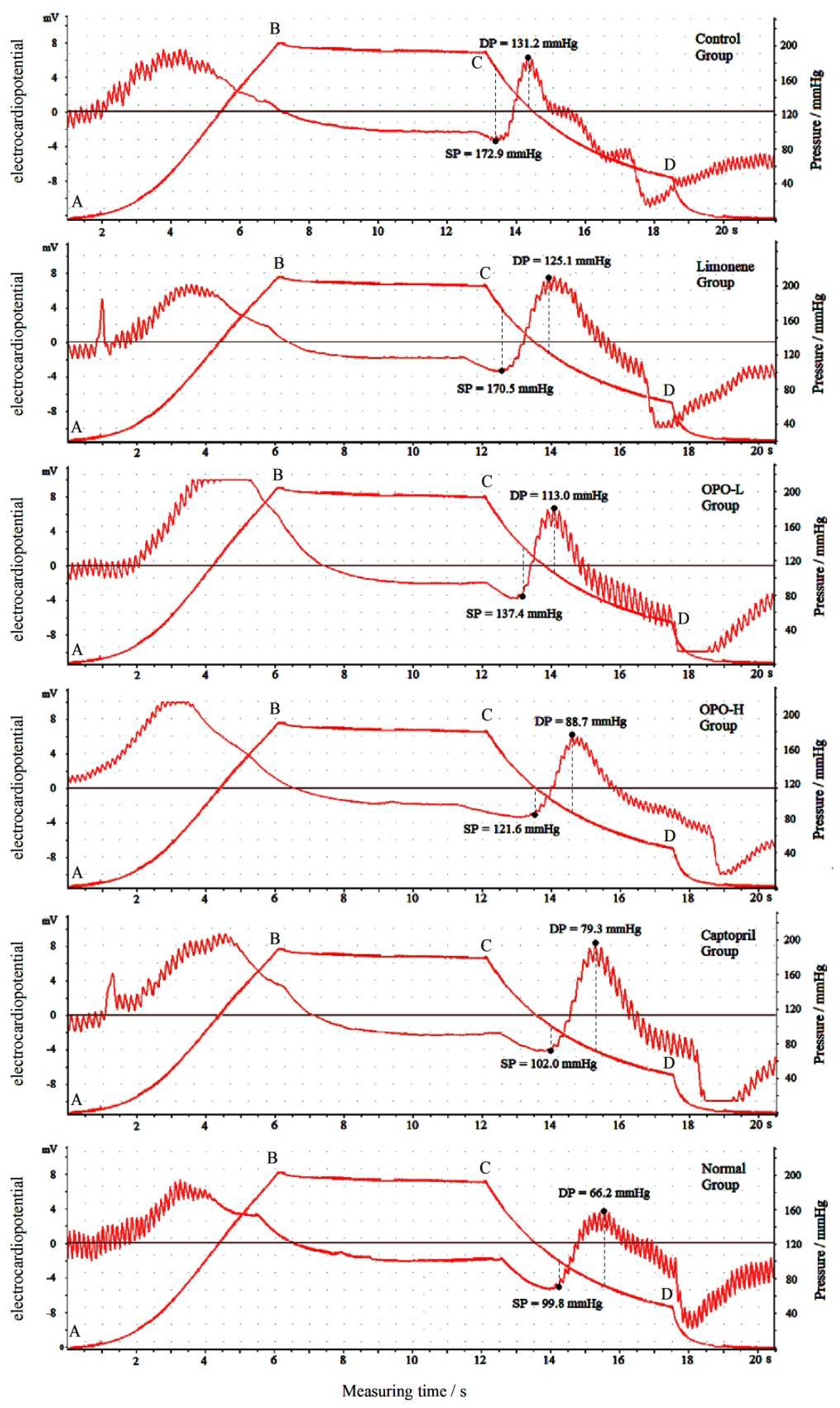
3.2. Effects of Orange Peel Oil on Blood Pressure

Figure 4 shows an example of the recorded working curves during one sphygmomanometric cycle (Day 61). The trapezoidal line segmented by Points A, B, C and D represents the inflation, static pressure, deflation and measurement and vent. The other curve shows the detected blood pressure and heart rate.
The changes in the SP and DP of each group are shown in Figure 5. After treatment with L-NNA, the SP and DP of all treated groups increased; the control group, which was treated with only L-NNA, showed the highest blood pressure with an SP of 174 ± 6 mmHg and a DP of 138 ± 4 mmHg on Day 61. The untreated rats (normal group) showed the lowest blood pressures, with SPs varying from 85 ± 3–99 ± 4 and DPs varying from 53 ± 5–74 ± 4. The 10-mL/kg limonene-treated group exhibited no significant changes in SP, but the DP was 11 mmHg lower than that of the control group. In comparison, ingestion of either a low dose or high dose of OPO or a medication containing captopril (15.6 mg/kg) significantly (p < 0.05) decreased blood pressure in the corresponding groups. The OPO high dose (10 mL/kg) treatment showed a greater reducing effect than the low dose of OPO (5 mL/kg). At the end of the experiment (Day 61), the SP/DP of the OPO high dose group (120 ± 5/95 ± 3 mmHg) was 15/16 mmHg higher than that of the captopril-treated group, but it was 54/43 mmHg lower than that of the control group. Typical examples of the blood pressure of each group on Day 61 are also shown in detail in Figure 3.

3.3. Nitric Oxide Content

As shown in Table 3, the NO contents in serum, heart, liver and kidney of rats in the normal group were the highest, and the contents in captopril-treated rats were the next lowest. The NO contents in rats in the OPO-H group were higher (p < 0.05) than those of rats in the OPO-L group, limonene group and control group.

3.4. Malondialdehyde Content

As shown in Table 4, the rats in the control group had the highest MDA contents, and the rats in the normal group had the lowest MDA contents. OPO-H-treated rats had lower MDA contents than OPO-L-treated rats, limonene-treated rats and rats in the control group, but the OPO-H-treated rats had higher MDA contents than captopril-treated rats and rats in the normal group.

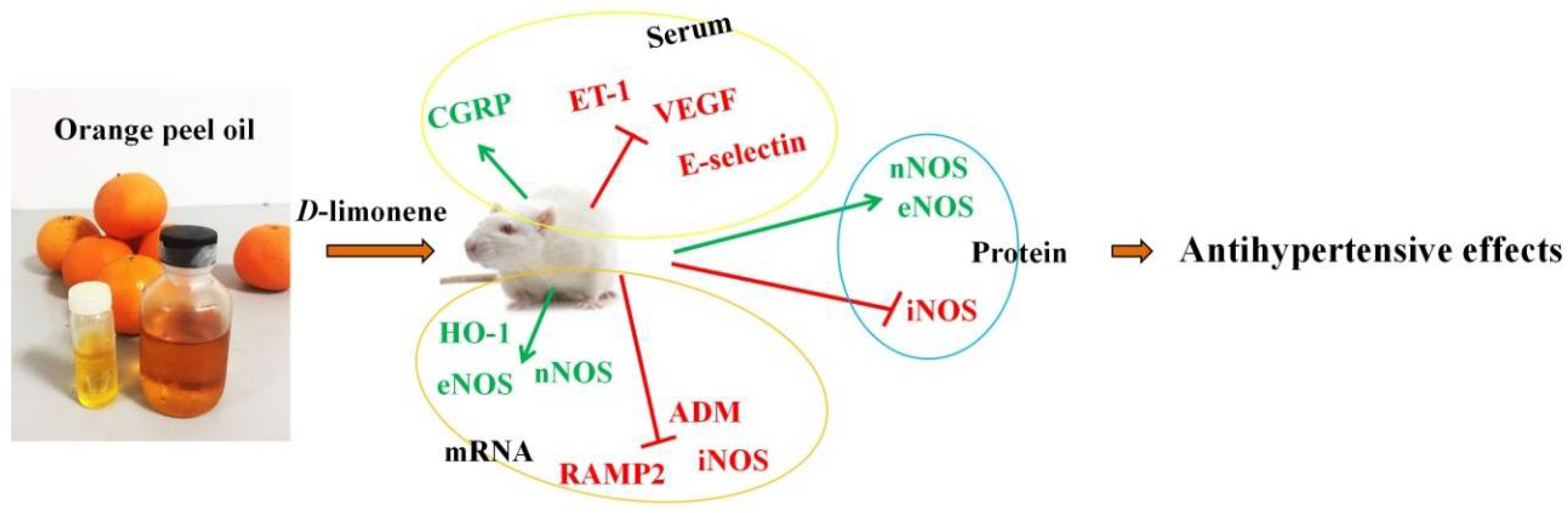
3.5. Serum Levels of ET-1, CGRP, VEGF and E-Selectin

As shown in Table 5, the rats in the normal group had the highest CGRP levels and the lowest ET-1, VEGF and E-selectin levels, and the levels of these compounds in captopril-treated rats were similar to those of the rats in the normal group; there were no significant differences (p > 0.05) between these values in the two groups of rats. Meanwhile, the CGRP levels in OPO-H-treated rats were higher than those in the OPO-L-treated rats, the limonene-treated rats and the rats in the control group; the ET-1, VEGF, E-selectin levels in OPO-H-treated rats were similar to those of the captopril-treated rats, but lower than those of the rats in the OPO-L, limonene and control groups.

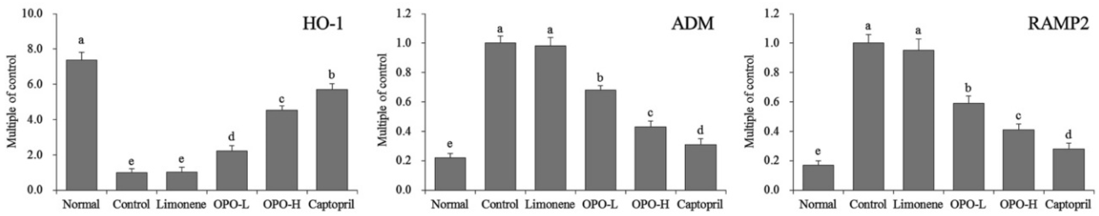
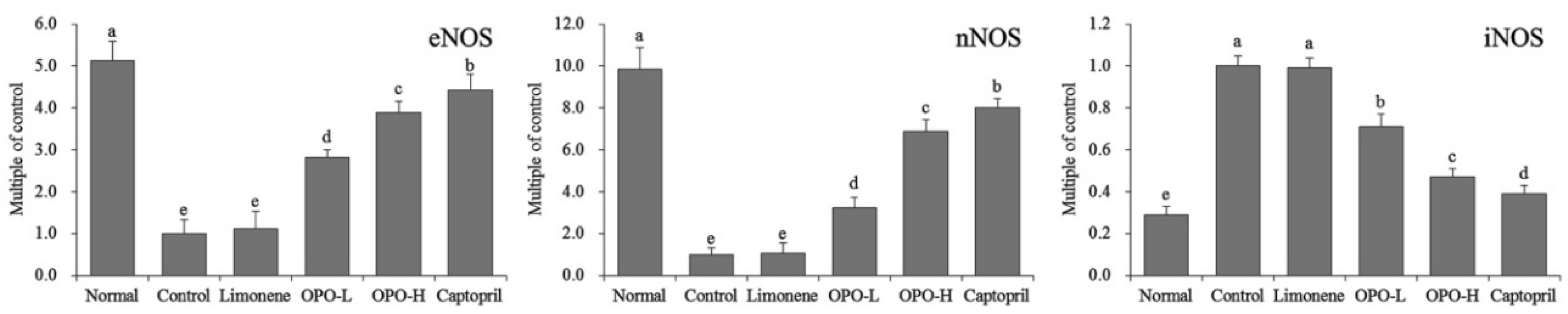
3.6. mRNA Expression of Myocardial Tissue

As shown in Figure 6, OPO could increase HO-1 mRNA expression and reduce ADM and RAMP2 expression in myocardial tissue of rats relative to the expression levels in rats in the control group, and the high dose of OPO (OPO-H) showed a greater effect than that observed with the low dose of OPO (OPO-L); the effects of OPO-H were only slightly weaker than those of the drug containing captopril. However, limonene had almost no effect on hypertensive rats (relative to the control group).
As shown in Figure 7, the rats in the normal group had the highest levels of nNOS and eNOS expression and the lowest level of iNOS expression in myocardial tissue, but the rats in the control group had the lowest levels of nNOS and eNOS expression and highest level of iNOS expression. OPO-H-treated rats also had higher levels of nNOS and eNOS expression and lower levels of iNOS expression than limonene-treated rats, OPO-L-treated rats and rats in the control group, but they had lower expression levels than captopril-treated rats and rats in the normal group. The iNOS expression in OPO-H-treated rats was lower than those in limonene-treated rats, OPO-L-treated rats and the rats in the control group.

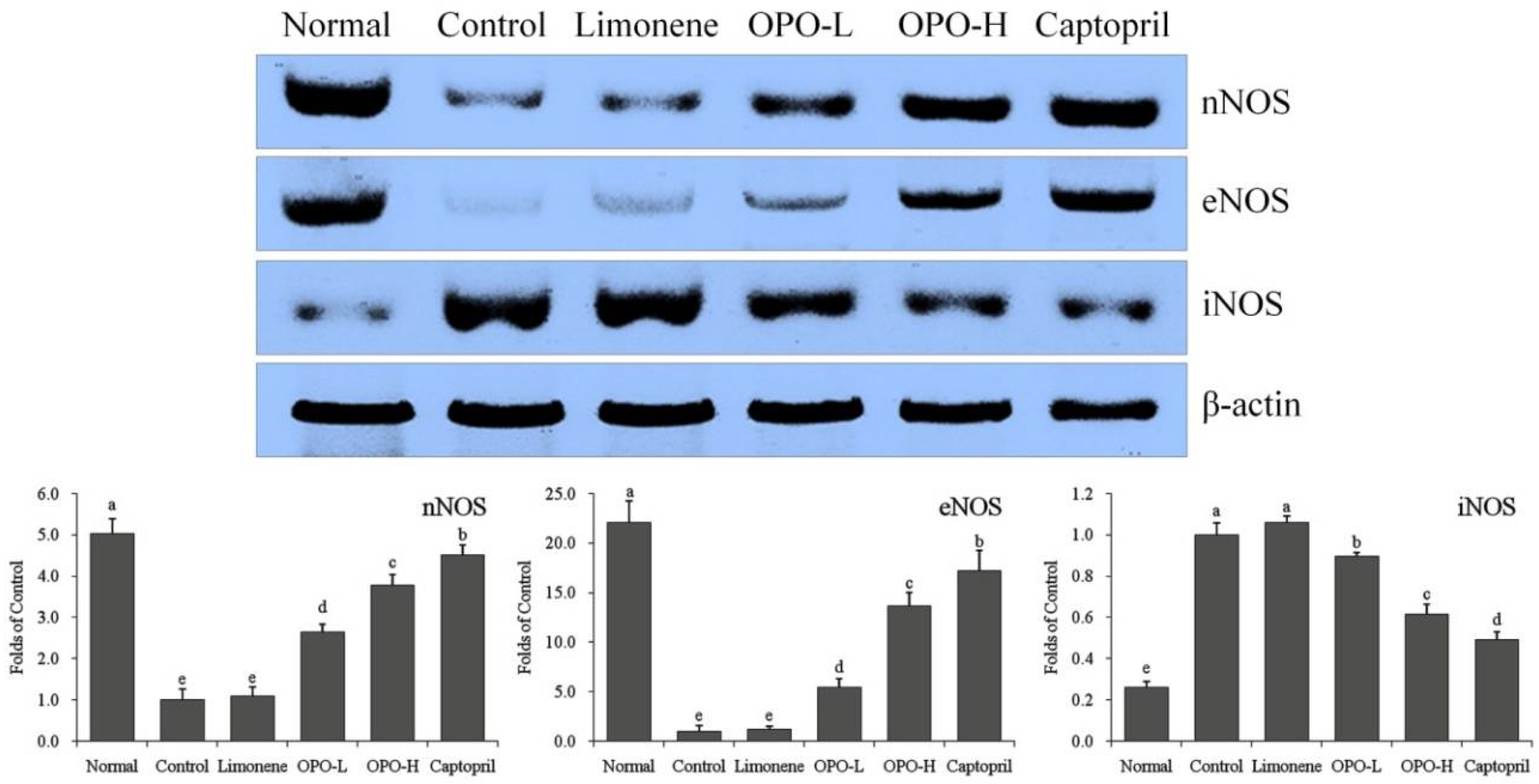
3.7. Protein Expression in Myocardial Tissue

As shown in Figure 8, the levels of nNOS, eNOS and iNOS protein expression in the myocardial tissue of OPO-H-treated rats showed the same trends as were seen for nNOS, eNOS and iNOS mRNA expression in OPO-H-treated rats. The levels of nNOS and eNOS protein expression in the myocardial tissue of OPO-H-treated rats were higher than those of limonene-treated rats, OPO-L-treated rats and rats in the control group, but the iNOS protein expression in OPO-H-treated rats was lower than those of the limonene-treated rats, OPO-L-treated rats and the rats in the control group.

4. Discussion

The pathogenesis of hypertension mainly involves dysfunction of the liver, kidney, heart and spleen, which cause imbalances in the body [21]. By restoring the immune function of the body and regulating the function of the viscera, hypertension can be relieved. Functional foods can reduce pain, fix hormone imbalance and repair blood, blood vessels and human gene damage in patients with hypertension [22]. Therefore, dietary therapy is used to promote blood circulation, regulate blood lipids, soften blood vessels, restore blood pressure regulation mechanisms, repair human gene defects and reduce the side effects of hypertension treatment [23].
ET-1 has the strongest and longest lasting effect of any vasoconstrictor peptide. It is also a strong promoter and driving factor behind cell proliferation. It can cause vascular smooth muscle hyperplasia and hypertrophy and promote proliferation and stenosis of the vascular wall [24]. CGRP is the most powerful vasodilator peptide in the body. It has been found that plasma CGRP levels in patients with essential hypertension are significantly lower than those of persons without hypertension, and thus, it is considered an important cause of hypertension [25]. CGRP also promotes NOS activity and the formation of NO in vascular smooth muscle cells, and its vasodilation mechanism involves NO. Low plasma CGRP secretion in patients with essential hypertension decreases the NOS activity and the content of NO and increases blood pressure [26]. VEGF, a glycosylated secretory polypeptide, is known to have the most direct angiogenic activity of any protein, and it is a mitogen and angiogenic factor, vascular endothelial cell-specific, selective enhancer of vascular endothelial cells, stimulator of endothelial cell proliferation and angiogenesis and increases vascular permeability and other biological functions [27]. The level of serum VEGF in patients with hypertension was significantly higher than that in persons without hypertension. This difference is due to local ischemia and hypoxia in the tissues of patients with hypertension. The ubiquity of ischemia and other cytokines is the main reason for increased serum VEGF levels in patients with hypertension [28]. E-selectin has endothelial specificity; therefore, the soluble E-selectin in peripheral blood can be used as a specific indicator of endothelial cell activation. The level of serum-soluble E-selectin in patients with hypertension is significantly increased, which is an indicator of vascular endothelial cell activation induced by hypertension [29]. E-selectin can mediate leukocyte adhesion. Leukocytes release chemical mediators that further damage vascular endothelial cells, resulting in degeneration, necrosis and abscission of endothelial cells and ultimately causing arteriosclerosis, stenosis and deformation of the arterial wall [30]. From the results in this study, OPO can alter these levels to inhibit hypertension, and these results are consistent with those of previous studies.
ROS plays an important role in the occurrence of hypertension. The increase in blood pressure is often accompanied by enhanced oxidative stress. Studies have shown that the occurrence and development of hypertension is closely related to oxidative stress, and free radical-induced injury is involved in the occurrence and development of hypertension [31]. MDA is a decomposition product of various unsaturated fatty acids that can serve as an indirect measure of the metabolism of oxygen free radicals in the body. Its content reflects the severity of free radical attack on cells. The results showed that the MDA content was lower in patients with hypertension and hypertensive ischemic stroke [32]. OPO could decrease MDA activity, thus reducing hypertension.
In pathophysiological studies, HO-1 can be induced by oxidative damage. The antioxidant environment induced by HO-1 is mainly due to its ability to degrade hemoglobin and increase the bilirubin level. HO-1 plays an important role in protecting the cardiovascular system. HO-1 protects endothelial cells and cardiomyocytes as an antioxidant [33]. HO-1 also directly inhibits cardiomyocyte hypertrophy and reduces the ratio of left ventricular weight to body weight. At the same time, the antihypertensive effect of HO-1 is also related to its ability to improve endothelial cell function [34]. The level of plasma ADM in patients with hypertension is higher than that of persons without hypertension. The mRNA of ADM and RAMP2 in rats with myocardial infarction and heart failure increased in both atrial and ventricular muscle. The mRNA levels of ADM and RAMP2 in infarcted and non-infarcted myocardium increased. In some disease states, such as shock, congestive heart failure and obstructive nephropathy, the expression levels of ADM and RAMP2 genes can be upregulated to different degrees in cardiovascular tissue, which plays an important compensatory protective role in the cardiovascular system [35]. Further studies have also showed that the expression levels of ADM and RAMP2 mRNA in heart and blood vessels of hypertensive rats were significantly higher than those in rats without hypertension, and similar results were obtained in this study [36].
NO is an effective vasodilator that can maintain normal tension in the blood vessels. The enzyme that catalyses the biosynthesis of NO is called NOS, and it is divided into nNOS, eNOS and iNOS based on the cell or tissue source of the prototype enzyme [37]. When hypertension occurs with decreasing NOS, vascular-dependent hypertension, the endothelial relaxant response becomes weaker. In the normal physiological state of the body, iNOS is generally not expressed, but cNOS (eNOS and nNOS) in endothelial cells continuously synthesizes a small amount of NO to regulate vascular tension and blood pressure [38]. Exposure to an endotoxin in pathological conditions involving various cytokines can induce the production of macrophages and iNOS gene expression in white blood cells; as a result, NO is produced to inhibit endotoxin and cytokines, and its hypotensive effect is strong and is involved in the pathological process [39]. NO produced by eNOS relaxes blood vessels, inhibits proliferation of vascular smooth muscle cells, inhibits platelet aggregation and maintains normal vascular tension and arterial blood pressure. However, in hypertension, atherosclerosis and other pathological conditions, the NO produced by eNOS cannot maintain the normal tension of blood vessels on its own; iNOS can activate and produce relatively large amounts of NO, so blood pressure does not increase malignancy [40,41]. In this study, OPO could also increase nNOS and eNOS expression levels and reduce iNOS expression in hypertensive rats.
Citrus juice consumption in China has been increasing, and juice processing generates millions of tons of peel every year. Citrus peel has long been recognized as a rich source of flavonoids that are important to human health and nutrition. Several studies have reported the effects of different flavonoids or peel phenolic extract on hypertension. Short-term and long-term administration of hesperidin and glucosyl hesperidin showed hypotensive effects on spontaneous hypertensive rats, and this effect was associated with nitric oxide-mediated vasodilation [42,43]. The phenolic extract from grapefruit peels was shown to inhibit α-amylase-, α-glucosidase- and angiotensin-I-converting enzymes in a dose-dependent manner [44]. In another study, a grapefruit peel extract was able to decrease coronary vascular resistance and the mean arterial pressure in a Langendorff isolated and perfused heart model [45]. Nevertheless, most of these studies used raw extract of citrus peel. In this study, the prophylactic effect of cold-pressed peel oil of long-leaf orange on hypertension was studied. Because of the hydrophobicity, the composition of the oil is much simpler than that of the whole peel extract; HPLC analysis showed that most of the non-volatile bioactive components were polymethoxyflavones. Thus, it is highly probable that the hypotensive effect of the long-leaf orange cold-pressed peel oil is related to the constituent PMFs (Figure 9).

5. Conclusions

In the present study, HPLC analysis showed that the OPO of long-leaf orange contained eight components, namely sinensetin, hexamethoxyflavone, tetramethyl-O-isoscutellarein, nobiletin, tetramethyl-O-scutellarein, heptamethoxyflavone, 5-demethylnobiletin and tangeretin. OPO could decrease the SP and DP of hypertensive rats. OPO could also increase the NO contents and reduce MDA contents in serum, heart, liver and kidney of hypertensive rats. After OPO treatment, the ET-1, VEGF and E-selectin serum levels were decreased, and the CGRP level was increased. Based on additional experiments, OPO could upregulate nNOS and eNOS mRNA and protein expression and upregulate HO-1 mRNA expression. Meanwhile, OPO could downregulate ADM and RAMP2 mRNA expression. From these results, OPO shows good antihypertensive effects, and the high dose of OPO, 10 mL/kg bw, showed more pronounced effects.

Author Contributions

Gui-Jie Li and Jun Wang performed the majority of the experiments and wrote the manuscript. Yu-Jiao Cheng, Xiang Tan, Yu-Lin Zhai and Qiang Wang contributed to the data analysis. Fang-Jin Gao and Guang-Lan Liu contributed to the making and component analysis of long-leaf orange cold-pressed oil. Xin Zhao and Hua Wang designed and supervised the study and checked the final manuscript.

Funding

This research was funded by the Special Fund for Public Welfare Industry of the Chinese Ministry of Agriculture grant number 201503142-12, the Recruitment Program of Foreign Expert of the China grant number WQ20135000161, the National Key Research and Development Plan of China grant number 2017YFD0400701-3, the Scientific Research Program of Chongqing University of Education grant number 17GZKP02, the Zhongxian Special Benefit Agriculture Project in 2015 grant number 2016001016, and the Scientific and Technological Research Program of Chongqing Municipal Education Commission grant number KJ1601413, China.

Conflicts of Interest

The authors declare no conflict of interest.

References

  1. Koes, R.E.; Quattrocchio, F.; Mol, J.N.M. The flavonoid biosynthetic pathway in plants: Function and evolution. Bioessays 1994, 1, 123–132. [Google Scholar] [CrossRef]
  2. Wang, L.; Wang, J.; Fang, L.; Zheng, Z.; Zhi, D.; Wang, S.; Li, S.; Ho, C.T.; Zhao, H. Anticancer activities of citrus peel polymethoxyflavones related to angiogenesis and others. Biomed. Res. Int. 2014, 2014, 453972. [Google Scholar] [CrossRef] [PubMed]
  3. Murakami, A.; Nakamura, Y.; Torikai, K.; Tanaka, T.; Koshiba, T.; Koshimizu, K.; Kuwahara, S.; Takahashi, Y.; Ogawa, K.; Yano, M.; et al. Inhibitory effect of citrus nobiletin on phorbol ester-induced skin inflammation, oxidative stress, and tumor promotion in rats. Cancer Res. 2000, 60, 5059–5066. [Google Scholar] [PubMed]
  4. Ou, M.C.; Liu, Y.H.; Sun, Y.W.; Chan, C.F. The composition, antioxidant and antibacterial activities of cold-pressed and distilled essential oils of Citrus paradisi and Citrus grandis (L.) osbeck. Evid. Based Complement. Alternat. Med. 2015, 2015, 804091. [Google Scholar] [CrossRef] [PubMed]
  5. Kobayashi, S.; Kato, T.; Azuma, T.; Kikuzaki, H.; Abe, K. Anti-allergenic activity of polymethoxyflavones from Kaempferia parviflora. J. Funct. Food. 2015, 13, 100–107. [Google Scholar] [CrossRef]
  6. Montanari, A.; Chen, J.; Widmer, W. Citrus flavonoids: Citrus flavonoids: A review of past biological activity against disease. Discovery of new flavonoids from Dancy tangerine cold pressed peel oil solids and leaves. Adv. Exp. Med. Biol. 1998, 439, 103–116. [Google Scholar] [PubMed]
  7. Manthey, J.A.; Guthrie, N. Antiproliferative activities of citrus flavonoids against six human cancer cell lines. J. Agric. Food Chem. 2002, 50, 5837–5843. [Google Scholar] [CrossRef] [PubMed]
  8. Morley, K.L.; Ferguson, P.J.; Koropatnick, J. Tangeretin and nobiletin induce G1 cell cycle arrest but not apoptosis in human breast and colon cancer cells. Cancer Lett. 2007, 251, 168–178. [Google Scholar] [CrossRef] [PubMed]
  9. Zhang, C.; Lu, Y.; Tao, L.; Tao, X.; Su, X.; Wei, D. Tyrosinase inhibitory effects and inhibition mechanisms of nobiletin and hesperidin from citrus peel crude extracts. J. Enzyme Inhibit. Med. Chem. 2007, 22, 91–98. [Google Scholar] [CrossRef] [PubMed]
  10. Jain, P.K.; Kharya, M.D.; Gajbhiye, A.; Sara, U.V.S.; Sharma, V.K. Flavonoids as nutraceuticals: Review. Herba Polonica 2010, 7, 1089–1099. [Google Scholar]
  11. Li, S.; Wang, Y.; Dushenkov, S.; Ho, C.T. Bioavailability of polymethoxyflavones. Acs Symp. 2008, 987, 233–245. [Google Scholar]
  12. Ohta, F.; Takagi, T.; Sato, H.; Ignarro, L.J. Low-dose L-arginine administration increases microperfusion of hindlimb muscle without affecting blood pressure in rats. Proc. Natl. Acad. Sci. USA 2007, 104, 1407–1411. [Google Scholar] [CrossRef] [PubMed]
  13. Thomsen, K.; Rubin, I.; Lauritzen, M. NO- and non-NO-, non-prostanoid-dependent vasodilatation in rat sciatic nerve during maturation and developing experimental diabetic neuropathy. J. Physiol. 2002, 543, 977–993. [Google Scholar] [CrossRef] [PubMed]
  14. Simmonds, M.J.; Detterich, J.A.; Connes, P. Nitric oxide, vasodilation and the red blood cell. Biorheology 2014, 51, 121–134. [Google Scholar] [PubMed]
  15. Gangula, P.; Ravella, K.; Chukkapalli, S.; Rivera, M.; Srinivasan, S.; Hale, A.; Channon, K.; Southerland, J.; Kesavalu, L. Polybacterial periodontal pathogens alter vascular and gut BH4/nNOS/NRF2-phase II enzyme expression. PLoS ONE 2015, 10, e0129885. [Google Scholar] [CrossRef] [PubMed]
  16. Fujiwara, A.; Hirawa, N.; Fujita, M.; Kobayashi, Y.; Okuyama, Y.; Yatsu, K.; Katsumata, M.; Yamamoto, Y.; Ichihara, N.; Saka, S.; et al. Impaired nitric oxide production and increased blood pressure in systemic heterozygous ATP2B1 null rats. J. Hypertens 2014, 32, 1415–1423. [Google Scholar] [CrossRef] [PubMed]
  17. Mahinur, B.; Yi, Y.; Chen, L.D.; Aisa, H.A.; Wang, M.H. Alkaloids of Nitraria sibirica Pall. decrease hypertension and albuminuria in angiotensin II-salt. Chin. J. Nat. Med. 2014, 12, 266–272. [Google Scholar]
  18. Li, L.; Yang, H.G.; Yuan, T.Y.; Zhao, Y.; Du, G.H. Rho kinase inhibition activity of pinocembrin in rat aortic rings contracted by angiotensin II. Chin. J. Nat. Med. 2013, 11, 258–263. [Google Scholar] [CrossRef] [PubMed]
  19. Sun, P.; Zhu, K.; Wang, C.; Liu, W.W.; Peng, D.G.; Zhao, X. Prophylactic effects of alkaloids from Ba lotus seeds on L-NNA-induced hypertension in rats. Chin. J. Nat. Med. 2016, 14, 835–843. [Google Scholar] [PubMed]
  20. Xue, B.J.; Zhang, Z.M.; Johnson, R.F.; Johnson, A.K. Sensitization of slow pressor angiotensin II (ANGII) initiated hypertension: Induction of sensitization by prior ANGII treatment. Hypertension 2012, 59, 459–466. [Google Scholar] [CrossRef] [PubMed]
  21. Gu, W.L.; Shi, Z.X.; Yu, Y.X.; Wu, Y.W.; Lu, B.W.; Hui, K.K. Distribution characteristics of syndrome types in essentialhypertension. Zhong Xi Yi Jie He Xue Bao 2010, 8, 842–847. [Google Scholar] [CrossRef] [PubMed]
  22. Xu, H.Y.; Wang, Y.Q. Investigation and application for the therapy of hypertension with traditional Chinese medicine. China Prac. Med. 2008, 3, 189–192. [Google Scholar]
  23. Shen, C.Z.; Peng, M.C.; Kuang, H.R.; Cheng, Z.Q. The effect of Chinese food therapy on the quality of life in patients with hypertension. Chin. J. Nurs. 2009, 44, 510–513. [Google Scholar]
  24. Silva, I.; Teixeira, A.; Oliveira, J.; Almeida, I.; Almeida, R.; Vasconcelos, C. Predictive value of vascular disease biomarkers for digital ulcers in systemic sclerosis patients. Clin. Exp. Rheumatol. 2015, 33, 127–130. [Google Scholar]
  25. Calò, L.A.; Dal Maso, L.; Pagnin, E.; Ravarotto, V.; Facco, M.; Boscaro, E.; Maiolino, G.; Pessina, A.C.; Rossi, G.P. Effect of olmesartan medoxomil on number and survival of circulating endothelial progenitor cells and calcitonin gene related peptide in hypertensive patients. J. Hypertens. 2014, 32, 193–199. [Google Scholar]
  26. Wang, Z.G.; Liu, J.L.; Liu, Y.; Wen, S.J.; Wen, J.; Liu, Y.P.; Chen, X.J.; Wu, Z.S. Increased plasma vasoactive substances and antioxidant enzymes levels in prehypertensive patients. Zhonghua Xin Xue Guan Bing Za Zhi 2007, 35, 719–722. [Google Scholar] [PubMed]
  27. Serban, D.; Leng, J.; Cheresh, D. H-ras regulates angiogenesis and vascular permeability by activation of distinct downstream effectors. Circ. Res. 2008, 102, 1350–1358. [Google Scholar] [CrossRef] [PubMed]
  28. Muti, L.A.; Pârvu, A.E.; Crăciun, A.M.; Miron, N.; Acalovschi, M. Nitro-oxidative stress, VEGF and MMP-9 in patients with cirrhotic and non-cirrhotic portal hypertension. Clujul. Med. 2015, 88, 140–145. [Google Scholar] [CrossRef] [PubMed]
  29. Zhang, J.L.; Qin, Y.W.; Zheng, X.; Qiu, J.L.; Zhang, L.Z.; Zhang, J.R.; Bian, J.L. Relevance of serum soluble E-selectin level and blood pressure in hypertensive patients. Shanghai Med. J. 2004, 27, 7–9. [Google Scholar]
  30. Chen, J.; Li, J. Clinical curative effect and influence on E-selectin, iNOS, eNOS in patient of essential hypertension with massotherapy. China J. Tradit. Chin. Med. Pharm. 2010, 25, 1708–1710. [Google Scholar]
  31. Zhou, Y.; Zhao, L.; Zhang, Z.; Lu, X. Protective effect of enalapril against methionine-enriched diet-induced hypertension: Role of endoplasmic reticulum and oxidative stress. Biomed. Res. Int. 2015, 2015, 724876. [Google Scholar] [CrossRef] [PubMed]
  32. Zhang, S.; Yang, T.; Xu, X.; Wang, M.; Zhong, L.; Yang, Y.; Zhai, Z.; Xiao, F.; Wang, C. Oxidative stress and nitric oxide signaling related biomarkers in patients with pulmonary hypertension: A case control study. BMC Pulm. Med. 2015, 15, 50. [Google Scholar] [CrossRef] [PubMed]
  33. Chao, H.M.; Chuang, M.J.; Liu, J.H.; Liu, X.Q.; Ho, L.K.; Pan, W.H.; Zhang, X.M.; Liu, C.M.; Tsai, S.K.; Kong, C.W.; et al. Baicalein protects against retinal ischemia by antioxidation, antiapoptosis, down-regulation of HIF-1α, VEGF, and MMP-9 and upregulation of HO-1. J. Ocul. Pharmacol. Ther. 2013, 29, 539–549. [Google Scholar] [CrossRef] [PubMed]
  34. Li, Z.; Wang, Y.; Man, R.Y.; Vanhoutte, P.M. Upregulation of heme oxygenase-1 potentiates EDH-type relaxations in the mesenteric artery of the spontaneously hypertensive rat. Am. J. Physiol. Heart Circ. Physiol. 2013, 305, 1471–1483. [Google Scholar] [CrossRef] [PubMed]
  35. Sano, M.; Kuroi, N.; Nakayama, T.; Sato, N.; Izumi, Y.; Soma, M.; Kokubun, S. Association study of calcitonin-receptor-like receptor gene in essential hypertension. Am. J. Hypertens. 2005, 18, 403–408. [Google Scholar] [CrossRef] [PubMed]
  36. Gong, Y.S.; Fan, X.F.; Wu, X.M.; Hu, L.G.; Tang, C.S.; Pang, Y.Z.; Qi, Y.F. Changes of intermedin/adrenomedullin 2 and its receptors in the right ventricle of rats with chronic hypoxic pulmonary hypertension. Sheng Li Xue Bao 2007, 59, 210–214. [Google Scholar] [PubMed]
  37. Basralı, F.; Nasırcılar Ülker, S.; Koçer, G.; Ülker Karadamar, P.; Özyurt, D.; Cengiz, M.; Şentürk, Ü.K. Effect of magnesium on vascular reactivity in NOS inhibition-induced hypertension. Magnes. Res. 2015, 28, 64–74. [Google Scholar] [PubMed]
  38. Nieto, C.I.; Cabildo, M.P.; Cornago, M.P.; Sanz, D.; Claramunt, R.M.; Torralba, M.C.; Torres, M.R.; Elguero, J.; García, J.A.; López, A.; Acuña-Castroviejo, D. Fluorination effects on NOS inhibitory activity of pyrazoles related to curcumin. Molecules 2015, 20, 15643–15665. [Google Scholar] [CrossRef] [PubMed]
  39. Huang, H.C.; Wang, S.S.; Chang, C.C.; Lee, F.Y.; Lin, H.C.; Hou, M.C.; Teng, T.H.; Chen, Y.C.; Lee, S.D. Evolution of portal-systemic collateral vasopressin response in endotoxemic portal hypertensive rats. Shock 2009, 32, 503–508. [Google Scholar] [CrossRef] [PubMed]
  40. Pulgar, V.M.; Yamaleyeva, L.M.; Varagic, J.; McGee, C.; Bader, M.; Dechend, R.; Brosnihan, K.B. Functional changes in the uterine artery precede the hypertensive phenotype in a transgenic model of hypertensive pregnancy. Am. J. Physiol. Endocrinol. Metab. 2015, 309, 811–817. [Google Scholar] [CrossRef] [PubMed]
  41. Chen, S.; Sun, P.; Zhao, X.; Yi, R.; Qian, J.; Shi, Y.; Wang, R. Gardenia jasminoides has therapeutic effects on L-NNA-induced hypertension in vivo. Mol. Med. Rep. 2017, 15, 4360–4373. [Google Scholar] [CrossRef] [PubMed]
  42. Yamamoto, M.; Suzuki, A.; Hase, T. Short-term effects of glucosyl hesperidin and hesperetin on blood pressure and vascular endothelial function in spontaneously hypertensive rats. J. Nutr. Sci. Vitaminol. 2008, 54, 95–98. [Google Scholar] [CrossRef] [PubMed]
  43. Ohtsuki, K.; Abe, A.H.; Kondo, M.; Uemura, K.; Iwasaki, Y.; Kondo, Y. Effects of long-term administration of hesperidin and glucosyl hesperidin to spontaneously hypertensive rats. J. Nutr. Sci. Vitaminol. 2002, 48, 420. [Google Scholar] [CrossRef] [PubMed]
  44. Oboh, G.; Ademosun, A.O. Phenolic extracts from grapefruit peels (Citrus paradisi) inhibit key enzymes linked with type 2 diabetes and hypertension. J. Food Biochem. 2011, 35, 1703–1709. [Google Scholar] [CrossRef]
  45. Díazjuárez, J.A.; Tenoriolópez, F.A.; Zarcoolvera, G.; Vallemondragón, L.D.; Torresnarváez, J.C.; Pastelínhernández, G. Effect of citrus paradisi extract and juice on arterial pressure both in vitro and in vivo. Phytother. Res. 2009, 23, 948. [Google Scholar] [CrossRef] [PubMed]
Sample Availability: Samples of the compounds are not available from the authors.
Figure 1. The process of animal experiment. L-NNA, Nω-nitro-L-arginine.
Applsci 08 00752 g001
Figure 2. HPLC chromatograms of long-leaf orange peel oil (OPO) showing polymethoxyflavones (PMFs). Notes: (A) The mixed standard chromatogram with the UV spectrum of Compound 1; (B) separation and identification of the non-volatile compounds in OPO and the UV spectrum of the peak with Rt = 3.2 min. Numbers 1–9: 1, isosinensetin; 2, sinensetin; 3, hexamethoxyflavone; 4, tetramethyl-O-isoscutellarein; 5, nobiletin; 6, tetramethyl-O-scutellarein; 7, heptamethoxyflavone; 8, 5-demethylnobiletin; and 9, tangeretin.
Applsci 08 00752 g002
Figure 3. Separation of the volatile components in long-leaf orange peel oil and identification of the major compound D-limonene using GC-MS analysis.
Applsci 08 00752 g003
Figure 4. Electrocardiograph and blood pressure of each group during one inflation/deflation cycle of the tail-cuff sphygmomanometry. Notes: The recorded voltage of the electrocardiograph corresponds to the left ordinate. The right ordinate corresponds to the blood pressure diagram, in which: A, start inflation; B, stop inflation and keep pressure; C, start deflation and measurement; and D, stop measurement and vent. SP, systolic pressure; DP, diastolic pressure. OPO-L group, orange peel oil low dose, 5 mL/kg bodyweight (bw); OPO-H group, orange peel oil high dose, 10 mL/kg bw; captopril group, 15.6 mg/kg bw; limonene group, 10 mL/kg bw.
Applsci 08 00752 g004
Figure 5. Effects of orange peel oil on the SP and DP of L-NNA-induced hypertensive rats. Notes: SP, systolic pressure; DP, diastolic pressure; OPO-L, orange peel oil low dose, 5 mL/kg bodyweight (bw); OPO-H, orange peel oil high dose, 10 mL/kg bw; captopril, 15.6 mg/kg bw; and limonene, 10 mL/kg bw.
Applsci 08 00752 g005
Figure 6. mRNA expression levels of HO-1, ADM and RAMP2 in rat myocardial tissue. For each group, n = 10 rats. a–e Bars with different letters in the chart are significantly different (p < 0.05) according to Duncan’s multiple range test, and the same letter indicates that the values are not significantly different. OPO-L, orange peel oil low dose, 5 mL/kg bodyweight (bw); OPO-H, orange peel oil high dose, 10 mL/kg bw; captopril, 15.6 mg/kg bw; and limonene, 10 mL/kg bw.
Applsci 08 00752 g006
Figure 7. mRNA expression levels of nNOS, eNOS and iNOS in rat myocardial tissue. For each group, n = 10 rats. a–e Bars with different letters in the chart are significantly different (p < 0.05) according to Duncan’s multiple range test, and the same letter indicates that the values are not significantly different. OPO-L, orange peel oil low dose, 5 mL/kg bodyweight (bw); OPO-H, orange peel oil high dose, 10 mL/kg bw; captopril, 15.6 mg/kg bw; and limonene, 10 mL/kg bw.
Applsci 08 00752 g007
Figure 8. Protein expression levels of nNOS, eNOS and iNOS in rat myocardial tissue. For each group, n = 10 rats. a–e Bars with different letters in the chart are significantly different (p < 0.05) according to Duncan’s multiple range test, and the same letter indicates that the values are significantly different. OPO-L, orange peel oil low dose, 5 mL/kg bodyweight (bw); OPO-H, orange peel oil high dose, 10 mL/kg bw; captopril, 15.6 mg/kg bw; and limonene, 10 mL/kg bw.
Applsci 08 00752 g008
Figure 9. Mechanism of antihypertensive effects of orange peel oil. Notes: red symbol, inhibitory effect; green symbol, promoting effect. Orange peel oil contains an important core component of D-limonene. Orange peel oil could regulate blood pressure-related factors in serum, mRNA and protein expression of tissue for reducing blood pressure through D-limonene in hypertensive rats.
Applsci 08 00752 g009
Table 1. Sequences of reverse transcription-polymerase chain reaction primers used in this study.
Gene NameSequence
HO-1Forward: 5′-GGAACTTTCAGAAGGGCCAG-3′
Reverse: 5′-GTCCTTGGTGTCATGGGTCA-3′
ADMForward: 5′-GCTGGTTTCCGTCGCCCTGATGT-3′
Reverse: 5′-CGTTGTCCTTGTCCTTATCTGTG-3′
RAMP2Forward: 5′-GGACGGTGAAGAACTATGAG-3′
Reverse: 5′-ATCATGGCCAGGAGTACATC-3′
nNOSForward: 5′-GAATACCAGCCTGATCCATGGAA-3′
Reverse: 5′-TCCTCCAGGAGGGTGTCCACCGCATG-3′
eNOSForward: 5′-GGAGAGGCTGCATGACATTG-3′
Reverse: 5′-GGTAGAGCCATAGTGGAATGAC-3′
iNOSForward: 5′-AGAGAGATCGGGTTCACA-3′
Reverse: 5′-CACAGAACTGAGGGTACA-3′
GAPDHForward: 5′-TGCACCACCAACTGCTTAG-3′
Reverse: 5′-GATGCAGGGATGATGTTC-3′
Table 2. Polymethoxyflavones and limonene contents in orange peel oil.
CompoundsQuant. MethodPeak Area %Calibration Equation *Concentration (mg/L)
isosinensetinHPLC-UVN.D.Y = 2.59X − 2.15N.D.
sinensetin2.2 ± 0.1Y = 3.33X + 0.235.10 ± 0.77
hexamethoxyflavone4.3 ± 0.2Y = 1.57X + 1.8420.23 ± 1.44
tetramethyl-O-isoscutellareinN.D.Y = 2.87X + 0.52N.D.
nobiletin16.7 ± 0.2Y = 2.64X + 4.4147.72 ± 1.69
tetramethyl-O-scutellarein16.0 ± 0.2Y = 5.19X + 3.4923.30 ± 1.25
heptamethoxyflavone28.6 ± 0.3Y = 5.29X + 0.3042.02 ± 1.78
5-demethylnobiletin0.9 ± 0.0Y = 2.41X + 5.730.44 ± 0.10
tangeretin22.7 ± 0.3Y = 6.88X + 1.3125.55 ± 1.13
total PMFs 91.4 ± 0.3-164.18 ± 3.14
D-limoneneGC-MS95.3 ± 0.5Y = 388,293X + 507,631809.10 ± 7.31 (g/L)
Notes: * For each calibration equation, correlation R2 > 0.9990.
Table 3. Nitric oxide (NO) contents in serum, heart, liver and kidney of L-NNA-induced hypertensive rats.
GroupSerum (μmol/L)Heart (µmol/gprot)Liver (µmol/gprot)Kidney (µmol/gprot)
Normal105.31 ± 4.92 a15.29 ± 1.35 a17.11 ± 0.46 a13.51 ± 0.57 a
Control58.91 ± 3.12 e7.35 ± 0.97 e5.83 ± 0.34 e6.65 ± 0.72 c
Limonene59.03 ± 2.70 e7.95 ± 0.40 e5.97 ± 1.00 e6.44 ± 1.85 c
OPO-L70.99 ± 2.54 d9.48 ± 1.13 d9.29 ± 0.82 d8.16 ± 0.28 b
OPO-H86.35 ± 3.48 c12.11 ± 1.76 c11.75 ± 0.76 c12.03 ± 1.34 a
Captopril97.37 ± 3.06 b13.54 ± 2.02 b14.63 ± 2.38 b13.21 ± 2.29 a
For each group, n = 10 rats. a–e Values with different letters in the same column are significantly different (p < 0.05) according to Duncan’s multiple range test, and the same letter means the values are not significantly different. OPO-L, orange peel oil low dose, 5 mL/kg bodyweight (bw); OPO-H, orange peel oil high dose, 10 mL/kg bw; captopril, 15.6 mg/kg bw; and limonene, 10 mL/kg bw.
Table 4. Malondialdehyde (MDA) content in serum, heart, liver and kidney of L-NNA-induced hypertensive rats.
GroupSerum (μmol/L)Heart (nmol/gprot)Liver (nmol/gprot)Kidney (nmol/gprot)
Normal2.23 ± 0.49 d2.12 ± 0.21 d0.50 ± 0.06 d1.35 ± 0.15 c
Control5.89 ± 0.31 a4.36 ± 0.23 a1.13 ± 0.08 a2.66 ± 0.17 a
Limonene5.69 ± 0.27 a4.33 ± 0.12 a1.14 ± 0.05 a2.40 ± 0.18 a
OPO-L4.09 ± 0.25 b3.75 ± 0.14 b0.92 ± 0.08 b1.81 ± 0.20 b
OPO-H3.68 ± 0.34 c2.98 ± 0.27 c0.72 ± 0.07 c1.62 ± 0.13 bc
Captopril2.97 ± 0.30 d2.70 ± 0.16 c0.58 ± 0.05 d1.52 ± 0.22 c
In each group, n = 10 rats. a–d Values with different letters in the same column are significantly different (p < 0.05) according to Duncan’s multiple range test, and the same letter indicates that the values are not significantly different. OPO-L, orange peel oil low dose, 5 mL/kg bodyweight (bw); OPO-H, orange peel oil high dose, 10 mL/kg bw; captopril, 15.6 mg/kg bw; and limonene, 10 mL/kg bw.
Table 5. Serum levels of ET-1 (endothelin-1), CGRP (calcium gene-related peptide), VEGF (vascular endothelial growth factor) and E-selectin in L-NNA-induced hypertensive rats.
GroupET-1 (pg/mL)CGRP (pg/mL)VEGF (pg/mL)E-selectin (ng/mL)
Normal62.47 ± 4.78 e125.72 ± 7.92 a82.36 ± 5.20 e231.28 ± 22.17 e
Control197.36 ± 12.79 a41.82 ± 3.37 e155.73 ± 11.38 a533.03 ± 38.29 a
Limonene195.71 ± 17.51 a43.45 ± 5.11 e152.39 ± 12.49 a528.67 ± 33.12 a
OPO-L151.38 ± 7.34 b72.82 ± 6.18 d137.72 ± 8.73 b451.91 ± 25.87 b
OPO-H122.03 ± 6.70 c93.45 ± 4.18 c107.81 ± 8.26 c348.75 ± 20.89 c
Captopril96.33 ± 3.89 d108.37 ± 5.65 b91.32 ± 4.77 d287.93 ± 24.62 d
In each group, n = 10 rats. a–e Values with different letters in the same column are significantly different (p < 0.05) according to Duncan’s multiple range test, and the same letter indicates that the values are not significantly different. OPO-L, orange peel oil low dose, 5 mL/kg bodyweight (bw); OPO-H, orange peel oil high dose, 10 mL/kg bw; captopril, 15.6 mg/kg bw; and limonene, 10 mL/kg bw.

