Article

The Effects of Remineralization via Fluoride Versus Low-Level Laser IR810 and Fluoride Agents on the Mineralization and Microhardness of Bovine Dental Enamel

by
Edith Lara-Carrillo
1,
Nayeli Lovera-Rojas
1,
Raúl Alberto Morales-Luckie
2,
Norma Leticia Robles-Bermeo
1,
María Magdalena García-Fabila
3,
Rubén De la Rosa-Santillana
4 and
Carlo Eduardo Medina-Solís
1,4,*
1
Advanced Studies and Research Center in Dentistry “Dr. Keisaburo Miyata” of School of Dentistry at Autonomous University State of Mexico, Toluca 50130, Mexico
2
Faculty of Chemistry, Department of Materials Science, Center for Research in Sustainable Chemistry (CIQS), Autonomous University State of Mexico-National Autonomous University of Mexico (UNAM), Toluca 50120, Mexico
3
Faculty of Chemistry, Instrumental Analysis Laboratory, Autonomous University State of Mexico, Toluca 50120, Mexico
4
Academic Area of Dentistry of Health Sciences Institute at the Autonomous University of Hidalgo State, Pachuca 42060, Mexico
*
Author to whom correspondence should be addressed.
Appl. Sci. 2018, 8(1), 78; https://doi.org/10.3390/app8010078
Submission received: 22 October 2017 / Revised: 1 January 2018 / Accepted: 3 January 2018 / Published: 9 January 2018
(This article belongs to the Section Materials Science and Engineering)

Abstract

:
The objective of this study was to assess the mineralization and microhardness of bovine dental enamel surfaces treated with fluoride, tri-calcium phosphate, and infrared (IR) 810 laser irradiation. The study used 210 bovine incisors, which were divided into six groups (n = 35 in each): Group A: Untreated (control), Group B: Fluoride (Durapath-Colgate), Group C: Fluoride+Tri-calcium phosphate (Clin-Pro White-3 M), Group D: Laser IR 810 (Quantum), Group E: Fluoride+laser, and Group F: Fluoride+tri-calcium phosphate+laser). Mineralization was measured via UV-Vis spectroscopy for phosphorus and via atomic absorption spectroscopy for calcium upon demineralization and remineralization with proven agents. Microhardness (SMH) was measured after enamel remineralization. Mineral loss data showed differences between the groups before and after the mineralizing agents were placed (p < 0.05). Fluoride presented the highest remineralization tendency for both calcium and phosphate, with a Vickers microhardness of 329.8 HV0.1/11 (p < 0.05). It was observed that, if remineralization solution contained fewer minerals, the microhardness surface values were higher (r = −0.268 and −0.208; p < 0.05). This study shows that fluoride has a remineralizing effect compared with calcium triphosphate and laser IR810. This in vitro study imitated the application of different remineralizing agents and showed which one was the most efficient for treating non-cavitated injuries. This can prevent the progression of lesions in patients with white spot lesions.

1. Introduction

Dental caries is one of the most prevalent diseases affecting humans worldwide. As technology advances, white spot lesions must be treated with non-invasive techniques to prevent further disease progression and preserve the integrity of tooth structure [1,2,3]. Remineralization of tooth enamel is defined as the process whereby calcium and phosphate ions are supplied from an external source to promote ion-deposition-demineralized enamel crystals to produce mineral gain [4]. Speaking about mineralization, it is fundamental to use the term biomineralization when referring to its formation. In particular, there are two kinds of dental hard tissue: enamel, which covers the tooth crown, and dentin, which constitutes the whole body of the tooth. Hard tissue formation involves two main processes: a biological one with cell signaling and a biochemical one with the interaction of biomolecules in crystal apatite formation [5].
Fluoride is the cornerstone of remineralization, but its ability to promote remineralization is limited by the availability of calcium and phosphate ions [4]. Recently, new alternatives that purport to be better than fluoride have appeared in the dentistry market. These include a calcium-phosphate-based delivery system and low-level lasers (LLLs). A laser is a device consisting of solid, liquid, or gas substances that produce a light beam when excited by a source of energy. This device can be classified into two categories: high-power lasers or surgical lasers, featuring thermal effects with cutting, vaporization, and hemostasis properties, and low-power lasers or therapeutic lasers, with analgesic, anti-inflammatory, and biostimulation properties [6]. The manufacturers claim that these products provide a new option for the remineralization of non-cavitated dental lesions. It has been reported that laser irradiation on enamel causes crystalline changes promoting significant acid resistance of dental hard tissue [7].
Producers of varnish based calcium and phosphate state that the crystalline system showed the potential to deliver calcium and phosphorous to enamel lesion. This tricalcium phosphate system is encapsulated in sodium lauryl sulfate. This is more efficient than using only fluoride on the lesion [4,8,9,10].
Similarly, LLLs have been proposed as a remineralization treatment when combined with fluoride to maximize their effects. It has been demonstrated that the application of a high-power lasers, such as CO2 and erbium lasers (erbium:yttrium-aluminium-garnet (Er:YAG) and erbium, chromium:yattrium-scandium-gallium-garnet (Er, Cr:YSGG)), are effective in white spot lesion prevention. These lasers absorb water from the hydroxyapatite of tooth tissues and can modify the crystalline structure, acid solubility, and permeability of the tooth surface to increase resistance against demineralization. However, high-power lasers are costly and not readily available in every practice [11,12,13,14].
LLLs are relatively inexpensive, small, and portable and have multiple applications in several areas of dentistry. Equally, their application in the prevention or arrestment of tooth caries is interesting. However, the efficacy of these lasers in remineralization has not yet been sufficiently studied.
Dental researchers have utilized several analytical techniques to quantify changes in the mineral content of enamel during white spot lesion formation. The most common are the Knoop and Vickers micro-hardness, polarized light microscopy, confocal laser scanning microscopy, and light-induced fluorescence.
In the present study, we primarily used the Vickers microhardness followed by UV-Vis spectroscopy to quantify the absence or presence of phosphorus. Atomic absorption spectroscopy was used to measure calcium. Bovine enamel was used as a model for human teeth. This model offers a large surface area and more uniform enamel thickness; previous studies have shown that they are very similar to human teeth [15,16,17].
The efficient treatment of enamel demineralization can prevent white spot lesions and be used in difficult children or post-orthodontic treatment. There is a need for less expensive and less invasive approaches for therapy. Therefore, the aim of this study was to assess mineralization and microhardness on bovine enamel surface treated by fluoride and tri-calcium phosphate exposed to LLL irradiation.

2. Materials and Methods

2.1. Materials

An experimental laboratory study was performed. Three remineralizing agents were evaluated in this study: (a) Duraphat (Colgate-Palmolive, New York, NY, USA) sodium fluoride to 22,600 ppm in content, (b) Clinpro White Varnish (3M ESPE, Saint Paul, MN, USA) functionalized with tri-calcium phosphate containing 22,600 ppm fluoride, and (c) LLL irradiation (IR810, Quantum, Queretaro, Mexico).

2.2. Specimens Preparation

Two hundred ten extracted bovine incisor teeth with no abnormalities were stored in thymol solution 0.2% until use. The root was removed with a low-speed turbine under water cooling. The palatal area was immersed in acrylic circles (a circular base of acrylic where the sample was placed to facilitate the manipulation), and the buccal surface was mounted horizontally. They were polished using 800, 1200 and 2400 grit silicon carbide paper. An acid-resistant nail varnish was applied around the exposed enamel surface, leaving an uncovered area of about 4 × 4 mm.

2.3. Demineralization

Following the proposal suggested by Prado et al. [2], the white spot lesions were created by individually immersing acrylic-mounted enamel specimens in a demineralization solution that had 2.2 mM CaCl2, 2.2 mM NaH2PO4, 0.05 M acetic acid, and a pH adjusted to 4.6 with 1 M KOH over two days at 37 °C; uniform demineralization was created on the surface of the enamel.

2.4. Remineralization

The remineralizing solution was prepared according to the formulation of Prado et al. [2] and consisted of 1.5 mM CaCl2, 0.9 mM Na2PO4, and 0.15 mM KCl, pH 7.0; the sample containers were kept at a constant temperature (37 °C). Mineralizing agents were applied every day for 15 days with pH cycling: 3 h demineralization and 21 h remineralization. Both solutions were changed daily. Specimens were randomly divided into six groups (n = 35) according to the treatment employed:
  • Group A: Control. No treatment was given to the enamel (but they underwent the cyclic pH as all the groups).
  • Group B: Fluoride (Duraphat varnish). The specimens were cleaned and dried with cotton; material was then applied on the surface and left for 1 min followed by storage in the remineralization solution.
  • Group C: Tricalcium phosphate (Clinpro White varnish). The sample was rinsed and dried, and the material was applied to the surface and left for 1 min and stored in a remineralization solution.
  • Group D: LLL (IR810 (Quantum)). The surface of the specimens were rinsed and dried, and it was exposed to infrared LLLs for 1 min at 810 nm and 200 mW in continuous wave mode. The window treatment received 6 J of energy.
  • Group E: Fluoride + LLL. The sample was rinsed and dried followed by fluoride for 1 min. This was then irradiated on the fluoride for another minute with the laser as mentioned earlier.
  • Group F: Tricalcium phosphate + LLL. The window treatment was rinsed, dried, mineralized for 1 min. It was then exposed to another minute of laser-like Group D with the remineralizing agent.

2.5. UV-Vis Spectroscopy for Phosphorus Determination

Measurements were made with a spectrophotometer UV-Vis (PerkinElmer, Inc., Lamda 25, Waltham, MA, USA) at a wavelength of 830–850 nm. All instruments were washed with chemicals before use to prevent contamination, and the solution standard was 1 M P2O5. The reducing solution contained ammonium molybdate, ascorbic acid, and sulfuric acid. Four milliliters of reducing solution with deionized water to the 10 mL capacity was added to 0.5 mL of the sample. This was stored at 50 °C for 45 min, and 1 mL of sample was used. The mixture was taken to quartz cell and read it at 830 nm.

2.6. Atomic Absorption Spectroscopy for Calcium Determination

All measurements were made via flame atomic absorption spectroscopy (PerkinElmer, Inc. Aanalyst 400, Waltham, MA, USA) using high purity air-acetylene. The radiation source was a calcium-based hollow cathode lamp operating at 4 mA. The wavelength was 422.7 nm with a bandwidth of 1.2 nm. Every sample was measured three times. The solution stock was 50 ppm for Ca. Hydrochloric acid and lanthanum oxide was used to prevent ionization and chemical interferences. One milliliter of 5% lanthanum oxide and 1 mL of hydrochloric acid was added to 1 mL of the sample, which was then diluted to 10 mL.

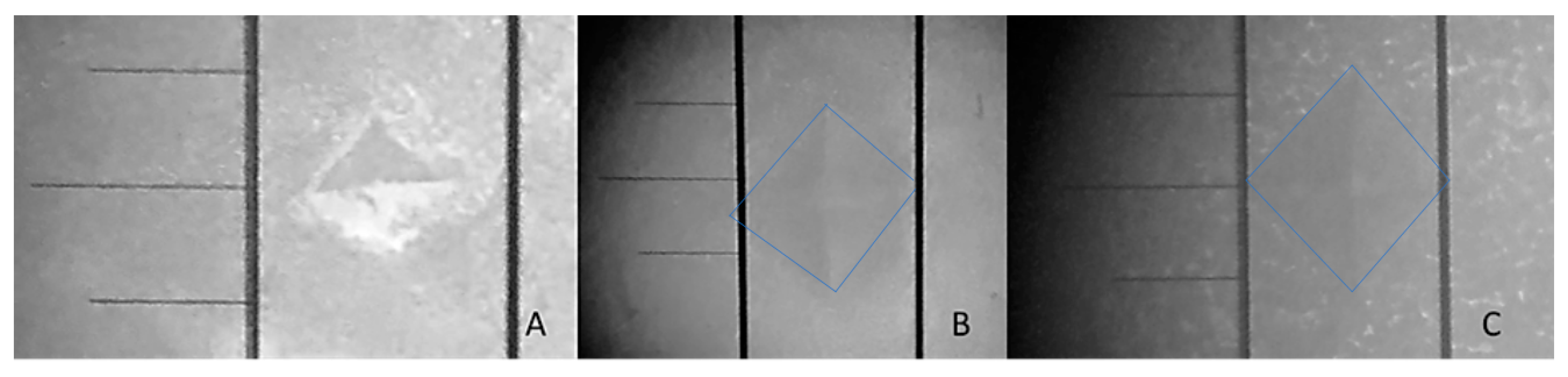
2.7. Surface Micro-Hardness (SMH)

Enamel surface micro-hardness was measured before applying remineralizing agents using the micro-hardness tester with a Vickers diamond indenter in three dental areas (Vickers diamond, 100 g, 11 s, HMV; Shimadzu Corporation Tokyo, Tokyo, Japan). All readings were performed by the same examiner, using the same calibrated machine.

2.8. Statistical Analyses

The mean values of mineral loss before and after placing the mineralizing agents were statistically analyzed via Wilcoxon. Comparison microhardness values between groups used the Kruskal–Wallis test, and the Spearman correlation determined whether there was a relationship between the decrease in the minerals in the sample solution and its influence over microhardness. All data were processed by SPSS (Statistical Package for the Social Sciences version 20.0) software package (SPSS Inc., Chicago, IL, USA).

2.9. Ethical Considerations

This article does not contain any studies with human participants or animals performed by any of the authors; when the teeth of the sample was extracted, the animals were already dead. All experiments were conducted and approved in accordance with the guidelines of the Bioethics Committee at Advanced Studies and Research Center in Dentistry “Dr. Keisaburo Miyata,” Autonomous University State of Mexico, and adhered to the principles of the Declaration of Helsinki.

3. Results

3.1. Phosphorus and Calcium Determination

The mean values of phosphorus and calcium loss in the demineralization and remineralization solution before and after treatment agents are shown in Table 1. There are significant differences between the pre- and post-treatment samples (p < 0.05). There are low levels of minerals in each group (p < 0.05, Wilcoxon), but Group B (fluoride) was the only one that had the least amount of both kinds of minerals. This was observed in the mineralizing solution after using the product on the treated surface. In Table 2, calcium and phosphorus ion content is presented.

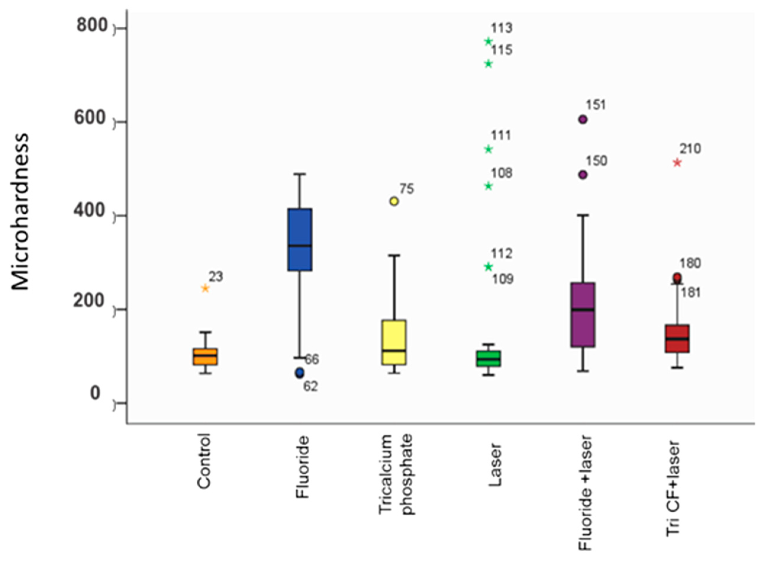
3.2. Enamel Surface Micro-Hardness

The treatment microhardness measurements were made to verify that the most remineralized surface would be the hardest. The mean micro-hardness values of the enamel surfaces are shown in Figure 1. The fluoride group had the highest micro-hardness after remineralization with 329.8 VH (p < 0.05). Groups A and D were significantly different (Control, Laser). Tri-calcium phosphate and tri-calcium phosphate + LLL showed no differences. Figure 2 shows indentation marks.
Spearman correlation determined a relationship between decreased mineralization in the remineralizing solution and the microhardness of the enamel surface (Table 3).

4. Discussion

There are many ways to treat demineralization. Here, fluoride and tri-calcium phosphate fluoride were added, followed by an LLL. These were evaluated in terms of micro-hardness and mineral content on bovine enamel.
Atomic absorption spectroscopy was used to measure calcium, and UV-Vis spectroscopy was used to measure phosphorus. These processes were used to determine mineral loss. Similarly, mineralizing agents were used in the remineralization solution. These methods were used to efficiently identify calcium and phosphorus and indirectly study the mineral components on dental surfaces [16,18,19,20].
The treatments offering the most calcium in the demineralization solution were the control, the tri-calcium phosphate, and the laser-irradiated tri-calcium phosphate; whereas the control, laser, and laser-irradiated tri-calcium phosphate offered the most phosphorus. This suggests that the minerals were not on the dental surface. These groups showed the lowest remineralizing potential. In other studies, tri-calcium phosphate was added to fluoride, and calcium was encapsulated in sodium lauryl sulfate, which was placed on white spot lesions and made dental enamel more resistant [8,9]. This contradicts the present study.
The laser increases hardness and prevents the treated area from becoming a cavitation. Moreover, the effects are higher when fluoride- or calcium-based compounds are irradiated concurrently [11,12,13,14,17]. Nevertheless, the data show that the mineral content of the laser group was similar to the control. In all examples where fluoride was exposed to laser irradiation, there was no increase in remineralization potential. This puts their effectiveness into question.
The group with the highest remineralizing effect was fluoride. Both calcium and phosphorus were found in low levels in the remineralization solution assuming that both components should be precipitated on enamel surfaces and transformed into more resistant tissue. Although tri-calcium phosphate and simple varnish fluoride had 22,600 ppm concentrations, fluoride was the most effective treatment for dental remineralization.
This was a model of constant remineralization, and cyclic pH (including periods of demineralization) was used over 15 days to emulate oral conditions in an optimal environment to determine the best performance of each of the mineralizing agents. An additional demineralizing solution was added at a critical pH to create a white spot lesion. The remineralization solution at pH 7.0 was similar to saliva. Despite this model’s chemical conditions, there are still biological differences, which might modify results in the laboratory.
The enamel surfaces of the samples were polished flat to homogenize them and create the area needed for testing microhardness. This can certainly be a factor because the studied area was susceptible to deep demineralization in accordance with Elkassas et al. [9]. The Vickers test was used to analyze the surface resistance because it provides indirect information about the mineral content and hardness of dental tissue [2,15].
The average microhardness of the control and laser groups had a similar remineralizing effect, which contradicts other studies that show that low-power lasers create a harder and more resistant surface than the additional calcium compounds or the control group [12,14]. The group with the best microhardness was fluoride, although the rate of application and fluoride content for all groups was the same (22,600 ppm). The group with tri-calcium phosphate that directly incorporated these components into the lesion is not shown here.
One limitation of this study is that baseline microhardness was not measured before the demineralization process. Additionally, bovine teeth may have important differences from human teeth. White spot lesions are formed much faster on bovine teeth than on human teeth and has more carbonate and less fluoride. This could affect remineralization and make it less efficient, although the distribution of minerals is almost the same in both tissues [15].

5. Conclusions

Even under the limitations of the present study, it can be concluded that fluoride is the most effective treatment for dental remineralization—more so than the addition of tri-calcium phosphate and low-level lasers. This in vitro study imitated the application of different remineralizing agents and showed which was the most efficient for treating non-cavitated injuries. This can help to prevent the progression of lesions in patients with white spot lesions.

Acknowledgments

We thank the National Council of Science and Technology (CONACYT Mexico) for their support for the development of this research. Publication was supported by the Ministry of Education, Mexican Federal Government, through the Faculty Development Program (PRODEP).

Author Contributions

E.L.-C., N.L.-R., R.A.M.-L. and N.L.R.-B. were involved in the design and development of the study. E.L.-C. designed the study, analyzed the data, and wrote the first draft of the manuscript. M.M.G.-F., R.d.l.R.-S. and C.E.M.-S. were involved in the conception of the paper and in the analysis and interpretation of the results. All authors were involved in the critical review and made intellectual contributions. They also approved the final version.

Conflicts of Interest

The authors declare that they have no conflict of interest.

References

  1. Peters, M. Strategies for noninvasive demineralized tissue repair. Dent. Clin. N. Am. 2010, 54, 507–525. [Google Scholar] [CrossRef] [PubMed]
  2. Murdoch-Kinch, C.A.; McLean, M.E. Minimally invasive dentistry. J. Am. Dent. Assoc. 2003, 134, 87–95. [Google Scholar] [CrossRef] [PubMed]
  3. Cochrane, N.; Cai, F.; Huq, N.; Burrow, M.; Reynolds, E. New Approaches to enhanced remineralization of tooth enamel. J. Dent. Res. 2010, 89, 1187–1197. [Google Scholar] [CrossRef] [PubMed]
  4. Zhou, S.; Zhou, J.; Watanabe, S.; Watanabe, K. In vitro study of the effects of fluoride-releasing dental materials on remineralization in an enamel erosion model. J. Dent. 2012, 40, 255–263. [Google Scholar] [CrossRef] [PubMed]
  5. Caruso, S.; Bernardi, S.; Pasini, M.; Giuca, M.R.; Docimo, R.; Continenza, M.A.; Gatto, R. The process of mineralisation in the development of human tooth. Eur. J. Paediatr. Dent. 2016, 17, 322–326. [Google Scholar] [PubMed]
  6. Lins, R.D.; Dantas, E.M.; Lucena, K.C.; Catão, M.H.; Granville-Garcia, A.F.; Carvalho Neto, L.G. Biostimulation effects of low-power laser in the repair process. An. Bras. Dermatol. 2010, 85, 849–855. [Google Scholar] [CrossRef] [PubMed]
  7. Matson, J.R.; Matson, E.; Navarro, R.S.; Bocangel, J.S.; Jaeger, R.G.; Eduardo, C.P. Er: YAG laser effects on enamel occlusal fissures: An in vitro study. J. Clin. Laser Med. Surg. 2002, 20, 27–35. [Google Scholar] [CrossRef] [PubMed]
  8. 3M ESPE. Solutions. Products 3M. Available online: http://solutions.3m.com.mx/wps/portal/3M/es_MX/3MESPE_LA/dental-professionals/ (accessed on 25 November 2014).
  9. Elkassas, D.; Arafa, A. Remineralizing efficacy of different calcium-phosphate and fluoride based delivery vehicles on artificial caries like enamel lesions. J. Dent. 2014, 42, 466–474. [Google Scholar] [CrossRef] [PubMed]
  10. Karlinsey, R.; Mackey, A.; Walker, E.; Frederick, K. Surfactant-modified beta-TCP: Structure, properties, and in vitro remineralization of subsurface enamel lesions. J. Mater. Sci. Mater. Med. 2010, 21, 415–420. [Google Scholar] [CrossRef] [PubMed]
  11. Vlacic, J.; Meyers, I.; Kim, J.; Walsh, L. Laser-activated fluoride treatment of enamel against and artificial caries challenge: Comparison of five wavelengths. Aust. Dent. J. 2007, 52, 101–105. [Google Scholar] [CrossRef] [PubMed]
  12. Muller, K.; Rodrigues, C.; Nunez, S.; Rocha, R.; Ribeiro, M. Effects of low-power red laser on induced-dental caries in rats. Arch. Oral Biol. 2007, 52, 648–654. [Google Scholar] [CrossRef] [PubMed]
  13. Laser Systems. Laser Dental Quantum® IR810. Available online: http://www.lasersystems.com.mx/dental.php (accessed on 20 October 2014).
  14. Heravi, F.; Farzaneh, A.; Mahdieh, M.; Sorouh, B. Comparative evaluation of the effect of Er: YAG laser and low level laser irradiation combined with CCP-ACPF cream on treatment of enamel caries. J. Clin. Exp. Dent. 2014, 6, 121–126. [Google Scholar] [CrossRef] [PubMed]
  15. Lippert, F.; Lynch, R. Comparison of Knoop and Vickers surface microhardness and transverse microradiography for the study of early caries lesion formation in human and bovine enamel. J. Arch. Oral Biol. 2014, 59, 704–710. [Google Scholar] [CrossRef] [PubMed]
  16. Díaz-Monroy, J.M.; Contreras-Bulnes, R.; Olea-Mejía, O.F.; García-Fabila, M.M.; Rodríguez-Vilchis, L.E.; Sánchez-Flores, I.; Centeno-Pedraza, C. Chemical changes associated with increased acid resistance of Er: YAG laser irradiated enamel. Sci. World J. 2014, 1, 1–6. [Google Scholar] [CrossRef] [PubMed]
  17. Lippert, F.; Hara, A. Fluoride dose-response of human and bovine enamel caries lesions under remineralizing conditions. Am. J. Dent. 2012, 25, 205–209. [Google Scholar] [PubMed]
  18. López, G.; De la Casa, M.; Sáez, M.; López, M. Decalcification of root dentin: Comparison of two methods for identification. Sci. Oral J. 2013, 10, 15–23. [Google Scholar]
  19. Hjortsjö, C.; Jonski, G.; Young, A.; Saxegaard, E. Etching effect of fluorides on human tooth enamel in vitro. Arch. Oral Biol. 2014, 59, 1328–1333. [Google Scholar] [CrossRef] [PubMed]
  20. Low, I.; Alhuthali, A. In-situ monitoring of dental erosion in tooth enamel when exposed to soft drinks. Mater. Sci. Eng. 2008, 28, 1322–1325. [Google Scholar] [CrossRef]
Figure 1. Mean microhardness values between groups (p < 0.05).
Applsci 08 00078 g001
Figure 2. (A) Indentation from Group B: (B) Indentation from Group D where the mark is bigger than the previous image, (C) Indentation from Group F, showing a softer surface for a greater indentation.
Applsci 08 00078 g002
Table 1. Solutions analysis of calcium and phosphorus content during treatment.
Calcium Content mg/LPhosphorus Content mg/L
GroupsDemineralRemineralGroupsDemineralRemineral
Before TreatmentAfter TreatmentBefore TreatmentAfter Treatment
A (Control)3.80 ± 0.671.00 ± 0.20A (Control)2.17 ± 0.220.52 ± 0.05
B (Fluoride)3.00 ± 0.630.88 ± 0.11 *B (Fluoride)1.45 ± 0.240.40 ± 0.05 *
C (TriFC)3.75 ± 0.751.34 ± 0.17C (TriFC)1.58 ± 0.290.41 ± 0.07
D (LLL)3.02 ± 0.580.99 ± 0.15D (LLL)1.94 ± 0.320.53 ± 0.08
E (Fluoride + LLL)2.52 ± 0.900.96 ± 0.16E (Fluoride + LLL)1.98 ± 0.190.42 ± 0.05
F (TriFC + LLL)2.70 ± 0.541.14 ± 0.19F (TriFC + LLL)1.98 ± 0.250.43 ± 0.07
Demineral = demineralization; Remineral = remineralization; TriFC = Fluoride + Tri-calcium phosphate; LLL = low-level laser, * shows the lowest amount of calcium and phosphorus ions in solution after the use of remineralizing agents. p < 0.05, Wilcoxon. All mineral levels decrease after placing remineralizants.
Table 2. Analysis of calcium and phosphorus ion content in solution after the use of remineralizing agents.
CalciumABCDEF
A-
B0.018271-
C0.000000 *0.000000 *-
D0.3990900.0094740.000002 *-
E0.000000 *0.000035 *0.000000 *0.000000 *-
F0.0033800.000001 *0.0139380.0070880.000000-
PhosphorusABCDEF
A-
B0.000000 *-
C0.000000 *0.199111-
D0.4440790.000000 *0.000000 *-
E0.000000 *0.0766460.2798800.000000 *-
F0.000001 *0.0199180.1130150.000002 *0.265181-
* Adjusted p-value for significance is 0.001667. One-way analysis of variance by ranks (Kruskal-Wallis Test). A = Control, B = Fluoride (Duraphat varnish), C = Tricalcium phosphate (Clinpro White varnish), D = LLL (IR810 (Quantum)), E = Fluoride + LLL, F = Tricalcium phosphate + LLL.
Table 3. Correlation between decreased mineralization and microhardness value.
VariableVariableRp
Remineralization solutionCalcium ionsMicrohardness−0.2680.0008
Phosphorus ionsMicrohardness−0.208<0.002
Negative correlation between the presence of minerals and microhardness (p < 0.05).

