Review

Visualizing the Knowledge Domain of Nanoparticle Drug Delivery Technologies: A Scientometric Review

1
Office of Research and Development, National Chiao Tung University, 1001, Ta-Hsueh Rd., Hsinchu 30010, Taiwan
2
College of Computing and Informatics, Drexel University, 3141 Chestnut Street, Philadelphia, PA 19104-2875, USA
3
Department of Foreign Languages and Literatures, National Chiao Tung University, 1001, Ta-Hsueh Rd., Hsinchu 30010, Taiwan
*
Author to whom correspondence should be addressed.
Appl. Sci. 2016, 6(1), 11; https://doi.org/10.3390/app6010011
Submission received: 19 November 2015 / Revised: 29 December 2015 / Accepted: 31 December 2015 / Published: 7 January 2016
(This article belongs to the Section Nanotechnology and Applied Nanosciences)

Abstract

:
The scientific literature of nanoparticle drug delivery technologies (NDDT) between 2005 and 2014 was reviewed. The visualized co-citation network of its knowledge domain was characterized in terms of thematic concentrations of co-cited references and emerging trends of surging keywords and citations to references through a scientometric review. The combined dataset of 25,171 bibliographic records were constructed through topic search and citation expansion to ensure adequate coverage of the field. While research in gold nanoparticle and magnetic nanoparticle remains the two most prominent knowledge domains in the NDDT field, research related to clinical and therapeutic applications has experienced a considerable growth. In particular, clinical and therapeutic developments in NDDT have demonstrated profound connections with the mesoporous silica nanoparticle research and microcrystal research. A rapid adaptation of mesoporous silica-based nanomaterials and rare earth fluoride nano-/microcrystal in NDDT is evident. Innovative strategies have been employed to exploit the multicomponent, chemical synthesis, surface modification, and controlled release imparting functionalized targeting capabilities. This study not only facilitated the connection of authors and research themes in the NDDT community, but also demonstrated how research interests and trends evolve over time, which greatly contributes to our understanding of the NDDT knowledge domains.

1. Introduction

In recent years, increasing attention has paid to the development of novel drug delivery systems (NDDS), because the average cost and time for the development of a new chemical or biochemical entity are much higher than those required to develop a NDDS [1]. Moreover, incorporating an existing medicine into a NDDS can significantly improve its performance in terms of efficacy, safety, and improved patient compliance [2].
Among the various NDDS, a considerable attention has focused on the development of therapeutic nanoparticle technologies, because they have the potential to revolutionize the drug development manner and alter the landscape of the pharmaceutical industry [3]. As nanoparticle drug delivery technologies (NDDT) begin to gain traction over the next several decades, it is important to visualize its knowledge domains. In this context, this paper aimed to explore the knowledge domains associated with NDDT, quantifying research patterns and trends in NDDT. Using CiteSpace as a visualization tool to analyze the scientific literature retrieved from the Web of Science Core Collection (WoSCC) [4], this paper reflected a number of remarkable connections and clusters of research in NDDT over the past decade (2005–2014).

2. The Visualization of Scientific Knowledge Domains

A knowledge domain is a particular field of study that creates a common ground and a sense of development of a common identity by affirming its purpose and value to members and stakeholders [5]. Moreover, intellectual relationships and collaboration networks are fundamental to a knowledge domain [6]. The visual representation of such “knowledge networks” contributes to the overall understanding of intellectual collaborations in a particular knowledge domain.
Scientific knowledge changes all the time. Most of the changes are incremental, but some are revolutionary and fundamental [7]. For example, in the field of NDDT, several subareas have formed through the years, ranging from drug incorporation and release, formulation stability and shelf life, biocompatibility, to biodistribution and targeting, and functionality [8]. With recent advances in computing power, scientific indexes, and bibliographic techniques, progress is being made and researchers are gradually piecing together this dilemma and exploring hidden connections and knowledge domains in the literature. In the next section, the process of exploring the knowledge domains associated with NDDT was described.

3. Method

3.1. Bibliographic Records

We collected the bibliographic records from the WoSCC of Thomson Reuters. We determined the time frame of this analysis to the last decade (2005–2014) due to a concerted effort to improve the clarity of derived results. The search consists of two subqueries about NDDT: “nanoparticle* drug delivery” in topic search and “nanoparticle* drug delivery” in title search. Two datasets of bibliographic records on NDDT were retrieved from the WoSCC, including both SSCI and SCI-Expanded subsidiary databases. The topic-search dataset is referred as the core dataset [9]. The query resulted in 1770 bibliographic records which includes 1178 original research articles. The expanded dataset is a superset of the core dataset with extra bibliographic records obtained by association through citation links. Any article citing at least one original article in the core set can be assumed that it may be thematically relevant to the subject matter underlying the core dataset [10]. The resultant expanded dataset consists of 23,993 unique records. Both datasets were merged as a whole for scientometric review. The whole bibliographic records were then exported to CiteSpace [11] for subsequent analysis.

3.2. CiteSpace

CiteSpace supports the visualization of a scientific field from bibliographic sources in terms of networks of several types of entities, including cited references, co-authors, and co-occurring keywords [12]. This paper centered on document co-citation networks and networks of co-occurring keywords in an effort to deliver more accurate and complete results for the NDDT knowledge domains. Individual nodes in the network can be aggregated into clusters based on their interconnectivity. Each cluster represents a distinct specialty or a thematic concentration. Other points of interest include highly cited landmark articles, articles with strong citation bursts, and keywords with a strong surge of frequency.
The aim of burst detection is to investigate whether the frequency of an entity increases abruptly with reference to its peers. If an article is found to have a steep increase of its citation counts, then the article is regarded as having a citation burst. Similarly, if the number of articles with a term in their titles or abstracts sharply increased at a much faster rate than other terms, then the term is defined as a burst term. For the purposes of this paper, we first identified key articles associated with NDDT. Next, we detected key clusters and emerging topics through citation bursts in the literature for exploring the knowledge domains of NDDT.

3.3. Network Analysis and Visualization

The data input of CiteSpace is a collection of scientific publications associated with a specific topic [4]. Through its network modeling and visualization, we can explore the knowledge domains in a specific topic. One of the important tools in CiteSpace helps identify betweenness centrality between pivotal points in the scientific literature [13]. The betweenness centrality of a node in a network functions as a measure to indicate the importance of nodes in a network.
Research fronts are a collection of articles that are actively cited by researchers and often display properties of domain specificity [14]. Research front terms generated by CiteSpace capture core concepts of co-citation clustering and provide a general overview of a knowledge domain and its associated network. Finally, time slices are powerful tools for providing a temporal perspective to the literature and identifying citation bursts [11].

4. Bibliographic Landscape

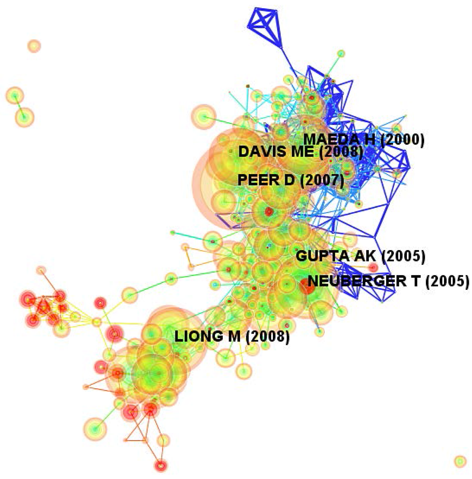
4.1. Document Co-Citation Analysis

The complete set of 25,171 bibliographic records combining both the core dataset and the expanded dataset were visualized and analyzed using CiteSpace. Next, the bibliographic records were extracted from the WoSCC and their document co-citation network was generated as shown in Figure 1.
Figure 1. Key articles in nanoparticle drug delivery technologies.
Applsci 06 00011 g001
In this network, there are 402 unique nodes and 2974 links for a one-year time slice. These nodes represent cited references from the collected articles, and the links in the network represent co-citation relationships. Each link colors correspond directly to each time slice. For example, blue links describe articles that were co-cited in 2005, and the most recent co-citation relationships are visualized as orange or red links. We can further conclude three focal points from Figure 1. First, larger node sizes imply that the article is an important one within the knowledge domain. Second, red rings around a node represent a citation burst. Third, purple rings indicate nodes that have a relatively high betweenness centrality in the network.
Table 1 presents the five top-cited articles associated with the term “nanoparticle drug delivery” between 2005 and 2014.
Table 1. Five critical articles in nanoparticle drug delivery technologies.
Cited FrequencyTitleAuthorYearBetweenness CentralityJournal
1014Nanocarriers as an emerging
platform for cancer therapy
Peer et al.20070.00Nature Nanotechnology
829Synthesis and surface engineering
of iron oxide nanoparticles for biomedical applications
Gupta and Gupta20050.02Biomaterials
764Multifunctional inorganic nanoparticles for imaging, targeting, and drug deliveryLiong et al.20080.00ACS Nano
755Superparamagnetic nanoparticles for biomedical applications: possibilities and limitations of a new drug delivery systemNeuberger et al.20050.01Journal of Magnetism and Magnetic Materials
712Tumor vascular permeability and the EPR effect in macromolecular therapeutics: a reviewMaeda et al.20000.08Journal of Controlled Release
The first is a paper by Peer et al. [15], which examined some of the approved formulations for clinical use and discussed the challenges in translating basic research to the clinic. The second is Gupta and Gupta [16] work, which discussed the synthetic chemistry, fluid stabilization and surface modification of superparamagnetic iron oxide nanoparticles. The other three papers focus on particular application areas or techniques. For example, Liong et al. [17] demonstrated the multifunctional inorganic nanoparticles can be monitored inside living cells by both magnetic resonance and fluorescence imaging methods. Neuberger et al. [18] discussed the characteristics and applications of superparamagnetic nanoparticles based on a core consisting of iron oxides (SPION). Finally, Maeda et al. [19] reviewed the basic characteristics of the enhanced permeability and retention (EPR) effect. In sum, regardless of the subareas, all five articles represent nanoparticle drug delivery as a key enabling technology for pursuing substantive research questions in physical or social environments.

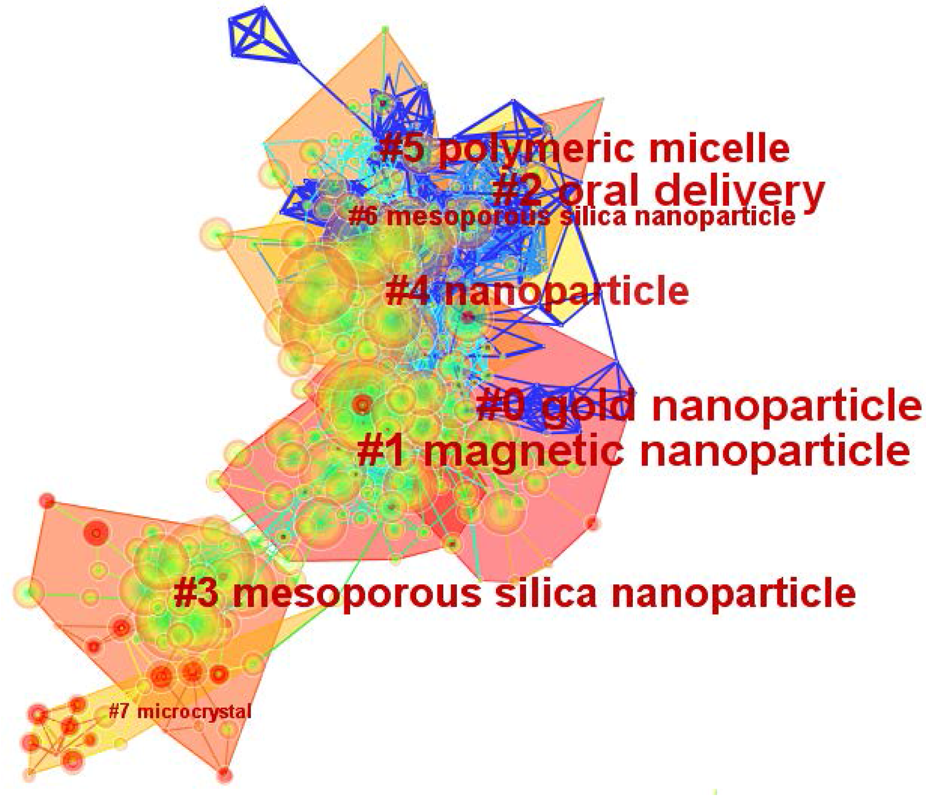
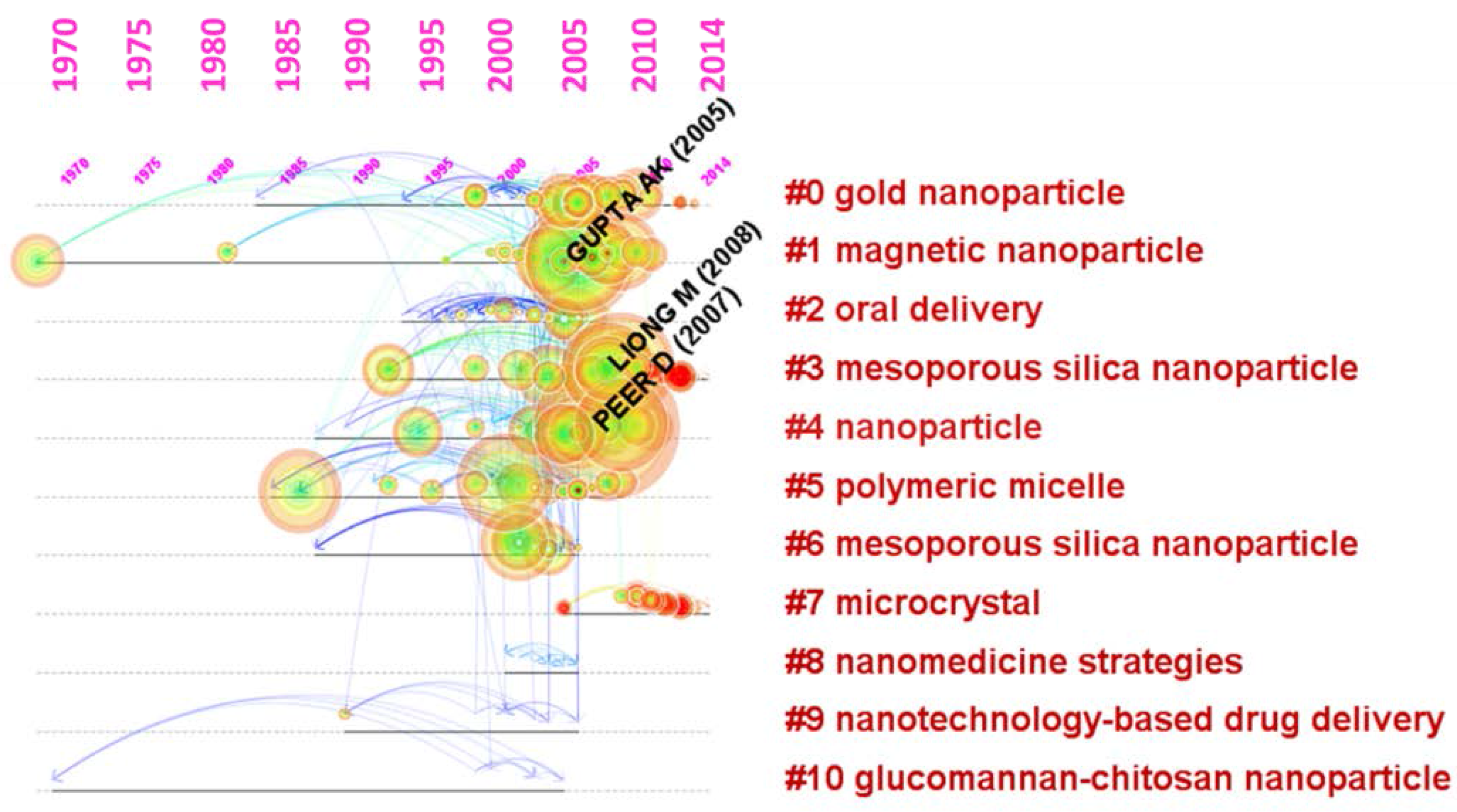
4.2. Identification and Interpretation of Clusters

We used CiteSpace to explore research patterns and emerging trends in the body of knowledge in terms of key clusters of articles. Figure 2 shows clusters labeled with title terms. The size of a cluster’s label is proportional to the size of the cluster.
Figure 2. Clusters visualization based on a document co-citation network.
Applsci 06 00011 g002
In this instance, there are total 55 clusters in the network. To characterize the nature of a cluster, CiteSpace can extract noun phrases from the titles of articles that cited the cluster based on three specialized metrics—TF*IDF, log-likelihood tests (LLR) and mutual information tests (MI). LLR usually gives the best result in terms of the uniqueness and coverage of themes associated with a cluster. Table 2 details the top 11 clusters in rank order.
Table 2. Top-ranked clusters in nanoparticle drug delivery technologies.
IDSizeSilhouetteLabel (TF*IDF)Label (LLR)Label (MI)Mean (Cited Year)
0550.789nanogold nanoparticlebiomaterial-based technologies2003
1530.853ionmagnetic nanoparticlebehavior2003
2510.776nanooral deliverygeneral strategy2001
3480.933silicamesoporous silica nanoparticleanticancer drug delivery system2007
4460.704nanonanoparticledrug discovery2003
5420.811nanopolymeric micellenanofibrous scaffold2001
6230.890nanomesoporous silica nanoparticleouzo region2002
7130.972upconversionmicrocrystaldrug-delivery system2010
881.000plaque angiogenesisnanomedicine strategiesmechanism2002
980.954nanonanotechnology-based drug deliverydrug-delivery system2000
1050.998carboxymethyl konjacglucomannan-chitosan nanoparticledrug delivery1994
As shown in Figure 2, gold nanoparticle and magnetic nanoparticle are the two largest clusters. Microcrystal and mesoporous silica nanoparticle are the two youngest clusters, and glucomannan-chitosan nanoparticle is the oldest cluster. The values of the silhouettes for each cluster are greater than 0.5, suggesting robust and meaningful results. The largest cluster, gold nanoparticle (#0), consists of 55 members. The three most active citers in this cluster are Aswathy et al. [20], Alkilany and Murphy [21], and Chithrani [22]. According to the titles of these citers in this cluster, research works related to gold nanoparticle shape a foundation of the knowledge domain. Researchers interested in gold nanoparticle are particularly concerned with near-infrared quantum dot, toxicity, and biomaterial-based technologies. Not surprisingly, this cluster covers a range of interests, reflecting the interdisciplinary nature of NDDT and their use.
The second largest cluster (#1) in this knowledge domain, magnetic nanoparticle, has 53 member articles and an average publication year of 2003. The three most active citers to this cluster are Hao et al. [23],Veiseh et al. [24], and Faraji et al. [25] accordingly. Because of their remarkable magnetic properties and biologically comparable sizes, these magnetic nanoparticles are very beneficial for biomedical applications [23]. In addition, these magnetic nanoparticles can also respond resonantly to an alternating magnetic field and function as a heater, offering a promising therapeutic solution by magnetic fluid hyperthermia [25]. In recent years, the synthesis, design, and fabrication of multifunctional magnetic nanoparticles for biomedical applications has become one of the most active research areas in this knowledge domain [24]. As shown in Figure 1 and Figure 2, this cluster has the top ranked burst item—Neuberger et al. [18] among all clusters, with bursts of 61.86. Thus, the magnetic nanoparticle cluster is essential to the literature represented by the datasets.
The third largest cluster (#2) is oral delivery which has 51 member articles and an average publication year of 2001. The three most active citers in this cluster are Ratzinger et al. [26], Roger et al. [27], and Patel et al. [28] accordingly. According to the titles of these citers in this cluster, nanometric-sized drug delivery systems are being extensively studied and provide promising potential for oral drug delivery. Researchers interested in oral delivery focus particularly on how technological solutions can enhance the bioavailability or the targeting of anticancer drug after oral administration.
There are other clusters worth mentioning. For example, the cluster (#3) for mesoporous silica nanoparticle consists of 48 member articles and an average publication year of 2007. According to the major citing articles [29,30], it is not surprising that previous advances push mesoporous silica nanoparticle to the research forefront of drug delivery development.
Another major cluster corresponds to the terms polymeric micelle. In most instances, polymeric micelles play a significant role in the advancement of NDDT by providing controlled release of therapeutic agents in constant doses over long periods [31,32]. Thus, polymeric micelle has become a substantive knowledge domain in NDDT research field.
Finally, the term microcrystal also represents a cluster which has 13 member articles and an average publication year of 2010. This cluster is the newest one in which the three most active citers in this cluster are Li and Lin [33], Chen et al. [34], and Liu et al. [35] accordingly. Dramatic efforts have been dedicated to the chemical synthesis of rare earth fluoride nano-/microcrystals with uniform size and shapes [33]. Hence, research works related to microcrystal reflect the recent knowledge domain in NDDT research field.
An alternative approach for viewing these clusters and their relationships is with timeline visualization as shown in Figure 3.
Figure 3. Timeline view for nanoparticle drug delivery technologies: 2005–2014.
Applsci 06 00011 g003
The most obvious trend in Figure 3 is that most of the documents cited were published after 1985, roughly corresponding to the rise and deployment of an existing drug molecule from a classic type to a novel nanoparticle drug delivery system. Interestingly, the earliest cited document in the derived network was published before 1970 [36] and is found in the cluster of magnetic nanoparticle. Moreover, as shown in Figure 2 and Figure 3, the top ranked item by centrality is Yoon et al. [37] in Cluster #1, with centrality of 0.26. The second one is Giri et al. [38] in Cluster #3, with centrality of 0.22. The third is Gao et al. [39] in Cluster #0, with centrality of 0.14. These nodes can be considered as pivotal points that provide important bridging connections between two research interests.

4.3. Most Active Clusters

Figure 3 shows two clusters, cluster #3 and cluster #7, with the strongest citation bursts. This means that cluster #3 and cluster #7 represent where the major efforts of the research in this field since 2010. Cluster #3 is labeled as mesoporous silica nanoparticle. Table 3 lists five articles in cluster #3 with the strongest citation bursts.
Table 3. Articles with the strongest citation bursts in cluster #3.
CitationBurstAuthorYearTitleSource
24846.48Tang et al. [40]2012Mesoporous silica nanoparticles: synthesis, biocompatibility and drug deliveryADV MATER
22244.12Yang et al. [41]2012Functionalized mesoporous silica materials for controlled drug deliveryCHEM SOC REV
23841.45Li et al. [42]2012Mesoporous silica nanoparticles in biomedical applicationsCHEM SOC REV
12720.30Liong et al. [43]2009Mesostructured multifunctional nanoparticles for imaging and drug deliveryJ MATER CHEM
29713.07Trewyn et al. [44]2007Mesoporous silica nanoparticle based controlled release, drug delivery, and biosensor systemsCHEM COMMUN
The title terms in Table 3 mainly include mesoporous silica nanoparticle, controlled release, and drug delivery. The highest bursted article in this cluster, Tang et al. [40], discussed the recent progress in the synthesis of mesoporous silica nanoparticles for drug delivery applications. The second highest bursted article in this cluster, Yang et al. [41], reviewed the most recent research progress on silica-based controlled drug delivery systems. The common theme in terms of the bursted articles to this cluster is the design, synthesis and functionalization of mesoporous silica nanoparticles for efficient drug delivery systems.
Cluster #7 is labeled as microcrystal. Table 4 lists five articles in cluster #7 with the strongest citation bursts.
Table 4. Articles with the strongest citation bursts in cluster #7.
CitationBurstAuthorYearTitleSource
20037.79Tian et al. [45]2012Mn2+ Dopant-Controlled Synthesis of NaYF4: Yb/Er Upconversion Nanoparticles for in vivo Imaging and Drug DeliveryADV MATER
18624.91Zhou et al. [46]2012Upconversion nanophosphors for small-animal imagingCHEM SOC REV
17819.64Haase and Schäfer [47]2011Upconverting nanoparticlesANGEW CHEM INT EDIT
23319.44Wang and Liu [48]2009Recent advances in the chemistry of lanthanide-doped upconversion nanocrystalsCHEM SOC REV
22113.97Wang et al. [49]2010Simultaneous phase and size control of upconversion nanocrystals through lanthanide dopingNATURE
The title terms in Table 4 mainly include nanocrystals, upconversion, and drug delivery. Tian et al. [45] has the strongest citation burst in the cluster. The work suggested that upconversion nanoparticles (UCNPs) could be used as potential bio-labels for in vivo imaging, and as promising drug carriers for intracellular drug delivery. The second highest bursted article in this cluster, Zhou et al. [46], considered a rational approach to obtain suitable UCNP nanoprobes for small animal bioimaging. A common theme among this group of articles appears to focus on applications of UCNP research for novel imaging agents and therapy.

4.4. References with Strong Citation Bursts

Significant increases of research interests within the NDDT knowledge domain are characterized by publications that experienced citation bursts. This was based on a total of 25,171 bibliographic records which were selected from 555,999 valid references. Figure 4 shows the top 30 references with the strongest citation bursts during the period between 2005 and 2014.
Figure 4. Top 30 references with strong citation bursts.
Applsci 06 00011 g004
As shown in Figure 4, most of the references started to burst in year 2005, two references started to burst in year 2006, and only one reference started to burst in year 2007. Table 5 shows the representative references for three groups by the beginning time of burst.
Table 5. Representative references with the strongest citation bursts.
ReferencesYearCitation Burst
StrengthBeginEnd
Chaw et al. [50]200419.6720052007
Feng et al. [51]200416.4720052006
Anderson et al. [52]200013.9520052007
Liu et al. [53]200315.2320062007
Little et al. [54]20046.5620072008
In the group of year 2005, the top three references with the strongest citation bursts are Chaw et al. [50], Feng et al. [51], and Anderson et al. [52] accordingly. Chaw et al. [50] analyzed the drug-loading process for understanding the effect of various fabrication parameters on drug encapsulation efficiency. The burst lasted for three years from 2005 till 2007. Feng et al. [51] article indicated that nanoparticles of biodegradable polymers can provide an ideal solution to clinical administration with better efficacy and less side effects. Anderson et al. [52] article demonstrated a new MRI method for visualizing the endothelial αvβ3 integrin in vivo using an antibody-targeted, site-directed contrast agent.
In the group of year 2006 by the beginning time of burst, Liu et al. [53] reported the synthesis, characterization and temperature sensitivity of thermally responsive polymeric micellar nanoparticles that were self-assembled from cholesteryl end-capped random poly. The article published in 2003 has the third strongest citation burst in the entire dataset. The burst lasted for two years from 2006 till 2007. Little et al. [54] article published in 2004 has the strongest citation burst in the group of year 2007 by the beginning time of burst. They described a microparticle-based DNA delivery system which is composed of pH-sensitive poly-β amino ester and poly lactic-co-glycolic acid.

4.5. References Bursted Since 2013

Table 6 shows the references with the recent citation bursts from 2010 onward. In this subsection, we will review key articles with the most recent citation bursts starting from 2013. Citation bursts starting from 2013 are associated with the main three 2012 articles.
Table 6. References with the most recent citation bursts since 2010.
ReferencesYearCitation Burst
StrengthBeginEndDuration
Liong et al. [43]200920.320102011 Applsci 06 00011 i001
Jain et al. [55]20086.220102011 Applsci 06 00011 i002
Winter et al. [56]20033.720102011 Applsci 06 00011 i003
McCarthy and Weissleder [57]20082.820102011 Applsci 06 00011 i004
Hood et al. [58]20022.520102011 Applsci 06 00011 i005
Lu et al. [59]20108.920122013 Applsci 06 00011 i006
Ashley et al. [60]20118.520122015 Applsci 06 00011 i007
Vivero-Escoto et al. [61]20107.720122013 Applsci 06 00011 i008
Meng et al. [62]20103.320122015 Applsci 06 00011 i009
Tang et al. [40]201246.520132015 Applsci 06 00011 i010
Yang et al. [41]201244.120132015 Applsci 06 00011 i011
Li et al. [42]201241.520132015 Applsci 06 00011 i012
Tian et al. [45]201237.820132015 Applsci 06 00011 i013
Pan et al. [63]201236.320132015 Applsci 06 00011 i014
Du et al. [64]201135.120132015 Applsci 06 00011 i015
Liu et al. [65]201134.220132015 Applsci 06 00011 i016
Albanese et al. [66]201229.020132015 Applsci 06 00011 i017
Zhang et al. [67]201225.820132015 Applsci 06 00011 i018
Zhou et al. [46]201224.920132015 Applsci 06 00011 i019
Wang et al. [68]201224.420132015 Applsci 06 00011 i020
Haase and Schäfer [47]201119.620132015 Applsci 06 00011 i021
Wang et al. [69]201119.620132015 Applsci 06 00011 i022
Wang and Liu [48]200919.420132015 Applsci 06 00011 i023
Wang et al. [49]201014.020132015 Applsci 06 00011 i024
Luo et al. [70]201112.820132015 Applsci 06 00011 i025
Auzel [71]200412.120132015 Applsci 06 00011 i026
He and Shi [72]201110.620132015 Applsci 06 00011 i027
Thomas et al. [73]20109.920132015 Applsci 06 00011 i028
Among the articles with strong citation bursts since 2013, Tang et al. [40] article, titled “Mesoporous silica nanoparticles: synthesis, biocompatibility and drug delivery”, has the strongest citation burst with a burst strength of 46.5. This article published in Advanced Materials discussed the biological barriers for nano-based targeted cancer therapy and mesoporous silica nanoparticle-based targeting strategies. The second article with the most recent citation burst studies the functionalized mesoporous silica materials [41]. The work reported several exciting achievements on mesoporous silica-based materials as sustained-release systems and stimuli-responsive controlled release systems. The third article that has drawn much attention is a 2012 article studied by Li et al. [42]. The authors described that the functionalization of mesoporous silica nanoparticles with molecular, supramolecular or polymer moieties provides the material with great versatility and makes the delivery operation highly controllable.

5. Conclusions

According to the network visualization and the document co-citation analysis supported by CiteSpace, we explored the key clusters of articles and identified research patterns and emerging trends in the literature. The top two clusters were labeled as gold nanoparticle and magnetic nanoparticle, suggesting that they are foundational to the knowledge domain. Not surprisingly, the two largest clusters cover a range of interests, reflecting the interdisciplinary nature of NDDT and their use. While research in gold nanoparticle and magnetic nanoparticle remains the two most prominent knowledge domains in the NDDT field, research related to clinical and therapeutic applications in NDDT has experienced a considerable growth. The detected surge of the two keywords—“mesoporous silica” and “microcrystal” in the literature of NDDT led us to investigate the nature and context of its use in NDDT. The investigation revealed a rapidly increasing number of studies that specifically used mesoporous silica-based nanomaterials and rare earth fluoride nano-/microcrystal in NDDT research. Similarly, a detected burst of citations underscores a fast-moving knowledge domain. For example, knowing exactly when citations to Chaw et al. [50] had surged (2005–2007) can improve our understanding of the complex adaptive behavior of the field. Knowing that Tang et al. [40] has been attracting much attention since 2012 can help us capitalize on the collective intelligence of the multidisciplinary scientific communities. The emergence of a new cluster indicates the beginning of a trend, for example, in microcrystal. A persistent cluster represents a continuation of an existing trend, for example, in mesoporous silica nanoparticle. In addition, the two most active clusters with the strongest citation since 2010 appear to mesoporous silica nanoparticle (cluster #3) and microcrystal (cluster #7). This means that both mesoporous silica nanoparticle and microcrystal represent where the major efforts of the research in this field.
Finally, considering the interdisciplinary characteristic of NDDT, it is not easy to obtain an overall picture of the research field. Hence, the contribution of this work was to explore an efficient and quantitative way of understanding the NDDT knowledge domain.

Author Contributions

Yen-Chun Lee designed and proposed the concept of this research. Both Yen-Chun Lee and Xing-Tzu Tsai wrote the paper. Chaomei Chen contributed in terms of guiding the use of CiteSpace and revising the final draft of the manuscript.

Conflicts of Interest

The authors declare no conflict of interest.

References

  1. Bhowmik, D.; Duraivel, S.; Kumar, K.S. Recent trends in challenges and opportunities in transdermal drug delivery system. Pharma Innov. 2012, 1, 9–23. [Google Scholar]
  2. NirvedV, U.; Lokesh, V.; Prasad, M.G.; Joshi, H.M. Formulation and evaluation of ethosomes of sesbania grandiflora linn. Seeds. Nov. Sci. Int. J. Pharm. Sci. 2012, 1, 274–275. [Google Scholar]
  3. Shi, J.; Votruba, A.R.; Farokhzad, O.C.; Langer, R. Nanotechnology in drug delivery and tissue engineering: From discovery to applications. Nano Lett. 2010, 10, 3223–3230. [Google Scholar] [CrossRef] [PubMed]
  4. Chen, C. The Citespace Manual. Available online: http://cluster.ischool.drexel.edu/~cchen/citespace/CiteSpaceManual.pdf (accessed on 2 May 2015).
  5. Wenger, E.; McDermott, R.; Snyder, W.M. Cultivating Communities of Practice: A Guide to Managing Knowledge; Harvard Business Press: Boston, MA, USA, 2002; pp. 202–258. [Google Scholar]
  6. Hu, C.; Racherla, P. Visual representation of knowledge networks: A social network analysis of hospitality research domain. Int. J. Hosp. Manag. 2008, 27, 302–312. [Google Scholar] [CrossRef]
  7. Chen, C. The Dynamics of Scientific Knowledge. In Mapping Scientific Frontiers; Springer: London, UK, 2013; pp. 1–46. [Google Scholar]
  8. De Jong, W.H.; Borm, P.J. Drug delivery and nanoparticles: Applications and hazards. Int. J. Nanomed. 2008, 3, 133–149. [Google Scholar] [CrossRef]
  9. Chen, C.; Dubin, R.; Kim, M.C. Emerging trends and new developments in regenerative medicine: A scientometric update (2000–2014). Expert Opin. Biol. Ther. 2014, 14, 1295–1317. [Google Scholar] [CrossRef] [PubMed]
  10. Chen, C.; Dubin, R.; Kim, M.C. Orphan drugs and rare diseases: A scientometric review (2000–2014). Expert Opin. Orphan Drugs 2014, 2, 709–724. [Google Scholar] [CrossRef]
  11. Chen, C. Citespace II: Detecting and visualizing emerging trends and transient patterns in scientific literature. J. Am. Soc. Inf. Sci. Technol. 2006, 57, 359–377. [Google Scholar] [CrossRef]
  12. Chen, C. The Structure and Dynamics of Scientific Knowledge. In Mapping Scientific Frontiers; Springer: London, UK, 2013; pp. 163–199. [Google Scholar]
  13. Wei, F.; Grubesic, T.H.; Bishop, B.W. Exploring the GIS knowledge domain using Citespace. Prof. Geographer 2015, 67, 1–11. [Google Scholar] [CrossRef]
  14. Yu, P.; van de Sompel, H. Networks of scientific papers. Science 1965, 169, 510–515. [Google Scholar]
  15. Peer, D.; Karp, J.M.; Hong, S.; Farokhzad, O.C.; Margalit, R.; Langer, R. Nanocarriers as an emerging platform for cancer therapy. Nat. Nanotechnol. 2007, 2, 751–760. [Google Scholar] [CrossRef] [PubMed]
  16. Gupta, A.K.; Gupta, M. Synthesis and surface engineering of iron oxide nanoparticles for biomedical applications. Biomaterials 2005, 26, 3995–4021. [Google Scholar] [CrossRef] [PubMed]
  17. Liong, M.; Lu, J.; Kovochich, M.; Xia, T.; Ruehm, S.G.; Nel, A.E.; Tamanoi, F.; Zink, J.I. Multifunctional inorganic nanoparticles for imaging, targeting, and drug delivery. ACS Nano 2008, 2, 889–896. [Google Scholar] [CrossRef] [PubMed]
  18. Neuberger, T.; Schöpf, B.; Hofmann, H.; Hofmann, M.; von Rechenberg, B. Superparamagnetic nanoparticles for biomedical applications: Possibilities and limitations of a new drug delivery system. J. Magn. Magn. Mater. 2005, 293, 483–496. [Google Scholar] [CrossRef]
  19. Maeda, H.; Wu, J.; Sawa, T.; Matsumura, Y.; Hori, K. Tumor vascular permeability and the epr effect in macromolecular therapeutics: A review. J. Controll. Release 2000, 65, 271–284. [Google Scholar] [CrossRef]
  20. Aswathy, R.G.; Yoshida, Y.; Maekawa, T.; Kumar, D.S. Near-infrared quantum dots for deep tissue imaging. Anal. Bioanal. Chem. 2010, 397, 1417–1435. [Google Scholar] [CrossRef] [PubMed]
  21. Alkilany, A.M.; Murphy, C.J. Toxicity and cellular uptake of gold nanoparticles: What we have learned so far? J. Nanopart. Res. 2010, 12, 2313–2333. [Google Scholar] [CrossRef] [PubMed]
  22. Chithrani, D.B. Intracellular uptake, transport, and processing of gold nanostructures. Mol. Membr. Biol. 2010, 27, 299–311. [Google Scholar] [CrossRef] [PubMed]
  23. Hao, R.; Xing, R.; Xu, Z.; Hou, Y.; Gao, S.; Sun, S. Synthesis, functionalization, and biomedical applications of multifunctional magnetic nanoparticles. Adv. Mater. 2010, 22, 2729–2742. [Google Scholar] [CrossRef] [PubMed]
  24. Veiseh, O.; Gunn, J.W.; Zhang, M. Design and fabrication of magnetic nanoparticles for targeted drug delivery and imaging. Adv. Drug Deliv. Rev. 2010, 62, 284–304. [Google Scholar] [CrossRef] [PubMed]
  25. Faraji, M.; Yamini, Y.; Rezaee, M. Magnetic nanoparticles: Synthesis, stabilization, functionalization, characterization, and applications. J. Iran. Chem. Soc. 2010, 7, 1–37. [Google Scholar] [CrossRef]
  26. Ratzinger, G.; Fillafer, C.; Kerleta, V.; Wirth, M.; Gabor, F. The role of surface functionalization in the design of plga micro-and nanoparticles. Crit. Rev. Ther. Drug 2010, 27, 1–83. [Google Scholar] [CrossRef]
  27. Roger, E.; Lagarce, F.; Garcion, E.; Benoit, J.-P. Biopharmaceutical parameters to consider in order to alter the fate of nanocarriers after oral delivery. Nanomedicine 2010, 5, 287–306. [Google Scholar] [CrossRef] [PubMed]
  28. Patel, N.R.; Damann, K.; Leonardi, C.; Sabliov, C.M. Itraconazole-loaded poly (lactic-co-glycolic) acid nanoparticles for improved antifungal activity. Nanomedicine 2010, 5, 1037–1050. [Google Scholar] [CrossRef] [PubMed]
  29. Rosenholm, J.M.; Sahlgren, C.; Lindén, M. Towards multifunctional, targeted drug delivery systems using mesoporous silica nanoparticles—Opportunities & challenges. Nanoscale 2010, 2, 1870–1883. [Google Scholar] [PubMed]
  30. Slowing, I.I.; Vivero-Escoto, J.L.; Trewyn, B.G.; Lin, V.S.-Y. Mesoporous silica nanoparticles: Structural design and applications. J. Mater. Chem. 2010, 20, 7924–7937. [Google Scholar] [CrossRef]
  31. Oerlemans, C.; Bult, W.; Bos, M.; Storm, G.; Nijsen, J.F.W.; Hennink, W.E. Polymeric micelles in anticancer therapy: Targeting, imaging and triggered release. Pharm. Res. 2010, 27, 2569–2589. [Google Scholar] [CrossRef] [PubMed]
  32. Kedar, U.; Phutane, P.; Shidhaye, S.; Kadam, V. Advances in polymeric micelles for drug delivery and tumor targeting. Nanomed. Nanotech. Biol. Med. 2010, 6, 714–729. [Google Scholar] [CrossRef] [PubMed]
  33. Li, C.; Lin, J. Rare earth fluoride nano-/microcrystals: Synthesis, surface modification and application. J. Mater. Chem. 2010, 20, 6831–6847. [Google Scholar] [CrossRef]
  34. Chen, F.; Zhang, S.; Bu, W.; Liu, X.; Chen, Y.; He, Q.; Zhu, M.; Zhang, L.; Zhou, L.; Peng, W. A “neck-formation” strategy for an antiquenching magnetic/upconversion fluorescent bimodal cancer probe. Chem. Eur. J. 2010, 16, 11254–11260. [Google Scholar] [CrossRef] [PubMed]
  35. Liu, Q.; Sun, Y.; Yang, T.; Feng, W.; Li, C.; Li, F. Sub-10 nm hexagonal lanthanide-doped NaLuF4 upconversion nanocrystals for sensitive bioimaging in vivo. J. Am. Chem. Soc. 2011, 133, 17122–17125. [Google Scholar] [CrossRef] [PubMed]
  36. Stöber, W.; Fink, A.; Bohn, E. Controlled growth of monodisperse silica spheres in the micron size range. J. Colloid Interface Sci. 1968, 26, 62–69. [Google Scholar] [CrossRef]
  37. Yoon, T.J.; Kim, J.S.; Kim, B.G.; Yu, K.N.; Cho, M.H.; Lee, J.K. Multifunctional nanoparticles possessing a “magnetic motor effect” for drug or gene delivery. Angew. Chem. Int. Ed. 2005, 117, 1092–1095. [Google Scholar] [CrossRef]
  38. Giri, S.; Trewyn, B.G.; Stellmaker, M.P.; Lin, V.S.Y. Stimuli-responsive controlled-release delivery system based on mesoporous silica nanorods capped with magnetic nanoparticles. Angew. Chem. Int. Ed. 2005, 44, 5038–5044. [Google Scholar] [CrossRef] [PubMed]
  39. Gao, X.; Cui, Y.; Levenson, R.M.; Chung, L.W.; Nie, S. In vivo cancer targeting and imaging with semiconductor quantum dots. Nat. Biotech. 2004, 22, 969–976. [Google Scholar] [CrossRef] [PubMed]
  40. Tang, F.; Li, L.; Chen, D. Mesoporous silica nanoparticles: Synthesis, biocompatibility and drug delivery. Adv. Mater. 2012, 24, 1504–1534. [Google Scholar] [CrossRef] [PubMed]
  41. Yang, P.; Gai, S.; Lin, J. Functionalized mesoporous silica materials for controlled drug delivery. Chem. Soc. Rev. 2012, 41, 3679–3698. [Google Scholar] [CrossRef] [PubMed]
  42. Li, Z.; Barnes, J.C.; Bosoy, A.; Stoddart, J.F.; Zink, J.I. Mesoporous silica nanoparticles in biomedical applications. Chem. Soc. Rev. 2012, 41, 2590–2605. [Google Scholar] [CrossRef] [PubMed]
  43. Liong, M.; Angelos, S.; Choi, E.; Patel, K.; Stoddart, J.F.; Zink, J.I. Mesostructured multifunctional nanoparticles for imaging and drug delivery. J. Mater. Chem. 2009, 19, 6251–6257. [Google Scholar] [CrossRef]
  44. Trewyn, B.G.; Giri, S.; Slowing, I.I.; Lin, V.S.-Y. Mesoporous silica nanoparticle based controlled release, drug delivery, and biosensor systems. Chem. Commun. 2007. [Google Scholar] [CrossRef] [PubMed]
  45. Tian, G.; Gu, Z.; Zhou, L.; Yin, W.; Liu, X.; Yan, L.; Jin, S.; Ren, W.; Xing, G.; Li, S. Mn2+ dopant-controlled synthesis of NaYF4: Yb/Er upconversion nanoparticles for in vivo imaging and drug delivery. Adv. Mater. 2012, 24, 1226–1231. [Google Scholar] [CrossRef] [PubMed]
  46. Zhou, J.; Liu, Z.; Li, F. Upconversion nanophosphors for small-animal imaging. Chem. Soc. Rev. 2012, 41, 1323–1349. [Google Scholar] [CrossRef] [PubMed]
  47. Haase, M.; Schäfer, H. Upconverting nanoparticles. Angew. Chem. Int. Ed. 2011, 50, 5808–5829. [Google Scholar] [CrossRef] [PubMed]
  48. Wang, F.; Liu, X. Recent advances in the chemistry of lanthanide-doped upconversion nanocrystals. Chem. Soc. Rev. 2009, 38, 976–989. [Google Scholar] [CrossRef] [PubMed]
  49. Wang, F.; Han, Y.; Lim, C.S.; Lu, Y.; Wang, J.; Xu, J.; Chen, H.; Zhang, C.; Hong, M.; Liu, X. Simultaneous phase and size control of upconversion nanocrystals through lanthanide doping. Nature 2010, 463, 1061–1065. [Google Scholar] [CrossRef] [PubMed]
  50. Chaw, C.-S.; Chooi, K.-W.; Liu, X.-M.; Tan, C.-W.; Wang, L.; Yang, Y.-Y. Thermally responsive core-shell nanoparticles self-assembled from cholesteryl end-capped and grafted polyacrylamides: Drug incorporation and in vitro release. Biomaterials 2004, 25, 4297–4308. [Google Scholar] [CrossRef] [PubMed]
  51. Feng, S.-S.; Mu, L.; Win, K.Y.; Huang, G. Nanoparticles of biodegradable polymers for clinical administration of paclitaxel. Curr. Med. Chem. 2004, 11, 413–424. [Google Scholar] [CrossRef] [PubMed]
  52. Anderson, S.A.; Rader, R.K.; Westlin, W.F.; Null, C.; Jackson, D.; Lanza, G.M.; Wickline, S.A.; Kotyk, J.J. Magnetic resonance contrast enhancement of neovasculature with αvβ3-targeted nanoparticles. Magn. Reson. Med. 2000, 44, 433–439. [Google Scholar] [CrossRef]
  53. Liu, X.-M.; Yang, Y.-Y.; Leong, K.W. Thermally responsive polymeric micellar nanoparticles self-assembled from cholesteryl end-capped random poly (N-isopropylacrylamide-co-N, N-dimethylacrylamide): Synthesis, temperature-sensitivity, and morphologies. J. Colloid Interface Sci. 2003, 266, 295–303. [Google Scholar] [CrossRef]
  54. Little, S.R.; Lynn, D.M.; Ge, Q.; Anderson, D.G.; Puram, S.V.; Chen, J.; Eisen, H.N.; Langer, R. Poly-β amino ester-containing microparticles enhance the activity of nonviral genetic vaccines. Proc. Natl. Acad. Sci. USA 2004, 101, 9534–9539. [Google Scholar] [CrossRef] [PubMed]
  55. Jain, T.K.; Richey, J.; Strand, M.; Leslie-Pelecky, D.L.; Flask, C.A.; Labhasetwar, V. Magnetic nanoparticles with dual functional properties: Drug delivery and magnetic resonance imaging. Biomaterials 2008, 29, 4012–4021. [Google Scholar] [CrossRef] [PubMed]
  56. Winter, P.M.; Morawski, A.M.; Caruthers, S.D.; Fuhrhop, R.W.; Zhang, H.; Williams, T.A.; Allen, J.S.; Lacy, E.K.; Robertson, J.D.; Lanza, G.M. Molecular imaging of angiogenesis in early-stage atherosclerosis with αvβ3-integrin-targeted nanoparticles. Circulation 2003, 108, 2270–2274. [Google Scholar] [CrossRef] [PubMed]
  57. McCarthy, J.R.; Weissleder, R. Multifunctional magnetic nanoparticles for targeted imaging and therapy. Adv. Drug Deliv. Rev. 2008, 60, 1241–1251. [Google Scholar] [CrossRef] [PubMed]
  58. Hood, J.D.; Bednarski, M.; Frausto, R.; Guccione, S.; Reisfeld, R.A.; Xiang, R.; Cheresh, D.A. Tumor regression by targeted gene delivery to the neovasculature. Science 2002, 296, 2404–2407. [Google Scholar] [CrossRef] [PubMed]
  59. Lu, J.; Liong, M.; Li, Z.; Zink, J.I.; Tamanoi, F. Biocompatibility, biodistribution, and drug-delivery efficiency of mesoporous silica anoparticles for cancer therapy in animals. Small 2010, 6, 1794–1805. [Google Scholar] [CrossRef] [PubMed]
  60. Ashley, C.E.; Carnes, E.C.; Phillips, G.K.; Padilla, D.; Durfee, P.N.; Brown, P.A.; Hanna, T.N.; Liu, J.; Phillips, B.; Carter, M.B. The targeted delivery of multicomponent cargos to cancer cells by nanoporous particle-supported lipid bilayers. Nat. Mater. 2011, 10, 389–397. [Google Scholar] [CrossRef] [PubMed]
  61. Vivero-Escoto, J.L.; Slowing, I.I.; Trewyn, B.G.; Lin, V.S.Y. Mesoporous silica nanoparticles for intracellular controlled drug delivery. Small 2010, 6, 1952–1967. [Google Scholar] [CrossRef] [PubMed]
  62. Meng, H.; Liong, M.; Xia, T.; Li, Z.; Ji, Z.; Zink, J.I.; Nel, A.E. Engineered design of mesoporous silica nanoparticles to deliver doxorubicin and p-glycoprotein sirna to overcome drug resistance in a cancer cell line. ACS Nano 2010, 4, 4539–4550. [Google Scholar] [CrossRef] [PubMed]
  63. Pan, L.; He, Q.; Liu, J.; Chen, Y.; Ma, M.; Zhang, L.; Shi, J. Nuclear-targeted drug delivery of tat peptide-conjugated monodisperse mesoporous silica nanoparticles. J. Am. Chem. Soc. 2012, 134, 5722–5725. [Google Scholar] [CrossRef] [PubMed]
  64. Du, J.-Z.; Du, X.-J.; Mao, C.-Q.; Wang, J. Tailor-made dual pH-sensitive polymer—doxorubicin nanoparticles for efficient anticancer drug delivery. J. Am. Chem. Soc. 2011, 133, 17560–17563. [Google Scholar] [CrossRef] [PubMed]
  65. Liu, J.; Qiao, S.Z.; Chen, J.S.; Lou, X.W.D.; Xing, X.; Lu, G.Q.M. Yolk/shell nanoparticles: New platforms for nanoreactors, drug delivery and lithium-ion batteries. Chem. Commun. 2011, 47, 12578–12591. [Google Scholar] [CrossRef] [PubMed]
  66. Albanese, A.; Tang, P.S.; Chan, W.C. The effect of nanoparticle size, shape, and surface chemistry on biological systems. Annu. Rev. Biomed. Eng. 2012, 14, 1–16. [Google Scholar] [CrossRef] [PubMed]
  67. Zhang, Z.; Wang, L.; Wang, J.; Jiang, X.; Li, X.; Hu, Z.; Ji, Y.; Wu, X.; Chen, C. Mesoporous silica-coated gold nanorods as a light-mediated multifunctional theranostic platform for cancer treatment. Adv. Mater. 2012, 24, 1418–1423. [Google Scholar] [CrossRef] [PubMed]
  68. Wang, A.Z.; Langer, R.; Farokhzad, O.C. Nanoparticle delivery of cancer drugs. Annu. Rev. Med. 2012, 63, 185–198. [Google Scholar] [CrossRef] [PubMed]
  69. Wang, F.; Wang, Y.-C.; Dou, S.; Xiong, M.-H.; Sun, T.-M.; Wang, J. Doxorubicin-tethered responsive gold nanoparticles facilitate intracellular drug delivery for overcoming multidrug resistance in cancer cells. Acs Nano 2011, 5, 3679–3692. [Google Scholar] [CrossRef] [PubMed]
  70. Luo, Z.; Cai, K.; Hu, Y.; Zhao, L.; Liu, P.; Duan, L.; Yang, W. Mesoporous silica nanoparticles end-capped with collagen: Redox-responsive nanoreservoirs for targeted drug delivery. Angew. Chem. Int. Ed. 2011, 50, 640–643. [Google Scholar] [CrossRef] [PubMed]
  71. Auzel, F. Upconversion and anti-stokes processes with f and d ions in solids. Chem. Rev. 2004, 104, 139–174. [Google Scholar] [CrossRef]
  72. He, Q.; Shi, J. Mesoporous silica nanoparticle based nano drug delivery systems: Synthesis, controlled drug release and delivery, pharmacokinetics and biocompatibility. J. Mater. Chem. 2011, 21, 5845–5855. [Google Scholar] [CrossRef]
  73. Thomas, C.R.; Ferris, D.P.; Lee, J.-H.; Choi, E.; Cho, M.H.; Kim, E.S.; Stoddart, J.F.; Shin, J.-S.; Cheon, J.; Zink, J.I. Noninvasive remote-controlled release of drug molecules in vitro using magnetic actuation of mechanized nanoparticles. J. Am. Chem. Soc. 2010, 132, 10623–10625. [Google Scholar] [CrossRef] [PubMed]

