Article

Stratum Corneum Hydration and Skin Surface pH Variation Indicate that Organ Blood Flow Is Regulated by Meridian Activity at Certain Hours

1
Institute of Fisheries Science, National Taiwan University, Da'an, Taipei 106, Taiwan
2
Department of Dermatology, Yung-Ming Branch, Taipei City Hospital, Shilin, Taipei 111, Taiwan
3
Graduate Institute of Physical Education, National Taiwan Sport University, Guishan, Taoyuan 333, Taiwan
4
Institute of Marine Biotechnology, National Dong Hwa University, Pingtung 944, Taiwan
5
Department of Pulmonology, Division of Internal Medicine, Yung-Ming Branch, Taipei City Hospital, Shilin, Taipei 111, Taiwan
*
Authors to whom correspondence should be addressed.
These authors contributed equally to this work.
Med. Sci. 2014, 2(4), 161-172; https://doi.org/10.3390/medsci2040161
Submission received: 5 September 2014 / Revised: 29 October 2014 / Accepted: 24 November 2014 / Published: 28 November 2014

Abstract

:
Day and night are regular occurrences in nature, and the organs and tissues in living bodies follow this cycle. The sympathetic nervous system (SNS) at various time points regulates organ excitation to maintain healthy functions in the living body. The energy required from basal metabolism can be used to explain living organisms according to the traditional Chinese medicine (TCM) concept of relationships between meridian directions and organs at various times (organs “at rest” and organs “in operation”). By monitoring skin reactions after applying a cream, we speculated regular blood flow changes, and established an animated hourglass-shaped trajectory diagram to visualize these changes. A combination of TCM and physiological perspectives were considered to explain how the cardiovascular system produces energy. These two perspectives were applied to interpret the correlation between the SNS and organ metabolism.

Graphical Abstract

1. Introduction

The basal metabolic rate (BMR) is also known as the “metabolic cost of living” and most of the energy involved is expended by organs, such as the heart, liver, kidneys, and brain [1]. Currently, thyroid hormones (T3 and T4) are the only known factors other than brain tissue that further enhance the BMR calorigenic effect by facilitating an increase in oxygen consumption and heat production in most bodily tissues [1]. Other factors influencing the metabolic rate include epinephrine, food-induced thermogenesis, and muscle activity [1]. In mammals, genes regulate liver and heart function and enable circadian clock performance [2,3]. The physiological implications of the circadian clock are related to the metabolic demands of organs, specifically for survival and organ integrity maintenance.
Tissue cell metabolism processes require oxygen and produce various metabolites. Thus, throughout the circulatory system, capillaries are supplied with sufficient blood to provide nutrients to cells and remove metabolites [4]. However, certain organs require particular control mechanisms to regulate the amount of blood flow [4]. Therefore, blood circulation plays a transmission role in regulating organ function, and the amount of organ blood content at various times is a form of metabolism. Two complementary regulation systems exist in the human body: the endocrine system, which operates slowly and comprises chemical messengers circulating in the blood, and the nervous system (NS), which exerts rapid control of the body [1]. The density of organic nutrients surrounding cells influences hormone secretions that are projected to the NS of secretory cells [5]. Thus, regulation of the metabolic energy requirements of cells is related to the regulation conducted by the NS.
Regarding the peripheral nervous system, which is one of the two largest NSs, the autonomic nervous system (ANS) located between the central nervous system and effector organs is a nerve chain comprising two neurons that innervate the smooth and cardiac muscles, glands, and gastrointestinal neurons [6]. Within the ANS, the sympathetic nervous system (SNS) possesses two characteristics that play critical roles in the metabolic rate and blood flow changes of organs. First, the SNS structure differs from that of the parasympathetic nervous system (PNS); specifically, the entire SNS is connected together, thereby enabling reactions as a single unit, although several parts of the system can be excited independently. This characteristic enables the SNS to conduct entire- and partial-body regulations simultaneously. Second, in the SNS branches, only one set of postganglionic neurons (i.e., the adrenal medullae) directly secretes neurotransmitters (comprising 80% adrenaline and 20% norepinephrine-like substances that circulate in the blood) that can enhance the regulation of organs possessing these receptors [6]. Moreover, people exhibit a cyclical rhythm comprising stages of sleeping and waking at certain times of the day; the neurosecretions vary among these stages, resulting in dissimilar day/night SNS performance [7].
The Chinese proverb “the face displays the heart” implies that a person’s personality, cognition, and behavior are manifested in their facial characteristics. Similarly, NS performance can be observed in the skin, and NS changes can be observed by how the skin reacts to the external environment. For example, a previous study indicated that the conditions of patients with atopic dermatitis and dry skin in the ANS indicate some form of dysfunction [8]. Furthermore, the SNS can regulate body temperature by adjusting the skin blood flow volume, thereby indirectly affecting the skin surface temperature [9,10]. Theories in traditional Chinese medicine (TCM) claim that acupuncture points and the NS are correlated [11]. In addition, the outermost skin layer to a depth of approximately 0.25–0.40 mm consumes external oxygen for respiration [12]. The skin surface pH (SSpH) affects gas solubility; consequently, it is an indicator of NS performance in skin metabolism.
We contend that stratum corneum hydration (SCH) and SSpH values correlate strongly with physiological changes. In human bodies, different organs affect each other. Different organs produce energy through basic metabolism and maintain integrity between one another in particular mode. According to TCM, an operation sequence exists between the organs and the level of energy activity at certain time points [13]. Specifically, regulation of SNS excitation has a critical effect on blood flow and velocity, which plays a vital role in energy operations. In this study, cream containing porphyra-334 (p-334), which is an energy conversion material in algae that is photostable and can convert UV energy into heat energy release, was applied to the skin of 25 participants over six weeks [14,15,16]. The p-334 was used to strengthen the reaction between the ANS and skin. We hypothesized that the ANS regulates skin changes according to a regular pattern, and established a signal operation diagram of the SNS excitement cycle based on SCH and SSpH values. Moreover, the proposed signal operation diagram was used to visualize the trajectories of SNS excitation changes for an average of 24 h. This article presents the proposed diagram by using animation and integrated TCM concepts to depict the entire physiological coordination process. In addition, the discrepancies between the average values and those derived from people with abnormal nerve conduction are compared.

2. Experimental Section

2.1. Test Preparations

Extraction and analysis of p-334 were conducted as follows. Water was added to dry Bangia atropurpurea (15 mL·g−1) and extracted for 30 min at room temperature. The extract was filtered using a Büchner funnel and 0.22-µm filter bowl. High-performance liquid chromatography was used to analyze the concentration of the extract the L-7100 pump (Hitachi Ltd., Tokyo, Japan) and the UV6000LP detector (Thermo Fisher Scientific Inc., Waltham, MA, USA) [17]. The concentration of p-334 in the extract was diluted to 0.01% as a cream additive. The cream was prepared as follows. An 85% cream base was mixed evenly with 15% avocado oil (First Cosmetics Manufacture Co., Ltd., Taoyuan, Taiwan), and 2.5% of the extract (v/v) was added, which was less than the concentration of p-334 used in a previous study [18].

2.2. Participants and Experimental Design

Twenty-five participants were recruited for this 6-week study (age range: 25–65 year). This study was approved by the Institutional Review Board of Taipei City Hospital (Taipei, Taiwan). Participants were permitted to continue using their daily skin care products during the experimental period. Dorsal cream was applied to the dorsal forearms. Participants were administered cream at a fixed time each week, and measurements were instrumentally recorded: (1) before the cream was applied; (2) after the participants’ forearms were washed with detergent and dried (20 min after the cream was applied); and (3) 10 min after a second application of cream.

2.3. Data Generation and Analysis

At a room temperature of 23 ± 1 °C and humidity of 52% ± 2%, the Callegari Soft Plus Skin Analyzer (Callegari SpA, Parma, Italy) was used to measure the SCH and SSpH of the participants’ dorsal forearms approximately 5 cm above the wrist joints and 5 cm below the elbow joints. The mean of the measured data obtained from the 2 areas of the forearms was used as the experimental data. The SCH-SSpH values collected at all 6 weeks were plotted as diagrams.

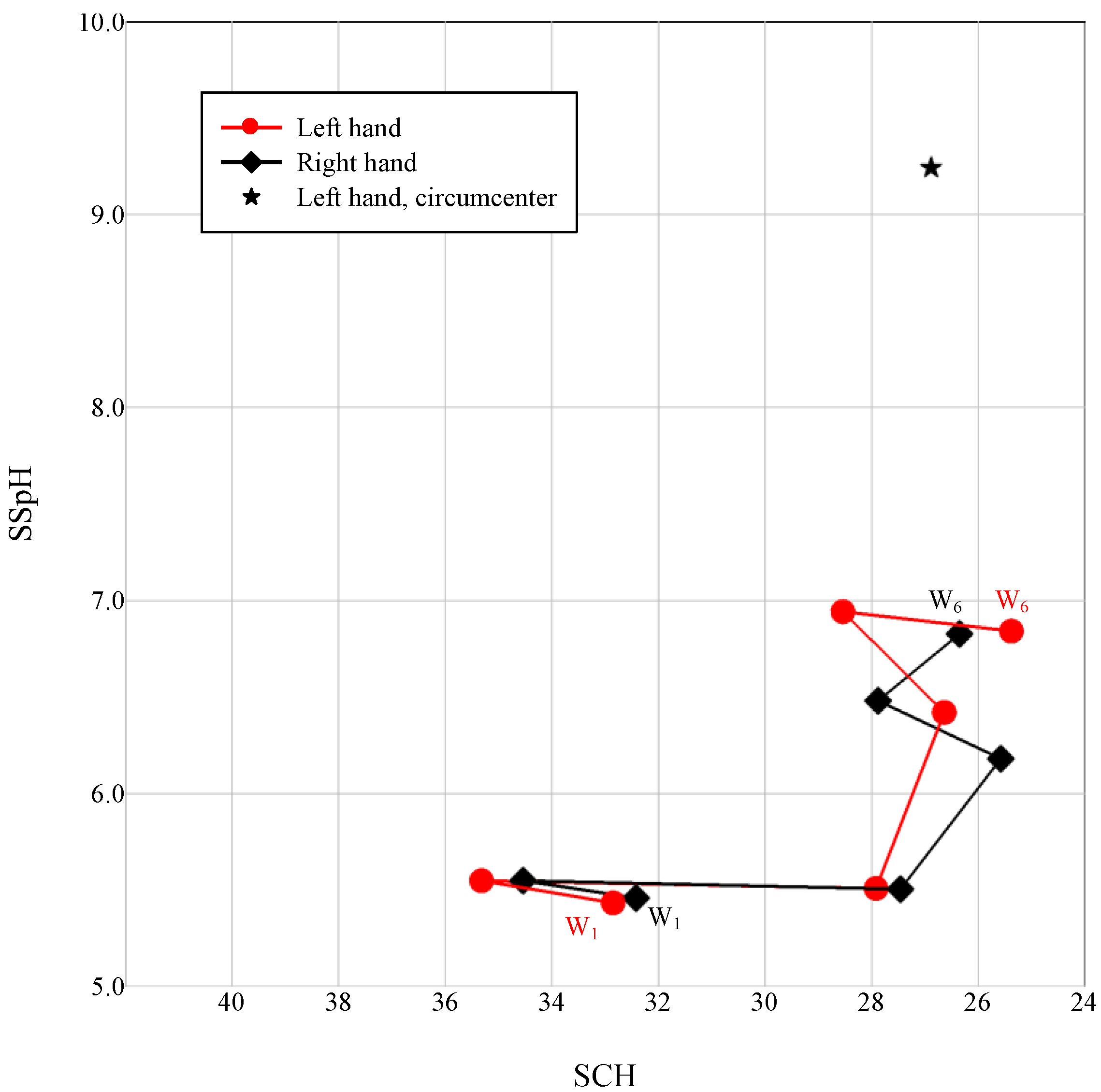
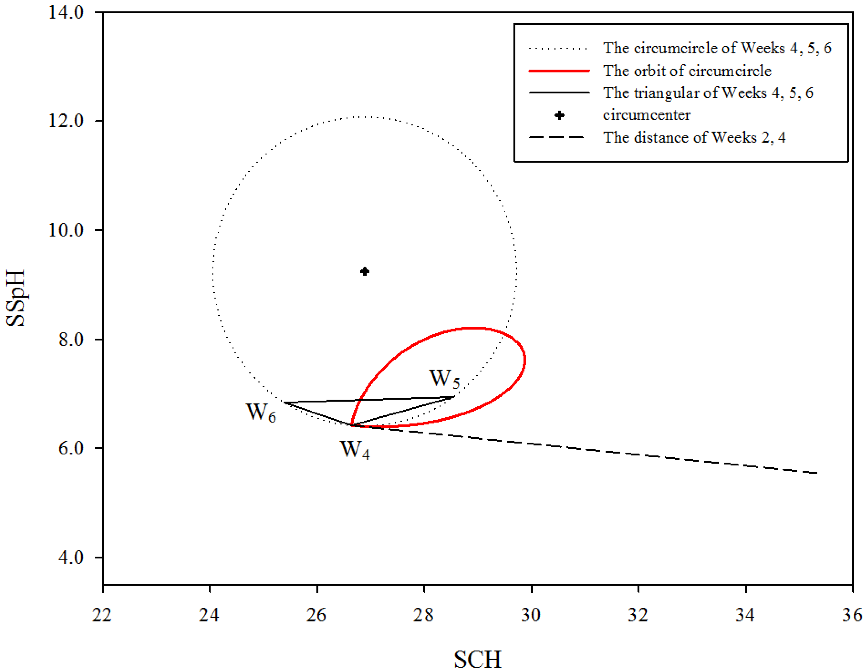
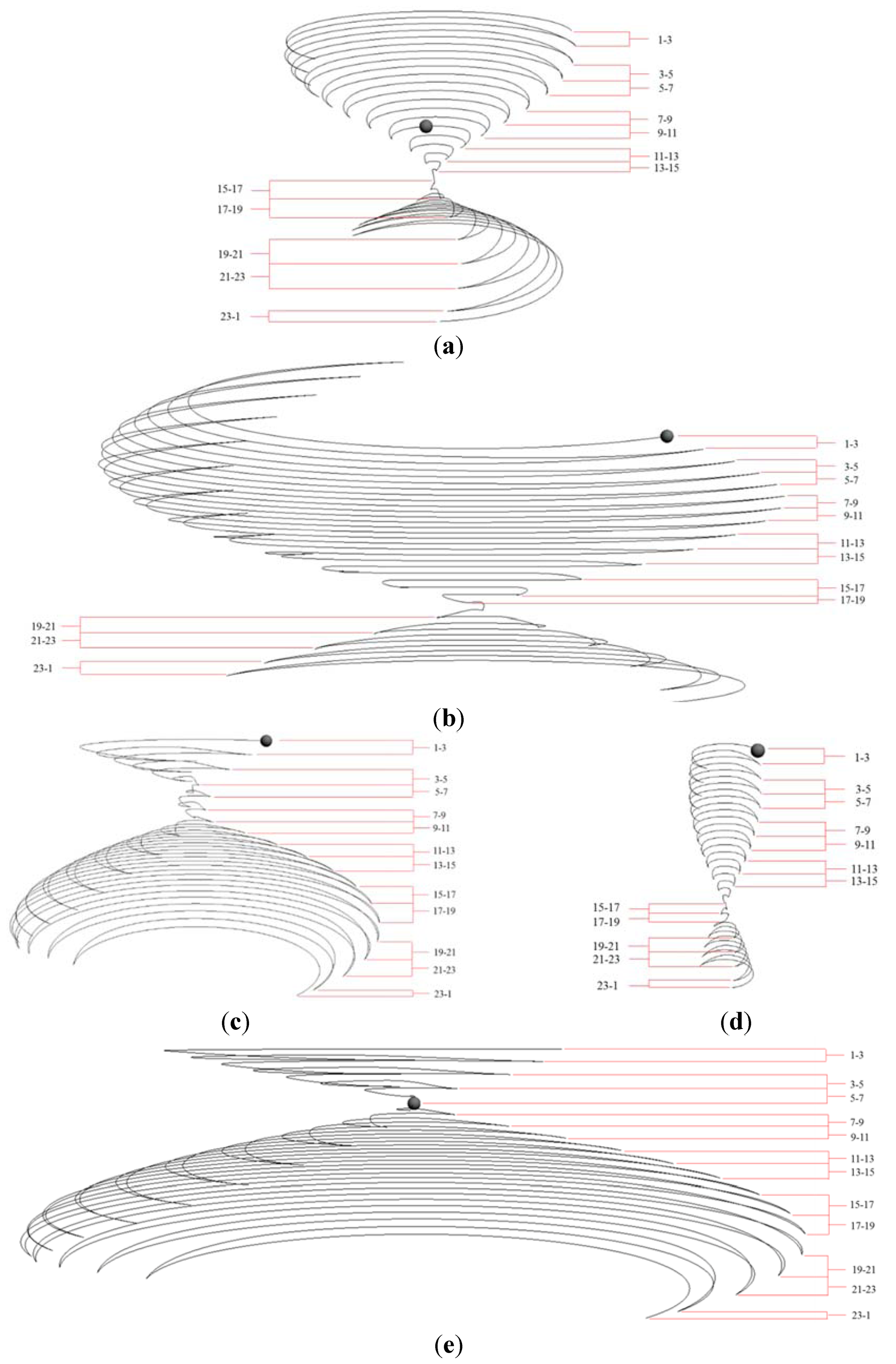
2.3.1. Spindle-Shaped Curve

The average SCH-SSpH values of the left dorsal forearms in Weeks 4 to 6 were used to form the circumcenter of a triangle (Figure 1). The value of the circumcenter was calculated using Excel 2010 (Microsoft Corporation, Redmond, WA, USA). The method of calculating the circumcenter is as follows: (1) Obtained mean values of the SCH and SSpH in 6 weeks of 25 participants per week; (2) The coordination of x and y represented the SCH and SSpH; (3) x4, x5, and x6 represented the SCH results from the left hands of Weeks 4, 5, and 6. y4, y5, and y6 represented the SSpH results from the left hands of Weeks 4, 5 and 6; (4) x = (((x4 × x4 + y4 × y4) × y5 + (x5 × x5 + y5 × y5) × y6 + (x6 × x6 + y6 × y6) × y4) − ((x6 × x6 + y6 × y6) × y5 + (x5 × x5 + y5 × y5) × y4 + (x4 × x4 + y4 × y4) × y6))/((x4 × y5 + x5 × y6 + x6 × y4) − (x6 × y5 + x5 × y4 + x4 × y6))/2; y = (((x5 × x5 + y5 × y5) × x4 + (x6 × x6 + y6 × y6) × x5 + (x4 × x4 + y4 × y4) × x6) − ((x5 × x5 + y5 × y5) × x6 + (x4 × x4 + y4 × y4) × x5 + (x6 × x6 + y6 × y6) × x4))/((x4 × y5 + x5 × y6 + x6 × y4) − (x6 × y5 + x5 × y4 + x4 × y6))/2.
The circumcenter from the SCH and SSpH values of Weeks 4 to 6 had a circumference. The points on the circumference (here represented by xm, yn) and the SCH and SSpH values from Weeks 2 and 4 (Week 2: x2, y2; Week 4: x4, y4) were used to form numerous triangles. The method of calculation for the points on the circumference is as follows: (1) calculated the radius of the circumcircle of Weeks 4, 5 and 6 (here represented by r). (2) xm = x + r* Cos(radians(m)), m is from 1 to 360; and yn = y + r* Sin(radians(n)), n is from 1 to 360. The 360 triangles had 360 incenters. These incenters formed a red spindle-shaped curve (Figure 2). The 360 points on the spindle-shaped curve were used to draw an hourglass trajectory diagram.

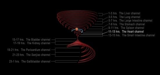
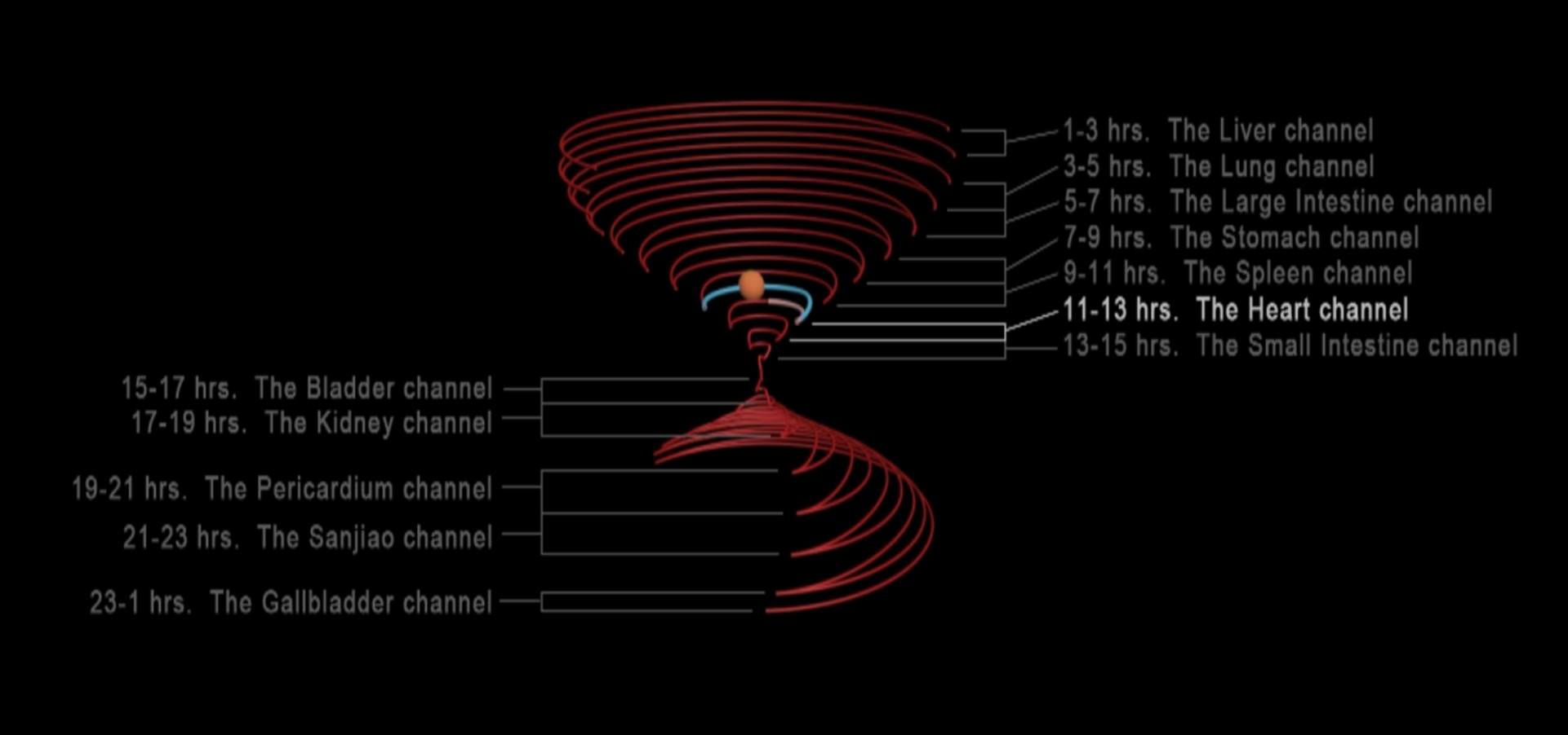
2.3.2. Design Procedure and Concepts of an Hourglass Trajectory Diagram

The 36 incenters on the spindle-shaped curve were selected at an equal distance between one another, and each point presented an inscribed circle. To produce an hourglass trajectory diagram, the circumferences of the 36 inscribed circles were used separately (222.48° was used for each circumference). Subsequently, Weeks 4 to 6 of the SCH-SSpH values were used to form the circumcircle of the triangle (Figure 2), which divided the 36 incenters into inner and outer parts. The 36 circumcircles were also divided into inner and outer parts. The inner and outer circle parts of the circumcircles were connected separately. Two parts of the circular segments were then connected together. Specifically, the lower part of Figure 3a comprises the outer circle parts of the circular segments, and the upper part comprises the inner circle parts. Both the circular segments progressively turned 45° counterclockwise. The intersection of the inner and outer circular segments was the narrowest point, which was a circumference near the SCH and SSpH values of Week 4 (Figure 2 and Figure 3a).
Figure 1. First measurement of the SCH–SSpH relationship in 6 weeks (N = 25). This 2-D layout was obtained by compressing the 3-D figure; W1: Week 1, W6: Week 6.
Medsci 02 00161 g001
A 2-colored ball was placed on the circular segments and its movement showed the changes in blood flow velocity. Figure 3 shows the starting point of the ball. This point was where the lines of Weeks 5 and 6 crossed, and was also derived from the red spindle-shaped curve (Figure 2). The required times for the ball in the lower part and upper part of Figure 3a were identical, thereby indicating that the speeds were different. In addition, participants with abnormal nerve conduction were analyzed. Because of the same animation conditions, the case in abnormal nerve conduction will follow the individual results and have different expression.
The hourglass trajectory concept is as follows (Figure 3a). First, the physiological operations recorded for 24 h were interpreted. Every third line indicated a 2-h change, with the trajectory widths representing changes in blood flow. The velocity of the moving ball signified blood flow velocity. Second, the upper section of the hourglass represented the physiological performance during the daytime and the lower section represented the performance at night. However, the daytime trajectories also included the nighttime trajectories, thereby representing the TCM concept that yin involves yang and yang requires yin [13]. Third, the trajectory line endpoints denoted tissues or organs. Nerve transmission was represented by the “go-back” mode of ball movement. Finally, the 222.48° circumference was applied based on the golden ratio. The spindle was rotated 45°; thus, the connection between each endpoint formed a DNA double helix. Rotating the DNA structure by 360° produced a diameter–height ratio that was also near the golden ratio [19].
Figure 2. SNS performance: a red spindle-shaped closed curve.
Medsci 02 00161 g002

3. Results and Discussion

The innermost layer of the skin epithelium is the proliferating layer. After two weeks, cells were transmitted to the differentiating layer, and then to the stratum corneum after another two weeks, reaching the end of the cell cycle approximately two weeks later [20]. Therefore, the adaptability of the NS to cream can be evaluated by separately observing the reaction of the skin in Weeks one to four, and the performance of the skin in Weeks four to six. Hence, the data of Weeks four to six were used in the analysis.

3.1. Energy Dependency for Organ Survival

The diagram can be explained in terms of the mean value spectrum. The intersection of the red spindle thread origins and the Week four SCH-SSpH value exhibited the highest energy and temperature and the lowest hydration level. Concurrently, the SNS exhibited a highly excited state; however, a decline in transmission speed was also observed, indicating that the excitation responses were concentrated in only a few areas of the body. However, an opposite result can be observed in the region where the origin of the red spindle threads extended from the Week four value to the vertex (Figure 2). The hourglass trajectory diagram extended from the red spindle-shaped curve to signify the energy requirements for physiological basal metabolic activity (Figure 3). Furthermore, the TCM concepts of the 12 official meridians and organs, which are based on an old Chinese timing system in which each “official” signifies 2 h, were used to explain the sequence by which SNS excitation provided survival energy to the organs, as well as the varying performance levels at different points in time [13,21].
Both the ends and the middle of the hourglass trajectories (at approximately 1:00 AM and 3:00 PM) indicated that the SNS was in an excited state, exhibiting strong energy and slight moisture (Figure 3a). However, notable blood flow differences were observed between these two trajectory parts, indicating that when the SNS was in an excited state, blood flow and velocity differed at the various time points. When people sleep at night, their SNS is excited, resulting in vasoconstriction and thus an increase in organ blood content [6,22]. This is the period when the meridian passes through the liver (Figure 3a), inducing high levels of activity in the liver. The consequent increased blood content facilitates metabolic activity. Another period for SNS excitation is when the meridian passes through the small intestine, activating the small intestine. Increased blood flow velocity in the small intestine facilitates the rapid transportation of absorbed nutrients.
The human circadian clock is primarily influenced by suprachiasmatic nuclei, which affect organ performance during the day and night through the ANS [23]. The hourglass trajectory diagram exhibited two regulation levels. In the animation shown in the Appendix, the ball trajectories are highlighted blue and red. The first level denotes the meridian that performs the operation of basic physiological energy; this performance simultaneously affects the operation of other meridians, which are represented by the red trajectories. The second level indicates the individual performance of each meridian over a 24-h period, represented by blue and red lines (see Supplementary Information). In this study, we focused on the first level, which featured a time range from 11:00 AM to 11:00 PM (Figure 3a). The operation followed the sequence from the heart channel to the small intestine, bladder, kidney, pericardium, and sanjiao channels. The SNS excitation cycle was observed during this period. The cycle supported the energy operation between organs through high–low degrees of excitation.
According to TCM, the operation of each meridian in the 12 officials signifies that a particular organ is active at a certain time and that a particular interrelationship exists between pairs of meridians [13]. The first level of regulation was observed in the period from 11:00 AM to 3:00 PM, which was the point at which ball movement began and the time of the heart and small intestine (Figure 3a), denoting NS and small intestine operations [13,24]. In other words, the operation of all living beings begins with NS operation, which regulates the operation of the small intestine. The SNS excitation accelerates the blood flow in the brain and small intestine, thereby reducing the amount of blood in the other organs. During this period, the body’s physiological energy is higher. In other words, the operation of the small intestine initiates the conversion of energy.
Between 3:00 PM and 7:00 PM, the meridian passes through the bladder and kidney channels [13]. The bladder meridian is the longest meridian, and it exerts the strongest and widest influence on the body. This meridian travels through 5 zàng and 6 ; therefore, it transports energy to all of the internal organs. Moreover, after energy metabolism, SNS excitation induces the relaxation of the bladder wall and contraction of the sphincter in preparation for storing water from the kidneys, indicating kidney operation [6]. During this period, because SNS excitation reduces the kidneys’ excretion of water, the degree of SNS excitation is reduced to facilitate water excretion [25]. Overall, the energy distribution exhibited a reducing trend based on the time points and meridian directions, and this distribution revived over the subsequent period through the feedback mechanism.
The first regulation level stopped between 7:00 PM to 11:00 PM when the meridian passed through the pericardium and sanjiao channel [13,24]. The pericardium meridian represents the cardiovascular system. Because of the low heartbeat frequency and mild direct influence of the SNS on the two cardiac muscles, SNS excitation is relatively low during this period, which is when the blood vessels dilate to increase the blood flow, thereby enabling the cardiovascular system to conduct metabolism [4]. The sanjiao channel represents the lymphatic system and its tissue spaces and microcycles. This period is when the interstitial fluid that has been distributed over the entire body flows back to the subclavian veins [4]. Thus, the volume of blood increases and stimulates the arterial baroreceptors, further prompting SNS excitation for the subsequent stage [4]. This entire process shows that the feedback mechanism produces energy, enabling life functions to continue.

3.2. Case Analysis

The trajectory diagram of the average values depicts normal SNS performance. The height–width ratio in the hourglass was a standard ratio. Abnormal SNS excitation can be explained by examining several cases. The following four cases were relative to abnormal nerve conduction. Case 1 was a patient who was blind in one eye because of damage to the optic nerve. The ratio of the widest point in the hourglass to the average trajectory values for Case 1 was 2.3:1.0; thus, the ball had to move at a high speed to complete the trajectory, indicating that over-excitation of the SNS occurred in this case. Moreover, fluctuations in the amount of blood flow between 11:00 AM and 11:00 PM occurred 4 h early in Case 1. In other words, the energy and SNS excitation at the peak heart-operation period was weak and deviated from the anticipated level of strength, indicating that the SNS feedback mechanism was dysfunctional (Figure 3b). Case 2 was a patient diagnosed with amyotrophic lateral sclerosis, and the trajectories of this case were larger than the average width value; the ratio of the widest point on the upper part of the hourglass to that on the lower part was 1.0:1.6. This suggested that the SNS did not undergo excitation, whereas the PNS exhibited relatively high excitation. In addition, the increase in blood flow amount for Case 2 was 8 h later than an average person’s would normally be, signifying that the SNS was weak at a time when it should have been active (Figure 3c). Case 3 was the sister of Case 2. Her trajectory diagram was another example of SNS over-excitation. The ratio of the widest point of her hourglass trajectory to that of the average-value trajectory diagram was 1:4; thus, the ball in the animation moved slowly (Figure 3d). Finally, Case 4 reported frequently experiencing left hand numbness from the cervical vertebra to the palm. The trajectories for this case showed that the ratio of the widest point on the upper section of the hourglass to that of the lower section was 1.0:1.5, providing another example where SNS excitation was low and PNS excitation was overly high. However, in contrast to Case 3, the ball on the trajectory lines moved rapidly for Case 4, indicating that the SNS experienced energy conversion problems, thus accumulating energy (Figure 3e).
Figure 3. Animation diagram of the physiological performance over a 24-h period. The various time points (from top to bottom) refer to the meridian performances in every official (i.e., the liver, lungs, large intestine, stomach, spleen, heart, small intestine, bladder, kidneys, pericardium, sanjiao, and gallbladder): (a) mean value spectrum; (b) Case 1; (c) Case 2; (d) Case 3; (e) Case 4.
Medsci 02 00161 g003

4. Conclusions

This study established an energy operation mode by applying p-334 cream to the skin and then extending the skin status to examine SNS performance in the organs. The meridian operation of the 12 officials was integrated by combining TCM meridian theory and the SNS organ regulation concept in Western physiology. In addition, this study explained the energy required for organ metabolism. Specifically, the performance of the SNS during the day and night coordinates organ function. The balancing system in the human body features a set of feedback mechanisms that can be transmitted to the SNS, enabling adjustment of the degree of excitation, which is a process completed through blood flow. Future studies can further investigate the following topics: (1) the amplification effects exerted by p-334 on NS in the field of molecular biology; (2) the correlation between case-specific SNS trajectories and diseases; (3) the correlations among p-334 energy conversion, the NS, and the cardiovascular system, as well as the possible pathways; and (4) using the correlation between skin and NS as well as the trajectory animation of blood flow to establish an automatic calculation system and database that are connected with disease incidence rates. In this study, we established a theory and contributed to the future development of medical science.

Acknowledgments

This research work was supported by grants from the National Science Council, Taiwan, ROC (NSC 99-2313-B-002-023). We thank the Yung-Ming Branch of Taipei City Hospital for their assistance.

Author Contributions

L.F.C., H.N.C. and F.G.C. designed the experiment, analyzed the data, and wrote the manuscript. L.F.C., C.K.H. and F.G.C. performed most of the experiments. H.S.C and P.J.S. contributed to writing the manuscript.

Conflicts of Interest

The authors declare no conflict of interest.

References

  1. Widmaier, E.P.; Raff, H.; Strang, K.T. Regulation of organic metabolism and energy balance. In Vander’s Human Physiology–The Mechanisms of Body Function, 13th ed.; Widmaier, E.P., Raff, H., Strang, K.T., Eds.; McGraw-Hill: New York, NY, USA, 2014; pp. 572–601. [Google Scholar]
  2. Storch, K.F.; Lipan, O.; Leykin, I.; Viswanathan, N.; Davis, F.C.; Wong, W.H.; Weitz, C.J. Extensive and divergent circadian gene expression in liver and heart. Nature 2002, 417, 78–83. [Google Scholar] [PubMed]
  3. Bray, M.S.; Shaw, C.A.; Moore, M.W.; Garcia, R.A.; Zanquetta, M.M.; Durgan, D.J.; Jeong, W.J.; Tsai, J.Y.; Bugger, H.; Zhang, D.; et al. Disruption of the circadian clock within the cardiomyocyte influences myocardial contractile function, metabolism, and gene expression. Am. J. Physiol. Heart Circ. Physiol. 2008, 294, 1036–1047. [Google Scholar]
  4. Widmaier, E.P.; Raff, H.; Strang, K.T. Cardiovascular and physiology. In Vander’s Human Physiology–The Mechanisms of Body Function, 13th ed.; Widmaier, E.P., Raff, H., Strang, K.T., Eds.; McGraw-Hill: New York, NY, USA, 2014; pp. 362–445. [Google Scholar]
  5. Widmaier, E.P.; Raff, H.; Strang, K.T. The endocrine system. In Vander’s Human Physiology–The Mechanisms of Body Function, 13th ed.; Widmaier, E.P., Raff, H., Strang, K.T., Eds.; McGraw-Hill: New York, NY, USA, 2014; pp. 319–361. [Google Scholar]
  6. Widmaier, E.P.; Raff, H.; Strang, K.T. Neuronal signaling and the structure of the nervous system. In Vander’s Human Physiology–The Mechanisms of Body Function, 13th ed.; Widmaier, E.P., Raff, H., Strang, K.T., Eds.; McGraw-Hill: New York, USA, 2014; pp. 138–190. [Google Scholar]
  7. Widmaier, E.P.; Raff, H.; Strang, K.T. Consciousness, the Brain, and Behavior. In Vander’s Human Physiology–The Mechanisms of Body Function, 13th ed.; Widmaier, E.P., Raff, H., Strang, K.T., Eds.; McGraw-Hill: New York, NY, USA, 2014; pp. 234–256. [Google Scholar]
  8. Cicek, D.; Kandi, B.; Berilgen, M.S.; Bulut, S.; Tekatas, A.; Dertlioglu, S.B.; Ozel, S.; Saral, Y. Does autonomic dysfunction play a role in atopic dermatitis? Br. J. Dermatol. 2008, 159, 834–838. [Google Scholar] [CrossRef] [PubMed]
  9. Charkoudian, N. Skin blood flow in adult human thermoregulation: How it works, when it does not, and why. Mayo Clin. Proc. 2003, 78, 603–612. [Google Scholar] [CrossRef] [PubMed]
  10. Pérgola, P.E.; Kellogg, D.L.; Johnson, J.M.; Kosiba, W.A.; Solomon, D.E. Role of sympathetic nerves in the vascular effects of local temperature in human forearm skin. Am. J. Physiol. 1993, 265, 785–792. [Google Scholar]
  11. Dung, H.C. Acupuncture points of the brachial plexus. Am. J. Chin. Med. 1985, 13, 49–64. [Google Scholar] [CrossRef] [PubMed]
  12. Stücker, M.; Struk, A.; Altmeyer, P.; Herde, M.; Baumgärtl, H.; Lübbers, D.W. The cutaneous uptake of atmospheric oxygen contributes significantly to the oxygen supply of human dermis and epidermis. J. Physiol. 2002, 538, 985–994. [Google Scholar] [CrossRef] [PubMed]
  13. Deadman, P.; Al-Khafaji, M.; Kevin, B. A Manual of Acupuncture. Available online: https://amanualofacupuncture.com/about-manual-of-acupuncture (accessed on 26 November 2014).
  14. Whitehead, K.; Hedges, J. Photodegradation and photosensitization of mycosporine-like amino acids. J. Photochem. Photobiol. B 2005, 80, 115–121. [Google Scholar] [CrossRef] [PubMed]
  15. Conde, F.R.; Churio, M.S.; Previtali, C.M. The deactivation pathways of the excited-states of the mycosporine-like amino acids shinorine and porphyra-334 in aqueous solution. Photochem. Photobiol. Sci. 2004, 3, 960–967. [Google Scholar] [CrossRef] [PubMed]
  16. Chuang, L.F.; Chou, H.N.; Sung, P.J. Porphyra-334 isolated from the marine algae Bangia atropurpurea: Conformational performance for energy conversion. Mar. Drugs 2014, 12, 4732–4740. [Google Scholar] [CrossRef] [PubMed]
  17. Volkmann, M.; Gorbushina, A.A. A broadly applicable method for extraction and characterization of mycosporines and mycosporine-like amino acids of terrestrial, marine and freshwater origin. FEMS Microbiol. Lett. 2006, 255, 286–295. [Google Scholar] [CrossRef] [PubMed]
  18. Schmid, D.; Schürch, C.; Zülli, F. UVA-screening compounds from red algae protect against photoageing. Personal Care 2004, 1, 29–31. [Google Scholar]
  19. Perez, J.C. Codon populations in single-stranded whole human genome DNA are fractal and fine-tuned by the Golden Ratio 1.618. Interdiscip. Sci. 2010, 2, 228–240. [Google Scholar] [CrossRef] [PubMed]
  20. Iizuka, H. Epidermal turnover time. J. Dermatol. Sci. 1994, 8, 215–217. [Google Scholar] [CrossRef] [PubMed]
  21. Zhou, B.; Zeng, Q.Q.; Peng, Z.Y. Explore the relationship between record of ZangFu and Jingmai in Huangdi Neijing and systematic anatomy entity. J. Liaoning Univ. TCM 2010, 12, 85–88. (In Chinese) [Google Scholar]
  22. Somers, V.K.; Dyken, M.E.; Mark, A.L.; Abboud, F.M. Sympathetic-nerve activity during sleep in normal subjects. N. Engl. J. Med. 1993, 328, 303–307. [Google Scholar] [CrossRef] [PubMed]
  23. Nader, N.; Chrousos, G.P.; Kino, T. Interactions of the circadian CLOCK system and the HPA axis. Trends Endocrin. Met. 2010, 21, 277–286. [Google Scholar] [CrossRef]
  24. Zhou, B.; Chen, Y.; Huang, W.Z.; Chen, S.X.; Zeng, Q.Q.; Chen, Q.S.; Li, W.; Sun, Z.F. Cardiovascular system and the nervous system has the identity in Huangdi Neijing–Comparing the difference between traditional Chinese medicine and modern medicine in the organizational structure of organs and explore the Luo Entity. J. Liaoning Univ. TCM 2011, 13, 115–118. (In Chinese) [Google Scholar]
  25. Widmaier, E.P.; Raff, H.; Strang, K.T. The kidneys and regulation of water and inorganic ions. In Vander’s Human Physiology–The Mechanisms of Body Function, 13th ed.; Widmaier, E.P., Raff, H., Strang, K.T., Eds.; McGraw-Hill: New York, NY, USA, 2014; pp. 490–532. [Google Scholar]

