Review

Evasion of Neutrophil Killing by Staphylococcus aureus

Laboratory of Bacteriology, Rocky Mountain Laboratories, National Institute of Allergy and Infectious Diseases, National Institutes of Health, 903 South 4th Street, Hamilton, MT 59840, USA
*
Author to whom correspondence should be addressed.
Pathogens 2016, 5(1), 32; https://doi.org/10.3390/pathogens5010032
Submission received: 23 February 2016 / Revised: 11 March 2016 / Accepted: 14 March 2016 / Published: 17 March 2016
(This article belongs to the Special Issue Staphylococcus Aureus Infection)

Abstract

:
Staphylococcus aureus causes many types of infections, ranging from self-resolving skin infections to severe or fatal pneumonia. Human innate immune cells, called polymorphonuclear leukocytes (PMNs or neutrophils), are essential for defense against S. aureus infections. Neutrophils are the most prominent cell type of the innate immune system and are capable of producing non-specific antimicrobial molecules that are effective at eliminating bacteria. Although significant progress has been made over the past few decades, our knowledge of S. aureus-host innate immune system interactions is incomplete. Most notably, S. aureus has the capacity to produce numerous molecules that are directed to protect the bacterium from neutrophils. Here we review in brief the role played by neutrophils in defense against S. aureus infection, and correspondingly, highlight selected S. aureus molecules that target key neutrophil functions.

1. Introduction

Staphylococcus aureus is adept at circumventing destruction by the human innate immune system, and the bacterium imposes a significant disease burden on at-risk and otherwise healthy populations worldwide [1]. S. aureus commonly resides as a quiescent commensal within the human nares, and, as such, is a frequent cause of opportunistic infections [2]. Indeed, S. aureus has remained a leading cause of healthcare and community associated infections in the United States over the past 60 years. The organism causes a broad array of diseases and syndromes, such as endocarditis, toxic-shock syndrome, bacteremia, soft-tissue infections, surgical-site infections, and pneumonia [3,4,5,6]. In addition, the pathogen is notorious for its ability to acquire antibiotic resistance, and S. aureus increasingly undermines treatment regimens already hampered by a substantial lack of novel therapeutics [7,8]. Taken together, the propensity for S. aureus to develop antibiotic resistance and cause significant morbidity, underscores the importance of developing new therapeutic approaches for treatment of infections.
Polymorphonuclear leukocytes (PMNs or neutrophils) are the most prominent cellular component of the host innate immune response to bacterial and fungal infections. The vast majority of healthy individuals are seemingly protected against severe S. aureus infections, and this characteristic is attributed largely to the role played by neutrophils in host defense. Indeed, individuals with neutrophil defects are highly susceptible to severe and life-threatening S. aureus infections [9]. This review highlights the role of neutrophils in innate immunity, and specific mechanisms used by S. aureus to circumvent elimination by PMNs.

2. Neutrophils in Host Defense

Neutrophils are the primary cellular constituent of the human innate immune system and are essential for host defense against invading microbial pathogens. PMNs originate and mature in the bone marrow, and are subsequently released into the peripheral vasculature. High numbers of neutrophils are maintained in circulation to facilitate rapid recruitment to infected tissue. Intravascular circulating neutrophils comprise approximately half of the entire granulocyte population, whereas marginated neutrophils maturing in the bone marrow or resident in organ systems make up the remaining half [10,11]. Mature neutrophils enter the vasculature from the bone marrow at a rate of approximately 109 cells/kg body weight every day in a healthy human adult [10,11]. Circulating neutrophils, by sheer force of presence, are poised for recruitment by chemotactic signals to areas of infection. The large pool of circulating PMNs is available to support the inflammatory response to infection.
Circulating neutrophils are recruited to infection sites through chemotactic signals produced by host cells and shed and/or secreted bacterial molecules. The ability of neutrophils to rapidly respond to invading microorganisms is a key facet of innate immunity. Neutrophils are highly sensitive to chemotactic signals that signify infection and promote migration. Host-derived chemotactic factors, such as interleukin 8, GRO-alpha, granulocyte chemotactic protein 2, and complement component C5a are potent proinflammatory mediators that are used to recruit additional PMNs to areas of infection [12,13,14]. Further, PMN migration can be elicited directly or indirectly by bacteria-derived stimuli, such as lipoteichoic acid or n-formyl peptides [15,16,17,18,19,20]. At sites of infection, a diversity of resident cells contribute to production of chemotactic factors including macrophages, endothelial, and epithelial cells [21,22,23].
Neutrophil recruitment from the bloodstream to infected tissue is governed by a series of complex signal transduction cascades that ultimately control the dynamics of PMN interaction with vascular cells and regulate chemotaxis [12,13]. The neutrophil recruitment process can be broken into four steps: Rolling adhesion, integrin activation, firm adhesion, and transmigration. PMN recruitment is initiated by inflammatory mediators, and is followed by the expression of surface ligands on the neutrophil plasma membrane [12,13,24]. During rolling adhesion, PMNs travel through the bloodstream by shear force within blood vessels, adherence to the blood vessel walls is maintained by repetitive ligand-receptor binding mediated by selectins allowing for reversible and bidirectional modulation of the binding process of neutrophils to endothelial cells [12,13,24]. Neutrophil-endothelial cell (blood vessel) adhesion interactions–mediated by expression of lymphocyte function-associated antigen-1 (LFA-1)–shift to a higher affinity state following stimulation by proinflammatory mediators. LFA-1 binds to intracellular adhesion molecule (ICAM)-1 and ICAM-2 present on endothelial cells, resulting in neutrophil arrest [13,24]. LFA-1 integrin clustering on the neutrophil plasma membrane increase the avidity of the binding process and stimulates cytoskeletal/receptor rearrangements that induce PMN morphological changes [24]. As the PMN morphology shifts from spherical to flat, shear force is no longer capable of initiating neutrophil movement through the blood vessel [12,13]. Following firm attachment to the endothelium, neutrophil transmigration from the endothelium into infected tissue is facilitated by several leukocyte and endothelial cell molecules including CD31, CD11b/CD18, CD47, and CD44 [12,13].
Neutrophils play a primary role in host defense and remove pathogens through a process known as phagocytosis. Phagocytosis is initiated by recognition and binding of bacteria, and is followed by internalization. Recognition of microbial pathogens is determined by pattern recognition receptors (PRRs) and opsonin receptors. PRRs bind microbe-associated molecules known as pathogen-associated molecular patterns (PAMPs), and PRRs are an essential component of the host-immune surveillance. The composition of PRRs includes a broad array of receptors, such as membrane-bound toll-like receptors (TLRs), that identify highly conserved bacterial surface and secreted molecules. PRR ligation induces intracellular signal transduction cascades that trigger enhanced phagocytosis and cytokine production, albeit pathogen recognition by PRRs alone is generally insufficient to stimulate phagocytosis [25,26,27]. The efficiency of phagocytosis is enhanced when invading microorganisms are opsonized with host serum molecules such as complement and antibody. Specific IgG, when bound to microbial surfaces, directs the deposition of serum complement. Distinct IgG and complement receptors on the neutrophil directly mediate uptake of opsonized microbes [28,29] (Figure 1). Fc receptor binding leads to the induction of highly complex signal transduction cascades that direct membrane reorganization, actin polymerization, and cytoskeletal rearrangements that are essential for phagocytosis. Electrophysiological alterations in transmembrane potential elicit lipid signaling cascades and actin polymerization that contribute to the advancing membrane cup. Complete microbial engulfment is accomplished when the membrane cup entirely surrounds the pathogen and closes to form a nascent phagosome.
Neutrophils use both oxygen-dependent and oxygen-independent processes to kill invading microbial pathogens (Figure 2). Phagocytosis of bacteria triggers assembly of a membrane-bound NADPH-dependent oxidase on PMN phagosomes [30,31,32]. The NADPH oxidase complex generates high levels of reactive oxygen species (ROS) in activated neutrophils [32,33]. The NADPH oxidase functions by shuttling electrons across the phagosomal membrane from cytosolic NADPH to intraphagosomal molecular oxygen to produce O2−. The superoxide anion is readily converted to hydrogen peroxide by superoxide dismutase. Notably, hydrogen peroxide and other secondary oxygen derivatives such hydroxyl radical, chloramines and hypochlorous acid (HOCl), which is generated by the myeloperoxidase-halide system, contribute to PMN microbicidal activity [33,34]. Neutrophil oxygen-independent microbicidal activity is effected by numerous degradative enzymes and cationic peptides contained in granules (primary, secondary, and tertiary) and secretory vesicles. Following phagocytosis, granules are trafficked to and fuse with PMN phagosomes in a process called degranulation [35,36,37]. Although the composition of granules varies by type, collectively they enrich the vacuole lumen with microbicidal agents such as cathepsins, elastase, proteinase-3, defensins, lysozyme, azurocidin and bacterial permeability increasing protein [35,36,38]. Collectively, PMN oxygen-dependent and oxygen-independent microbicidal systems are highly efficient for elimination of invading bacterial pathogens.
The rapid influx of large numbers of neutrophils to sites of infection is a hallmark of the acute inflammatory response. The combination of ROS, degradative enzymes, and cationic peptides create a potent microbicidal environment confined within neutrophil phagosomal membranes. Inasmuch as these molecules do not discriminate between host and microbe targets, the potential of host-tissue damage is high should the neutrophil undergo inappropriate cell lysis [39]. To mitigate tissue damage and control inflammation, activated neutrophils rapidly undergo apoptosis and effete cells are recognized and safely removed by macrophages through a process known as efferocytosis. Resolution of acute inflammation is coordinated through the activities of proresolving lipid mediators, such as resolvins, protectins, and maresins [40].

3. Evasion of S. aureus Killing by Neutrophils

S. aureus is capable of producing numerous freely secreted and surface-bound molecules that have potential to alter and/or limit the functional capacity of neutrophils. In vitro, these molecules have been shown to inhibit neutrophil recruitment, bacterial binding and phagocytosis, and killing by ROS and antimicrobial peptides (AMPs) (reviewed in [41,42]) (Figure 3). Here, we review some of the S. aureus molecules and processes that contribute to evasion of killing by neutrophils.

3.1. Inhibition of Neutrophil Recruitment

Chemotaxis inhibiting protein of S. aureus (CHIPS) is a freely secreted protein that inhibits neutrophil and monocyte chemotaxis [43,44]. CHIPS binds directly to the C5a and formyl peptide receptors (FPRs), and thereby inhibits phagocyte recruitment [44]. The gene encoding CHIPS, along with those for 3 other immune evasion molecules—staphylococcal complement inhibitor (SCIN), staphylokinase (SAK), and S. aureus enterotoxin A or P (SEA or SEP)—is located on a mobile genetic element known as a β-hemolysin-converting bacteriophage [45]. SAK binds and inhibits the ability of neutrophil defensins to kill bacteria [46], and SEA or SEP are superantigens with known capacity to modulate phagocyte function [47,48,49]. Collectively, the genes encoding these molecules comprise an immune evasion cluster (IEC) that is present in many S. aureus strains tested to date [45]. More recently, Stapels et al. reported that S. aureus extracellular adherence protein (Eap) and two homologs, EapH1 and EapH2, inhibit neutrophil elastase, proteinase-3, and cathepsin G [50]. These neutrophil serine proteases can cleave S. aureus immune evasion molecules, such as SCIN and CHIPS, and this process is blocked by the Eap molecules [51]. The extent to which these molecules contribute to pathogenesis in humans remains to be determined.
There is also a family of staphylococcal-like proteins (Ssls) that has been reported to function as immune evasion molecules. For example, Ssl7 binds host IgA and complement component C5, inhibiting generation of C5a, phagocytosis, and production of phagocyte reactive oxygen species [52,53]. In addition, Bestebroer et al. found that Ssl7 blocked neutrophil recruitment in a mouse model of inflammation [52]. This same research group reported Ssl5 associates with P-selectin glycoprotein ligand-1 (PSGL-1, also known as CD162) and inhibits neutrophil rolling in vitro [54]. In subsequent studies, they demonstrated Ssl5 bound the amino terminus G protein-coupled receptors, thereby inhibiting leukocyte activation by host inflammatory molecules such as cytokines [55].
S. aureus produces at least five molecules that inhibit serum complement convertases—host enzyme complexes important for a fully functional complement system. The activity of host C3 convertase is essential for the opsonization of microbes with complement components such as C3b and iC3b, which promote phagocytosis by complement receptors. Moreover, activation of the complement system ultimately generates C5a, a neutrophil chemoattractant. Staphylococcal complement inhibitor (SCIN) and homologues (SCIN–B and SCIN–C), extracellular fibrinogen-binding protein (Efb), and extracellular complement-binding protein (Ecb), bind and inhibit the C3 convertase (SCIN proteins), or alternatively, C3b–containing convertases and C5 convertases (Efb and Ecb) [56,57]. The interaction of these S. aureus molecules with the host complement system inhibits leukocyte recruitment and neutrophil phagocytosis of S. aureus in vitro and ex vivo in human blood [58,59]. Although SCIN and homologues are human-specific, and thus cannot be tested effectively in animal infection models, Efb has been shown to inhibit phagocytosis of S. aureus in a mouse infection model [59]. The ability of Efb to block phagocytosis is independent of its ability to inhibit complement. Rather, Efb forms a complex with fibrinogen and C3b on the microbe surface, and in doing so blocks phagocytosis [59]. Jongerius et al. found that SCIN-B, SCIN-C, Efb and Ecb are located within a segment of the S. aureus genome that also encodes alpha-hemolysin (Hla), Ssl12, Ssl13, Ssl14, FPR-like 1 inhibitory protein (FLIPr), and a FLIPr homologue (FLIPr-like) [57]. Inasmuch as these genes contribute to S. aureus evasion of host defense, this segment of the genome was named immune evasion cluster 2 (IEC-2) [57].
Laarman et al. recently demonstrated that staphopain A (ScpA), a secreted S. aureus cysteine protease, inhibits in vitro neutrophil chemotaxis mediated by CXCR2 [60]. ScpA cleaves an N-terminal region of CXCR2, and this phenomenon blocks signal transduction triggered by key proinflammatory chemokines such as IL-8 and GROα [60]. The redundancy of molecules directed to inhibit neutrophil chemotaxis and complement activation is curious, especially for a commensal microbe that infrequently causes severe infections in otherwise healthy individuals.

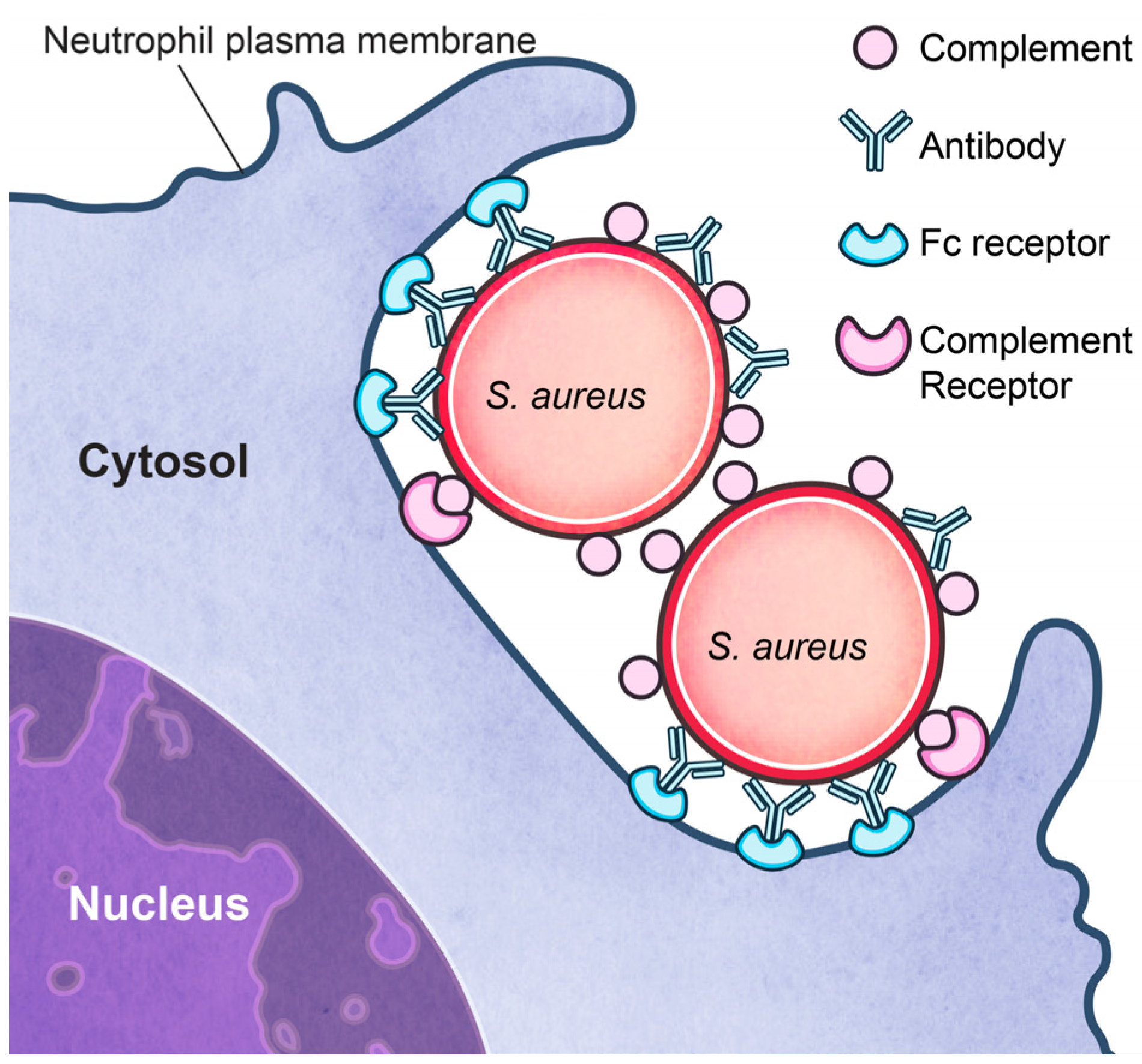
3.2. Inhibition of Phagocytosis?

In addition to the complement inhibitors discussed above, several other S. aureus molecules have been shown to inhibit neutrophil phagocytosis in vitro or in whole blood. For instance, S. aureus protein A (SpA) and the second binding protein of immunoglobulin (Sbi) associate with the Fc-region of immunoglobulin and can inhibit phagocytosis in vitro [61,62,63,64]. S. aureus capsule polysaccharide [65,66], clumping factor A [67], and iron-regulated surface determinant protein H (IsdH) [68], have also been shown to inhibit S. aureus phagocytosis in vitro. S. aureus strains lacking at least two of these molecules—e.g., those lacking Spa and ClfA—are ingested to a greater extent by neutrophils than those lacking only one of these molecules [67]. It is noteworthy that none of these molecules, either singly or collectively, inhibit in vitro uptake by neutrophils completely. The ability of these molecules to block uptake is also probably dependent on assay conditions. The vast majority of the studies of S. aureus phagocytosis by neutrophils have been performed with cells in suspension. The extent to which phagocytosis assays with neutrophils in suspension approximate the interaction of bacteria with neutrophils in the bloodstream is unclear. It has long been known that neutrophils are largely responsible for clearance of S. aureus from the bloodstream in animal infection models [69]. However, neutrophils in suspension are relatively inefficient at phagocytosis, and published studies have shown that blood-borne bacteria are largely cleared by phagocytes (neutrophils) in tissues such as the liver [70]. Importantly, phagocytosis of S. aureus by adherent human neutrophils—which are representative of phagocytes at sites of infection—is highly efficient and seemingly unimpeded by anything produced by the microbe [71,72]. The rapid ingestion of S. aureus by neutrophils was noted by Rogers and Tompsett many years ago, and they also found that phagocytosis of S. aureus was significantly greater than that of Klebsiella pneumoniae, group A streptococcus, or Streptococcus pneumoniae [69,73]. Therefore, the role of S. aureus “antiphagocytic” molecules in the pathogenesis of human infections remains unclear. The problem with S. aureus and human infections is not inefficient phagocytosis (David Rogers had an interesting view on the topic [74]). Rather, a key issue is that some of the ingested bacteria are not killed by the microbicidal systems that operate within the neutrophil phagosome [71,73,75,76]. These ingested microbes ultimately cause lysis of neutrophils and, in turn, escape and disseminate [71,75,77].

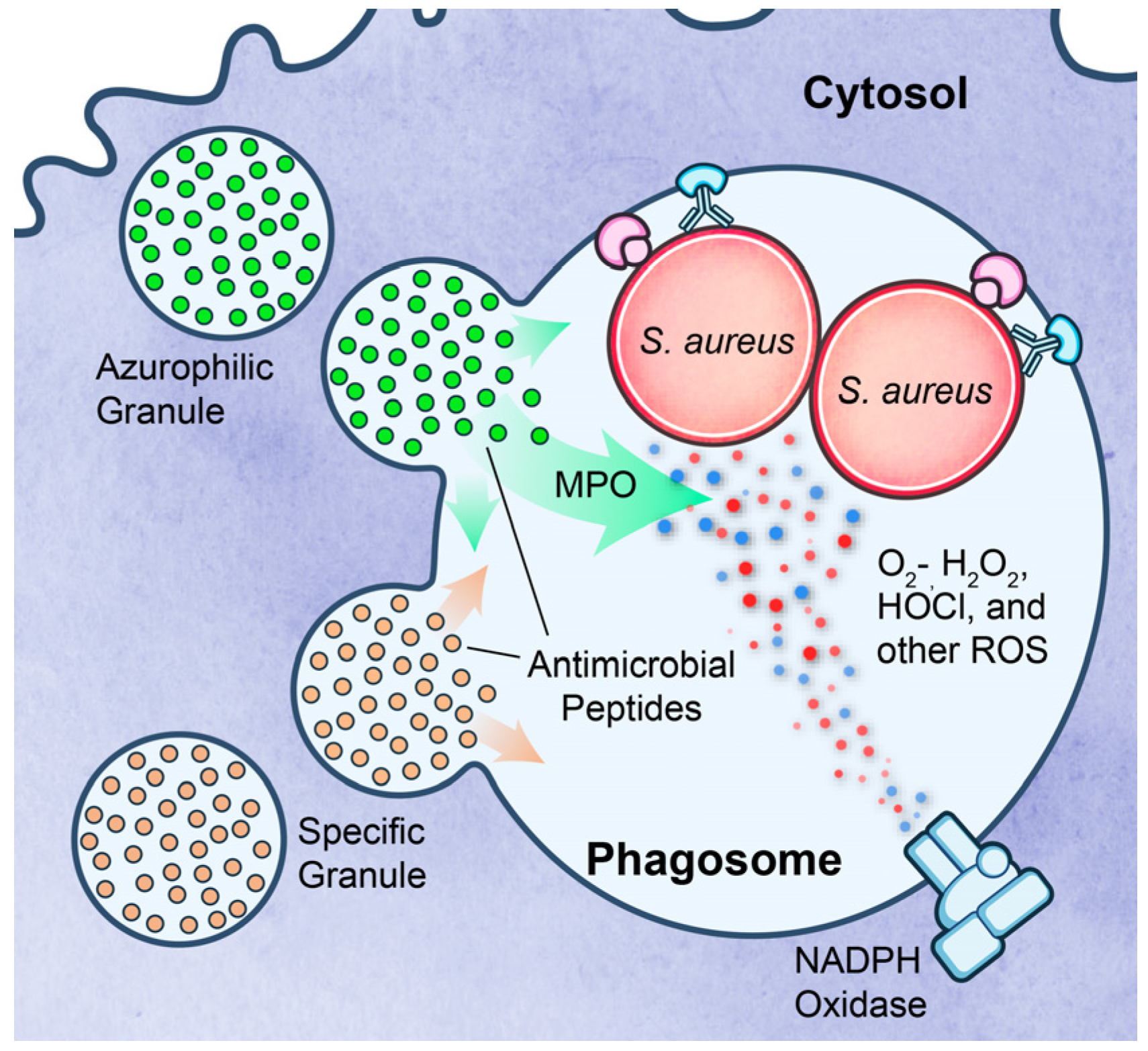
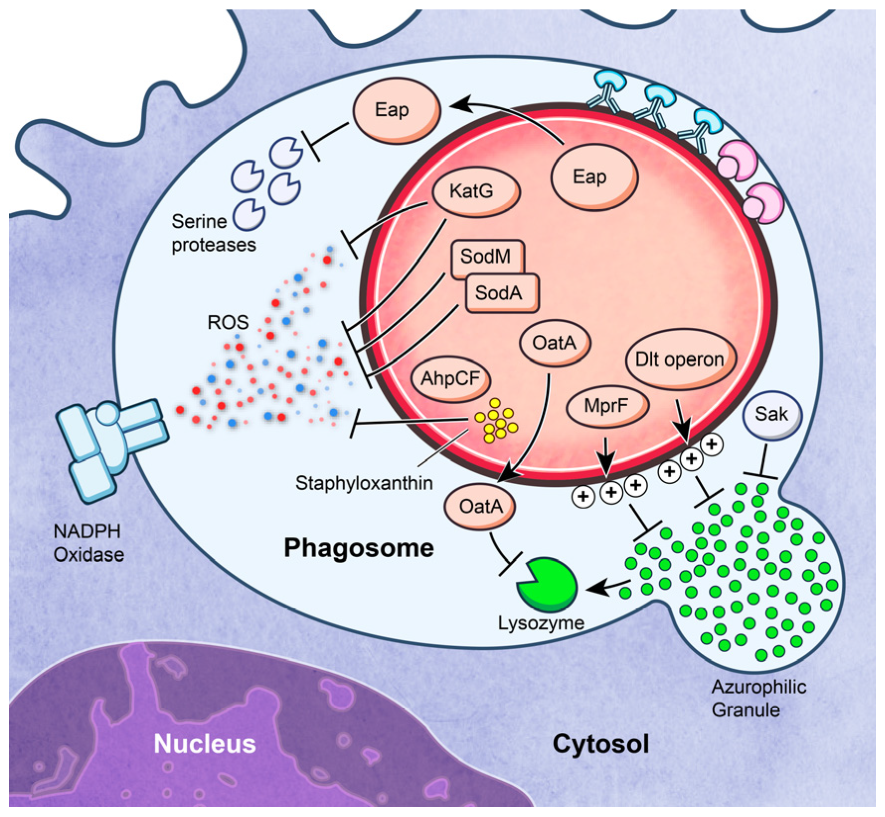
3.3. Survival after Phagocytosis

S. aureus has the capacity to produce molecules that can moderate the cytotoxic effects of ROS and AMPs. Survival of S. aureus inside the phagosome is directly linked to the ability to mitigate the effects of host–derived ROS and AMPs. Superoxide dismutases (SodA and SodM), catalase (KatA), alkyl hydroperoxide reductase (AhpCF), and staphyloxanthin (encoded by crtOPQMN), have been shown to protect S. aureus from ROS [78,79,80]. S. aureus also mitigates the effects of ROS by means of proteins and enzymes involved in metal ion homeostasis (reviewed by Cassat and Skaar [81]). Although these systems moderate the bactericidal effects of ROS to some extent, it is well known that the NADPH oxidase of phagocytes is essential for defense against S. aureus infections. Individuals with chronic granulomatous disease, a disorder caused by inherited deficiency of one or more protein components of NADPH oxidase, are highly susceptible to severe S. aureus infections in the absence of antibiotic prophylaxis [82]. Most evidence to date indicates secondarily-derived ROS, such as HOCl produced by the MPO-halide system (rather than superoxide or H2O2 directly), contribute to killing of ingested S. aureus [83,84,85].
The dlt operon of S. aureus (dltABCD) encodes a cell envelope modification system that attaches d-alanine to cell wall teichoic acids [86]. Incorporation of d-alanine into teichoic acids decreases the negative charge on the bacterial surface and limits susceptibility of the microbe to cationic AMPs, including human neutrophil alpha-defensins [86,87]. Collins et al. used a mouse infection model to demonstrate that the dlt operon is important for S. aureus virulence [87]. Multiple peptide resistance factor (MprF) of S. aureus transfers l–lysine to phosphatidylglycerol to produce lysylphosphatidylglycerol, which contributes to neutralization of the negative charge of the bacterial cell surface [88]. S. aureus strains with mprF genetically inactivated are significantly more susceptible to killing by human neutrophils, a phenomenon linked to increased binding to host AMPs [88]. S. aureus mprF mutants also have a decreased virulence phenotype in mouse infection models, which further underscores the importance of the resistance to AMPs in S. aureus pathogenesis [88]. In general, DltABCD and MprF–mediated resistance to cationic AMPs works by charge neutralization—i.e., the AMPs no longer interact with the charge-neutral membrane of the microbe. There are several other notable molecules and immune evasion systems utilized by S. aureus to promote survival after phagocytosis by neutrophils. For example, lysozyme is a prominent antimicrobial agent present in neutrophil granules. It functions as an antibacterial agent by hydrolyzing beta-glyosidic linkages in cell wall peptidoglycan. Notably, S. aureus is completely resistant to lysozyme. This a characteristic conferred by O-acetylation of peptidoglycan, a process catalyzed by a protein known as OatA [89].

3.4. Lysis after Phagocytosis

S. aureus is capable of producing an extensive arsenal of cytolytic toxins. Several of these molecules, including alpha-type phenol soluble modulins (PSMα) [90], Panton-Valentine leukocidin (PVL) [91,92,93], leukocidin GH (LukGH; also named leukocidin AB (LukAB)) [94,95], and gamma-hemolysin (Hlg) [96], cause osmotic lysis of neutrophils. The role played by these molecules as secreted cytolysins in the pathogenesis of S. aureus infections is an interesting topic, but outside of the scope of this review. Here we focus our brief discussion on the contribution of molecules that contribute to lysis of neutrophils after phagocytosis.
Rogers and colleagues discovered that S. aureus within granulocytes can be trafficked to tissues and promote low-grade persistent infection [69,74]. This same phenomenon was reported many years later by Gresham and colleagues using a mouse infection model [75]. Rogers and Tompsett were among the first to show that S. aureus causes lysis of neutrophils after phagocytosis [73]. Voyich et al. reported that prominent community-associated MRSA strains such as USA300 and USA400 have high capacity to cause rapid lysis of human neutrophils after phagocytosis [71]. Recent work has shown that LukGH/AB and PSMα to contribute to lysis of human neutrophils after phagocytosis [94,97]. However, the contribution of these molecules is strain-dependent or partial, and therefore cannot explain the phenomenon in full. Studies by Kobayashi et al. indicated that S. aureus—in this case USA300—is contained within the phagosome to the point of neutrophil lysis [77]. The observed neutrophil lysis was caspase and ROS-independent, findings incompatible with pyroptosis or NETosis [77]. Rather, the authors suggested that lysis of neutrophils after phagocytosis of S. aureus is consistent with programmed necrosis. Indeed, Greenlee-Wacker et al. then demonstrated that neutrophils undergo programmed necrosis following phagocytosis of S. aureus [98]. Such cytolysis presumably contributes to the pathogenesis of S. aureus infections, although direct evidence is still lacking and more work is needed.

4. Conclusions

To summarize, S. aureus has capacity to produce many molecules directed to inhibit neutrophil functions such as recruitment and phagocytosis. Despite this repertoire of immune evasion molecules, severe invasive S. aureus disease is infrequent in otherwise healthy individuals who lack risk factors for infection. In addition, the localized presence of neutrophils is a hallmark of S. aureus infections and neutrophils readily phagocytose S. aureus. These observations bring into question the role of the S. aureus immune evasion molecules in human disease (outside of the at-risk individual). Finally, the possible utility of an opsonophagocytic vaccine for S. aureus is confounded by two key features of S. aureus-neutrophil interactions: (1) phagocytosis is already efficient in the absence of a vaccine; and (2) neutrophils can undergo rapid lysis after phagocytosis of S. aureus. Despite the noted caveats, research in these areas must move forward. Advances in basic research that continue to elucidate the complex interactions between S. aureus and the human host will likely offer new inroads into developing more efficacious therapeutic treatments and perhaps, a preventative vaccine.

Acknowledgments

The authors are supported by the Intramural Program of the National Institute of Allergy and Infectious Diseases, National Institutes of Health. We thank Ryan Kissinger (NIAID) for assistance with graphic illustration.

Author Contributions

Will A. McGuinness, Scott D. Kobayashi, and Frank R. DeLeo wrote the manuscript.

Conflicts of Interest

The authors declare no conflict of interest.

Abbreviations

The following abbreviations are used in this manuscript:
PMN
Polymorphonuclear leukocyte
LFA-1
Lymphocyte function-associated antigen-1
PRR
Pattern recognition receptor
PAMP
Pathogen-associated molecular pattern
TLR
Toll-like receptor
ROS
Reactive oxygen species
AMP
Antimicrobial peptide
CHIPS
Chemotaxis inhibiting protein of S. aureus
FPR
Formyl peptide receptor
SCIN
Staphylococcal complement inhibitor
SAK
Staphylokinase
SEA
Staphylococcal enterotoxin A
IEC
Immune evasion cluster
Eap
Extracellular adherence protein
Ssls
Staphylococcal-like protein
PSGL-1
P-selectin glycoprotein ligand-1
Efb
Extracellular fibrinogen-binding protein
Ecb
Extracellular complement-binding protein
FLIPr
FPR-like 1 inhibitory protein
Hla
alpha-hemolysin
SpA
S. aureus protein A
ScpA
Staphopain A
Sbi
Second binding protein of immunoglobulin
IsdH
Iron-regulated surface determinant protein H
SOD
Superoxide dismutase
MPO
Myeloperoxidase
PSM
Phenol soluble modulin
PVL
Panton-Valentine leukocidin
Hlg
Gamma-hemolysin

References

  1. Diekema, D.J.; Pfaller, M.A.; Schmitz, F.J.; Smayevsky, J.; Bell, J.; Jones, R.N.; Beach, M.; Group, S.P. Survey of infections due to staphylococcus species: Frequency of occurrence and antimicrobial susceptibility of isolates collected in the united states, canada, latin america, europe, and the western pacific region for the sentry antimicrobial surveillance program, 1997–1999. Clin. Infect. Dis. 2001, 32 (Suppl. 2), S114–S132. [Google Scholar] [PubMed]
  2. Gorwitz, R.J.; Kruszon-Moran, D.; McAllister, S.K.; McQuillan, G.; McDougal, L.K.; Fosheim, G.E.; Jensen, B.J.; Killgore, G.; Tenover, F.C.; Kuehnert, M.J. Changes in the prevalence of nasal colonization with Staphylococcus aureus in the united states, 2001–2004. J. Infect. Dis. 2008, 197, 1226–1234. [Google Scholar] [CrossRef] [PubMed]
  3. DeLeo, F.R.; Otto, M.; Kreiswirth, B.N.; Chambers, H.F. Community-associated meticillin-resistant Staphylococcus aureus. Lancet 2010, 375, 1557–1568. [Google Scholar] [CrossRef]
  4. Talan, D.A.; Krishnadasan, A.; Gorwitz, R.J.; Fosheim, G.E.; Limbago, B.; Albrecht, V.; Moran, G.J.; Group, E.M.I.N.S. Comparison of Staphylococcus aureus from skin and soft-tissue infections in us emergency department patients, 2004 and 2008. Clin. Infect. Dis. 2011, 53, 144–149. [Google Scholar] [CrossRef] [PubMed]
  5. Klevens, R.M.; Morrison, M.A.; Nadle, J.; Petit, S.; Gershman, K.; Ray, S.; Harrison, L.H.; Lynfield, R.; Dumyati, G.; Townes, J.M.; et al. Invasive methicillin-resistant Staphylococcus aureus infections in the united states. JAMA 2007, 298, 1763–1771. [Google Scholar] [CrossRef] [PubMed]
  6. Fridkin, S.K.; Hageman, J.C.; Morrison, M.; Sanza, L.T.; Como-Sabetti, K.; Jernigan, J.A.; Harriman, K.; Harrison, L.H.; Lynfield, R.; Farley, M.M.; et al. Methicillin-resistant Staphylococcus aureus disease in three communities. N Engl. J. Med. 2005, 352, 1436–1444. [Google Scholar] [CrossRef] [PubMed]
  7. Chambers, H.F.; Deleo, F.R. Waves of resistance: Staphylococcus aureus in the antibiotic era. Nat. Rev. Microbiol. 2009, 7, 629–641. [Google Scholar] [CrossRef] [PubMed]
  8. DeLeo, F.R.; Chambers, H.F. Reemergence of antibiotic-resistant Staphylococcus aureus in the genomics era. J. Clin. Investig. 2009, 119, 2464–2474. [Google Scholar] [CrossRef] [PubMed]
  9. Lekstrom-Himes, J.A.; Gallin, J.I. Immunodeficiency diseases caused by defects in phagocytes. N. Engl. J. Med. 2000, 343, 1703–1714. [Google Scholar] [PubMed]
  10. Rankin, S.M. The bone marrow: A site of neutrophil clearance. J. Leukoc. Biol. 2010, 88, 241–251. [Google Scholar] [CrossRef] [PubMed]
  11. Summers, C.; Rankin, S.M.; Condliffe, A.M.; Singh, N.; Peters, A.M.; Chilvers, E.R. Neutrophil kinetics in health and disease. Trends. Immunol. 2010, 31, 318–324. [Google Scholar] [CrossRef] [PubMed]
  12. Phillipson, M.; Kubes, P. The neutrophil in vascular inflammation. Nat. Med. 2011, 17, 1381–1390. [Google Scholar] [CrossRef] [PubMed]
  13. Ley, K.; Laudanna, C.; Cybulsky, M.I.; Nourshargh, S. Getting to the site of inflammation: The leukocyte adhesion cascade updated. Nat. Rev. Immunol. 2007, 7, 678–689. [Google Scholar] [CrossRef] [PubMed]
  14. Sadik, C.D.; Kim, N.D.; Luster, A.D. Neutrophils cascading their way to inflammation. Trends Immunol. 2011, 32, 452–460. [Google Scholar] [CrossRef] [PubMed]
  15. Gillrie, M.R.; Zbytnuik, L.; McAvoy, E.; Kapadia, R.; Lee, K.; Waterhouse, C.C.; Davis, S.P.; Muruve, D.A.; Kubes, P.; Ho, M. Divergent roles of toll-like receptor 2 in response to lipoteichoic acid and Staphylococcus aureus in vivo. Eur. J. Immunol. 2010, 40, 1639–1650. [Google Scholar] [CrossRef] [PubMed]
  16. Von Aulock, S.; Morath, S.; Hareng, L.; Knapp, S.; Van Kessel, K.P.; Van Strijp, J.A.; Hartung, T. Lipoteichoic acid from Staphylococcus aureus is a potent stimulus for neutrophil recruitment. Immunobiology 2003, 208, 413–422. [Google Scholar] [CrossRef] [PubMed]
  17. Leemans, J.C.; Heikens, M.; van Kessel, K.P.; Florquin, S.; van der Poll, T. Lipoteichoic acid and peptidoglycan from Staphylococcus aureus synergistically induce neutrophil influx into the lungs of mice. Clin. Diagn. Lab. Immunol. 2003, 10, 950–953. [Google Scholar] [CrossRef] [PubMed]
  18. Lotz, S.; Aga, E.; Wilde, I.; van Zandbergen, G.; Hartung, T.; Solbach, W.; Laskay, T. Highly purified lipoteichoic acid activates neutrophil granulocytes and delays their spontaneous apoptosis via cd14 and tlr2. J. Leukoc. Biol. 2004, 75, 467–477. [Google Scholar] [CrossRef] [PubMed]
  19. Mullaly, S.C.; Kubes, P. The role of tlr2 in vivo following challenge with Staphylococcus aureus and prototypic ligands. J. Immunol. 2006, 177, 8154–8163. [Google Scholar] [CrossRef] [PubMed]
  20. Hoogerwerf, J.J.; de Vos, A.F.; Bresser, P.; van der Zee, J.S.; Pater, J.M.; de Boer, A.; Tanck, M.; Lundell, D.L.; Her-Jenh, C.; Draing, C.; et al. Lung inflammation induced by lipoteichoic acid or lipopolysaccharide in humans. Am. J. Respir. Crit. Care Med. 2008, 178, 34–41. [Google Scholar] [CrossRef] [PubMed]
  21. Standiford, T.J.; Arenberg, D.A.; Danforth, J.M.; Kunkel, S.L.; VanOtteren, G.M.; Strieter, R.M. Lipoteichoic acid induces secretion of interleukin-8 from human blood monocytes: A cellular and molecular analysis. Infect. Immun. 1994, 62, 119–125. [Google Scholar] [PubMed]
  22. Soell, M.; Diab, M.; Haan-Archipoff, G.; Beretz, A.; Herbelin, C.; Poutrel, B.; Klein, J.P. Capsular polysaccharide types 5 and 8 of Staphylococcus aureus bind specifically to human epithelial (kb) cells, endothelial cells, and monocytes and induce release of cytokines. Infect. Immun. 1995, 63, 1380–1386. [Google Scholar] [PubMed]
  23. Yao, L.; Lowy, F.D.; Berman, J.W. Interleukin-8 gene expression in Staphylococcus aureus-infected endothelial cells. Infect. Immun. 1996, 64, 3407–3409. [Google Scholar] [PubMed]
  24. Abram, C.L.; Lowell, C.A. The ins and outs of leukocyte integrin signaling. Annu. Rev. Immunol. 2009, 27, 339–362. [Google Scholar] [CrossRef] [PubMed]
  25. Akira, S.; Takeda, K. Toll-like receptor signalling. Nat. Rev. Immunol. 2004, 4, 499–511. [Google Scholar] [CrossRef] [PubMed]
  26. Kanneganti, T.D.; Lamkanfi, M.; Nunez, G. Intracellular nod-like receptors in host defense and disease. Immunity 2007, 27, 549–559. [Google Scholar] [CrossRef] [PubMed]
  27. Liu, C.; Xu, Z.; Gupta, D.; Dziarski, R. Peptidoglycan recognition proteins: A novel family of four human innate immunity pattern recognition molecules. J. Biol. Chem. 2001, 276, 34686–34694. [Google Scholar] [CrossRef] [PubMed]
  28. McKenzie, S.E.; Schreiber, A.D. Fc gamma receptors in phagocytes. Curr. Opin. Hematol. 1998, 5, 16–21. [Google Scholar] [CrossRef] [PubMed]
  29. Sengelov, H. Complement receptors in neutrophils. Crit. Rev. Immunol. 1995, 15, 107–131. [Google Scholar] [CrossRef] [PubMed]
  30. Nunes, P.; Demaurex, N.; Dinauer, M.C. Regulation of the nadph oxidase and associated ion fluxes during phagocytosis. Traffic 2013, 14, 1118–1131. [Google Scholar] [CrossRef] [PubMed]
  31. DeLeo, F.R.; Allen, L.A.; Apicella, M.; Nauseef, W.M. Nadph oxidase activation and assembly during phagocytosis. J. Immunol. 1999, 163, 6732–6740. [Google Scholar] [PubMed]
  32. Nauseef, W.M.; Borregaard, N. Neutrophils at work. Nat. Immunol. 2014, 15, 602–611. [Google Scholar] [CrossRef] [PubMed]
  33. Nauseef, W.M. Detection of superoxide anion and hydrogen peroxide production by cellular nadph oxidases. Biochim. Biophys. Acta 2014, 1840, 757–767. [Google Scholar] [CrossRef] [PubMed]
  34. Winterbourn, C.C.; Kettle, A.J. Redox reactions and microbial killing in the neutrophil phagosome. Antioxid. Redox Signal. 2013, 18, 642–660. [Google Scholar] [CrossRef] [PubMed]
  35. Faurschou, M.; Borregaard, N. Neutrophil granules and secretory vesicles in inflammation. Microbes Infect. 2003, 5, 1317–1327. [Google Scholar] [CrossRef] [PubMed]
  36. Borregaard, N.; Sorensen, O.E.; Theilgaard-Monch, K. Neutrophil granules: A library of innate immunity proteins. Trends Immunol. 2007, 28, 340–345. [Google Scholar] [CrossRef] [PubMed]
  37. Hirsch, J.G.; Cohn, Z.A. Degranulation of polymorphonuclear leucocytes following phagocytosis of microorganisms. J. Exp. Med. 1960, 112, 1005–1014. [Google Scholar] [CrossRef] [PubMed]
  38. Lominadze, G.; Powell, D.W.; Luerman, G.C.; Link, A.J.; Ward, R.A.; McLeish, K.R. Proteomic analysis of human neutrophil granules. Mol. Cell. Proteom. 2005, 4, 1503–1521. [Google Scholar] [CrossRef] [PubMed]
  39. Kobayashi, S.D.; DeLeo, F.R. Role of neutrophils in innate immunity: A systems biology-level approach. Wiley Interdiscip. Rev. Syst. Biol. Med. 2009, 1, 309–333. [Google Scholar] [CrossRef] [PubMed]
  40. Buckley, C.D.; Gilroy, D.W.; Serhan, C.N. Proresolving lipid mediators and mechanisms in the resolution of acute inflammation. Immunity 2014, 40, 315–327. [Google Scholar] [CrossRef] [PubMed]
  41. DeLeo, F.R.; Diep, B.A.; Otto, M. Host defense and pathogenesis in Staphylococcus aureus infections. Infect. Dis. Clin. N. Am. 2009, 23, 17–34. [Google Scholar] [CrossRef] [PubMed]
  42. Rigby, K.M.; DeLeo, F.R. Neutrophils in innate host defense against Staphylococcus aureus infections. Semin. Immunopathol. 2012, 34, 237–259. [Google Scholar] [CrossRef] [PubMed]
  43. De Haas, C.J.; Veldkamp, K.E.; Peschel, A.; Weerkamp, F.; Van Wamel, W.J.; Heezius, E.C.; Poppelier, M.J.; Van Kessel, K.P.; van Strijp, J.A. Chemotaxis inhibitory protein of Staphylococcus aureus, a bacterial antiinflammatory agent. J. Exp. Med. 2004, 199, 687–695. [Google Scholar] [CrossRef] [PubMed]
  44. Postma, B.; Poppelier, M.J.; van Galen, J.C.; Prossnitz, E.R.; van Strijp, J.A.; de Haas, C.J.; van Kessel, K.P. Chemotaxis inhibitory protein of Staphylococcus aureus binds specifically to the c5a and formylated peptide receptor. J. Immunol. 2004, 172, 6994–7001. [Google Scholar] [CrossRef] [PubMed]
  45. Van Wamel, W.J.; Rooijakkers, S.H.; Ruyken, M.; van Kessel, K.P.; van Strijp, J.A. The innate immune modulators staphylococcal complement inhibitor and chemotaxis inhibitory protein of Staphylococcus aureus are located on beta-hemolysin-converting bacteriophages. J. Bacteriol. 2006, 188, 1310–1315. [Google Scholar] [CrossRef] [PubMed]
  46. Jin, T.; Bokarewa, M.; Foster, T.; Mitchell, J.; Higgins, J.; Tarkowski, A. Staphylococcus aureus resists human defensins by production of staphylokinase, a novel bacterial evasion mechanism. J. Immunol. 2004, 172, 1169–1176. [Google Scholar] [CrossRef] [PubMed]
  47. Xu, S.X.; Gilmore, K.J.; Szabo, P.A.; Zeppa, J.J.; Baroja, M.L.; Haeryfar, S.M.; McCormick, J.K. Superantigens subvert the neutrophil response to promote abscess formation and enhance Staphylococcus aureus survival in vivo. Infect. Immun. 2014, 82, 3588–3598. [Google Scholar] [CrossRef] [PubMed]
  48. Lei, L.; Altstaedt, J.; von der Ohe, M.; Proft, T.; Gross, U.; Rink, L. Induction of interleukin-8 in human neutrophils after mhc class ii cross-linking with superantigens. J. Leukoc. Biol. 2001, 70, 80–86. [Google Scholar] [PubMed]
  49. Hopkins, P.A.; Fraser, J.D.; Pridmore, A.C.; Russell, H.H.; Read, R.C.; Sriskandan, S. Superantigen recognition by hla class ii on monocytes up-regulates toll-like receptor 4 and enhances proinflammatory responses to endotoxin. Blood 2005, 105, 3655–3662. [Google Scholar] [CrossRef] [PubMed]
  50. Stapels, D.A.; Ramyar, K.X.; Bischoff, M.; von Kockritz-Blickwede, M.; Milder, F.J.; Ruyken, M.; Eisenbeis, J.; McWhorter, W.J.; Herrmann, M.; van Kessel, K.P.; et al. Staphylococcus aureus secretes a unique class of neutrophil serine protease inhibitors. Proc. Natl. Acad. Sci. USA 2014, 111, 13187–13192. [Google Scholar] [CrossRef] [PubMed]
  51. Stapels, D.A.; Kuipers, A.; von Kockritz-Blickwede, M.; Ruyken, M.; Tromp, A.T.; Horsburgh, M.J.; de Haas, C.J.; van Strijp, J.A.; van Kessel, K.P.; Rooijakkers, S.H. Staphylococcus aureus protects its immune-evasion proteins against degradation by neutrophil serine proteases. Cell. Microbiol. 2015. [Google Scholar] [CrossRef] [PubMed]
  52. Bestebroer, J.; Aerts, P.C.; Rooijakkers, S.H.; Pandey, M.K.; Kohl, J.; van Strijp, J.A.; de Haas, C.J. Functional basis for complement evasion by staphylococcal superantigen-like 7. Cell. Microbiol. 2010, 12, 1506–1516. [Google Scholar] [CrossRef] [PubMed]
  53. Langley, R.; Wines, B.; Willoughby, N.; Basu, I.; Proft, T.; Fraser, J.D. The staphylococcal superantigen-like protein 7 binds iga and complement c5 and inhibits iga-fc alpha ri binding and serum killing of bacteria. J. Immunol. 2005, 174, 2926–2933. [Google Scholar] [CrossRef] [PubMed]
  54. Bestebroer, J.; Poppelier, M.J.; Ulfman, L.H.; Lenting, P.J.; Denis, C.V.; van Kessel, K.P.; van Strijp, J.A.; de Haas, C.J. Staphylococcal superantigen-like 5 binds psgl-1 and inhibits p-selectin-mediated neutrophil rolling. Blood 2007, 109, 2936–2943. [Google Scholar] [CrossRef] [PubMed]
  55. Bestebroer, J.; van Kessel, K.P.; Azouagh, H.; Walenkamp, A.M.; Boer, I.G.; Romijn, R.A.; van Strijp, J.A.; de Haas, C.J. Staphylococcal ssl5 inhibits leukocyte activation by chemokines and anaphylatoxins. Blood 2009, 113, 328–337. [Google Scholar] [CrossRef] [PubMed]
  56. Rooijakkers, S.H.; Ruyken, M.; Roos, A.; Daha, M.R.; Presanis, J.S.; Sim, R.B.; van Wamel, W.J.; van Kessel, K.P.; van Strijp, J.A. Immune evasion by a staphylococcal complement inhibitor that acts on c3 convertases. Nat. Immunol. 2005, 6, 920–927. [Google Scholar] [CrossRef] [PubMed]
  57. Jongerius, I.; Kohl, J.; Pandey, M.K.; Ruyken, M.; van Kessel, K.P.; van Strijp, J.A.; Rooijakkers, S.H. Staphylococcal complement evasion by various convertase-blocking molecules. J. Exp. Med. 2007, 204, 2461–2471. [Google Scholar] [CrossRef] [PubMed]
  58. Rooijakkers, S.H.; Ruyken, M.; van Roon, J.; van Kessel, K.P.; van Strijp, J.A.; van Wamel, W.J. Early expression of scin and chips drives instant immune evasion by staphylococcus aureus. Cell. Microbiol. 2006, 8, 1282–1293. [Google Scholar] [CrossRef] [PubMed]
  59. Ko, Y.P.; Kuipers, A.; Freitag, C.M.; Jongerius, I.; Medina, E.; van Rooijen, W.J.; Spaan, A.N.; van Kessel, K.P.; Hook, M.; Rooijakkers, S.H. Phagocytosis escape by a Staphylococcus aureus protein that connects complement and coagulation proteins at the bacterial surface. PLoS Pathog. 2013, 9, e1003816. [Google Scholar] [CrossRef] [PubMed]
  60. Laarman, A.J.; Mijnheer, G.; Mootz, J.M.; van Rooijen, W.J.; Ruyken, M.; Malone, C.L.; Heezius, E.C.; Ward, R.; Milligan, G.; van Strijp, J.A.; et al. Staphylococcus aureus staphopain a inhibits cxcr2-dependent neutrophil activation and chemotaxis. EMBO J. 2012, 31, 3607–3619. [Google Scholar] [CrossRef] [PubMed]
  61. Dossett, J.H.; Kronvall, G.; Williams, R.C., Jr.; Quie, P.G. Antiphagocytic effects of staphylococcal protein a. J. Immunol. 1969, 103, 1405–1410. [Google Scholar] [PubMed]
  62. Peterson, P.K.; Verhoef, J.; Sabath, L.D.; Quie, P.G. Effect of protein a on staphylococcal opsonization. Infect. Immun. 1977, 15, 760–764. [Google Scholar] [PubMed]
  63. Zhang, L.; Jacobsson, K.; Vasi, J.; Lindberg, M.; Frykberg, L. A second igg-binding protein in Staphylococcus aureus. Microbiology 1998, 144, 985–991. [Google Scholar] [CrossRef] [PubMed]
  64. Smith, E.J.; Visai, L.; Kerrigan, S.W.; Speziale, P.; Foster, T.J. The sbi protein is a multifunctional immune evasion factor of Staphylococcus aureus. Infect. Immun. 2011, 79, 3801–3809. [Google Scholar] [CrossRef] [PubMed]
  65. Thakker, M.; Park, J.S.; Carey, V.; Lee, J.C. Staphylococcus aureus serotype 5 capsular polysaccharide is antiphagocytic and enhances bacterial virulence in a murine bacteremia model. Infect. Immun. 1998, 66, 5183–5189. [Google Scholar] [PubMed]
  66. Nanra, J.S.; Buitrago, S.M.; Crawford, S.; Ng, J.; Fink, P.S.; Hawkins, J.; Scully, I.L.; McNeil, L.K.; Aste-Amezaga, J.M.; Cooper, D.; et al. Capsular polysaccharides are an important immune evasion mechanism for Staphylococcus aureus. Hum. Vaccines Immunother. 2013, 9, 480–487. [Google Scholar] [CrossRef]
  67. Higgins, J.; Loughman, A.; van Kessel, K.P.; van Strijp, J.A.; Foster, T.J. Clumping factor a of Staphylococcus aureus inhibits phagocytosis by human polymorphonuclear leucocytes. FEMS Microbiol. Lett. 2006, 258, 290–296. [Google Scholar] [CrossRef] [PubMed]
  68. Visai, L.; Yanagisawa, N.; Josefsson, E.; Tarkowski, A.; Pezzali, I.; Rooijakkers, S.H.; Foster, T.J.; Speziale, P. Immune evasion by Staphylococcus aureus conferred by iron-regulated surface determinant protein isdh. Microbiology 2009, 155, 667–679. [Google Scholar] [CrossRef] [PubMed]
  69. Rogers, D.E. Studies on bacteriemia. I. Mechanisms relating to the persistence of bacteriemia in rabbits following the intravenous injection of staphylococci. J. Exp. Med. 1956, 103, 713–742. [Google Scholar] [CrossRef] [PubMed]
  70. Gregory, S.H.; Sagnimeni, A.J.; Wing, E.J. Bacteria in the bloodstream are trapped in the liver and killed by immigrating neutrophils. J. Immunol. 1996, 157, 2514–2520. [Google Scholar] [PubMed]
  71. Voyich, J.M.; Braughton, K.R.; Sturdevant, D.E.; Whitney, A.R.; Said-Salim, B.; Porcella, S.F.; Long, R.D.; Dorward, D.W.; Gardner, D.J.; Kreiswirth, B.N.; et al. Insights into mechanisms used by Staphylococcus aureus to avoid destruction by human neutrophils. J. Immunol. 2005, 175, 3907–3919. [Google Scholar] [CrossRef] [PubMed]
  72. Lu, T.; Porter, A.R.; Kennedy, A.D.; Kobayashi, S.D.; DeLeo, F.R. Phagocytosis and killing of Staphylococcus aureus by human neutrophils. J. Innate. Immun. 2014, 6, 639–649. [Google Scholar] [CrossRef] [PubMed]
  73. Rogers, D.E.; Tompsett, R. The survival of staphylococci within human leukocytes. J. Exp. Med. 1952, 95, 209–230. [Google Scholar] [CrossRef] [PubMed]
  74. Rogers, D.E. Experimental observations on staphylococcal disease. Postep. Mirobiologii. 1966, 5, 279–296. [Google Scholar]
  75. Gresham, H.D.; Lowrance, J.H.; Caver, T.E.; Wilson, B.S.; Cheung, A.L.; Lindberg, F.P. Survival of Staphylococcus aureus inside neutrophils contributes to infection. J. Immunol. 2000, 164, 3713–3722. [Google Scholar] [CrossRef] [PubMed]
  76. Kapral, F.A.; Shayegani, M.G. Intracellular survival of staphylococci. J. Exp. Med. 1959, 110, 123–138. [Google Scholar] [CrossRef] [PubMed]
  77. Kobayashi, S.D.; Braughton, K.R.; Palazzolo-Ballance, A.M.; Kennedy, A.D.; Sampaio, E.; Kristosturyan, E.; Whitney, A.R.; Sturdevant, D.E.; Dorward, D.W.; Holland, S.M.; et al. Rapid neutrophil destruction following phagocytosis of Staphylococcus aureus. J. Innate. Immun. 2010, 2, 560–575. [Google Scholar] [CrossRef] [PubMed]
  78. Karavolos, M.H.; Horsburgh, M.J.; Ingham, E.; Foster, S.J. Role and regulation of the superoxide dismutases of Staphylococcus aureus. Microbiology 2003, 149, 2749–2758. [Google Scholar] [CrossRef] [PubMed]
  79. Cosgrove, K.; Coutts, G.; Jonsson, I.M.; Tarkowski, A.; Kokai-Kun, J.F.; Mond, J.J.; Foster, S.J. Catalase (kata) and alkyl hydroperoxide reductase (ahpc) have compensatory roles in peroxide stress resistance and are required for survival, persistence, and nasal colonization in Staphylococcus aureus. J. Bacteriol. 2007, 189, 1025–1035. [Google Scholar] [CrossRef] [PubMed]
  80. Liu, G.Y.; Essex, A.; Buchanan, J.T.; Datta, V.; Hoffman, H.M.; Bastian, J.F.; Fierer, J.; Nizet, V. Staphylococcus aureus golden pigment impairs neutrophil killing and promotes virulence through its antioxidant activity. J. Exp. Med. 2005, 202, 209–215. [Google Scholar] [CrossRef] [PubMed]
  81. Cassat, J.E.; Skaar, E.P. Iron in infection and immunity. Cell Host Microbe 2013, 13, 509–519. [Google Scholar] [CrossRef] [PubMed]
  82. Holland, S.M. Chronic granulomatous disease. Hematol. Oncol. Clin. N. Am. 2013, 27, 89–99. [Google Scholar] [CrossRef] [PubMed]
  83. Pincus, S.H.; Klebanoff, S.J. Quantitative leukocyte iodination. N. Engl. J. Med. 1971, 284, 744–750. [Google Scholar] [CrossRef] [PubMed]
  84. Klebanoff, S.J.; Kettle, A.J.; Rosen, H.; Winterbourn, C.C.; Nauseef, W.M. Myeloperoxidase: A front-line defender against phagocytosed microorganisms. J. Leukoc. Biol. 2013, 93, 185–198. [Google Scholar] [CrossRef] [PubMed]
  85. Chapman, A.L.; Hampton, M.B.; Senthilmohan, R.; Winterbourn, C.C.; Kettle, A.J. Chlorination of bacterial and neutrophil proteins during phagocytosis and killing of Staphylococcus aureus. J. Biol. Chem. 2002, 277, 9757–9762. [Google Scholar] [CrossRef] [PubMed]
  86. Peschel, A.; Otto, M.; Jack, R.W.; Kalbacher, H.; Jung, G.; Gotz, F. Inactivation of the dlt operon in Staphylococcus aureus confers sensitivity to defensins, protegrins, and other antimicrobial peptides. J. Biol. Chem. 1999, 274, 8405–8410. [Google Scholar] [CrossRef] [PubMed]
  87. Collins, L.V.; Kristian, S.A.; Weidenmaier, C.; Faigle, M.; Van Kessel, K.P.; Van Strijp, J.A.; Gotz, F.; Neumeister, B.; Peschel, A. Staphylococcus aureus strains lacking d-alanine modifications of teichoic acids are highly susceptible to human neutrophil killing and are virulence attenuated in mice. J. Infect. Dis. 2002, 186, 214–219. [Google Scholar] [CrossRef] [PubMed]
  88. Peschel, A.; Jack, R.W.; Otto, M.; Collins, L.V.; Staubitz, P.; Nicholson, G.; Kalbacher, H.; Nieuwenhuizen, W.F.; Jung, G.; Tarkowski, A.; et al. Staphylococcus aureus resistance to human defensins and evasion of neutrophil killing via the novel virulence factor mprf is based on modification of membrane lipids with l-lysine. J. Exp. Med. 2001, 193, 1067–1076. [Google Scholar] [CrossRef] [PubMed]
  89. Bera, A.; Herbert, S.; Jakob, A.; Vollmer, W.; Gotz, F. Why are pathogenic staphylococci so lysozyme resistant? The peptidoglycan o-acetyltransferase oata is the major determinant for lysozyme resistance of Staphylococcus aureus. Mol. Microbiol. 2005, 55, 778–787. [Google Scholar] [CrossRef] [PubMed]
  90. Wang, R.; Braughton, K.R.; Kretschmer, D.; Bach, T.H.; Queck, S.Y.; Li, M.; Kennedy, A.D.; Dorward, D.W.; Klebanoff, S.J.; Peschel, A.; et al. Identification of novel cytolytic peptides as key virulence determinants for community-associated mrsa. Nat. Med. 2007, 13, 1510–1514. [Google Scholar] [CrossRef] [PubMed]
  91. Gladstone, G.P.; Van Heyningen, W.E. Staphylococcal leucocidins. Br. J. Exp. Pathol. 1957, 38, 123–137. [Google Scholar] [PubMed]
  92. Woodin, A.M. Staphylococcal leukocidin. Ann. N Y Acad. Sci. 1965, 128, 152–164. [Google Scholar] [CrossRef] [PubMed]
  93. Panton, P.N.; Valentine, F.C.O. Staphylococcal toxin. Lancet 1932, 219, 506–508. [Google Scholar] [CrossRef]
  94. Ventura, C.L.; Malachowa, N.; Hammer, C.H.; Nardone, G.A.; Robinson, M.A.; Kobayashi, S.D.; DeLeo, F.R. Identification of a novel Staphylococcus aureus two-component leukotoxin using cell surface proteomics. PLoS ONE 2010, 5, e11634. [Google Scholar] [CrossRef] [PubMed]
  95. Dumont, A.L.; Nygaard, T.K.; Watkins, R.L.; Smith, A.; Kozhaya, L.; Kreiswirth, B.N.; Shopsin, B.; Unutmaz, D.; Voyich, J.M.; Torres, V.J. Characterization of a new cytotoxin that contributes to Staphylococcus aureus pathogenesis. Mol. Microbiol. 2011, 79, 814–825. [Google Scholar] [CrossRef] [PubMed]
  96. Malachowa, N.; Whitney, A.R.; Kobayashi, S.D.; Sturdevant, D.E.; Kennedy, A.D.; Braughton, K.R.; Shabb, D.W.; Diep, B.A.; Chambers, H.F.; Otto, M.; et al. Global changes in Staphylococcus aureus gene expression in human blood. PLoS ONE 2011, 6, e18617. [Google Scholar] [CrossRef] [PubMed]
  97. Surewaard, B.G.; de Haas, C.J.; Vervoort, F.; Rigby, K.M.; DeLeo, F.R.; Otto, M.; van Strijp, J.A.; Nijland, R. Staphylococcal alpha-phenol soluble modulins contribute to neutrophil lysis after phagocytosis. Cell. Microbiol. 2013, 15, 1427–1437. [Google Scholar] [CrossRef] [PubMed]
  98. Greenlee-Wacker, M.C.; Rigby, K.M.; Kobayashi, S.D.; Porter, A.R.; DeLeo, F.R.; Nauseef, W.M. Phagocytosis of Staphylococcus aureus by human neutrophils prevents macrophage efferocytosis and induces programmed necrosis. J. Immunol. 2014, 192, 4709–4717. [Google Scholar] [CrossRef] [PubMed]
Figure 1. Neutrophil phagocytosis of S. aureus. S. aureus opsonized with serum complement components (complement) and specific antibody (antibody) are efficiently bound by complement receptors and Fc receptors on the surface of neutrophils. Ligation of these receptors drives the process of phagocytosis.
Pathogens 05 00032 g001
Figure 2. Neutrophil microbicidal mechanisms. Bacteria are exposed to PMN-derived oxygen-dependent and –independent factors within the phagosome. See text for details.
Pathogens 05 00032 g002
Figure 3. S. aureus evasion of killing by neutrophils. S. aureus produces several secreted and surface-bound molecules that are capable of interfering with neutrophil function. See text for details.
Pathogens 05 00032 g003

