Article

Differences in the Dominant and Non-Dominant Knee Valgus Angle in Junior Elite and Amateur Soccer Players after Unilateral Landing

1
Sportwissenschaftliches Institut, Universität des Saarlandes, Geb B 8.1, 66041 Saarbrücken, Germany
2
Fachbereich Sportwissenschaft, Technische Universität Kaiserslautern, 67663 Kaiserslautern, Germany
*
Author to whom correspondence should be addressed.
Sports 2017, 5(1), 14; https://doi.org/10.3390/sports5010014
Submission received: 29 October 2016 / Revised: 31 January 2017 / Accepted: 7 February 2017 / Published: 13 February 2017
(This article belongs to the Special Issue Performance in Soccer)

Abstract

:
More than 70% of all knee injuries in soccer occur in non-contact situations. It is known that increased lower limb dynamic knee valgus is associated with such situations. Little has been found out about differences in knee kinematics of the dominant (kicking) and non-dominant (supporting) leg during a single leg landing. A total of 114 male adolescent soccer players (age 14.6 ± 1.1 years) from elite (N = 66) and amateur soccer clubs (N = 48) performed a single leg drop landing down from a box. For each leg, the two-dimensional dynamic knee valgus angle (DKVA) was calculated. Paired t-tests were used to statistically determine significant differences between dominant and non-dominant leg DKVA, and t-tests were calculated between the two performance groups. Statistically significant differences (p < 0.05) were identified for the DKVA between the dominant and non-dominant leg for both amateur and elite players, showing a greater DKVA for the dominant leg. Group differences for the DKVA between amateur and elite players were not found, neither for the dominant, nor for the non-dominant leg. It can be concluded that the non-dominant leg showed more stable dynamics than the dominant leg during unilateral landing regardless of the player’s performance level. This could be due to adaptions to sport-specific requirements. Therefore, it is recommended that programs to prevent knee injuries among soccer players consider the dynamics of each leg individually.

1. Introduction

Stabilization disorders in the knee joint in jump landings are associated with injuries of the anterior cruciate ligament (ACL) and patellofemoral pain [1,2]. An enlarged knee valgus in combination with hip adduction and knee flexion seem to increase the probability of a major knee injury [3,4].
In soccer, ACL injuries rank among the most severe injuries involving long periods of convalescence [5,6,7,8,9,10]. Interestingly, most ACL injuries occur in situations without external influence [11,12]. These particularly include quick changes of direction and jump landings [13]. When landing after a jump, such as in header situations, muscular activity must ensure stabilization of the knee joint in all directions [14]. A lack of neuromuscular stability seems to increase the risk of injuries to the knee joint [15,16,17].
Especially in soccer, the dominance of one leg becomes obvious [18]. The neuromuscular requirements of the kicking leg are entirely different from those of the supporting leg [19,20]. It can therefore be assumed that different muscular activity in both extremities may lead to a different degree of muscular stabilization of the knee joint when landing on one leg. Since in men’s soccer the frequency of ACL injuries on the side of the dominant, kicking leg is slightly higher than that on the non-dominant side [21,22], the question arises whether different landing kinematics might play a role in this situation [23].
Muscular asymmetries already occur in juniors [24], which might be of medical relevance. Individual balancing exercises are often part of professionals’ training programs. At the same time, the higher training intensity in the professional area can also promote the occurrence of muscular asymmetries resulting in asymmetrical landing kinematics. In previous studies, the dynamic knee valgus angle in the frontal plane has been proven suitable for the analysis of landing kinematics [25,26].
This study is aiming to answer the following questions:
(1)
Are there any statistically significant differences between the frontal knee angles of the dominant leg (DOM) and the non-dominant leg (NON) in junior soccer players when they land on one leg?
(2)
Are there any differences between the knee angles of the dominant and non-dominant leg in professional and amateur junior soccer players?

2. Materials and Methods

A total of 117 male junior soccer players (age: 14.6 ± 1.1 years, height: 171.3 ± 8.2 cm, weight: 59.0 ± 10.6 kg) from seven different junior teams participated in the tests (see Table 1). Three test persons were not included in the evaluation due to wrong or incomplete performances, thus 114 test persons were evaluated. The junior players of the top performance class (N = 66) were members of professional soccer clubs of the German national soccer league (‘Bundesliga’) which run youth academies. They play in the top class of their respective age group and perform at least four soccer-specific training sessions as well as one athletic training each week. In contrast, only two to three training sessions without any specific athletic training are held per week in the amateur clubs (second lowest local league, N = 48).
Exclusion criteria were acute injuries or complaints as well as two-footedness. The participants and their parents or guardians were informed prior to the trial, in accordance with the requirements of the Helsinki Declaration, about the trial objective and trial procedure and gave their written informed consent. The local ethics commission had approved the study.

2.1. Test Procedure

The tests in the amateur clubs were carried out on separate dates in the respective club houses. The elite junior players were tested during an international tournament in a mobile test station before the actual tournament matches in a non-exhausted condition. The players of all teams were identically prepared and instructed for the tests. Weight and height were measured on site, and information on leg dominance was provided by both player and coach. The dominant leg was defined as the leg that the player usually kicks with.
The test persons wore sports shorts, no shirt, and were barefoot in order to prevent influences of the shoes on leg kinematics. Marker points with a diameter of 12 mm were fixed to six anatomic landmarks (anterior superior iliac spine (ASIS), middle of the patella (CP), middle of the connecting line between the malleoli on the foot’s instep (MM); Figure 1a). The same person performed palpation and fixed the markers.
Before testing started, a photograph was taken of the test persons in order to measure their natural leg axis, standing with their feet at hip width apart and kneecaps pointing forward.
The players were introduced to the test procedure according to a specific, standardized pattern. The single leg drop jumps were performed from a 30 cm high box (PlyoBox®). The arms were crossed behind the back because preliminary measurements showed this to be an appropriate method to eliminate arm swing during the landing process. Otherwise, arm movement would have been an additional uncontrollable parameter as regards trunk stabilization. The drop jump was carried out in a forward direction with both feet, and the landing took place on one foot. A 40 cm × 60 cm mat was placed directly in front of the box to mark the landing zone.
After the standardized instruction, the test person had two trial jumps, the performance of which was corrected, if required. Subsequently, they jumped twice, once landing on the dominant and once on the non-dominant leg. An attempt was deemed a failure and the test had to be repeated if the test person performed an evasive movement when landing (such as a change of the foot position), carried out a balancing movement of the arms, or correction of the jump after landing.

2.2. Data Analysis

The image recordings were transferred to a PC and analyzed using the Dartfish 7 Pro Suite (Dartfish, Fribourg, Switzerland) software. For each leg, the time of the highest degree of knee flexion while landing was identified. In the still images, the knee angles in the frontal plane (angle between ASIS, CP and MM; Figure 1a) were calculated for each side. Valgus angles were marked with a positive sign and varus angles were marked with a negative sign. The evaluation distinguished between the dominant (kicking leg) and non-dominant leg (supporting leg).
Data processing and graphical representation were executed using Microsoft Excel 14.1.0. Statistics were calculated by means of XLSTAT 2016 (Addinsoft, New York, NY, USA). To compare the dominant and non-dominant leg, dependent samples t-tests were calculated. t-Tests were calculated to identify possible differences between the amateur and elite group. Equality of variance was tested in advance, applying Fisher’s F-test, and the degree of freedom was corrected according to Welch-Satterthwaite, if required.
The significance level was set to 5%. The calculated p-values were adjusted according to Bonferroni-Holm. The effect size was determined by Cohen’s d. Values between 0.2 and 0.4 signify weak effects, up to 0.7 they point to medium-sized effects, and if they are >0.7 they imply strong effects.

3. Results

There is a statistically significant difference in the dynamic valgus angles when landing on the dominant, kicking leg and on the non-dominant, supporting leg in both the amateur players group (t(47) = 2.051, p = 0.046) and the elite players group (t(65) = 4.001, p = 0.000). In the first case, a weak-sized effect occurs, and in the second case, a medium-sized effect occurs (Table 2). The share of left-footed players in the professionals’ group was 29% (19 left-footed vs. 47 right-footed players) and 17% (8 left-footed, 40 right-footed players) in the amateurs’ group. The comparison of the knee angles between the amateur and elite groups does not exhibit any statistically significant differences, neither for the dominant leg (t(82) = 1.009, p = 0.316) nor for the non-dominant leg (t(112) = −0.450, p = 0.653).

4. Discussion

This study aimed at finding possible differences in the landing kinematics of the dominant and non-dominant leg in junior soccer players. A statistically significant effect of small and medium size was identified in the groups analyzed. The dominant leg showed a larger knee valgus angle than the non-dominant leg, which, at the time of maximum flexion, is generally stabilized in a slight varus position. Figure 1b shows an example of a right-footed soccer player. All subgroups showed large coefficients of variance (Table 2). This was caused by the large inter-individual variation of the knee angles, which might probably have been caused by the individual differences in the players’ static leg axes. The static leg axes and their deviations, such as knock knees (genu valgum) or bowed legs (genu varum) influence the initial position from which a dynamic movement will start. Therefore, further studies should examine the influence of the individual static leg axes on the dynamic valgus angles.
We let the players perform only one jump for each leg after having finished the trial jumps. Although other studies analyzed the mean value of several jumps [25,26], we decided not do so as we wanted to minimize possible learning effects.
Obviously, the kinematic differences reflect the different motor requirements of the supporting and the kicking leg in soccer. Differences between the two extremities can be considered specializations of the sensorimotor system [27]. The supporting leg is optimized in a way to ensure a stable mechanical anchor point for the movement of the kicking leg. This happens through neuromuscular movement programs that dynamically stabilize the foot, knee, and hip joints. The kicking leg’s motion is very complex and depends on the situational motor requirements when guiding or kicking the ball. Simply put, the non-dominant (supporting) leg is optimized for stabilizing the leg axis and hip, also during a one-legged landing. This happens through intensified activation of the hip’s outer rotators and hip abductors [28]. In contrast, the dominant (kicking) leg exhibited a potential instability during our tests when serving as a landing leg. Muscular imbalances between the two sides are known in soccer and are the subject of controversial debate in terms of their manifestation and potential influence on injuries [29,30,31].
Since dynamic knee valgus positions in combination with hip adduction and hip flexion (“medial knee collapse”) seem to promote ACL injuries [23], these kinematic cases must be given special attention. Nevertheless, this study cannot answer the question of whether the increased probability of injury of the knee joint of the dominant leg [22] is also caused by more unfavorable landing kinematics. However, the results show that more research is required in this field in the future.
The differences between the supporting and kicking leg were identified in both groups of players with the effect being larger in the elite player group. On the one hand, it would be plausible if those differences occur more prominently in junior professionals because in their age group, the degree of specialization and training is already very intense, which leads to muscular adaptations [18]. Differences in posture stability when standing on one leg are already known for professional and amateur players [32]. On the other hand, all professional soccer clubs that participated in this study offer a special athletic training at least once a week to compensate for muscular imbalances. Both aspects possibly keep a balance. In any case, the problem of the differences between the two sides concerns both performance groups.
Determining the two-dimensional (2D) knee angle in the frontal plane to assess knee kinematics is well-known from other studies [25,26]. Even though it is impossible to infer the three-dimensional knee angle from the 2D analysis [33], the 2D knee angle still supplies important and usable information [34,35]. Geometrically, this angle is a result of the projection of the knee position into the frontal plane. It therefore includes rotation movement in the hip joint as well as knee flexion, rotation, and adduction [36]. The combination of these movements is known as “dynamic knee valgus” and represents a sum parameter that, in itself, does not say anything about the biomechanical knee valgus [37], but seems to be associated to injuries of the joint. Therefore, this parameter seems to be a time- and cost-efficient measure and supplies useful data on a complex knee movement [35].
Differences between the two extremities during movement exist in many types of sports and are the result of neuromuscular adaptation [38,39]. It is therefore not necessary to consider them basically negative. However, since correlations to injuries are known [40], kinematic and muscular asymmetries should continue to receive their due attention in both amateur and professional training and performance diagnostics. To ensure effective injury prevention, coaches should emphasize not only a suitable warm-up training [15,40,41], but also a separate training of the musculature that stabilizes the knees and hip, which includes coordinate aspects [42,43,44,45,46].

5. Conclusions

The differences between the kinematics of the dominant and non-dominant leg during a single leg landing need to be critically assessed, especially in view of injury prevention, and should be reflected in corresponding training programs.

Acknowledgments

The authors gratefully acknowledge the participation of the players in this project and wish to thank Monika Schutz for translating the manuscript.

Author Contributions

O.L. and F.M. conceived and designed the experiments; S.S, J.P., S.B. and O.L. performed the experiments; O.L., S.S., S.B. and F.M. analyzed the data; O.L. and F.M. contributed analysis tools; O.L. wrote the paper.

Conflicts of Interest

The authors declare no conflict of interest.

References

  1. Hewett, T.E.; Myer, G.D.; Ford, K.R.; Heidt, R.S., Jr.; Colosimo, A.J.; McLean, S.G.; van den Bogert, A.J.; Paterno, M.V.; Succop, P. Biomechanical measures of neuromuscular control and valgus loading of the knee predict anterior cruciate ligament injury risk in female athletes: A prospective study. Am. J. Sports Med. 2005, 33, 492–501. [Google Scholar] [CrossRef] [PubMed]
  2. Powers, C.M. The influence of altered lower-extremity kinematics on patellofemoral joint dysfunction: A theoretical perspective. J. Orthop. Sports Phys. Ther. 2003, 33, 639–646. [Google Scholar] [CrossRef] [PubMed]
  3. Aerts, I.; Cumps, E.; Verhagen, E.; Verschueren, J.; Meeusen, R. A systematic review of different jump-landing variables in relation to injuries. J. Sports Med. Phys. Fit. 2013, 53, 509–519. [Google Scholar]
  4. Dai, B.; Mao, D.; Garrett, W.E.; Yu, B. Anterior cruciate ligament injuries in soccer: Loading mechanisms, risk factors, and prevention programs. J. Sport Health Sci. 2014, 3, 299–306. [Google Scholar] [CrossRef]
  5. Pfirrmann, D.; Herbst, M.; Ingelfinger, P.; Simon, P.; Tug, S. Analysis of injury incidences in male professional adult and elite youth soccer players: A systematic review. J. Athl. Train. 2016, 51, 410–424. [Google Scholar] [CrossRef] [PubMed]
  6. Nilsson, T.; Ostenberg, A.H.; Alricsson, M. Injury profile among elite male youth soccer players in a Swedish first league. J. Exerc. Rehabil. 2016, 12, 83–89. [Google Scholar] [CrossRef] [PubMed]
  7. Brito, J.; Malina, R.M.; Seabra, A.; Massada, J.L.; Soares, J.M.; Krustrup, P.; Rebelo, A. Injuries in portuguese youth soccer players during training and match play. J. Athl. Train. 2012, 47, 191–197. [Google Scholar] [PubMed]
  8. Rossler, R.; Junge, A.; Chomiak, J.; Dvorak, J.; Faude, O. Soccer injuries in players aged 7 to 12 years: A descriptive epidemiological study over 2 seasons. Am. J. Sports Med. 2016, 44, 309–317. [Google Scholar] [CrossRef] [PubMed]
  9. Junge, A.; Cheung, K.; Edwards, T.; Dvorak, J. Injuries in youth amateur soccer and rugby players—Comparison of incidence and characteristics. Br. J. Sports Med. 2004, 38, 168–172. [Google Scholar] [CrossRef] [PubMed]
  10. Junge, A.; Dvorak, J. Soccer injuries: A review on incidence and prevention. Sports Med. 2004, 34, 929–938. [Google Scholar] [CrossRef] [PubMed]
  11. Alentorn-Geli, E.; Myer, G.D.; Silvers, H.J.; Samitier, G.; Romero, D.; Lazaro-Haro, C.; Cugat, R. Prevention of non-contact anterior cruciate ligament injuries in soccer players. Part 1: Mechanisms of injury and underlying risk factors. Knee Surg. Sports Traumatol. Arthrosc. 2009, 17, 705–729. [Google Scholar] [CrossRef] [PubMed]
  12. Fauno, P.; Wulff Jakobsen, B. Mechanism of anterior cruciate ligament injuries in soccer. Int. J. Sports Med. 2006, 27, 75–79. [Google Scholar] [CrossRef]
  13. Shimokochi, Y.; Shultz, S.J. Mechanisms of noncontact anterior cruciate ligament injury. J. Athl. Train. 2008, 43, 396–408. [Google Scholar] [CrossRef] [PubMed]
  14. Kagaya, Y.; Fujii, Y.; Nishizono, H. Association between hip abductor function, rear-foot dynamic alignment, and dynamic knee valgus during single-leg squats and drop landings. J. Sport Health Sci. 2015, 4, 182–187. [Google Scholar] [CrossRef]
  15. Barber-Westin, S.D.; Noyes, F.R.; Smith, S.T.; Campbell, T.M. Reducing the risk of noncontact anterior cruciate ligament injuries in the female athlete. Phys. Sportsmed. 2009, 37, 49–61. [Google Scholar] [CrossRef] [PubMed]
  16. Claiborne, T.L.; Armstrong, C.W.; Gandhi, V.; Pincivero, D.M. Relationship between hip and knee strength and knee valgus during a single leg squat. J. Appl. Biomech. 2006, 22, 41–50. [Google Scholar] [CrossRef] [PubMed]
  17. Santamaria, L.J.; Webster, K.E. The effect of fatigue on lower-limb biomechanics during single-limb landings: A systematic review. J. Orthop. Sports Phys. Ther. 2010, 40, 464–473. [Google Scholar] [CrossRef] [PubMed]
  18. Kearns, C.F.; Isokawa, M.; Abe, T. Architectural characteristics of dominant leg muscles in junior soccer players. Eur. J. Appl. Physiol. 2001, 85, 240–243. [Google Scholar] [CrossRef] [PubMed]
  19. Kellis, E.; Katis, A.; Gissis, I. Knee biomechanics of the support leg in soccer kicks from three angles of approach. Med. Sci. Sports Exerc. 2004, 36, 1017–1028. [Google Scholar] [CrossRef] [PubMed]
  20. Knight, A.C.; Weimar, W.H. Difference in response latency of the peroneus longus between the dominant and nondominant legs. J. Sport Rehab. 2011, 20, 321–332. [Google Scholar] [CrossRef]
  21. Brophy, R.; Silvers, H.J.; Gonzales, T.; Mandelbaum, B.R. Gender influences: The role of leg dominance in acl injury among soccer players. Br. J. Sports Med. 2010, 44, 694–697. [Google Scholar] [CrossRef] [PubMed]
  22. Brophy, R.H.; Schmitz, L.; Wright, R.W.; Dunn, W.R.; Parker, R.D.; Andrish, J.T.; McCarty, E.C.; Spindler, K.P. Return to play and future ACL injury risk after ACL reconstruction in soccer athletes from the Multicenter Orthopaedic Outcomes Network (MOON) group. Am. J. Sports Med. 2012, 40, 2517–2522. [Google Scholar] [CrossRef] [PubMed]
  23. Munro, A.; Herrington, L.; Comfort, P. Comparison of landing knee valgus angle between female basketball and football athletes: Possible implications for anterior cruciate ligament and patellofemoral joint injury rates. Phys. Ther. Sport 2012, 13, 259–264. [Google Scholar] [CrossRef] [PubMed]
  24. Śliwowski, R.; Jadczak, Ł.; Hejna, R.; Wieczorek, A. The effects of individualized resistance strength programs on knee muscular imbalances in junior elite soccer players. PLoS ONE 2015, 10, e0144021. [Google Scholar]
  25. Herrington, L.; Munro, A. Drop jump landing knee valgus angle; normative data in a physically active population. Phys. Ther. Sport 2010, 11, 56–59. [Google Scholar] [CrossRef] [PubMed]
  26. Munro, A.; Herrington, L.; Carolan, M. Reliability of 2-dimensional video assessment of frontal-plane dynamic knee valgus during common athletic screening tasks. J. Sport Rehab. 2012, 21, 7–11. [Google Scholar] [CrossRef]
  27. Vieira, O., Jr.; Coelho, D.B.; Teixeira, L.A. Asymmetric balance control between legs for quiet but not for perturbed stance. Exp. Brain Res. 2014, 232, 3269–3276. [Google Scholar] [CrossRef] [PubMed]
  28. Thorborg, K.; Couppé, C.; Petersen, J.; Magnusson, P.; Holmich, P. Eccentric hip adduction and abduction strength in elite soccer players and matched controls a cross-sectional study. Br. J. Sports Med. 2011, 45, 10–13. [Google Scholar] [CrossRef] [PubMed]
  29. Ergün, M.; İşlegen, Ç.; Taşkıran, E. A cross-sectional analysis of sagittal knee laxity and isokinetic muscle strength in soccer players. Int. J. Sports Med. 2004, 25, 594–598. [Google Scholar] [CrossRef] [PubMed]
  30. Gstöttner, M.; Neher, A.; Scholtz, A.; Millonig, M.; Lembert, S.; Raschner, C. Balance ability and muscle response of the preferred and nonpreferred leg in soccer players. Motor Control 2009, 13, 218–231. [Google Scholar] [CrossRef] [PubMed]
  31. Rahnama, N.; Lees, A.; Bambaecichi, E. A comparison of muscle strength and flexibility between the preferred and non-preferred leg in English soccer players. Ergonomics 2005, 48, 1568–1575. [Google Scholar] [CrossRef] [PubMed]
  32. Paillard, T.; Noé, F.; Rivière, T.; Marion, V.; Montoya, R.; Dupui, P. Postural performance and strategy in the unipedal stance of soccer players at different levels of competition. J. Athl. Train. 2006, 41, 172–176. [Google Scholar] [PubMed]
  33. Sorenson, B.; Kernozek, T.W.; Willson, J.D.; Ragan, R.; Hove, J. Two- and three-dimensional relationships between knee and hip kinematic motion analysis: Single-leg drop-jump landings. J. Sport Rehab. 2015, 24, 363–372. [Google Scholar] [CrossRef] [PubMed]
  34. McLean, S.G.; Walker, K.; Ford, K.R.; Myer, G.D.; Hewett, T.E.; van den Bogert, A.J. Evaluation of a two dimensional analysis method as a screening and evaluation tool for anterior cruciate ligament injury. Br. J. Sports Med. 2005, 39, 355–362. [Google Scholar] [CrossRef] [PubMed]
  35. Willson, J.D.; Davis, I.S. Utility of the frontal plane projection angle in females with patellofemoral pain. J. Orthop. Sports Phys. Ther. 2008, 38, 606–615. [Google Scholar] [CrossRef] [PubMed]
  36. Bittencourt, N.F.N.; Ocarino, J.M.; Mendonça, L.D.; Hewett, T.E.; Fonseca, S.T. Foot and hip contributions to high frontal plane knee projection angle in athletes: A classification and regression tree approach. J. Orthop. Sports Phys. Ther. 2012, 42, 996–1004. [Google Scholar] [CrossRef]
  37. Donohue, M.R.; Ellis, S.M.; Heinbaugh, E.M.; Stephenson, M.L.; Zhu, Q.; Dai, B. Differences and correlations in knee and hip mechanics during single-leg landing, single-leg squat, double-leg landing, and double-leg squat tasks. Res. Sport Med. 2015, 23, 394–411. [Google Scholar] [CrossRef] [PubMed]
  38. Edwards, S.; Steele, J.R.; Cook, J.L.; Purdam, C.R.; McGhee, D.E. Lower limb movement symmetry cannot be assumed when investigating the stop-jump landing. Med. Sci. Sports Exerc. 2012, 44, 1123–1130. [Google Scholar] [CrossRef] [PubMed]
  39. Sugiyama, T.; Kameda, M.; Kageyama, M.; Kiba, K.; Kanehisa, H.; Maeda, A. Asymmetry between the dominant and non-dominant legs in the kinematics of the lower extremities during a running single leg jump in collegiate basketball players. J. Sports Sci. Med. 2014, 13, 951–957. [Google Scholar] [PubMed]
  40. Faude, O.; Rossler, R.; Junge, A. Football injuries in children and adolescent players: Are there clues for prevention? Sports Med. 2013, 43, 819–837. [Google Scholar] [CrossRef] [PubMed]
  41. Van Beijsterveldt, A.M.; van der Horst, N.; van de Port, I.G.; Backx, F.J. How effective are exercise-based injury prevention programmes for soccer players? A systematic review. Sports Med. 2013, 43, 257–265. [Google Scholar] [CrossRef] [PubMed]
  42. Cashman, G.E. The effect of weak hip abductors or external rotators on knee valgus kinematics in healthy subjects: A systematic review. J. Sport Rehab. 2012, 21, 273–284. [Google Scholar] [CrossRef]
  43. Greska, E.K.; Cortes, N.; Van Lunen, B.L.; Oñate, J.A. A feedback inclusive neuromuscular training program alters frontal plane kinematics. J. Strength Cond Res. 2012, 26, 1609–1619. [Google Scholar] [CrossRef] [PubMed]
  44. Grimm, N.L.; Jacobs, J.C., Jr.; Kim, J.; Denney, B.S.; Shea, K.G. Anterior cruciate ligament and knee injury prevention programs for soccer players: A systematic review and meta-analysis. Am. J. Sports Med. 2015, 43, 2049–2056. [Google Scholar] [CrossRef] [PubMed]
  45. Herrington, L.; Munro, A.; Comfort, P. A preliminary study into the effect of jumping-landing training and strength training on frontal plane projection angle. Man Ther. 2015, 20, 680–685. [Google Scholar] [CrossRef] [PubMed]
  46. Munro, A.; Herrington, L. The effect of videotape augmented feedback on drop jump landing strategy: Implications for anterior cruciate ligament and patellofemoral joint injury prevention. Knee 2014, 21, 891–895. [Google Scholar] [CrossRef] [PubMed]
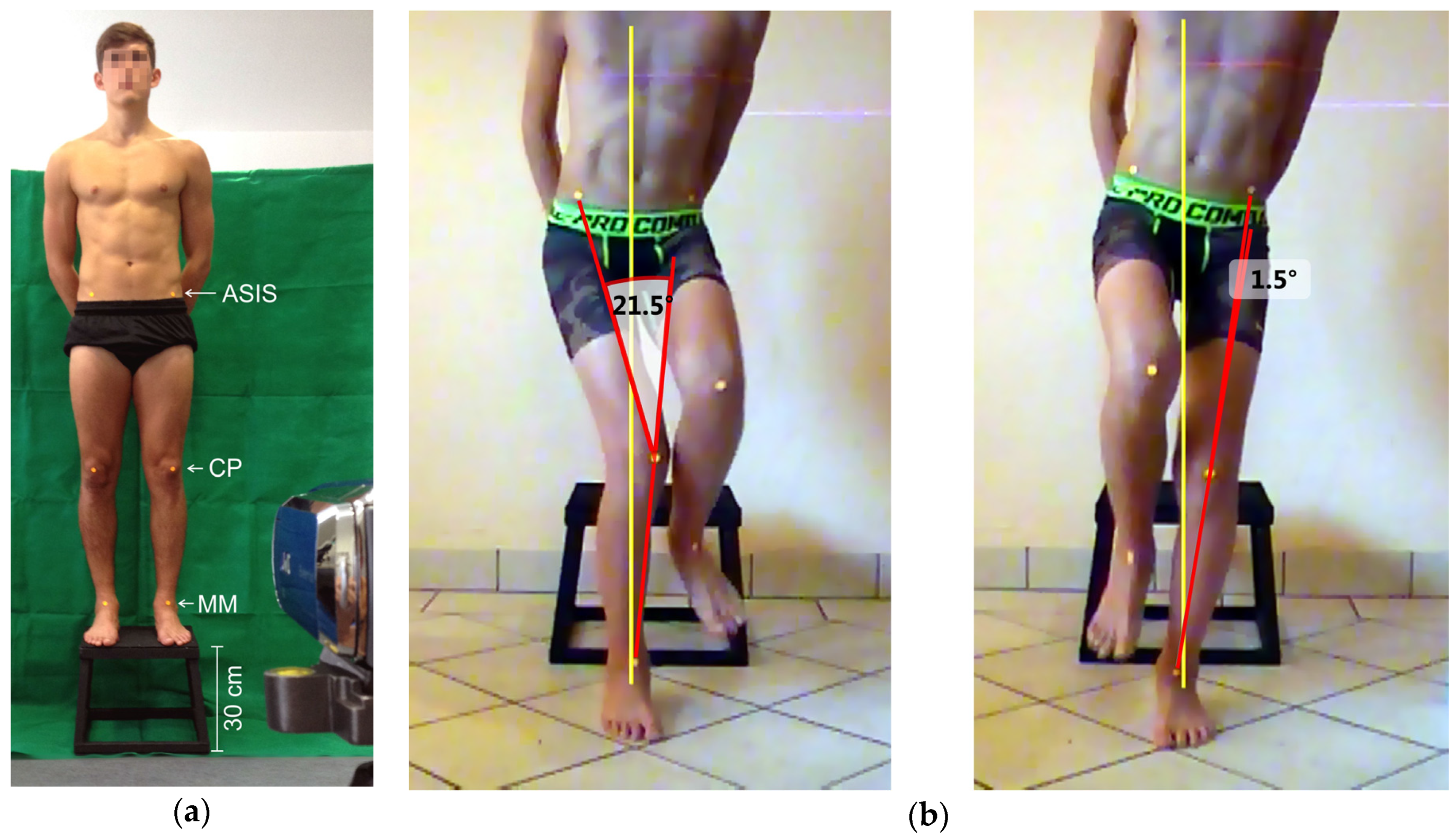
Figure 1. (a) Experimental setup: ASIS = marker on the anterior superior iliac spine, CP = marker on the center of the patella, MM = marker on the instep; (b) Example of the landing positions of a right-footed soccer player: the dominant (right) leg shows an increased dynamic knee valgus angle (note that the left hip rotates backwards).
Sports 05 00014 g001
Table 1. Anthropometric data of the 144 test persons.
LevelNAge (years)Height (cm)Weight (kg)
Amateur4815.4 ± 1.4174.1 ± 6.864.1 ± 10.9
Elite6614.0 ± 0.2169.3 ± 8.655.3 ± 8.8
Table 2. Differences in the dynamic knee valgus angles between the dominant (DOM) and non-dominant (NON) leg in the groups examined, CV = coefficient of variation, d = Cohen’s d. Negative values mark varus angles and positive values signify valgus angles.
LevelLegdfMSDCVpd
AmateurDOM471.41913.5979.580.0460.28
NON47−1.8569.729−5.24
EliteDOM653.75810.0362.670.0000.64
NON65−2.71510.2813.79