Interesting Images

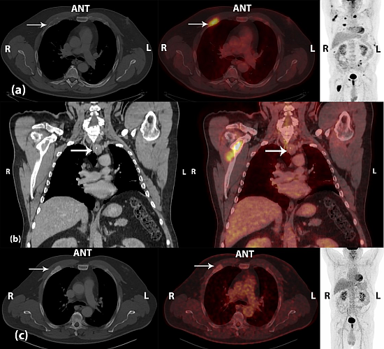
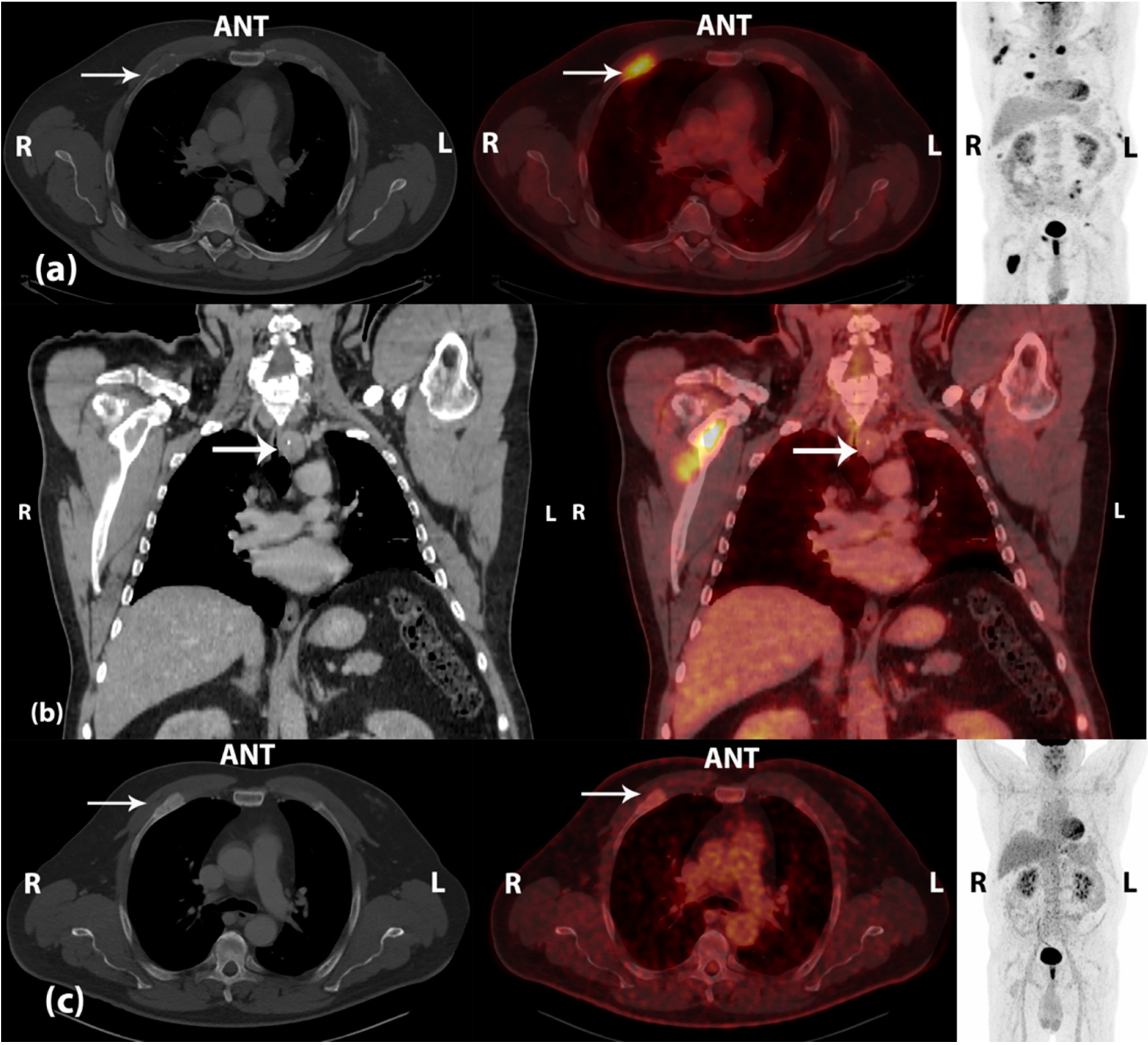
Brown Tumors Due to Primary Hyperparathyroidism in a Patient with Parathyroid Carcinoma Mimicking Skeletal Metastases on 18F-FDG PET/CT

by
Kim Francis Andersen
* and
Elisabeth Albrecht-Beste
Department of Clinical Physiology, Nuclear Medicine & PET, Rigshospitalet, Copenhagen University Hospital, Blegdamsvej 9—PET 3982, DK-2100 Copenhagen, Denmark
*
Author to whom correspondence should be addressed.
Diagnostics 2015, 5(3), 290-293; https://doi.org/10.3390/diagnostics5030290
Submission received: 23 June 2015 / Revised: 6 July 2015 / Accepted: 6 July 2015 / Published: 9 July 2015
(This article belongs to the Collection Hybrid Imaging in Medicine)

Abstract

:
Parathyroid carcinoma only represents <1% of all cases of primary hyperparathyroidism (PHPT). Even rare, chronic PHPT may lead to excessive osteoclast activity, and the increased resorption leads to destruction of cortical bone and formation of fibrous cysts with deposits of hemosiderin—so-called brown tumors. These benign, osteolytic lesions may demonstrate FDG-avidity on 18F-FDG PET/CT, and as such are misinterpreted as skeletal metastases. Regression of the lesions may occur following successful treatment. We present a case demonstrating the diagnostic work-up and follow-up of a patient with PHPT due to parathyroid carcinoma and with presence of brown tumors on 18F-FDG PET/CT, visualizing the possible role of this imaging modality in the evaluation of treatment response in these patients.

Graphical Abstract

Figure 1. (a) Initial scan—left and middle: CT (bone window) and fused 18F-FDG PET/CT, transaxial view; right: 18F-FDG PET, maximal intensity projection. A 53-year old man with persistent bone pain localized to the knees, fatigue, and memory loss. Diagnostic work-up came out with strongly elevated plasma levels of parathyroid hormone (PTH, 180 pmol/L (normal range 1.18–8.43 pmol/L)) and ionized calcium (2.27 mmol/L (normal range 1.18–1.32 mmol/L)). Medical treatment with calcimimetics was initialized. The patient was referred to a fluorine-18 fluoro-2-deoxy-d-glucose positron emission tomography/computed tomography (18F-FDG PET/CT), which demonstrated multiple FDG-avid osteolytic and destructive lesions located to the peripheral and axial skeleton, some of the lesions with an extra-skeletal compartment. The lesions were suspected of being skeletal metastases. The white arrows point at a FDG-avid costal osteolytic lesion on the right side with maximal standardized uptake value (SUVmax) of 7.2; (b) Initial scan—CT (mediastinal window) and fused 18F-FDG PET/CT, coronal view. In addition, a 2.5 cm paratracheal lesion with central calcifications located inferior to the left lobe of the thyroid gland was visualized on CT. The lesion demonstrated no pathological FDG uptake (SUVmax 2.8) and was concluded as being a parathyroid tumor (white arrows). Due to suspiciousness of primary hyperparathyroidism (PHPT), surgery was performed in terms of left-sided hemithyroidectomy and en bloc resection of the parathyroid tumor including regional lymph nodes. A spontaneous drop in PTH plasma levels of approximately 85% was seen 5 min after removal of the parathyroid tumor. Histopathology demonstrated parathyroid carcinoma with infiltrative growth pattern and invasion of vessels. The surgical margins were negative, and there were no lymph node metastases in the resected tissue; (c) Follow-up scan—left and middle: CT (bone window) and fused 18F-FDG PET/CT, transaxial view; right: 18F-FDG PET, maximal intensity projection. As staging of the disease was essential in terms of choice of treatment, a core needle biopsy from a FDG-avid osteolytic lesion in the left ilium seen on the pre-operative 18F-FDG PET/CT scan was performed. Histopathology was inconclusive due to lack of representative tissue. A follow-up 18F-FDG PET/CT scan was performed 3 months after surgery, and the previously seen osteolytic lesions were sclerotized with normal or only slightly increased FDG uptake. SUVmax of the previously mentioned costal lesion on the right side was now 2.5 (white arrows). In addition, both the patient’s symptoms as well as plasma levels of PTH and ionized calcium were normalized on further medical treatment. The lesions initially seen on 18F-FDG PET/CT were concluded as being benign brown tumors due to PHPT in this patient with parathyroid carcinoma. This case illustrates two clinical dilemmas: (1) A patient with parathyroid carcinoma, which was not visible on 18F-FDG PET due to lack of pathological FDG uptake in the tumor detected on CT; (2) The presence of multiple benign bone lesions with high FDG-avidity, which mimicked metastatic disease and as such could be misinterpreted. Parathyroid carcinoma is rare, representing <1% of all cases of PHPT [1]. Even though 18F-FDG PET/CT can offer information regarding lesion metabolism and potential loco-regional and/or distant spread of malignant disease [2], it probably has no place in the diagnostic work-up in a group of unselected patients with PHPT [3]. Despite the nomenclature, brown tumors are not true neoplasms, but rare, benign osteolytic lesions, which arise in the setting of excessive osteoclast activity in patients with hyperparathyroidism. The increased resorption leads to destruction of cortical bone and formation of fibrous cysts. Microscopically, hemosiderin deposition in the cysts gives a characteristic brown coloration and osteoclast-like giant cells may be present. The increased FDG uptake, which can be seen in these lesions (possibly due to the presence of osteoclast-like giant cells and macrophage glucose metabolism [4]), may mimic skeletal metastasis on 18F-FDG PET/CT. However, regression of the lesions has been reported after successful parathyroidectomy [5,6,7], supported by the findings in our case report. This indicates a possible role of 18F-FDG PET/CT in the evaluation of treatment response in these patients.
Diagnostics 05 00290 g001

Conflicts of Interest

The authors declare no conflict of interest.

References

  1. Villar-del-Moral, J.; Jimenez-Garcia, A.; Salvador-Egea, P.; Martos-Martinez, J.M.; Nuno-Vazquez-Garza, J.M.; Serradilla-Martin, M.; Gomez-Palacios, A.; Moreno-Llorente, P.; Ortega-Serrano, J.; de la Quintana-Basarrate, A. Prognostic factors and staging systems in parathyroid cancer: A multicenter cohort study. Surgery 2014, 156, 1132–1144. [Google Scholar] [CrossRef] [PubMed]
  2. Evangelista, L.; Sorgato, N.; Torresan, F.; Boschin, I.M.; Pennelli, G.; Saladini, G.; Piotto, A.; Rubello, D.; Pelizzo, M.R. FDG-PET/CT and parathyroid carcinoma: Review of literature and illustrative case series. World J. Clin. Oncol. 2011, 2, 348–354. [Google Scholar] [CrossRef] [PubMed]
  3. Hindie, E.; Zanotti-Fregonara, P.; Tabarin, A.; Rubello, D.; Morelec, I.; Wagner, T.; Henry, J.F.; Taieb, D. The role of radionuclide imaging in the surgical management of primary hyperparathyroidism. J. Nucl. Med. 2015, 56, 737–744. [Google Scholar] [CrossRef] [PubMed]
  4. Gamelli, R.L.; Liu, H.; He, L.K.; Hofmann, C.A. Augmentations of glucose uptake and glucose transporter-1 in macrophages following thermal injury and sepsis in mice. J. Leukoc. Biol. 1996, 59, 639–647. [Google Scholar] [PubMed]
  5. Kerstens, M.N.; de Vries, R.; Plukker, J.T.; Slart, R.H.; Dullaart, R.P. Multiple skeletal lesions on FDG PET in severe primary hyperparathyroidism. Eur. J. Nucl. Med. Mol. Imaging 2014, 41, 182–183. [Google Scholar] [CrossRef] [PubMed]
  6. Kuwahara, K.; Izawa, S.; Murabe, H.; Murakami, N.; Yokota, T.; Wani, Y.; Notohara, K.; Tsukayama, C.; Ishimori, T.; Watanabe, Y. Increased 18F-fluorodeoxyglucose uptake in a brown tumor in a patient with primary hyperparathyroidism. J. Clin. Endocrinol. Metab. 2007, 92, 2408–2409. [Google Scholar] [CrossRef] [PubMed]
  7. Shim, H.K.; Kim, B.S. 18F-FDG PET findings of a parathyroid cancer with cortical skeletal demineralization. Clin. Nucl. Med. 2012, 37, 293–295. [Google Scholar] [CrossRef] [PubMed]

