Review

Survival Strategies in the Aquatic and Terrestrial World: The Impact of Second Messengers on Cyanobacterial Processes

by
Marco Agostoni
1,2 and
Beronda L. Montgomery
1,3,*
1
Cell and Molecular Biology Graduate Program, 322 Plant Biology Building, Michigan State University, East Lansing, MI 48824, USA
2
Department of Energy Plant Research Laboratory, 106 Plant Biology Building, Michigan State University, East Lansing, MI 48824, USA
3
Department of Biochemistry and Molecular Biology, Michigan State University, 612 Wilson Road, East Lansing, MI 48823, USA
*
Author to whom correspondence should be addressed.
Life 2014, 4(4), 745-769; https://doi.org/10.3390/life4040745
Submission received: 3 September 2014 / Revised: 31 October 2014 / Accepted: 5 November 2014 / Published: 18 November 2014
(This article belongs to the Special Issue Cyanobacteria: Ecology, Physiology and Genetics)

Abstract

:
Second messengers are intracellular substances regulated by specific external stimuli globally known as first messengers. Cells rely on second messengers to generate rapid responses to environmental changes and the importance of their roles is becoming increasingly realized in cellular signaling research. Cyanobacteria are photooxygenic bacteria that inhabit most of Earth’s environments. The ability of cyanobacteria to survive in ecologically diverse habitats is due to their capacity to adapt and respond to environmental changes. This article reviews known second messenger-controlled physiological processes in cyanobacteria. Second messengers used in these systems include the element calcium (Ca2+), nucleotide-based guanosine tetraphosphate or pentaphosphate (ppGpp or pppGpp, represented as (p)ppGpp), cyclic adenosine 3’,5’-monophosphate (cAMP), cyclic dimeric GMP (c-di-GMP), cyclic guanosine 3’,5’-monophosphate (cGMP), and cyclic dimeric AMP (c-di-AMP), and the gaseous nitric oxide (NO). The discussion focuses on processes central to cyanobacteria, such as nitrogen fixation, light perception, photosynthesis-related processes, and gliding motility. In addition, we address future research trajectories needed to better understand the signaling networks and cross talk in the signaling pathways of these molecules in cyanobacteria. Second messengers have significant potential to be adapted as technological tools and we highlight possible novel and practical applications based on our understanding of these molecules and the signaling networks that they control.

Graphical Abstract

1. Introduction

Microorganisms need to cope with variations in the external environment and rely on signaling molecules to translate these changes into intracellular responses to mediate adaptation to the new condition. After cells sense an external stimulus (i.e., first messenger), a second messenger will be synthesized or degraded to rapidly amplify the first messenger signal and initiate physiological changes. Proteins involved in the synthesis and degradation of second messengers are generally constitutively present in the cell to support rapid activation. First messenger-induced fluctuations of second messengers are propagated in cells through binding to DNA, RNA, or proteins/protein complexes. The ligand-effector complex will then trigger a signal cascade involving specific receptors, outputs and feedback processes. This signal cascade is common for all known second messengers (Figure 1).
Figure 1. Specific second messengers are regulated by an external stimulus (i.e., first messenger) and will bind specific effectors. In turn, the effector will initiate a signal cascade, which leads to organism-specific outputs (examples shown for cyanobacteria). This mechanism is common for all known second messengers.
Life 04 00745 g001
There is a rich variation of second messengers in prokaryotic organisms, from cyclic nucleotides to gases. In cyanobacteria, the most intensely studied second messengers are calcium (Ca2+), guanosine tetraphosphate or pentaphosphate (ppGpp or pppGpp; hereafter (p)ppGpp), and cyclic adenosine 3’,5’-monophosphate (cAMP). Lesser-studied second messengers in cyanobacteria are cyclic dimeric GMP (c-di-GMP), cyclic guanosine 3’,5’-monophosphate (cGMP) and nitric oxide (NO). By contrast, cyclic dimeric AMP (c-di-AMP) remains to be characterized in cyanobacteria. All of these second messengers are commonly studied in pathogenic bacteria. Calcium has been considered for its ability to influence cell structure and differentiation, motility, and gene expression in pathogenic bacteria [1]. The molecule (p)ppGpp is mainly studied in these bacteria for its involvement under cell starvation stress [2]. Cyclic AMP has been implicated in sugar metabolism [3], motility [4] and virulence [5] in pathogenic bacterial systems. Cyclic di-GMP impacts several processes in bacterial cells including transcription, RNA turnover, biofilm formation, protein synthesis, motility, and virulence [6]. NO serves as an important factor in host-pathogen interactions [7]. Finally, the most recently discovered nucleotide second messengers are c-di-AMP, which affects ion transport, membrane lipid homeostasis, DNA integrity [8], and cGMP, which has been implicated in the control of cyst formation [9].
Cyanobacteria are an ancient and distinct group of gram-negative photoautotrophic bacteria for which there have been limited investigations into the roles of most of these second messengers. Cyanobacteria are one of the most abundant photoautotrophic organisms in oceans [10] and are able to fix both carbon and nitrogen, thereby playing key roles in global carbon and nitrogen cycles. Elucidation of the processes controlling cyanobacterial adaption to aquatic habitats is critical for understanding their roles as primary producers controlling global carbon and nitrogen cycles. Cyanobacteria exhibit extraordinary diversity in terms of genome plasticity, morphological characteristics, ecological niches, and physiological properties [11,12,13]. These organisms generally rely on complex signal transduction systems, which reflect their diverse ecophysiological origins and abilities to colonize a wide range of habitats. Cyanobacteria, largely with the exception of marine Prochlorococcus and Synechococcus species, possess a much larger repertoire of two-component proteins compared to other bacteria [14], and they rely heavily on cyclic nucleotide signaling proteins [15,16,17].
In recent years, there has been a growing interest in utilizing cyanobacteria as systems for the production of valuable bioindustrial compounds from sugars to biofuels [18,19]. A number of natural physiological processes of cyanobacterial systems could be regulated to improve their use as bioproduction platforms. Sugar metabolism, motility, and biofilm production are just some examples of physiological processes under the control of second messengers. Regulatable control of these processes could lead to improvements in the efficiency of growing photosynthetic bacteria in partially or fully enclosed photobioreactors or other production platforms.
This review is designed to highlight the major advances in knowledge about the second messengers Ca2+, (p)ppGpp, cAMP, c-di-GMP, cGMP, c-di-AMP, and NO and their roles in cyanobacteria. Compared to other bacteria, the major contribution of these second messengers in cyanobacteria is to the regulation of key processes, such as nitrogen fixation, the perception of a variety of light qualities, photosynthesis-related processes, and gliding motility. This review aims to emphasize continuing areas of needed investigation for these signaling molecules and to address useful applications of knowledge about the signaling pathways to practical biotechnological interventions.

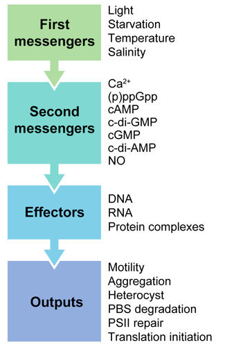
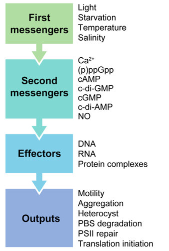
2. Second Messengers in Cyanobacteria

Studies on second messengers in cyanobacteria started ~40 years ago and have demonstrated that these molecules can influence several physiological processes. Genetic studies on the functional roles of second messengers have highlighted the involvement of these molecules in controlling physiological processes and biochemical studies and have described complex interactions between second messengers with DNA, RNA, proteins, and protein complexes.

2.1. Calcium, Ca2+

One of the most intensely studied second messengers in cyanobacteria is the ion/element Ca2+. A role for Ca2+ as a second messenger in stimulus–response coupling has been correlated frequently with a variety of environmental stresses, such as heat and cold [20], oxygen stress [21], and osmotic stress [22]. It can impact a number of physiological responses, including motility, nitrogen fixation, and responses to stress [1]. Calcium must be tightly regulated to create a concentration gradient utilized by the cells to transfer information to downstream processes. Internal Ca2+ levels can be increased by an influx of Ca2+ present in the external medium, or by releasing intracellular stores of bound Ca2+ from Ca2+-binding proteins (Figure 2).
Figure 2. External signals controlling free intracellular Ca2+ levels and phenotypes or processes that are controlled by Ca2+ in cyanobacteria. Levels of free intracellular Ca2+ are regulated externally by influx(es) of extracellular Ca2+ through ion channels or internally by release of Ca2+ from Ca2+-binding proteins. Green arrow from free [Ca2+] indicates increased Ca2+ levels from external sources leads to noted phenotypes, whereas the red arrow indicates that increased Ca2+ levels from internal sources leads to the noted phenotypes. Blue arrow indicates that referenced studies did not demonstrate whether the noted phenotypes are under external or internal control. +, indicates process promoted by increased cellular Ca2+ levels; −, indicates process inhibited by increased intracellular Ca2+ levels.
Life 04 00745 g002

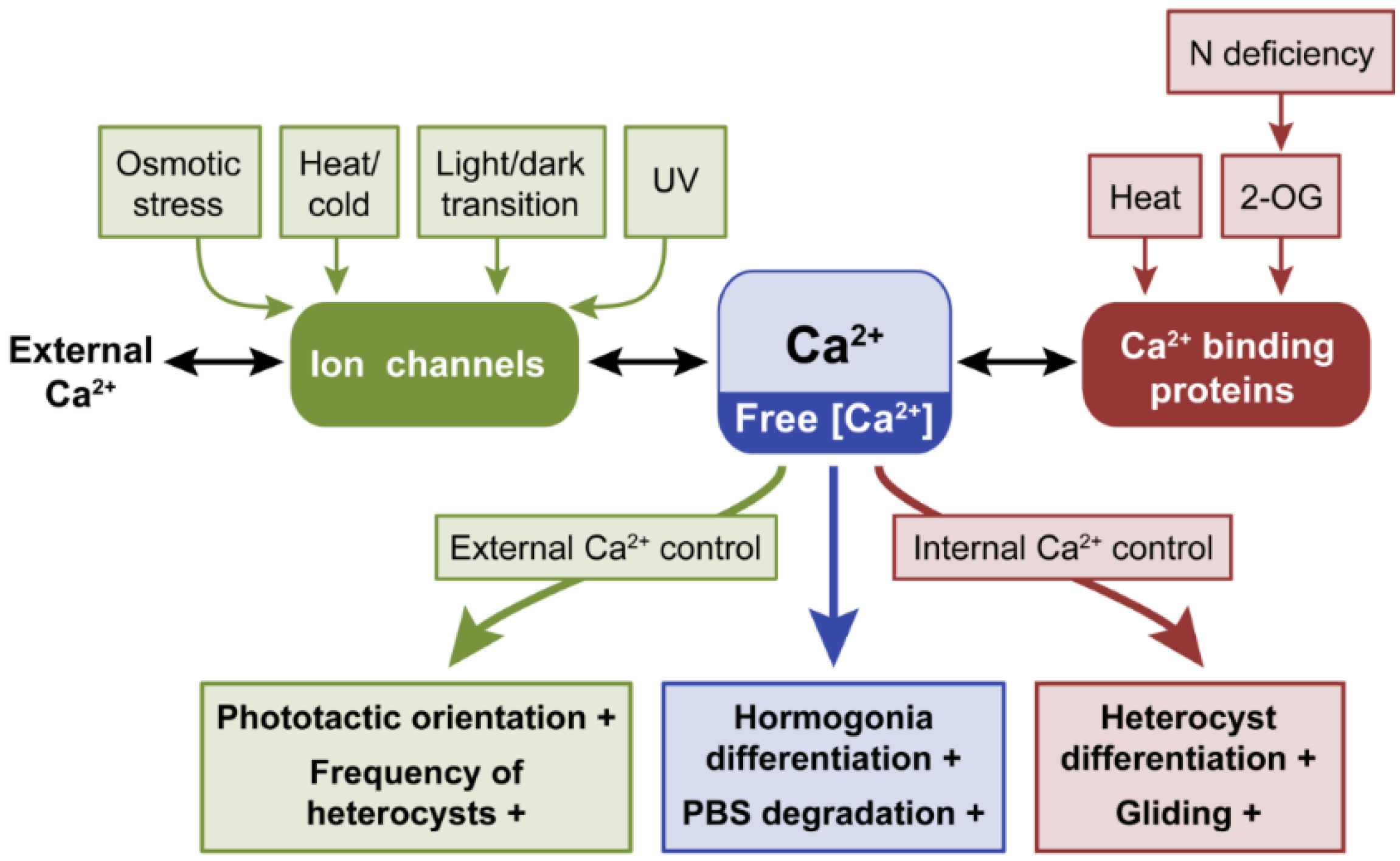
2.1.1. Calcium Controls Motility

Calcium is involved in behavioral responses in prokaryotes, including directional motility in cyanobacteria [1,23,24,25]. The first reported study to investigate the effects of Ca2+ as a second messenger in cyanobacteria showed that gliding was attributed to an increase in cellular Ca2+ concentration [26]. A study of hormogonia, which are motile filaments of cells, confirmed that Ca2+ was critical to promote the differentiation of hormogonia and sustain motility [27]. In Synechocystis sp. PCC 6803 (hereafter Synechocystis), depletion of free Ca2+ resulted in diminished photoorientation and gliding speed [28]. Based on Ca2+ chelation and calcium ionophore studies, the authors concluded that phototactic orientation was likely caused by the uptake of extracellular Ca2+, whereas gliding motility was dependent on internal Ca2+ mobilization or release from Ca2+-binding proteins [28]. Using genetic and biochemical approaches, proteins containing Ca2+-binding domains were discovered [29,30,31]. A key step to understand gliding motility in cyanobacteria was to isolate surface fibrils. Fibrils in some cyanobacteria are composed of a single type of protein called oscillin, which contains multiple Ca2+-binding motifs [29]. Phormidium uncinatum filaments that did not accumulate oscillin were not able to glide [29]. Similar cell surface-associated glycoproteins function in this manner in other cyanobacteria [29]. Motility in Synechococcus sp. WH8102 depended on the protein SwmA, a homolog of oscillin that contains Ca2+-binding motifs. A mutant lacking swmA could still rotate but no longer exhibited swimming motility [30]. Calcium was also shown to promote swimming in Synechococcus strain WH8113 [31].

2.1.2. The Role of Ca2+ in Heterocyst Differentiation

Calcium is required for the activity of the nitrogen-fixation enzyme nitrogenase in vivo and is purported to have a role in protecting nitrogenase from oxygen inactivation [32,33,34,35]. Moreover, the frequency of heterocysts found in a filament varies with different Ca2+ concentrations in the growth medium, which indicates a Ca2+-regulated mechanism for determining heterocyst abundance and placement [35]. These early experiments provided evidence that Ca2+ was involved in nitrogen fixation.
The Ca2+-binding photoprotein aequorin can be used to measure intracellular levels of Ca2+ [36]. Determining the levels of free cytosolic Ca2+ is fundamental for establishing the role of Ca2+ as second messenger. Expression of an exogenous aequorin in Anabaena sp. PCC7120 (hereafter Anabaena) resulted in the detection of a distinct Ca2+ transient after nitrogen deprivation [37]. Alteration of the amplitude or duration of the Ca2+ transient using pharmacological treatments arrested heterocyst differentiation at an early stage [37]. Thus, proper regulation of the timing and amplitude of the transient promotes heterocyst differentiation [37]. Notably, an increase of Ca2+ in the cell after nitrogen deficiency originated from an intracellular source of Ca2+ [37].
Cyanobacteria can regulate Ca2+ homeostasis by using mechanosensitive ion channels and through protein-Ca2+ complexes that may serve in Ca2+ storage (Figure 2). The gene hetR in Anabaena encodes a calcium-stimulated protease essential for vegetative cells to differentiate into heterocysts [38]. In vegetative cells, intracellular free Ca2+ levels are ten times lower than in mature heterocysts [39]. Overexpression of ccbP, which encodes a Ca2+-binding protein that is localized in vegetative cells, suppressed heterocyst formation, whereas a ccbP mutant exhibited multiple contiguous heterocysts [39]. Thus, accumulation of CcbP in vegetative cells may contribute to sequestration of Ca2+ in these cells and its absence in heterocysts likely allows the accumulation of Ca2+ and associated induction of hetR and heterocyst differentiation [31].
Nitrogen deficiency is also an important signal mediated by Ca2+ in the unicellular, non-diazotrophic cyanobacterium Synechococcus elongatus PCC 7942 [40]. Cells under nitrogen deficiency produce 2-oxoglutarate (2-OG), an important biological compound involved in the carbon-nitrogen status signal. The anion 2-OG can trigger Ca2+ accumulation transiently in S. elongatus as an increase in 2-OG occurs in cells before observed changes in intracellular Ca2+ levels [40]. In response to nitrogen starvation, S. elongatus can degrade phycobilisomes to recyclenitrogen-rich amino acids through activating two transcriptional regulators, NtcA and NblR [41,42]. Under nitrogen deficiency, a transiently increased level of intracellular Ca2+ is NtcA dependent [40]. NtcA, which is a member of the cAMP-receptor transcriptional regulator protein family, may contribute to transient accumulation of 2-OG followed by Ca2+ in regulating the expression of NtcA-dependent genes involved in the process of phycobiliprotein degradation [40].

2.1.3. Responses to Temperature Stress and Other Stresses Are Mediated by Ca2+

Calcium is also an important second messenger for cells to regulate responses to stresses, such as temperature shock, osmotic stress, and light-to-dark transitions [20,22,43]. The mechanosensitive ion channel protein MscL that is found in the plasma membrane of Synechocystis is involved in Ca2+ homeostasis regulation [44]. In Anabaena, heat shock at 44 °C resulted in an induction of intracellular Ca2+ levels that had a higher amplitude when Ca2+ was present in the external medium; similarly, a cold shock at 10 °C induced an increased magnitude of intracellular Ca2+ accumulation in cells [20]. The use of inhibitors or pharmacological agents indicated that the source of Ca2+ in the heat shock-induced elevation of cellular Ca2+ levels is both from Ca2+ in the external medium and internal stores, whereas the cold-shock induced elevation of Ca2+ levels results primarily from import of Ca2+ from the external medium [20].
Calcium is also induced by other stresses, including salinity, osmotic stress, light-to-dark transitions, and UV irradiation [22,43,45]. Intracellular levels of Ca2+ increased when Anabaena was exposed to salt or osmotic stresses. The source of increased intracellular Ca2+ levels was external as inhibiting calcium channels or the use of Ca2+-free medium eliminated this response [22]. Detecting changes in external light availability are critical for photosynthetic organisms, such as cyanobacteria. An increase of internal Ca2+ levels was observed in heterocysts in Anabaena when cells were exposed to UV irradiation [45]. A Ca2+ transient also occurred during a light-to-dark transition in Anabaena [43]. The observed elevation of intracellular Ca2+ levels was not associated with a specific photoreceptor, but more likely occurs in response to changes in the redox state of components of the photosynthetic electron transport chain [43]. Biochemical assays indicated that the source of the Ca2+ was external during the response to UV irradiation and light-to-dark transitions [43,45]. Together, the rapid Ca2+ transients that occur in cyanobacteria under temperature, osmotic, nitrogen and light stresses provide evidence that Ca2+ signaling is involved in early responses to these environmental stimuli.

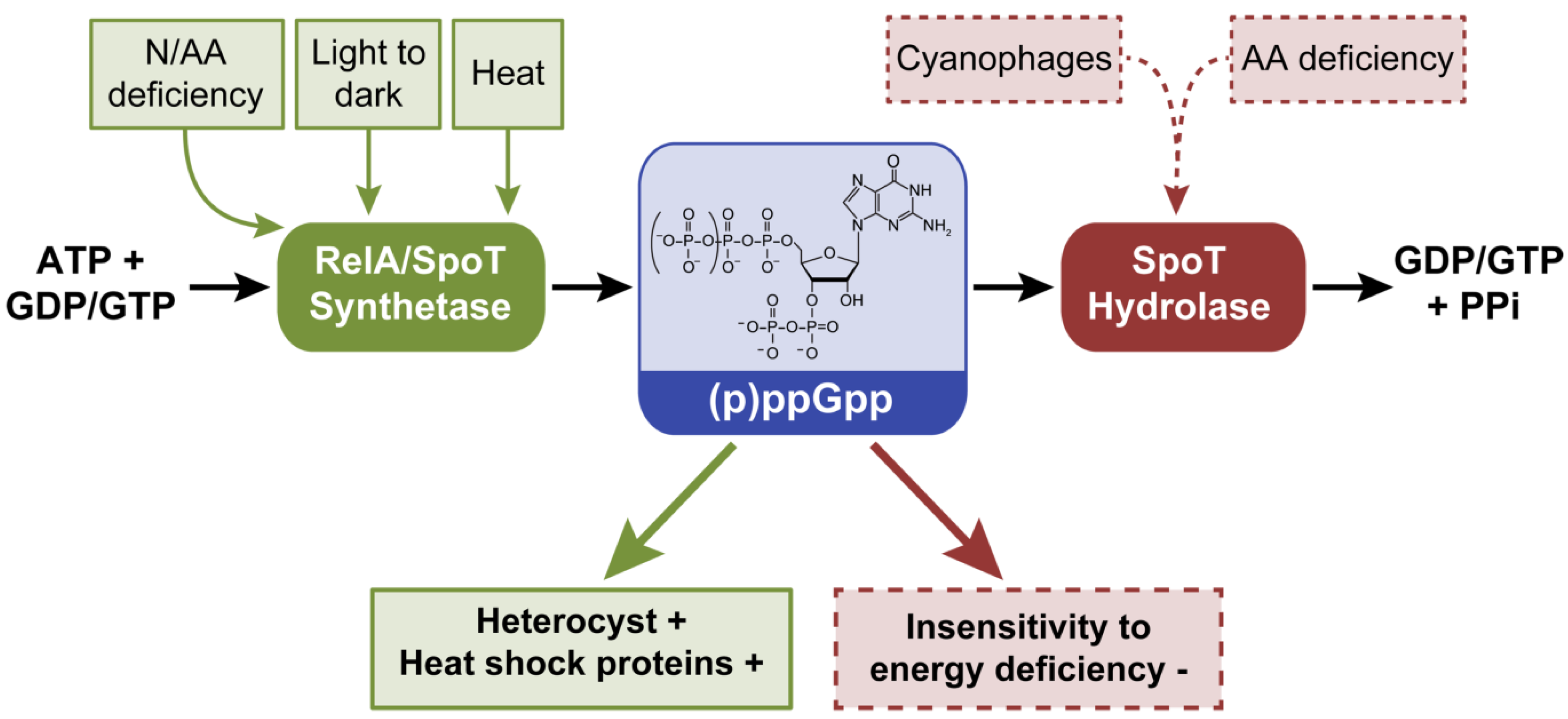
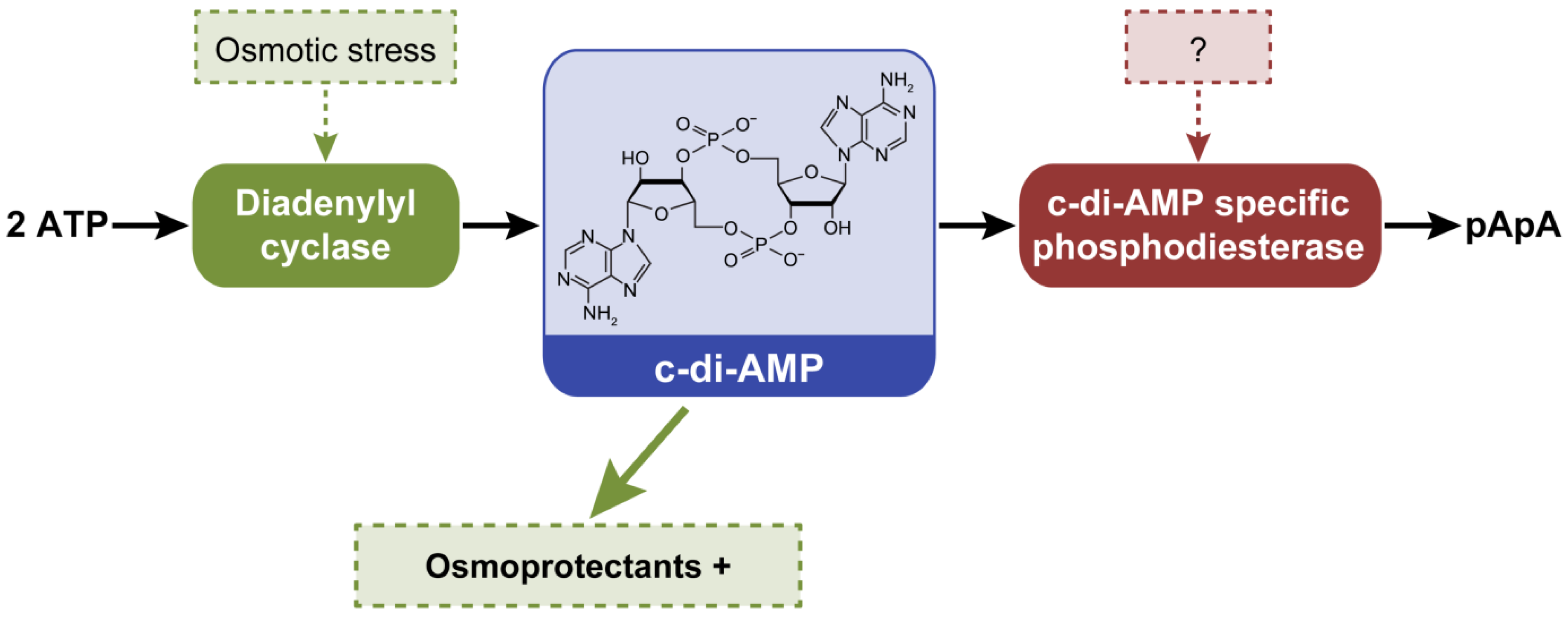
2.2. Guanosine-3’, 5’-(bis) Pyrophosphate, (p)ppGpp

Guanosine-3’, 5’-(bis) pyrophosphate, (p)ppGpp, was the first second messenger characterized in cyanobacteria ~40 years ago. This molecule is involved in the stringent response, during which alterations in metabolism and gene expression occur due to limitation in the availability of amino acids or nutrition stress [39]. During the stringent response, resources are diverted away from growth towards amino acid synthesis to support survival until unfavorable conditions improve. During this process, ppGpp or pppGpp is synthesized from ATP and GTP/GDP by a (p)ppGpp synthetase, RelA [46], and can be degraded to GDP/GTP and pyrophosphate by a (p)ppGpp hydrolase, SpoT [46] (Figure 3). (p)ppGpp inhibits translation initiation to limit excessive protein synthesis during nutritional deficiencies [47]. Recently, more details of the regulation of (p)ppGpp and downstream processes emerged. In E. coli (p)ppGpp binds the β’ subunit of RNA polymerase and decreases the half-life of rRNA, resulting in decreased transcription [48]. (p)ppGpp also binds the β’ subunit of the plastid RNA polymerase in chloroplasts of plants [49], suggesting that (p)ppGpp likely binds the β’ subunit of the RNA polymerase in cyanobacteria, which are widely recognized as the progenitor of plastids [50].
Figure 3. External factors controlling intracellular levels of (p)ppGpp and phenotypes or processes that are controlled by (p)ppGpp in cyanobacteria. (p)ppGpp is synthesized from GDP or GTP together with ATP by RelA or SpoT proteins and degraded to GDP or GTP and the by-product pyrophosphate (PPi) by SpoT. Dashed lines denote hypothetical or suggested role of cyanophages under nutrient-deficient growth conditions in controlling internal (p)ppGpp levels. Green arrow indicates that increased synthesis of (p)ppGpp leads to noted phenotypes, whereas red arrow indicates that degradation of (p)ppGpp leads to noted phenotypes. +, indicates process promoted by increased (p)ppGpp synthesis; −, indicates process inhibited by increased (p)ppGpp levels.
Life 04 00745 g003
The (p)ppGpp homeostasis enzymes RelA/SpoT (PF04607) are present as bifunctional gene homologs, sometimes referred to as RSH (Rel Spo homologs) genes [47], in each of the 83 finished genomes present in the Integrated Microbial Genomes (IMG) database [51] (genomes listed in Table S1) suggesting a widespread utilization of (p)ppGpp as a signaling molecule in cyanobacteria (Table 1). Cyanobium gracile PCC 6307, Cyanothece sp. BH68 ATCC 51142, and Nodularia spumigena CCY9414 possess two copies of the RSH genes annotated as RelA/SpoT (Table S2).
Anabaena possesses a single relA/spoT homolog (all1549 or Ana-RSH) that regulates intracellular levels of (p)ppGpp in response to amino acid deprivation [52]. Ana-RSH was determined to be essential, as it could not be deleted from the genome [52]. Ana-RSH is maintained at a basal level in Anabaena under non-stressful conditions and appears to be regulated at the enzymatic rather than at a transcriptional level under amino acid deprivation [52]. As noted above, nitrogen deprivation leads to transient increases in (p)ppGpp levels in some cyanobacteria [53,54]; thus, the role of Ana-RSH during nitrogen deprivation and heterocyst development was explored. Although Ana-RSH was enriched in vegetative cells relative to heterocysts, the regulation of Ana-RSH or (p)ppGpp was not correlated with heterocyst formation and nitrogen fixation [52]. A more recent study, however, reported up-regulation of Ana-RSH under nitrogen deprivation and confirmed enrichment in vegetative cells [55]. These authors also were not able to delete Ana-RSH from the genome, but isolated a non-functional insertion mutant of Ana-RSH that exhibited reduced (p)ppGpp levels in response to amino acid starvation and failed to form heterocysts in nitrogen-limited conditions [55].
Table 1. Total number of genomes with specific pfam domains present in the finished genomes (n = 83) represented in the IMG database a.
Second messengerPfamFunctionNumber of genomes
(p)ppGpp04607(p)ppGpp synthesis and degradation83
cAMP or cGMP00211Adenylate and guanylate cyclase65
c-di-GMP00990Diguanylate cyclase61
00563Diguanylate phosphodiesterase60
Nitric oxide00394Nitrite reductase27
07731Nitrite reductase49
07732Nitrite reductase47
13442Nitrite reductase80
00115Nitric oxide reductase83
c-di-AMP02457Diadenylyl cyclase83
Note: a IMG, Integrated Microbial Genomes database [51].

2.2.1. The Role of (p)ppGpp in Cyanobacterial Cells

The sources of carbon and energy are usually one and the same in heterotrophic organisms; however, in phototrophic organisms the source of energy, i.e., light, differs from the source of carbon. Initial studies of (p)ppGpp in phototrophic organisms compared the effects of variations in light to nutrient starvation. The cyanobacterium Synechococcus sp. PCC 6301 (formerly Anacystis nidulans) responds to a reduction of ambient light, which is equivalent to an energy reduction, with reduced growth and an associated decrease in RNA synthesis and a transient increase in GTP levels. The levels of GTP decreased as (p)ppGpp began to accumulate [56]. Notably, amino acid deprivation induced similar responses in Synechococcus sp. PCC 6301 [57]. Nitrogen deprivation also transiently increased intracellular levels of (p)ppGpp in Anabaena cylindrica [53] and in Synechococcus sp. PCC 6301 [54]. Under nitrogen starvation in Synechococcus sp. PCC 6301, the regulation of (p)ppGpp is primarily due to (p)ppGpp synthesis rather than decreased degradation of (p)ppGpp [54]. In this organism, light-to-dark shifts and temperature stress led to accumulation of (p)ppGpp with an associated accumulation of heat shock proteins [58]. The effects of these two environmental factors could be separated, as ppGpp levels also increased when this species was exposed to 47 °C in the dark [58]. Thus, although the source of energy and carbon differ in cyanobacteria, a limitation of either results in a role of (p)ppGpp in transducing the environmental change.

2.2.2. Cyanophage and (p)ppGpp

A fascinating aspect of (p)ppGpp regulation in cyanobacteria was observed when Synechococcus sp. PCC 6301 cells were infected with the AS-1 cyanophage (a cyanomyovirus). Cyanophage-infected Synechococcus sp. PCC 6301 cells failed to exhibit significantly increased intracellular (p)ppGpp levels under amino acid or energy deficiency as did uninfected cells, suggesting that infected cells did not perceive starvation [59]. One way for a cyanomyovirus to maintain low levels of (p)ppGpp is to express the protein MazG in the host; MazG is a protein found in all cyanomyovirus isolates [60]. In E. coli, MazG can hydrolyse (p)ppGpp [61], and if this is also true for MazG in cyanomyovirus-infected cyanobacteria, expression of MazG could allow the cyanomyoviruses to induce host cells to maintain basal levels of (p)ppGpp. Such a virus-induced response would support a normal cellular growth rate of the host, where the physiological state is expected to be optimal for production of progeny phage [62].

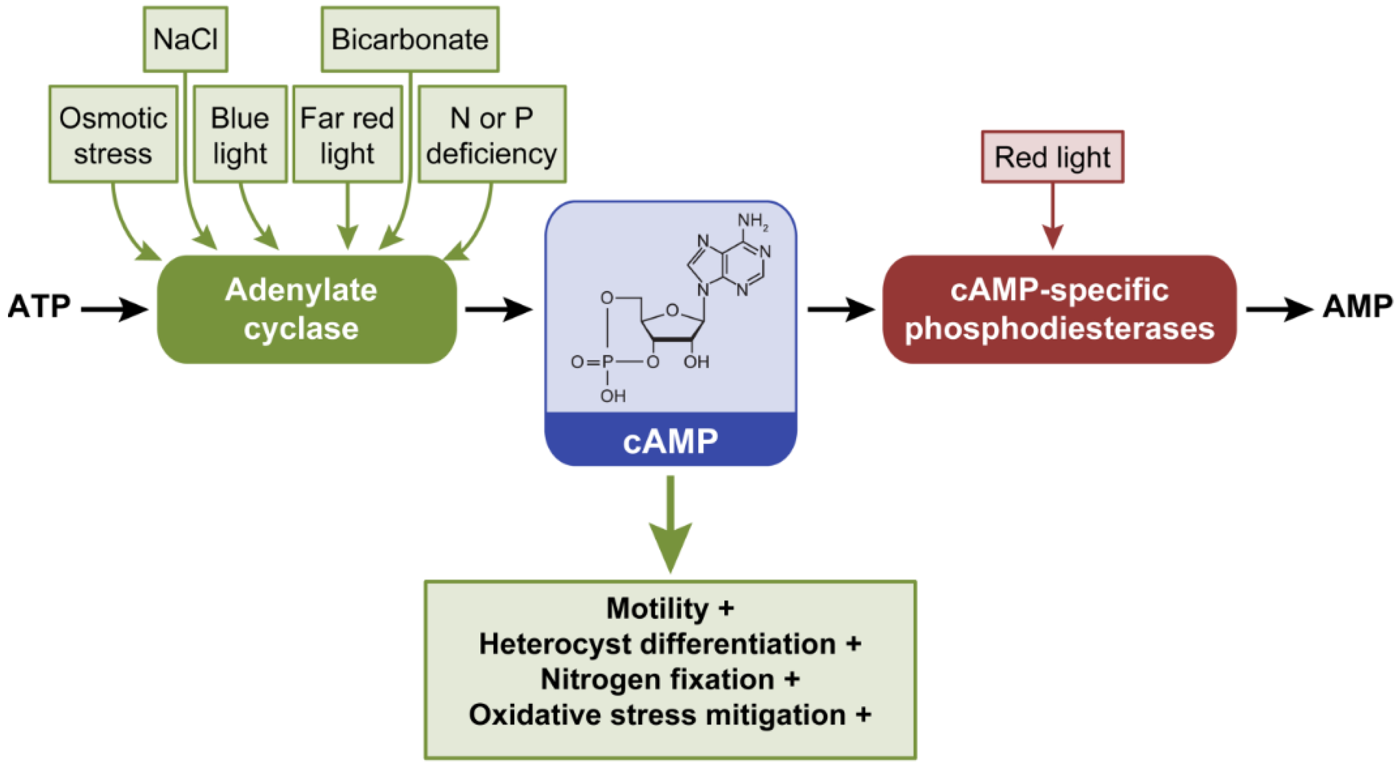
2.3. Cyclic Adenosine 3’,5’-Monophosphate, cAMP

Cyclic AMP is synthesized by adenylate cyclase (AC) (class III nucleotidyl cyclases family, Pfam: Pfam00211) proteins using ATP as a substrate and hydrolyzed to AMP by cAMP-specific phosphodiesterases (PDE) (Figure 4). Cyclic AMP is a widespread molecule in cyanobacteria. In an analysis of cyanobacterial genomes, only the picocyanobacteria Prochlorococcus and Synechococcus were reported to lack cAMP receptors, which can bind cAMP and serve as transcriptional regulators that impact diverse responses [17]. Species lacking cAMP receptors likely lost them during the course of evolution to adapt to new environments [17]. In an assessment of 83 finished cyanobacterial genomes present in the IMG database, AC genes homologous to the class III AC family (Pfam00211) were also found to be widespread (Table 1; Table S3), except in some strains that include mostly Prochlorococcus and Synechococcus strains (Table S4).
Figure 4. External factors controlling intracellular levels of cAMP and phenotypes or processes that are controlled by cAMP in cyanobacteria. Cyclic AMP is synthesized from ATP by adenylate cyclases (AC) and degraded to AMP by cAMP-specific phosphodiesterases. Green arrow indicates that increased synthesis of cAMP leads to noted phenotypes. +, indicates process promoted by increased cAMP levels.
Life 04 00745 g004

2.3.1. Cyclic AMP Regulates Motility Under Blue, Red, and Far Red Light

Cyclic AMP was initially recognized for its importance in controlling motility. In the filamentous cyanobacterium Spirulina platensis, external cAMP supplementation stimulated gliding motility and algal mat formation [63]. A Synechocystis mutant lacking the AC gene cya1 exhibited lower intracellular levels of cAMP and did not exhibit movement on agar plates under standard white light growth conditions [64]. Further studies showed the blue light significantly and rapidly increased intracellular cAMP levels in Synechocystis compared to red or far red light [65]. Motility was enhanced by blue light in a fluence- and Cya1-dependent manner [65], indicating that a blue-light dependent photoreceptor was involved in the regulation of Cya1 during this process.
Intracellular levels of cAMP are regulated by light in several cyanobacteria. Similar to Synechocystis, blue light serves as a crucial signal for cAMP regulation in Microcoleus chthonoplastes PCC 7420. A protein encoding a blue light-responsive LOV (light, oxygen, voltage) domain with an associated AC domain supported increased synthesis of cAMP in vitro upon exposure to blue light [66]. Whereas, blue light increases cAMP levels in Synechocystis and M. chthonoplastes, Anabaena cylindrica exhibits reversible red light-mediated reductions in cAMP levels and far-red light-induced increases in cAMP content [67]. Similarly, in Anabaena, red light decreased intracellular levels of cAMP, and far-red light levels increased cellular cAMP content [68]. Together, these findings suggest photoreceptor regulation of cAMP levels. Indeed, a mutant lacking a functional aphC gene, which encodes a phytochrome histidine kinase-like protein, did not exhibit FR-induced accumulation of cAMP [68]. In its photoregulation of intracellular cAMP levels, AphC purportedly phosphorylates and activates the AC CyaC, thereby inducing cAMP synthesis [68].

2.3.2. cAMP-dependent Transcriptional Regulation of Motility

In Synechocystis, the cAMP-receptor protein SYCRP1 is required for motility and appears to impact pili biogenesis [69]. Non-motile sycrp1 mutant cells showed a drastic change in the pili phenotype as the length and number of pili were extremely reduced. SYCRP1 exhibits cAMP-dependent binding to E. coli CRP consensus DNA binding sites [70,71]. Additional insights into the signaling pathway that controls motility emerged when it was demonstrated that several SYCRP1-regulated genes were under the control of Hfq, a homolog of a bacterial RNA-binding protein [72]. Notably, a mutant lacking hfq lost motility, which was correlated with the absence of pili on the cell surface. When SYCRP1 is activated it controls cell surface proteins CccS and CccP [73], both of which were also identified as targets of Hfq [69]. Similar to Δsycrp1 and Δhfq mutants, a Synechocystis mutant lacking cccS showed an absence of thick pili [73].

2.3.3. Cyclic AMP as Nutrient Deficiency Signal

Not only does cAMP control motility, but it is also an important signal for environmental nutrient deficiencies [74]. Nitrate and phosphate deficiencies resulted in transient increases in cAMP levels in Anabaena flos-aquae [74]. In Anabaena variabilis, an increased intracellular cAMP concentration was observed under nitrogen starvation that coincided with early heterocyst development [75]. Notably, cAMP can bind to AnCrpA, which in turn binds the 5’ upstream region of nifB, a nitrogen fixation-related gene, impacting its expression and that of other genes related to nitrogen fixation and heterocyst differentiation in the presence of nitrate [76]. Similar to SYCRP1, AnCrpA binds to a consensus CRP DNA sequence in a cAMP-dependent manner [77].

2.3.4. The Role of cAMP under Other Stresses

An additional link between NaCl-induced cAMP signaling and heterocyst formation was also suggested [78]. NaCl, preferential to KCl or LiCl, transiently increased intracellular cAMP levels and induced expression of genes related to heterocyst formation [78].
cAMP levels transiently increase during rehydration following desiccation in Anabaena [79]. AC CyaC is important for this cellular response as a cyaC mutant is disrupted in cAMP accumulation and associated regulation of recovery from desiccation during rehydration [79]. This mutant exhibited impairments in oxygen evolution, increased ROS levels and increased respiration compared to wild-type cells [79]. Respiration in cyanobacteria can yield CO2, which equilibrates to bicarbonate in solution. Of note, bicarbonate causes a structural change in a cyanobacterial CyaC enzyme that has been shown to stimulate its AC activity and thereby result in increased cAMP synthesis [80]. A bicarbonate-stimulated induction of cAMP accumulation during rehydration could serve a protective role, as cAMP treatment of the cyaC mutant mitigated oxidative stress and growth impairments [79].

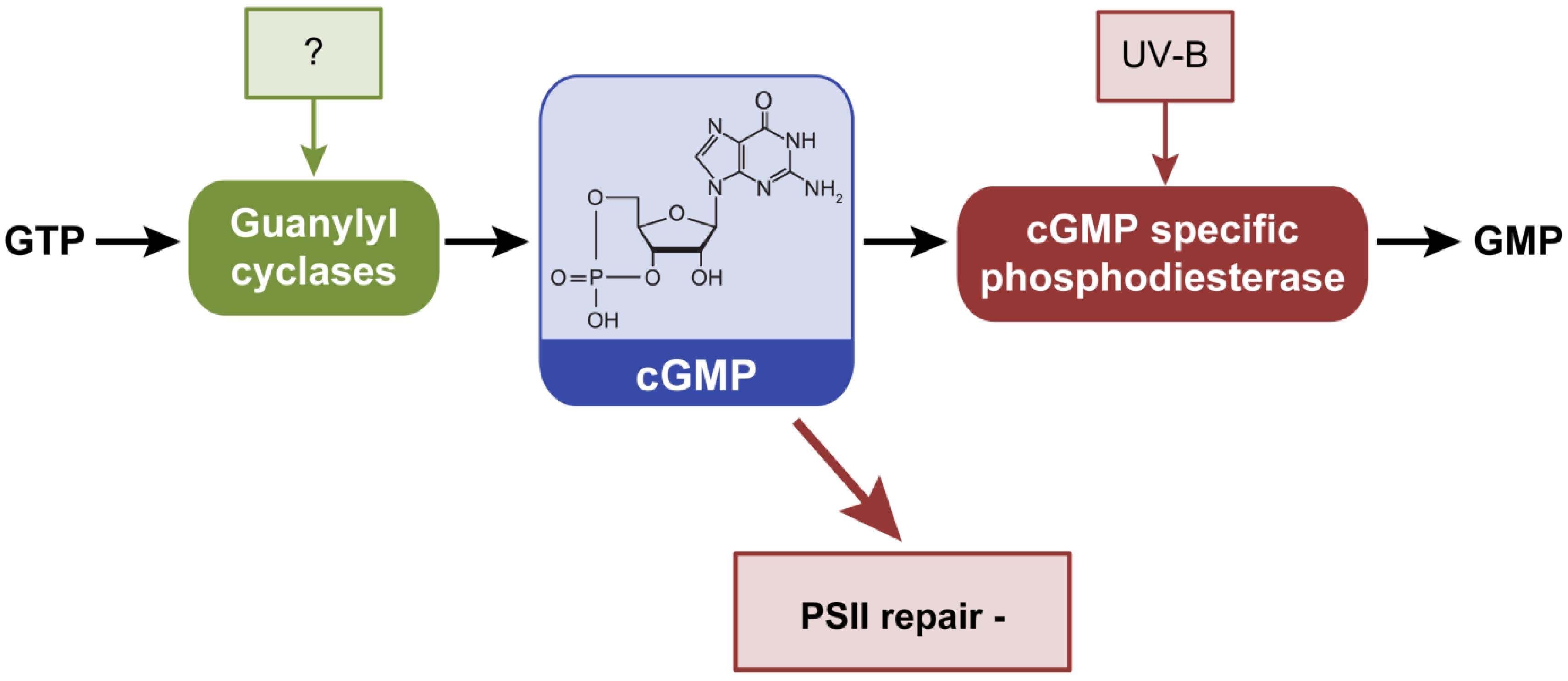
2.4. Cyclic Guanosine 3’,5’-Monophosphate, cGMP

Cyclic GMP (cGMP) is a fairly recently confirmed second messenger in bacteria [81], although it has been well characterized in eukaryotic systems. Cyclic GMP is synthesized from GTP by guanylyl cyclases (GC), which are homologous to the class III adenylate cyclases. Class III AC and GC enzymes share similar catalytic domains and are thought to have evolved from a common ancestor [82]. Cyclic GMP is hydrolyzed to GMP by cGMP-specific phosphodiesterases (PDE) (Figure 5). Cyanobacteria contain higher levels of cGMP compared to other bacteria [83]. To date the only confirmed bacterial GC reported is the protein Cya2 from Synechocystis [84]. Mutational analysis of cya2 indicated that the encoded protein contributes to intracellular cGMP levels, but does not impact cAMP levels [84]. Cya2 has higher specific activity for synthesizing cGMP from GTP than for the production of cAMP from ATP [85]. This cGMP specificity has been attributed to faster turnover of GTP than ATP by Cya2 rather than preferential affinity for GTP [85]. The only cyanobacterial phosphodiesterase (PDE) known to degrade cGMP is encoded by the gene slr2100 in Synechocystis [86]. This cGMP-specific PDE is required for the adaptation of the cells to UV-B radiation. Intracellular cGMP levels decreased in wild-type after exposure to UV-B radiation, but not in the slr2100 mutant. The slr2100 mutant exhibited reduced transcripts of genes encoding components essential for PSII repair [86].
Figure 5. External factors controlling intracellular levels of cGMP and phenotypes or processes that are controlled by cGMP in cyanobacteria. Cyclic GMP is synthesized by guanylyl cyclases and degraded by cGMP-specific phosphodiesterases. −, indicates process inhibited by increased cGMP levels.
Life 04 00745 g005

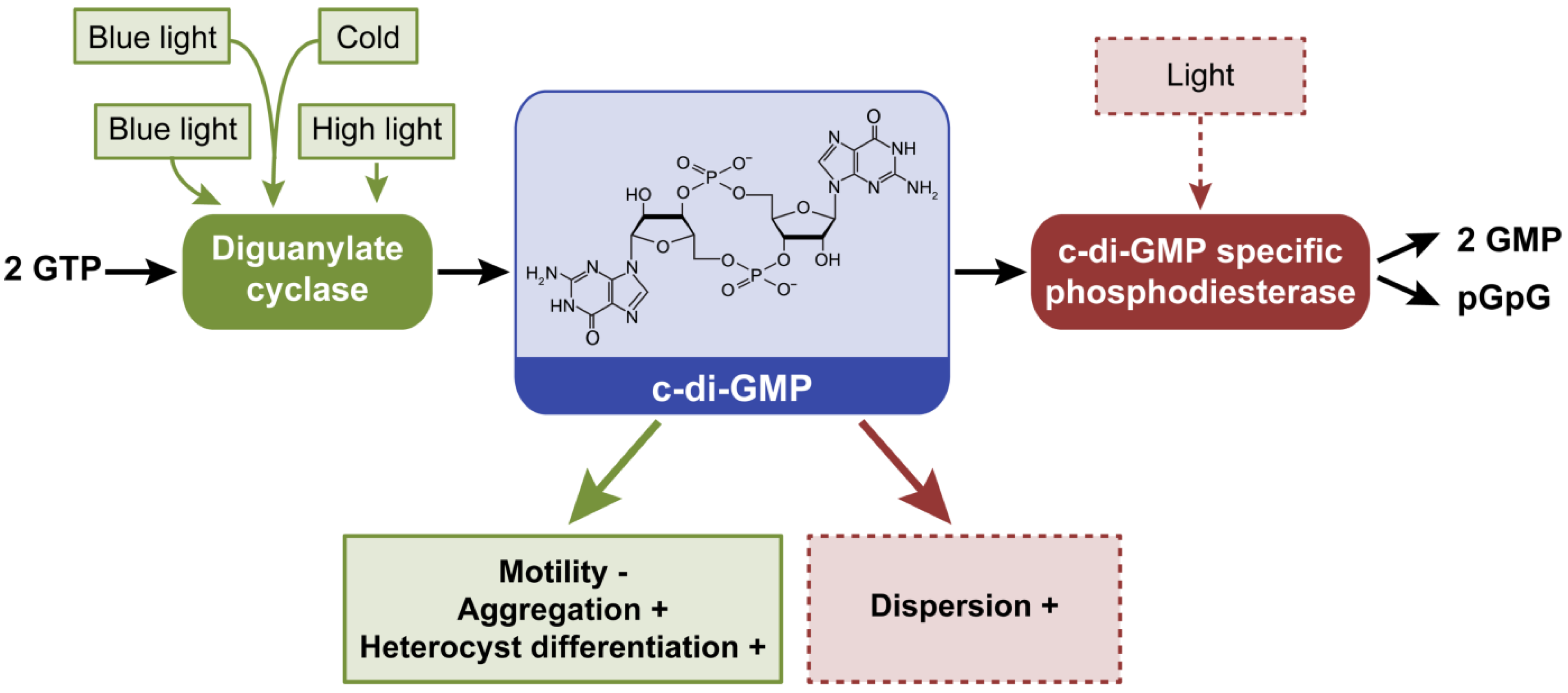
2.5. Cyclic Dimeric Guanosine 3’,5’-Monophosphate, c-di-GMP

Cyclic di-GMP is synthesized from two GTP molecules by diguanylate cylase (DGC) (Pfam 00990) activity, whereas c-di-GMP-specific PDE (Pfam00563 and Pfam01966) proteins degrade the molecule into pGpG or GMP (Figure 6). Proteins that can impact the synthesis or degradation of c-di-GMP and c-di-GMP-based signaling exist in a range of cyanobacterial species, yet are largely underexplored in these systems relative to their characterization in pathogenic bacteria [16]. Many cyanobacterial proteins contain light-responsive domains linked to domains that can impact c-di-GMP synthesis or degradation, suggesting that light is an important signal for altering c-di-GMP homeostasis and associated development, physiology and metabolism in cyanobacteria [16]. Among the species present in the CyanoBase database [87], the only species found to lack c-di-GMP signaling systems were Prochlorococcus and some strains of Synechococcus [16], similar to a report for cAMP [17]. DGC and PDE domain sequences (i.e., Pfam00990 and Pfam00563) were used to identify conserved c-di-GMP domains in the 83 finished genomes in IMG (Table 1; Tables S5 and S6). These new results confirmed that only Prochlorococcus and some strains of Synechococcus lack c-di-GMP domains, with the exception of UCYN-A, an uncultured unicellular cyanobacterium associated with a eukaryotic cell [88] (Table S7). Similar to cAMP receptors [17], species adapted to stable habitats may have lost genes encoding c-di-GMP-modulating proteins [16].
Figure 6. External factors controlling intracellular levels of c-di-GMP and phenotypes or processes that are controlled by c-di-GMP in cyanobacteria. Cyclic di-GMP is synthesized from two GTP by diguanylate cyclases and degraded to two GMP or pGpG by phosphodiesterases (PDE). Dashed lines denote hypothetical or suggested roles of light in activating PDEs and resulting phenotypes; these relationships between light absorption and c-di-GMP degradation are proposed as PDEs are often associated with photoreceptors in cyanobacteria [16] and this class of proteins induces motility and promotes dispersion in several pathogenic bacteria [6]. Green arrow indicates that increased c-di-GMP synthesis supports the noted phenotypes, whereas a red arrow indicates that increased degradation of c-di-GMP is associated with the noted phenotypes. +, indicates process promoted by altered c-di-GMP levels; −, indicates process inhibited by altered c-di-GMP levels.
Life 04 00745 g006
To date, the protein encoded by gene all2874 in Anabaena, the phytochrome-like protein Cph2 in Synechocystis, and the cyanobacteriochrome SesA in Thermosynechococcus elongatus are the only reported cyanobacterial proteins with a functional DGC activity characterized in vitro [89,90,91]. Mutation of the gene all2874 resulted in decreased heterocyst differentiation and reduced vegetative cell size under relatively high light intensity [91]. Cph2 has three GAF domains, two DGCs and one PDE domain. GAF domains bind a light-absorbing chromophore in phytochrome family proteins [92,93]. Cph2 has been studied for its involvement in inhibiting phototaxis toward blue light in Synechocystis [89]. Although wild-type Synechocystis cells did not move toward blue light, mutants lacking Cph2 showed phototaxis toward the light source. Covalent binding of a tetrapyrrole to conserved cysteine residues has been shown for two of the GAF domains of Cph2, as has light-induced photoconversion [89,94]. The DGC SesA possesses one GAF domain that can sense green and blue light [90]. SesA is responsible for cell aggregation under blue light at relatively low temperature [90]. Although c-di-GMP levels were not measured in vivo in any of these studies, these results suggest that light can serve as a signal for regulating c-di-GMP levels in these three species.
Recently, we demonstrated that intracellular levels of c-di-GMP are regulated by light in some cyanobacteria in vivo [16]. Levels of c-di-GMP were higher under blue light than other qualities of light in Synechocystis, whereas c-di-GMP levels were lower under blue light and higher under white and red light in the chromatically-acclimating Fremyella diplosiphon. Intracellular c-di-GMP levels in F. diplosiphon were overall higher than those measured in Synechocystis. These data confirmed that light is an important first messenger for regulating this second messenger in cyanobacteria.

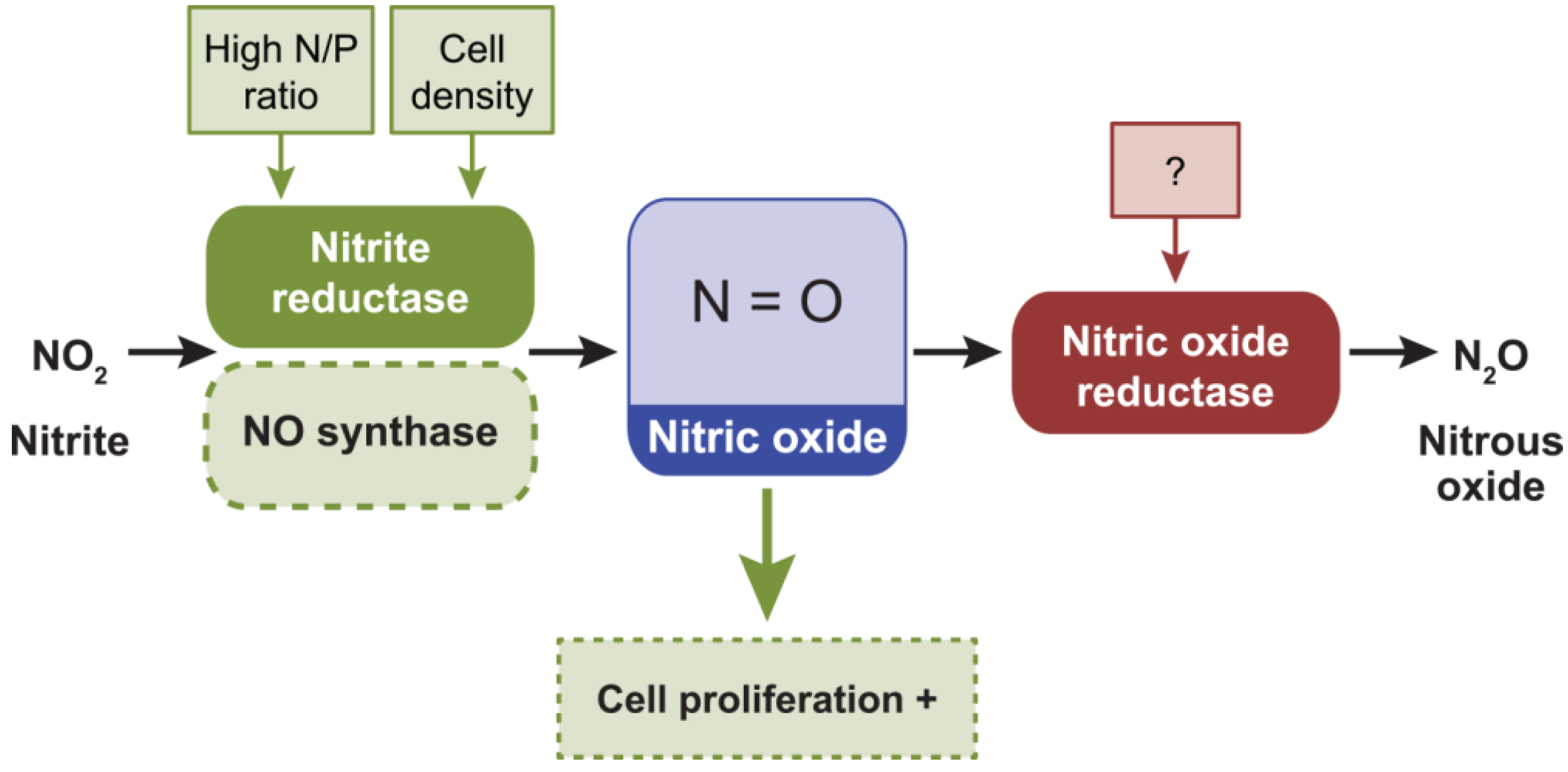
2.6. Nitric Oxide, NO

Cyanobacteria are proposed to have contributed to the rise of NO in the atmosphere due to the production of ozone from the photolysis of O2, the latter of which is generated as a by-product of oxygenic photosynthesis [95]. NO is able to neutralize ozone, scavenge reactive oxygen species and mediate their potentially damaging effects [95]. NO is an intermediate of denitrification produced through reduction of nitrite by nitrite reductase by bacteria; NO can be reduced to nitrous oxide by nitric oxide reductase (Figure 7). Although exogenous NO can be a toxic gas, at low concentrations NO can be used as signaling molecule involved in the regulation of diverse biochemical and physiological processes.
Figure 7. External factors controlling intracellular levels of nitric oxide (NO) and phenotypes or processes that are controlled by NO in cyanobacteria. NO is synthesized from nitrite (NO2) by nitrite reductase (and/or NO synthase) during denitrification and reduced to nitrous oxide (N2O) by nitric oxide reductase. Dashed lines denote hypothetical roles of NO synthase in controlling NO concentration. Green arrow pointing to the dashed green box indicates that cell proliferation could be induced (+) by high intracellular levels of NO [96].
Life 04 00745 g007
A limited number of cyanobacteria have been shown to produce NO when grown in nitrate-containing media [97]. However, there are limited insights into the in vivo role(s) of NO as a second messenger in cyanobacteria. Increasing concentrations of NO were produced and released at higher cell densities in the cyanobacterium Microcystis aeruginosa [96]. Increasing the nitrogen-to-phosphorous ratio of the growth medium also supported higher levels of NO accumulation in this organism [96]. NO accumulation appears to be primarily due to the activity of nitrate reductase, rather than NO synthase in Microcystis aeruginosa [96]. In the 83 finished cyanobacterial genomes, nitrite reductase (Pfam00394, Pfam07731, Pfam07732, or Pfam13442) homologs are largely present in cyanobacteria (Table 1; Tables S8–S11). The nitric oxide reductase (Pfam00115) is present in all finished cyanobacterial genome (Table 1; Table S12).

2.7. New Second Messenger-Dependent Phenotypes in Cyanobacteria

The DisA_N domain shows diadenylyl cyclase (DAC) activity and synthesizes c-di-AMP from two molecules of ATP; c-di-AMP is degraded to pApA by c-di-AMP specific PDE enzymes [8] (Figure 8). All cyanobacteria with a finished genome in IMG possess at least one DAC (PF02457) (Table 1). Cyanothece sp. PCC 7424, Cyanothece sp. PCC 7822, Gloeobacter kilaueensis JS1, Gloeobacter violaceus PCC 7421, and Synechococcus sp. PCC 7002 instead possess two DAC genes (Table S13). The presence of DAC in sequenced cyanobacterial genomes suggests an important role for c-di-AMP in these organisms. Surprisingly, DAC are orphan proteins; they have not been reported to be associated with other sensing domains. Cyclic di-AMP plays a central metabolic regulatory role in bacteria [98]. It has been suggested that c-di-AMP functions under osmotic stress in cyanobacteria based on assessment of regulons of riboswitches involved in binding c-di-AMP that include targets implicated in the transport and synthesis of osmoprotectants [99]. Cyclic di-AMP could also control the synthesis of c-di-GMP in some organisms, thereby potentially unveiling additional new roles of this widespread nucleotide [99].
Figure 8. Suggested external factors controlling intracellular levels of c-di-AMP and phenotypes or processes that are proposed to be controlled by c-di-AMP in cyanobacteria. Cyclic di-AMP is synthesized from two ATP by diadenylyl cyclases and degraded to pApA by a putative phosphodiesterase. Green arrow pointing to the dashed green box indicates that the production of osmoprotectants could be induced (+) by increased c-di-AMP synthesis.
Life 04 00745 g008
Recently the hybrid cyclic dinucleotide c-AMP-GMP was discovered in Vibrio cholerae [100]. The protein DncV can produce c-AMP-GMP from ATP and GTP. To date, the physiological function of c-AMP-GMP remains unsubstantiated. Using the program BLAST, the best matching sequences from the 83 finished genomes present in the IMG database were the genes Nos7107_0246 from Nostoc sp. PCC 7107 and Glo7428_5202 from Gloeocapsa sp. PCC 7428. The statistically significant E-values for these genes were 9e−11 and 9e−10, respectively, with identities around 27% for both.

2.8. Cross Talk in Second Messenger Signaling

Interactions between signaling pathways creates a flexible signaling network that allows an organism to finely tune its responses to complex, and perhaps interacting, external stimuli. There are reported examples of regulatory cross talk between second messenger signaling pathways in cyanobacteria. For example, a partially purified adenylate cyclase was activated by Ca2+ in Anabaena sp. ATCC 29151 [101]. As cyanobacteria have been demonstrated to possess both cAMP and cGMP, a potential for cross talk between their regulatory networks emerges in such organisms. Indeed, in a cGMP PDE mutant that exhibits elevated cGMP levels, transcript levels were increased for AC gene cya1, guanylyl cyclase gene cya2, and a cAMP receptor protein-encoding gene [86]. These results provide evidence for crosstalk between some second messenger signaling pathways in cyanobacterial systems.

3. Challenges for the Future

3.1. The Complexity of Second Messenger Regulatory Networks

Second messenger homeostasis genes are widespread in the genomes of cyanobacteria and a single second messenger molecule can regulate several phenotypes. For instance, Ca2+ can control gliding motility, heterocyst differentiation, and degradation of phycobilisomes (Figure 2). (p)ppGpp regulates ribosomal RNA accumulation, heat shock proteins, and heterocyst differentiation (Figure 3). Cyclic AMP is involved in photoprotection, heterocyst differentiation, photoheterotrophic growth, and nitrogen and phosphorous uptake (Figure 4). In addition to a single second messenger controlling multiple phenotypes, multiple genes may encode proteins that regulate the synthesis or degradation of a single second messenger molecule. One extreme case is apparent with the c-di-GMP signaling system in bacteria. Among cyanobacteria possessing c-di-GMP proteins, there is an average of 20 enzymes that synthesize or degrade c-di-GMP for each species in which they are found [16]. There are several mechanisms by which c-di-GMP signaling specificity is achieved in systems exhibiting such complexity. One mechanism involves regulating the timing of accumulation of c-di-GMP relative to the presence of c-di-GMP receptors or effectors in cells. For instance, expression of c-di-GMP homeostasis enzymes could occur under environmental conditions that support accumulation of c-di-GMP specific targets. This has been demonstrated in E. coli for c-di-GMP in the control of biofilm formation during stationary phase [102]. Alternatively, in light of evidence that c-di-GMP enzymes are constitutively expressed in many species, a different mechanism could explain signaling specificity. In this instance, temporal or spatial sequestration of individual c-di-GMP components could be used to control signaling, in which case c-di-GMP molecules would target co-localized receptors. The use of c-di-GMP sensors has demonstrated distinct patterns of spatial localization of c-di-GMP in some bacterial cells [103]. Individual second messenger homeostasis enzymes also could distinctly effect second messenger pools and thereby control distinct phenotypes in the organism. Such signaling is referred to as high-specificity signaling and has been reported for distinct c-di-GMP synthesis enzymes in Vibrio cholera [104]. Also, by controlling the presence of receptors of a second messenger that have differences in binding affinities, the activation of distinct receptors could be achieved at different intracellular second messenger concentrations, thereby allowing specificity of control of distinct phenotypes [105]. Although compartmentalization of several second messengers has been well demonstrated in eukaryotic cells [106], signaling specificity in cyanobacterial systems has not been well investigated. Technologies already in use in pathogenic bacteria, such as fluorescence resonance energy transfer (FRET) [103] or single fluorescent protein-based indicators [107] could be used to monitor c-di-GMP or cAMP concentrations to permit visualization of asymmetrical distributions of these second messengers in cyanobacteria.

3.2. Second Messengers and Practical Application in Biotechnology or Therapeutics

Cyanobacteria have a realistic potential to generate fuels and high-value bioindustrial products using partially or fully enclosed bioreactors [108,109]. Biofuel, ethanol, isobutanol, alkanes, biodiesel, hydrogen, sugars, and medicinal products are just a few examples of compounds that cyanobacteria can produce (reviewed by [18,19]). Engineering cyanobacteria for efficient growth and harvesting is a priority to decrease the costs and reduce environmental trade-offs. Additional insights will provide a better understanding into the roles of second messengers in regulating specific aspects of cyanobacterial growth, including the impact of environmental factors and the ability to induce cellular floating or biofilm formation. Introducing exogenous enzymes that could regulate intracellular levels of second messengers under the control of inducible promoters could be an attractive tool for regulated growth of these organisms. For instance, cells could be induced to aggregate and deposit in the bottom or to float to the surface in partially or fully enclosed bioreactors by expressing exogenous enzymes to promote energy-efficient harvesting of biomass. In addition, the induction of biofilm formation could be applied in environments contaminated by positively charged heavy metal ions, as biofilms include exopolysaccharides, which have been considered useful for metal biosorption [110].
Second messengers that are not synthesized by eukaryotes have enormous engineering potential for use in mammalian cell therapeutics. Many potential applications are possible, including controlling the levels of second messengers through light-dependent mechanisms by associating second messenger homeostasis domains with a photoreceptor [111]. Proteins containing cAMP and cGMP synthesizing domains associated with photoreceptors have been engineered for potential optogenetic applications [111,112,113,114,115]; yet, as cAMP and cGMP are present in mammals these two second messengers may not be optimal for cell-based therapeutics. However, there may be potential for the development and use of light-activatable, nucleotide second messenger-degrading enzymes to regulate levels of these molecules in vivo for therapeutic purposes. Cyclic di-GMP or c-di-AMP are particularly interesting therapeutic targets as they are not present in mammalian cells and therefore are not expected to interfere with or alter native physiological processes [116]. Cyclic di-GMP and c-di-AMP can directly induce a STING protein-dependent response, which leads to the production of cytokines essential for the induction of an innate immunity response to bacterial infection [117]. Furthermore, cyanobacterial photoreceptor domains, such as the GAF domain can absorb a wide range of light wavelengths [118]; including wavelengths that represent regions of low light absorption in mammalian tissues [119]. Thus, there is significant potential for the development of optogentic tools based on the large number of putative light-responsive c-di-GMP homeostasis proteins found in cyanobacteria [16].

4. Concluding Remarks

To date, second messengers have been shown to play key roles in controlling fundamental and perhaps underappreciated aspects of photosynthetic-related processes. Heterocyst differentiation is regulated by a complex signaling network involving cAMP, Ca2+, and (p)ppGpp. Phototaxis is regulated by both cAMP and c-di-GMP, whereas UV photoprotective mechanisms are regulated by cAMP and cGMP. Second messengers in cyanobacteria also function in pathways mediating cellular responses to oxidative stress, nutrient imbalances, and temperature variations in the environment. In the future, new studies are anticipated to shed light on additional phenotypes that are under control of second messenger molecules. Elucidation of the roles of uncharacterized second messengers, including c-di-AMP, is anticipated to provide additional insights into the complex biological networks and physiological responses regulated by second messengers. Such additional knowledge gained about second messenger signaling pathways may support the development of new tools for biotechnological, optogenic, and therapeutic applications.

Acknowledgments

This work was supported by the National Science Foundation to Beronda L. Montgomery (grant No. MCB–1243983), and support to Marco Agostoni from the U.S. Department of Energy (Chemical Sciences, Geosciences and Biosciences Division, Office of Basic Energy Sciences, Office of Science, grant No. DE-FG02-91ER20021 to Beronda L. Montgomery) and from a continuation fellowship from the College of Natural Science at Michigan State University. We are grateful to Christopher M. Waters for engaging conversations and input into discussions about the role of c-di-GMP.

Author Contributions

Marco Agostoni conducted the primary literature search and contributed to the development, organization and writing of the manuscript. Beronda L. Montgomery contributed to the organization, writing and editing of the manuscript. Both authors have read and approved the final manuscript.

Supplementary Materials

Supplementary materials can be accessed at: https://www.mdpi.com/2075-1729/4/4/745/S1.

Conflicts of Interest

The authors declare no conflict of interest.

References and Notes

  1. Dominguez, D.C. Calcium signalling in bacteria. Mol. Microbiol. 2004, 54, 291–297. [Google Scholar] [CrossRef] [PubMed]
  2. Kanjee, U.; Ogata, K.; Houry, W.A. Direct binding targets of the stringent response alarmone (p)ppGpp. Mol. Microbiol. 2012, 85, 1029–1043. [Google Scholar] [CrossRef] [PubMed]
  3. You, C.; Okano, H.; Hui, S.; Zhang, Z.; Kim, M.; Gunderson, C.W.; Wang, Y.P.; Lenz, P.; Yan, D.; Hwa, T. Coordination of bacterial proteome with metabolism by cyclic AMP signalling. Nature 2013, 500, 301–306. [Google Scholar] [CrossRef] [PubMed]
  4. Soutourina, O.; Kolb, A.; Krin, E.; Laurent-Winter, C.; Rimsky, S.; Danchin, A.; Bertin, P. Multiple control of flagellum biosynthesis in Escherichia coli: Role of H-NS protein and the cyclic AMP-catabolite activator protein complex in transcription of the flhDC master operon. J. Bacteriol. 1999, 181, 7500–7508. [Google Scholar] [PubMed]
  5. Wolfgang, M.C.; Lee, V.T.; Gilmore, M.E.; Lory, S. Coordinate regulation of bacterial virulence genes by a novel adenylate cyclase-dependent signaling pathway. Dev. Cell 2003, 4, 253–263. [Google Scholar] [CrossRef] [PubMed]
  6. Römling, U.; Galperin, M.Y.; Gomelsky, M. Cyclic di-GMP: The first 25 years of a universal bacterial second messenger. Microbiol. Mol. Biol. Rev. 2013, 77, 1–52. [Google Scholar] [CrossRef] [PubMed]
  7. Marletta, M.A.; Spiering, M.M. Trace elements and nitric oxide function. J. Nutr. 2003, 133, 1431S–1433S. [Google Scholar] [PubMed]
  8. Corrigan, R.M.; Gründling, A. Cyclic di-AMP: Another second messenger enters the fray. Nat. Rev. Microbiol. 2013, 11, 513–524. [Google Scholar] [CrossRef] [PubMed]
  9. Berleman, J.E.; Hasselbring, B.M.; Bauer, C.E. Hypercyst mutants in Rhodospirillum centenum identify regulatory loci involved in cyst cell differentiation. J. Bacteriol. 2004, 186, 5834–5841. [Google Scholar] [CrossRef] [PubMed]
  10. Garcia-Pichel, F.; Belnap, J.; Neuer, S.; Schanz, F. Estimates of global cyanobacterial biomass and its distribution. Algol. Stud. 2003, 109, 213–227. [Google Scholar] [CrossRef]
  11. Beck, C.; Knoop, H.; Axmann, I.M.; Steuer, R. The diversity of cyanobacterial metabolism: Genome analysis of multiple phototrophic microorganisms. BMC Genomics 2012, 13. [Google Scholar] [CrossRef] [PubMed]
  12. Sánchez-Baracaldo, P.; Hayes, P.K.; Blank, C.E. Morphological and habitat evolution in the cyanobacteria using a compartmentalization approach. Geobiology 2005, 3, 145–165. [Google Scholar]
  13. Bryant, D.A.; Frigaard, N.U. Prokaryotic photosynthesis and phototrophy illuminated. Trends Microbiol. 2006, 14, 488–496. [Google Scholar] [CrossRef] [PubMed]
  14. Ashby, M.K.; Houmard, J. Cyanobacterial two-component proteins: Structure, diversity, distribution, and evolution. Microbiol. Mol. Biol. Rev. 2006, 70, 472–509. [Google Scholar] [CrossRef] [PubMed]
  15. Wu, J.; Bai, J.; Bao, Q.; Zhao, F. Lineage-specific domain fusion in the evolution of purine nucleotide cyclases in cyanobacteria. J. Mol. Evol. 2008, 67, 85–94. [Google Scholar] [CrossRef] [PubMed]
  16. Agostoni, M.; Koestler, B.J.; Waters, C.M.; Williams, B.L.; Montgomery, B.L. Occurrence of cyclic di-GMP-modulating output domains in cyanobacteria: An illuminating perspective. mBio 2013, 4. [Google Scholar] [CrossRef]
  17. Xu, M.; Su, Z. Computational prediction of cAMP receptor protein (CRP) binding sites in cyanobacterial genomes. BMC Genomics 2009, 10. [Google Scholar] [CrossRef] [PubMed]
  18. Lindblad, P.; Lindberg, P.; Oliveira, P.; Stensjö, K.; Heidorn, T. Design, engineering, and construction of photosynthetic microbial cell factories for renewable solar fuel production. Ambio 2012, 41, 163–168. [Google Scholar] [CrossRef] [PubMed]
  19. Oliver, J.W.K.; Atsumi, S. Metabolic design for cyanobacterial chemical synthesis. Photosyn. Res. 2014, 120, 249–261. [Google Scholar] [CrossRef] [PubMed]
  20. Torrecilla, I.; Leganés, F.; Bonilla, I.; Fernández-Piñas, F. Use of recombinant aequorin to study calcium homeostasis and monitor calcium transients in response to heat and cold shock in cyanobacteria. Plant Physiol. 2000, 123, 161–176. [Google Scholar] [CrossRef] [PubMed]
  21. Herbaud, M.L.; Guiseppi, A.; Denizot, F.; Haiech, J.; Kilhoffer, M.C. Calcium signalling in Bacillus subtilis. Biochim. Biophys. Acta 1998, 1448, 212–226. [Google Scholar] [CrossRef]
  22. Torrecilla, I.; Leganés, F.; Bonilla, I.; Fernández-Piñas, F. Calcium transients in response to salinity and osmotic stress in the nitrogen-fixing cyanobacterium Anabaena sp. PCC7120, expressing cytosolic apoaequorin. Plant Cell Environ. 2001, 24, 641–648. [Google Scholar] [CrossRef]
  23. Ordal, G.W. Calcium ion regulates chemotactic behaviour in bacteria. Nature 1977, 270, 66–67. [Google Scholar] [CrossRef] [PubMed]
  24. Tisa, L.S.; Adler, J. Calcium ions are involved in Escherichia coli chemotaxis. Proc. Natl. Acad. Sci. USA 1992, 89, 11804–11808. [Google Scholar] [CrossRef] [PubMed]
  25. Baryshev, V.A.; Glagolev, A.N.; Skulachev, V.P. Interrelationship between Ca2+ and a methionine-requiring step in Halobacterium Halobium taxis. FEMS Microbiol. Lett. 1982, 13, 47–50. [Google Scholar] [CrossRef]
  26. Murvanidze, G.V.; Glagolev, A.N. Electrical nature of the taxis signal in cyanobacteria. J. Bacteriol. 1982, 150, 239–244. [Google Scholar] [PubMed]
  27. Hernández-Muñiz, W.; Stevens, S.E., Jr. Characterization of the motile hormogonia of Mastigocladus laminosus. J. Bacteriol. 1987, 169, 218–223. [Google Scholar] [PubMed]
  28. Moon, Y.J.; Park, Y.M.; Chung, Y.H.; Choi, J.S. Calcium is involved in photomovement of cyanobacterium Synechocystis sp PCC 6803. Photochem. Photobiol. 2004, 79, 114–119. [Google Scholar] [CrossRef] [PubMed]
  29. Hoiczyk, E.; Baumeister, W. Oscillin, an extracellular, Ca2+-binding glycoprotein essential for the gliding motility of cyanobacteria. Mol. Microbiol. 1997, 26, 699–708. [Google Scholar] [CrossRef] [PubMed]
  30. Brahamsha, B. An abundant cell-surface polypeptide is required for swimming by the nonflagellated marine cyanobacterium Synechococcus. Proc. Natl. Acad. Sci. USA 1996, 93, 6504–6509. [Google Scholar] [CrossRef] [PubMed]
  31. Pitta, T.P.; Sherwood, E.E.; Kobel, A.M.; Berg, H.C. Calcium is required for swimming by the nonflagellated cyanobacterium Synechococcus strain WH8113. J. Bacteriol. 1997, 179, 2524–2528. [Google Scholar] [PubMed]
  32. Huang, T.-C.; Chow, T.-J. Comparative studies of some nitrogen-fixing unicellular cyanobacteria isolated from rice fields. J. Gen. Microbiol. 1988, 134, 3089–3097. [Google Scholar]
  33. Chen, T.-H.; Huang, T.-C.; Chow, T.-J. Calcium requirement in nitrogen fixation in the cyanobacterium Synechococcus RF-1. Planta 1988, 173, 253–256. [Google Scholar] [CrossRef] [PubMed]
  34. Gallon, J.R.; Hamadi, A.F. Studies on the effects of oxygen on acetylene reduction (nitrogen fixation) in Gloeothece sp. ATCC 27152. J. Gen. Microbiol. 1984, 130, 495–503. [Google Scholar]
  35. Smith, R.; Hobson, S.; Ellis, I. Evidence for calcium-mediated regulation of heterocyst frequency and nitrogenase activity in Nostoc 6720. New Phytol. 1987, 105, 531–541. [Google Scholar] [CrossRef]
  36. Knight, M.R.; Campbell, A.K.; Smith, S.M.; Trewavas, A.J. Recombinant aequorin as a probe for cytosolic free Ca2+ in Escherichia coli. FEBS Lett. 1991, 282, 405–408. [Google Scholar] [CrossRef] [PubMed]
  37. Torrecilla, I.; Leganés, F.; Bonilla, I.; Fernández-Piñas, F. A calcium signal is involved in heterocyst differentiation in the cyanobacterium Anabaena sp. PCC7120. Microbiology 2004, 150, 3731–3739. [Google Scholar] [CrossRef] [PubMed]
  38. Zhou, R.; Wei, X.; Jiang, N.; Li, H.; Dong, Y.; Hsi, K.; Zhao, J. Evidence that HetR protein is an unusual serine-type protease. Proc. Natl. Acad. Sci. USA 1998, 95, 4959–4963. [Google Scholar] [CrossRef] [PubMed]
  39. Zhao, Y.; Shi, Y.; Zhao, W.; Huang, X.; Wang, D.; Brown, N.; Brand, J.; Zhao, J. CcbP, a calcium-binding protein from Anabaena sp. PCC 7120, provides evidence that calcium ions regulate heterocyst differentiation. Proc. Natl. Acad. Sci. USA 2005, 102, 5744–5748. [Google Scholar] [CrossRef] [PubMed]
  40. Leganés, F.; Forchhammer, K.; Fernández-Piñas, F. Role of calcium in acclimation of the cyanobacterium Synechococcus elongatus PCC 7942 to nitrogen starvation. Microbiology 2009, 155, 25–34. [Google Scholar] [CrossRef] [PubMed]
  41. Herrero, A.; Muro-Pastor, A.M.; Valladares, A.; Flores, E. Cellular differentiation and the NtcA transcription factor in filamentous cyanobacteria. FEMS Microbiol. Rev. 2004, 28, 469–487. [Google Scholar] [CrossRef] [PubMed]
  42. Schwarz, R.; Forchhammer, K. Acclimation of unicellular cyanobacteria to macronutrient deficiency: Emergence of a complex network of cellular responses. Microbiology 2005, 151, 2503–2514. [Google Scholar] [CrossRef] [PubMed]
  43. Torrecilla, I.; Leganés, F.; Bonilla, I.; Fernández-Piña, F. Light-to-dark transitions trigger a transient increase in intracellular Ca2+ modulated by the redox state of the photosynthetic electron transport chain in the cyanobacterium Anabaena sp. PCC7120. Plant Cell Environ. 2004, 27, 810–819. [Google Scholar] [CrossRef]
  44. Nazarenko, L.V.; Andreev, I.M.; Lyukevich, A.A.; Pisareva, T.V.; Los, D.A. Calcium release from Synechocystis cells induced by depolarization of the plasma membrane: MscL as an outward Ca2+ channel. Microbiology 2003, 149, 1147–1153. [Google Scholar] [CrossRef] [PubMed]
  45. Richter, P.; Krywult, M.; Sinha, R.P.; Häder, D.-P. Calcium signals from heterocysts of Anabaena sp. after UV irradiation. J. Plant. Physiol. 1999, 154, 137–139. [Google Scholar] [CrossRef]
  46. Srivatsan, A.; Wang, J.D. Control of bacterial transcription, translation and replication by (p)ppGpp. Curr. Opin. Microbiol. 2008, 11, 100–105. [Google Scholar] [CrossRef] [PubMed]
  47. Potrykus, K.; Cashel, M. (p)ppGpp: Still magical? Annu. Rev. Microbiol. 2008, 62, 35–51. [Google Scholar] [CrossRef] [PubMed]
  48. Toulokhonov, I.I.; Shulgina, I.; Hernandez, V.J. Binding of the transcription effector ppGpp to Escherichia coli RNA polymerase is allosteric, modular, and occurs near the N terminus of the β’-subunit. J. Biol. Chem. 2001, 276, 1220–1225. [Google Scholar] [CrossRef] [PubMed]
  49. Sato, M.; Takahashi, K.; Ochiai, Y.; Hosaka, T.; Ochi, K.; Nabeta, K. Bacterial alarmone, guanosine 5’-diphosphate 3’-diphosphate (ppGpp), predominantly binds the β’ subunit of plastid-encoded plastid RNA polymerase in chloroplasts. Chembiochem 2009, 10, 1227–1233. [Google Scholar] [CrossRef] [PubMed]
  50. Bhattacharya, D.; Archibald, J.M.; Weber, A.P.; Reyes-Prieto, A. How do endosymbionts become organelles? Understanding early events in plastid evolution. Bioessays 2007, 29, 1239–1246. [Google Scholar] [CrossRef] [PubMed]
  51. Markowitz, V.M.; Chen, I.M.; Palaniappan, K.; Chu, K.; Szeto, E.; Grechkin, Y.; Ratner, A.; Jacob, B.; Huang, J.; Williams, P.; et al. IMG: The Integrated Microbial Genomes database and comparative analysis system. Nucleic Acids Res. 2012, 40, D115–D122. [Google Scholar] [CrossRef] [PubMed]
  52. Ning, D.; Qian, Y.; Miao, X.; Wen, C. Role of the all1549 (ana-rsh) gene, a relA/spoT homolog, of the cyanobacterium Anabaena sp. PCC 7120. Curr. Microbiol. 2011, 62, 1767–1773. [Google Scholar] [CrossRef] [PubMed]
  53. Akinyanju, J.; Smith, R.J. Accumulation of ppGpp and pppGpp during nitrogen deprivation of the cyanophyte Anabaena cylindrica. FEBS Lett. 1979, 107, 173–176. [Google Scholar] [CrossRef] [PubMed]
  54. Friga, G.M.; Borbély, G.; Farkas, G.L. Accumulation of guanosine tetraphosphate (ppGpp) under nitrogen starvation in Anacystis nidulans, a cyanobacterium. Arch. Microbiol. 1981, 129, 341–343. [Google Scholar] [CrossRef] [PubMed]
  55. Zhang, S.-R.; Lin, G.-M.; Chen, W.-L.; Wang, L.; Zhang, C.-C. ppGpp metabolism is involved in heterocyst development in the cyanobacterium Anabaena sp. strain PCC 7120. J. Bacteriol. 2013, 195, 4536–4544. [Google Scholar] [CrossRef] [PubMed]
  56. Mann, N.; Carr, N.G.; Midgley, J.E. RNA synthesis and the accumulation of guanine nucleotides during growth shift down in the blue-green alga Anacystis nidulans. Biochim. Biophys. Acta 1975, 402, 41–50. [Google Scholar] [CrossRef] [PubMed]
  57. Smith, R.J.; Carr, N.G. The regulation of stable RNA synthesis in the blue-green alga Anacystis nidulans: effect of leucine deprivation and 5-methyltryptophan inhibition. J. Gen. Microbiol. 1977, 103, 61–68. [Google Scholar] [CrossRef] [PubMed]
  58. Surányi, G.; Korcz, A.; Pálfi, Z.; Borbély, G. Effects of light deprivation on RNA synthesis, accumulation of guanosine 3’(2’)-diphosphate 5’-diphosphate, and protein synthesis in heat-shocked Synechococcus sp. strain PCC 6301, a cyanobacterium. J. Bacteriol. 1987, 169, 632–639. [Google Scholar] [PubMed]
  59. Borbély, G.; Kaki, C.; Gulyás, A.; Farkas, G.L. Bacteriophage infection interferes with guanosine 3’-diphosphate-5’-diphosphate accumulation induced by energy and nitrogen starvation in the cyanobacterium Anacystis nidulans. J. Bacteriol. 1980, 144, 859–864. [Google Scholar] [PubMed]
  60. Bryan, M.J.; Burroughs, N.J.; Spence, E.M.; Clokie, M.R.; Mann, N.H.; Bryan, S.J. Evidence for the intense exchange of MazG in marine cyanophages by horizontal gene transfer. PLoS One 2008, 3, e2048. [Google Scholar] [CrossRef] [PubMed]
  61. Gross, M.; Marianovsky, I.; Glaser, G. MazG–a regulator of programmed cell death in Escherichia coli. Mol. Microbiol. 2006, 59, 590–601. [Google Scholar] [CrossRef] [PubMed]
  62. Clokie, M.R.; Mann, N.H. Marine cyanophages and light. Environ. Microbiol. 2006, 8, 2074–2082. [Google Scholar] [CrossRef] [PubMed]
  63. Ohmori, K.; Hirose, M.; Ohmori, M. Function of cAMP as a mat-forming factor in the cyanobacterium Spirulina platensis. Plant Cell Physiol. 1992, 33, 21–25. [Google Scholar]
  64. Terauchi, K.; Ohmori, M. An adenylate cyclase, Cya1, regulates cell motility in the cyanobacterium Synechocystis sp. PCC 6803. Plant Cell Physiol. 1999, 40, 248–251. [Google Scholar] [CrossRef] [PubMed]
  65. Terauchi, K.; Ohmori, M. Blue light stimulates cyanobacterial motility via a cAMP signal transduction system. Mol. Microbiol. 2004, 52, 303–309. [Google Scholar] [CrossRef] [PubMed]
  66. Raffelberg, S.; Wang, L.; Gao, S.; Losi, A.; Gärtner, W.; Nagel, G. A LOV-domain-mediated blue-light-activated adenylate (adenylyl) cyclase from the cyanobacterium Microcoleus chthonoplastes PCC 7420. Biochem. J. 2013, 455, 359–365. [Google Scholar] [CrossRef] [PubMed]
  67. Ohmori, M.; Terauchi, K.; Okamoto, S.; Watanabe, M. Regulation of cAMP-mediated photosignaling by a phytochrome in the cyanobacterium Anabaena cylindrica. Photochem. Photobiol. 2002, 75, 675–679. [Google Scholar] [PubMed]
  68. Okamoto, S.; Kasahara, M.; Kamiya, A.; Nakahira, Y.; Ohmori, M. A phytochrome-like protein AphC triggers the cAMP signaling induced by far-red light in the cyanobacterium Anabaena sp. strain PCC7120. Photochem. Photobiol. 2004, 80, 429–433. [Google Scholar] [CrossRef] [PubMed]
  69. Yoshimura, H.; Yoshihara, S.; Okamoto, S.; Ikeuchi, M.; Ohmori, M. A cAMP receptor protein, SYCRP1, is responsible for the cell motility of Synechocystis sp. PCC 6803. Plant Cell Physiol. 2002, 43, 460–463. [Google Scholar] [CrossRef] [PubMed]
  70. Yoshimura, H.; Yoshihara, S.; Okamoto, S.; Ikeuchi, M.; Ohmori, M. A cAMP receptor protein, SYCRP1, is responsible for the cell motility of Synechocystis sp. PCC 6803. Plant Cell Physiol. 2002, 43, 460–463. [Google Scholar] [PubMed]
  71. Omagari, K.; Yoshimura, H.; Takano, M.; Hao, D.; Ohmori, M.; Sarai, A.; Suyama, A. Systematic single base-pair substitution analysis of DNA binding by the cAMP receptor protein in cyanobacterium Synechocystis sp. PCC 6803. FEBS Lett. 2004, 563, 55–58. [Google Scholar] [CrossRef] [PubMed]
  72. Dienst, D.; Dühring, U.; Mollenkopf, H.J.; Vogel, J.; Golecki, J.; Hess, W.R.; Wilde, A. The cyanobacterial homologue of the RNA chaperone Hfq is essential for motility of Synechocystis sp PCC 6803. Microbiology 2008, 154, 3134–3143. [Google Scholar] [CrossRef] [PubMed]
  73. Yoshimura, H.; Kaneko, Y.; Ehira, S.; Yoshihara, S.; Ikeuchi, M.; Ohmori, M. CccS and CccP are involved in construction of cell surface components in the cyanobacterium Synechocystis sp. strain PCC 6803. Plant Cell Physiol. 2010, 51, 1163–1172. [Google Scholar] [CrossRef] [PubMed]
  74. Francko, D.A.; Wetzel, R.G. Dynamics of cellular and extracellular cAMP in Anabaena flos-aquae (Cyanophyta): Intrinsic culture variability and correlation with metabolic variables. J. Phycol. 1981, 17, 129–134. [Google Scholar] [CrossRef]
  75. Hood, E.E.; Armour, S.; Ownby, J.D.; Handa, A.K.; Bressan, R.A. Effect of nitrogen starvation on the level of adenosine 3’,5’-monophosphate in Anabaena variabilis. Biochim. Biophys. Acta 1979, 588, 193–200. [Google Scholar] [CrossRef] [PubMed]
  76. Suzuki, T.; Yoshimura, H.; Ehira, S.; Ikeuchi, M.; Ohmori, M. AnCrpA, a cAMP receptor protein, regulates nif-related gene expression in the cyanobacterium Anabaena sp. strain PCC 7120 grown with nitrate. FEBS Lett. 2007, 581, 21–28. [Google Scholar] [CrossRef] [PubMed]
  77. Suzuki, T.; Yoshimura, H.; Hisabori, T.; Ohmori, M. Two cAMP receptor proteins with different biochemical properties in the filamentous cyanobacterium Anabaena sp. PCC 7120. FEBS Lett. 2004, 571, 154–160. [Google Scholar] [CrossRef] [PubMed]
  78. Imashimizu, M.; Yoshimura, H.; Katoh, H.; Ehira, S.; Ohmori, M. NaCl enhances cellular cAMP and upregulates genes related to heterocyst development in the cyanobacterium, Anabaena sp. strain PCC 7120. FEMS Microbiol. Lett. 2005, 252, 97–103. [Google Scholar] [CrossRef] [PubMed]
  79. Higo, A.; Ikeuchi, M.; Ohmori, M. cAMP regulates respiration and oxidative stress during rehydration in Anabaena sp. PCC 7120. FEBS Lett. 2008, 582, 1883–1888. [Google Scholar] [CrossRef] [PubMed]
  80. Chen, Y.; Cann, M.J.; Litvin, T.N.; Iourgenko, V.; Sinclair, M.L.; Levin, L.R.; Buck, J. Soluble adenylyl cyclase as an evolutionarily conserved bicarbonate sensor. Science 2000, 289, 625–628. [Google Scholar] [CrossRef] [PubMed]
  81. Gomelsky, M. cAMP, c-di-GMP, c-di-AMP and now cGMP: bacteria use them all! Mol. Microbiol. 2011, 79, 562–565. [Google Scholar] [CrossRef] [PubMed]
  82. Kasahara, M.; Unno, T.; Yashiro, K.; Ohmori, M. CyaG, a novel cyanobacterial adenylyl cyclase and a possible ancestor of mammalian guanylyl cyclases. J. Biol.Chem. 2001, 276, 10564–10569. [Google Scholar] [CrossRef] [PubMed]
  83. Herdman, M.; Elmorjani, K. Cyclic nucleotides. Methods Enzymol. 1988, 167, 584–591. [Google Scholar]
  84. Ochoa de Alda, J.A.; Ajlani, G.; Houmard, J. Synechocystis strain PCC 6803 cya2, a prokaryotic gene that encodes a guanylyl cyclase. J. Bacteriol. 2000, 182, 3839–3842. [Google Scholar]
  85. Rauch, A.; Leipelt, M.; Russwurm, M.; Steegborn, C. Crystal structure of the guanylyl cyclase Cya2. Proc. Natl. Acad. Sci. USA 2008, 105, 15720–15725. [Google Scholar] [CrossRef]
  86. Cadoret, J.C.; Rousseau, B.; Perewoska, I.; Sicora, C.; Cheregi, O.; Vass, I.; Houmard, J. Cyclic nucleotides, the photosynthetic apparatus and response to a UV-B stress in the cyanobacterium Synechocystis sp. PCC 6803. J. Biol. Chem. 2005, 280, 33935–33944. [Google Scholar] [CrossRef] [PubMed]
  87. Cyanobase. Available online: http://genome.kazusa.or.jp/cyanobase (accessed on 5 November 2014).
  88. Krupke, A.; Lavik, G.; Halm, H.; Fuchs, B.M.; Amann, R.I.; Kuypers, M.M. Distribution of a consortium between unicellular algae and the N2 fixing cyanobacterium UCYN-A in the North Atlantic Ocean. Environ. Microbiol. 2014, 16, 3153–3167. [Google Scholar] [CrossRef] [PubMed]
  89. Savakis, P.; De Causmaecker, S.; Angerer, V.; Ruppert, U.; Anders, K.; Essen, L.O.; Wilde, A. Light-induced alteration of c-di-GMP level controls motility of Synechocystis sp. PCC 6803. Mol. Microbiol. 2012, 85, 239–251. [Google Scholar] [CrossRef] [PubMed]
  90. Enomoto, G.; Nomura, R.; Shimada, T.; Win, N.-N.; Narikawa, R.; Ikeuchi, M. Cyanobacteriochrome SesA is a diguanylate cyclase that induces cell aggregation in Thermosynechococcus. J. Biol. Chem. 2014, 289, 24801–24809. [Google Scholar] [CrossRef] [PubMed]
  91. Neunuebel, M.R.; Golden, J.W. The Anabaena sp. strain PCC 7120 gene all2874 encodes a diguanylate cyclase and is required for normal heterocyst development under high-light growth conditions. J. Bacteriol. 2008, 190, 6829–6836. [Google Scholar] [CrossRef] [PubMed]
  92. Wu, S.H.; Lagarias, J.C. Defining the bilin lyase domain: lessons from the extended phytochrome superfamily. Biochemistry 2000, 39, 13487–13495. [Google Scholar] [CrossRef] [PubMed]
  93. Montgomery, B.L.; Lagarias, J.C. Phytochrome ancestry: sensors of bilins and light. Trends Plant Sci. 2002, 7, 357–366. [Google Scholar] [CrossRef] [PubMed]
  94. Anders, K.; von Stetten, D.; Mailliet, J.; Kiontke, S.; Sineshchekov, V.A.; Hildebrandt, P.; Hughes, J.; Essen, L.-O. Spectroscopic and photochemical characterization of the red-light sensitive photosensory module of Cph2 from Synechocystis PCC 6803. Photochem. Photobiol. 2011, 87, 160–173. [Google Scholar] [CrossRef] [PubMed]
  95. Feelisch, M.; Martin, J.F. The early role of nitric oxide in evolution. Trends Ecol. Evol. 1995, 10, 496–499. [Google Scholar] [CrossRef] [PubMed]
  96. Tang, X.; Chen, J.; Wang, W.H.; Liu, T.W.; Zhang, J.; Gao, Y.H.; Pei, Z.M.; Zheng, H.L. The changes of nitric oxide production during the growth of Microcystis aerugrinosa. Environ. Pollut. 2011, 159, 3784–3792. [Google Scholar] [CrossRef] [PubMed]
  97. Mallick, N.; Rai, L.C.; Mohn, F.H.; Soeder, C.J. Studies on nitric oxide (NO) formation by the green alga Scenedesmus obliquus and the diazotrophic cyanobacterium Anabaena doliolum. Chemosphere 1999, 39, 1601–1610. [Google Scholar] [CrossRef] [PubMed]
  98. Sureka, K.; Choi, P.H.; Precit, M.; Delince, M.; Pensinger, D.A.; Huynh, T.N.; Jurado, A.R.; Goo, Y.A.; Sadilek, M.; Iavarone, A.T.; et al. The cyclic dinucleotide c-di-AMP is an allosteric regulator of metabolic enzyme function. Cell 2014, 158, 1389–1401. [Google Scholar] [CrossRef] [PubMed]
  99. Nelson, J.W.; Sudarsan, N.; Furukawa, K.; Weinberg, Z.; Wang, J.X.; Breaker, R.R. Riboswitches in eubacteria sense the second messenger c-di-AMP. Nat. Chem. Biol. 2013, 9, 834–839. [Google Scholar] [CrossRef] [PubMed]
  100. Davies, B.W.; Bogard, R.W.; Young, T.S.; Mekalanos, J.J. Coordinated regulation of accessory genetic elements produces cyclic di-nucleotides for V. cholerae virulence. Cell 2012, 149, 358–370. [Google Scholar] [CrossRef] [PubMed]
  101. Bianchini, G.M.; Pastini, A.C.; Muschietti, J.P.; Téllez-Iñón, M.T.; Martinetto, H.E.; Torres, H.N.; Flawiá, M.M. Adenylate cyclase activity in cyanobacteria: Activation by Ca2+-calmodulin and a calmodulin-like activity. Biochim. Biophys. Acta 1990, 1055, 75–81. [Google Scholar] [CrossRef]
  102. Weber, H.; Pesavento, C.; Possling, A.; Tischendorf, G.; Hengge, R. Cyclic-di-GMP-mediated signalling within the σS network of Escherichia coli. Mol. Microbiol. 2006, 62, 1014–1034. [Google Scholar] [CrossRef] [PubMed]
  103. Christen, M.; Kulasekara, H.D.; Christen, B.; Kulasekara, B.R.; Hoffman, L.R.; Miller, S.I. Asymmetrical distribution of the second messenger c-di-GMP upon bacterial cell division. Science 2010, 328, 1295–1297. [Google Scholar] [CrossRef] [PubMed]
  104. Massie, J.P.; Reynolds, E.L.; Koestler, B.J.; Cong, J.P.; Agostoni, M.; Waters, C.M. Quantification of high-specificity cyclic diguanylate signaling. Proc. Natl. Acad. Sci. USA 2012, 109, 12746–12751. [Google Scholar] [CrossRef] [PubMed]
  105. Pultz, I.S.; Christen, M.; Kulasekara, H.D.; Kennard, A.; Kulasekara, B.; Miller, S.I. The response threshold of Salmonella PilZ domain proteins is determined by their binding affinities for c-di-GMP. Mol. Microbiol. 2012, 86, 1424–1440. [Google Scholar] [CrossRef] [PubMed]
  106. McCormick, K.; Baillie, G.S. Compartmentalisation of second messenger signalling pathways. Curr. Opin. Genet. Dev. 2014, 27, 20–25. [Google Scholar] [CrossRef] [PubMed]
  107. Odaka, H.; Arai, S.; Inoue, T.; Kitaguchi, T. Genetically-encoded yellow fluorescent cAMP indicator with an expanded dynamic range for dual-color imaging. PLoS One 2014, 9, e100252. [Google Scholar] [CrossRef] [PubMed]
  108. Ducat, D.C.; Way, J.C.; Silver, P.A. Engineering cyanobacteria to generate high-value products. Trends Biotechnol. 2011, 29, 95–103. [Google Scholar] [CrossRef] [PubMed]
  109. Nozzi, N.E.; Oliver, J.W.; Atsumi, S. Cyanobacteria as a platform for biofuel production. Front. Bioeng. Biotechnol. 2013, 1. [Google Scholar] [CrossRef]
  110. De Philippis, R.; Colica, G.; Micheletti, E. Exopolysaccharide-producing cyanobacteria in heavy metal removal from water: Molecular basis and practical applicability of the biosorption process. Appl. Microbiol. Biotechnol. 2011, 92, 697–708. [Google Scholar] [CrossRef] [PubMed]
  111. Ryu, M.H.; Moskvin, O.V.; Siltberg-Liberles, J.; Gomelsky, M. Natural and engineered photoactivated nucleotidyl cyclases for optogenetic applications. J. Biol. Chem. 2010, 285, 41501–41508. [Google Scholar] [CrossRef] [PubMed]
  112. Gasser, C.; Taiber, S.; Yeh, C.M.; Wittig, C.H.; Hegemann, P.; Ryu, S.; Wunder, F.; Möglich, A. Engineering of a red-light-activated human cAMP/cGMP-specific phosphodiesterase. Proc. Natl. Acad. Sci. USA 2014, 111, 8803–8808. [Google Scholar] [CrossRef] [PubMed]
  113. Ryu, M.H.; Kang, I.H.; Nelson, M.D.; Jensen, T.M.; Lyuksyutova, A.I.; Siltberg-Liberles, J.; Raizen, D.M.; Gomelsky, M. Engineering adenylate cyclases regulated by near-infrared window light. Proc. Natl. Acad. Sci. USA 2014, 111, 10167–10172. [Google Scholar] [CrossRef] [PubMed]
  114. Stierl, M.; Penzkofer, A.; Kennis, J.T.; Hegemann, P.; Mathes, T. Key residues for the light regulation of the blue light-activated adenylyl cyclase from Beggiatoa sp. Biochemistry 2014, 53, 5121–5130. [Google Scholar] [CrossRef] [PubMed]
  115. Chen, Z.; Raffelberg, S.; Losi, A.; Schaap, P.; Gärtner, W. A cyanobacterial light activated adenylyl cyclase partially restores development of a Dictyostelium discoideum, adenylyl cyclase a null mutant. J. Biotechnol. 2014. [Google Scholar] [CrossRef]
  116. Ryu, M.-H.; Gomelsky, M. Near-infrared light responsive synthetic c-di-GMP module for optogenetic applications. ACS Synth. Biol. 2014. [Google Scholar] [CrossRef]
  117. Burdette, D.L.; Monroe, K.M.; Sotelo-Troha, K.; Iwig, J.S.; Eckert, B.; Hyodo, M.; Hayakawa, Y.; Vance, R.E. STING is a direct innate immune sensor of cyclic di-GMP. Nature 2011, 478, 515–518. [Google Scholar] [CrossRef]
  118. Rockwell, N.C.; Martin, S.S.; Feoktistova, K.; Lagarias, J.C. Diverse two-cysteine photocycles in phytochromes and cyanobacteriochromes. Proc. Natl. Acad. Sci. USA 2011, 108, 11854–11859. [Google Scholar] [CrossRef] [PubMed]
  119. Petrov, G.I.; Doronin, A.; Whelan, H.T.; Meglinski, I.; Yakovlev, V.V. Human tissue color as viewed in high dynamic range optical spectral transmission measurements. Biomed. Opt. Express. 2012, 3, 2154–2161. [Google Scholar] [CrossRef] [PubMed]

