Effect of Ethylene Diamine Phosphate on the Sulfidization Flotation of Chrysocolla

State Key Laboratory of Complex Nonferrous Metal Resources Clean Utilization, Faculty of Land Resource Engineering, Kunming University of Science and Technology, Kunming 650000, China
*
Author to whom correspondence should be addressed.
Minerals 2018, 8(5), 216; https://doi.org/10.3390/min8050216
Submission received: 29 April 2018
/
Revised: 16 May 2018
/
Accepted: 16 May 2018
/
Published: 18 May 2018
Abstract
:In this study, ethylene diamine phosphate (EDP) was employed as an activator to improve the sulfidization and flotation of chrysocolla. The micro-flotation experiment results indicated that EDP could greatly increase the flotation recovery of chrysocolla. BET and TEM measurements confirmed that the porous structure of the chrysocolla’s surface would lead to large amounts of the reagents. ICP-AES analysis revealed that the addition of EDP caused more active Cu sites formed on the chrysocolla’s surface, enhancing the adsorption of S2− on its surface. Meanwhile, a redox reaction occurred between the S2− and [Cu(en)2]2+ ions causing the Cu, S, and N in the solution to counter-adsorb onto the chrysocolla’s surface by forming new complexes. During this reaction, the Cu(II) species reduced to Cu(I) species and the sulfide ions in the form of S2−, S22−, Sn2−, and SO42− appeared on the mineral surface. The zeta potential measurements further revealed that the EDP-activated chrysocolla surfaces adsorbed more sulfide species and xanthate species, thereby improving the floatability of the chrysocolla.
1. Introduction
The majority of mined copper is extracted from copper sulfide minerals. The development and utilization of copper oxide minerals are particularly important with the increasing depletion of sulfide mineral resources. Copper oxide minerals include malachite (CuCO3·Cu(OH)2), chrysocolla ((Cu, Al)2H2Si2O5(OH)4·nH2O), cuprite (Cu2O), azurite (2CuCO3·Cu(OH)2), tenorite (CuO), and atacamite (Cu2(OH)3Cl). Among these, malachite and chrysocolla are the most common oxidized copper ore minerals, and the reserves of chrysocolla in the Earth’s crust are lesser than those of malachite. Malachite and chrysocolla are very different in nature. Chrysocolla is the most refractory copper mineral, and the difficulties in its flotation are well known. These difficulties are due to its varying crystallo-chemical structure and the porous nature of its surface [1]. The most common formula for chrysocolla is (Cu, Al)2H2Si2O5(OH)4·nH2O [2,3,4]. Because of this, chrysocolla is often discarded as tailings, leading to a waste of resources. Thus, the effective flotation recovery of chrysocolla is a difficult and important problem to solve.
In the current study, the most common methods of recovering copper oxide minerals are hydrometallurgy and flotation [5]. Although the leaching technology with acids that is involved in hydrometallurgy can recover copper oxide minerals, it is noteworthy that these acids also interact with large amounts of gangue material, such as the calcium and magnesium in the ores, which results in an increase in acid consumption. Another problem concerning chrysocolla is that the silicates interact with the acids to form the colloid H2SiO3 in solution, making solid-liquid separation very difficult. Leaching technology that uses alkalis could avoid these problems, but the leaching rate and efficiency of chrysocolla recovery is very slow when alkalis are used [6,7]. Hence, it is difficult to achieve efficient industrial production using hydrometallurgy. In recent years, bioleaching technology has also been applied to the recovery of copper oxide minerals [8,9]. However, the research results have not been satisfactory due to the lack of effective and economical autotrophic bacteria, which has limited the industrialization of this method [10]. Few studies have been conducted on the flotation of chrysocolla, but many have been conducted on the properties of chrysocolla and the effect of collectors on its flotation. A study by Gonzalez et al. [11] concluded that using a carboxylic acid or its salts, sodium dodecyl sulfate (SDS), and long-chain cationic collectors could achieve good flotation recovery of chrysocolla at pH 6–9. Aplan and Fuerstenau [12] used mercaptan as a collector to directly float chrysocolla in water, proving that the formation of copper mercaptide on its surface improved the hydrophobicity of chrysocolla. More recently, chelating collectors have been employed in the froth flotation of metal minerals [13]. This technique involves the formation of metal chelates on the mineral’s surface to improve the mineral’s hydrophobicity, and it has attracted a great deal of attention. These chelating collectors, such as 1-phenylthiosemicarbazide and potassium octyl hydroxamate, can be used to change the contact angle of the chrysocolla’s surface [13]. Hope et al. [14,15] also confirmed that n-octanohydroxamate can act as a collector because the formation of multiple layers of copper hydroxamate can result in good chrysocolla recovery. These collectors exhibit superior affinities for copper oxide minerals, but their poor selectivity for gangue minerals and the expensive reagents restrict their industrial application.
Flotation by sulfidization using xanthate as a collector remains the most promising and economical method for the industrial recovery of chrysocolla. However, using xanthate or higher xanthate as a collector cannot directly float chrysocolla [12], while Na2S affects the ability of the collector to stably coat the chrysocolla’s surface. However, during this process, the weak adsorption capacity of the sulfide ions on the chrysocolla’s surface results in the presence of large amounts of residual sulfide ions in the solution, and some oxidized species such as thiosulfate have a depressing effect on flotation, which is difficult to eliminate, limiting the chrysocolla’s floatability [16,17]. Parks et al. [18] and Gonzalez et al. [19] determined that heating chrysocolla to 500 °C–600 °C prior to sulfidization with xanthate as a collector can to obtaining good chrysocolla flotation recovery. Although thermal activation can recover copper minerals well, heating the minerals to 500 °C–600 °C before flotation is difficult to do economically at an industrial scale. Hence, the selection of cost-effective chemicals as mineral activators for chrysocolla sulfidization and xanthate flotation is a better solution for copper recovery.
Thus, in this study, an effective activator, ethylene diamine phosphate (EDP), was used to enhance the sulfidization reaction in chrysocolla flotation with xanthate as the collector, and the activation mechanism was investigated through a series of flotation experiments and analytical techniques. Understanding the enhancement of the sulfidization mechanism using EDP would provide an economical and effective method for the industrial production of chrysocolla by flotation.
2. Materials and Methods
2.1. Minerals and Reagents
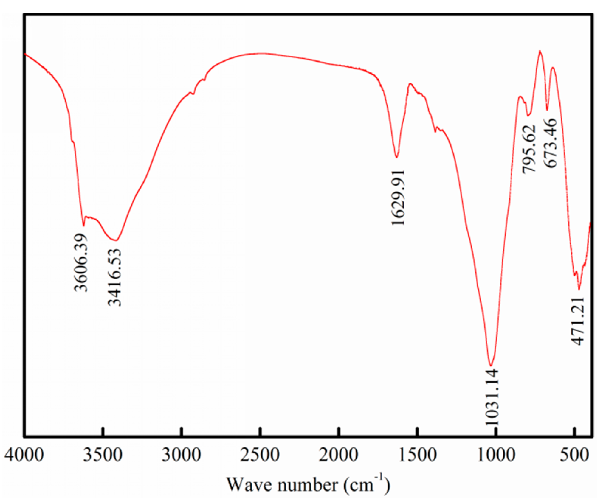
Pure chrysocolla ((Cu, Al)2H2Si2O5(OH)4·nH2O) was collected from Dongchuan, Yunnan Province, China, was crushed to 45–74 μm using a hammer. Then, it was pulverized in an agate mortar for several minutes and dry screened using a Tyler sieve. Due to chrysocolla’s varying crystallo-chemical structure, it is difficult to qualitatively analyze chrysocolla. A Fourier Transform Infrared Spectrometer (FTIR) was used to identity its composition, the results of which are presented in Figure 1.
Figure 1 shows the H–O–H stretching vibrations and the bending vibrations of bound water in chrysocolla at 3416.53 cm−1 and 1629.91 cm−1. The Si–O stretching vibration absorption region is seen at 1031.14 cm−1, which is the typical characteristic peak of silicate minerals. Therefore, this sample is present in an SiO4 framework. The characteristic vibration peak of Si–O–Si in quartz appears at 795.62 cm−1, and a strong absorption region is seen at 471.21 cm−1 due to the Si–O bending vibration and the lattice vibration. The FTIR analysis indicates that this sample exhibits silicate mineral characteristics, which is consistent with the results of other authors [20]. In addition, the chemical analysis of chrysocolla (Table 1) reveals that the sample is composed of 31.02% Cu, 5. 77% Al2O3, and 40.57% SiO2. This combined with the FTIR analysis reveals that the sample used has a high degree of purity.
Later, some of the chrysocolla particles 45–74 μm in size and 13 μm in size were used for BET and transmission electron microscopy (TEM) analyses, respectively. Particle in the size range of 45–74 μm were used in the flotation tests, inductively coupled plasma-atomic emission spectrometry (ICP-AES) and X-ray photoelectron spectroscopy (XPS) analyses. Particles less than 5 μm in size were prepared for zeta potential analysis.
The EDP used as the activator was a white solid analytical grade powder. Sodium isoamyl xanthate (NaIX, C6H12OCSSNa, commercial grade) and Na2S·9H2O (purchased from Tianjin Jinhuitaiya Chemical Reagents Co. Ltd., Tianjin, China) were used as the collector and sulfidizing agent, respectively. The other chemicals used in this study were all analytical grade chemicals.
2.2. Micro-Flotation Experiments
The chrysocolla flotation experiments were conducted in a Hallimond tube with a total pulp volume of 50 mL. Prior to flotation, 2.0 g of pure chrysocolla mineral particles was conditioned in deionized water, EDP at the concentration of 2 × 10⁻³ M was added and allowed to activate the chrysocolla for 3 min, followed by the addition of 5 × 10⁻⁴ M and 4 × 10⁻³ M Na2S·9H2O and sulfidization for 10 min. Subsequently, the pulp’s pH was adjusted from 5.09 and 5.22 to 9.0 using sodium hydroxide and hydrochloric acid, respectively, and various concentrations of NaIX were added. Finally, bubbling air flotation occurred for 8 min with N2 as the air supply at a rate of 20 mL/min, which was repeated 3 times for each experiment.
After drying the collected products, the weight distribution of the floated and unfloated minerals was used to calculate the flotation recovery. All of the reagents used for experiment were fresh and all of the flotation experiments were conducted at ambient temperature.
2.3. TEM and BET Measurements
TEM (JEM-2100) and BET were used to investigate the microstructures of the chrysocolla’s surface. The TEM observations were carried out under an accelerating voltage of 200 kV, a column vacuum pressure of 10−5 Pa, and a beam current density of 15 pA/cm2. The BET analyses were conducted using QUADRASORB evo (Quantachrome, Boynton Beach, FL, USA) equipment and 0.2891 g of chrysocolla at less than 1 × 10⁻³ Pa. N2 was used as the adsorbate. The sample was statically adsorbed at a liquid nitrogen saturation temperature of 77.3 K, outgas time was 6 h and outgas temperature was 200 °C. The specific surface area, pore volume, and pore size were calculated using the BET theoretical model and the BJH theoretical formula.
2.4. Adsorption Experiments
The sulfur adsorption experiments on the chrysocolla’s surface after the chrysocolla was treated with EDP solutions as well as when it was not treated were also conducted in the water bath reactor. 2.0 g of chrysocolla particles was conditioned in 20 mL of deionized water with 5 × 10⁻⁴ M and 4 × 10⁻³ M Na2S·9H2O solutions in the absence and presence of EDP, respectively. The activation and sulfidization times were 3 min and 10 min, respectively. Finally, 15 mL of the liquid obtained from the pulp was stored in closed vials. Then, solid-liquid separation was conducted using a filter machine and the obtained solid products were thoroughly rinsed with pure deionized water to eliminate the effect of the weak adsorption of other impurities.
After drying, the solid samples were analyzed using XPS, which was performed using a K-Alpha+ (Thermo fisher Scientific, Waltham, MA, USA) and a monochromatized Al-Kα X-ray source. The operating parameters were as follows: an energy of 1486.6 eV and 4.2 mA × 12 kV, and an optimal energy resolution of <0.5 eV. The analysis chamber’s vacuum pressure was 5 × 10⁻⁹ mbar, and the pass energies of the survey scan and element scan were 100 eV and 30 eV, respectively. The samples were scanned 5 times and the x-ray exposure time was 6 min for each sample. The C1s spectrum at 284.80 eV was used as an internal standard for the charge compensation to calibrate all of the measured spectra.
The 15 mL liquid samples were separated using a centrifuge (9000 r/min), then 3 mL of supernatant was analyzed using ICP-AES (ICPS-1000II, Shimadzu, Japan), and the remaining solids in the solution were dried and weighed using an electronic balance (FA-JY).
2.5. Zeta Potential Measurements
Zeta potential measurements were conducted using a NanoBrook ZetaPlus electrophoretic analyzer (Brookhaven Instruments, Holtsville, NY, USA). One tenth of a gram of chrysocolla was suspended in 100 mL of 2 × 10⁻³ M KCl solution. The pH range investigated was from pH 2 to 12. When necessary, EDP solutions of 2 × 10⁻³ M were added to the suspension at the beginning of the conditioning period. Freshly prepared Na2S·9H2O solutions were added to the mixture. The resultant suspensions in the absence and presence of NaIX were allowed to settle for 10 min, and the supernatant of the dilute fine particle suspension was transferred to the measurement vessel for zeta potential measurements at room temperature. The zeta potential of each sample was measured 3 times.
3. Results and Discussion
3.1. Surface Structure and Properties of Chrysocolla
Mineral surface properties were found to have a direct effect on the flotation recovery. The mineral surface micro-structures not only affected the surface’s hydrophobicity, but also affected the adsorption of the flotation reagents onto the surface. Therefore, we investigated surface micro-structures of the chrysocolla to interpret the relationship between the surface properties and mineral flotation. The results of the BET analyses are shown in Figure 2.
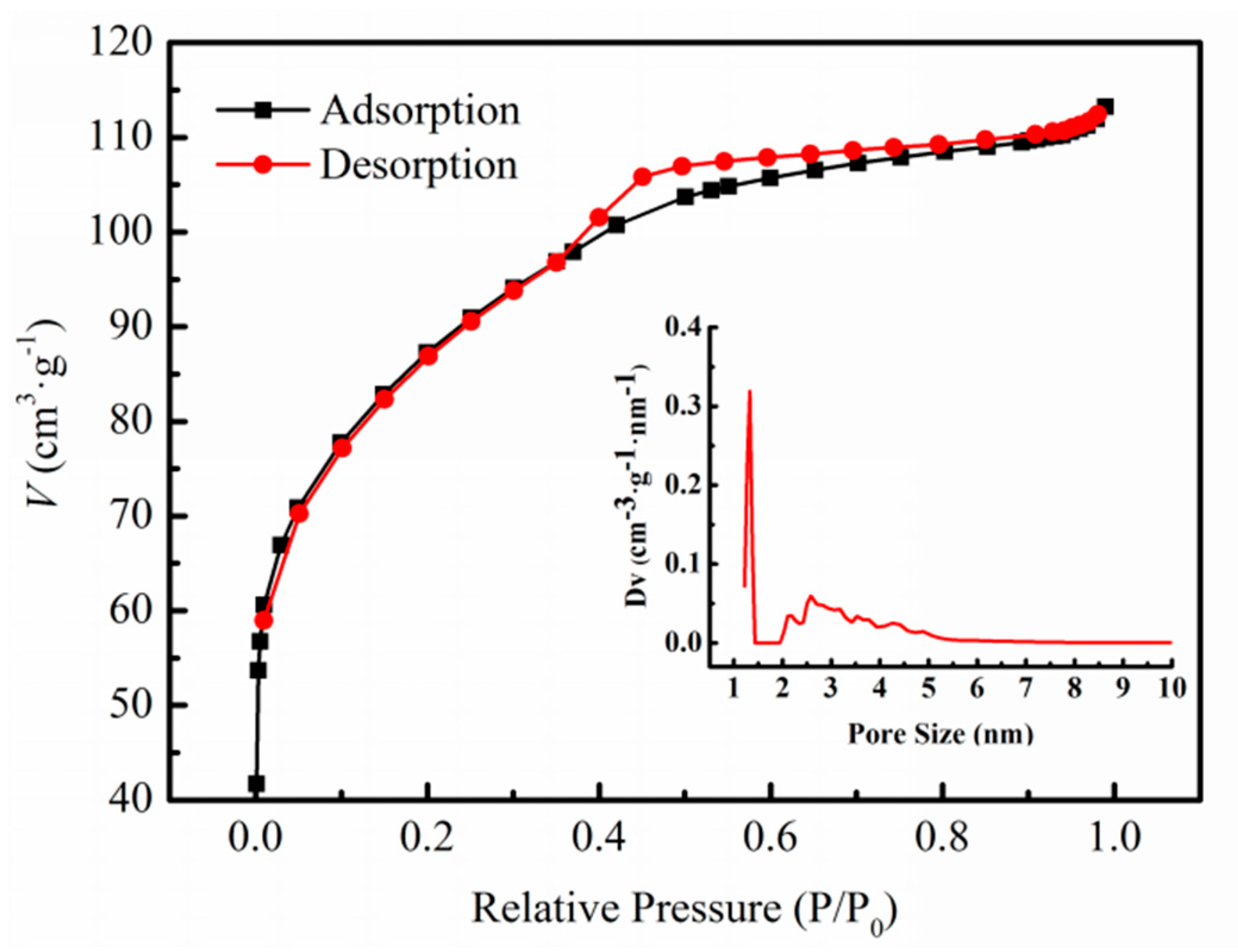
According to the IUPAC classification, the N2 adsorption/desorption isotherms in Figure 2 correspond to Type I. Type I isotherms are mainly characteristic of microporous materials. N2 adsorption/desorption isotherms may also occur when the pore size of the mesoporous material is close to that of the micropores. The chrysocolla isotherms have a hysteresis loop, which is associated with the capillary condensation that occurs in the mesopores. For chrysocolla, the hysteresis loops correspond to Type H2. A Type H2 hysteresis loop is characteristic of solids consisting of particles crossed by nearly cylindrical channels and the pores that are not uniform in size or shape. This is usually attributed to a difference in size between the narrow mouth and the wide body of the pore. We used Equation (1) to calculate the value of the specific surface area and the BJH method to calculate the pore size and pore volume of chrysocolla.
The linearized form of the BET equation is
where p is the nitrogen partial pressure, p0 is the saturated vapor pressure of N2 at the liquid nitrogen temperature, p/p0 is the relative vapor pressure of the adsorbate, v is the volume of the adsorbed gas, vm is the volume of the adsorbed gas in a monolayer, and c is a constant related to the energy of adsorption.
The calculation results are shown in Figure 2 and Table 2, the pore size of the chrysocolla fell mainly within the range of 1–5 nm with one significant peaks near 1.2 nm, indicating that the amount of micropores (<2 nm) existed in the chrysocolla. In addition, the pore size of the chrysocolla within the range of 2–5 nm was mesopores (2–50 nm), the amount of mesopores were second to micropores. Although the mesopores account for the large proportion, their size indicates that they are almost micropore. Therefore, the chrysocolla sample is a porous mineral rich in micropores and with an approximately microporous structure. The data from Table 2 shows that the specific surface area of the chrysocolla particles 45–74 μm in size was 242.51 m2·g−1 and that of malachite particles in same size was 0.363 m2·g−1. This indicates that chrysocolla has a larger specific surface area than malachite, which may be the reason that enormous amounts of EDP, sodium sulfide, and xanthate are needed to float chrysocolla compared to that required to float malachite [21,22].
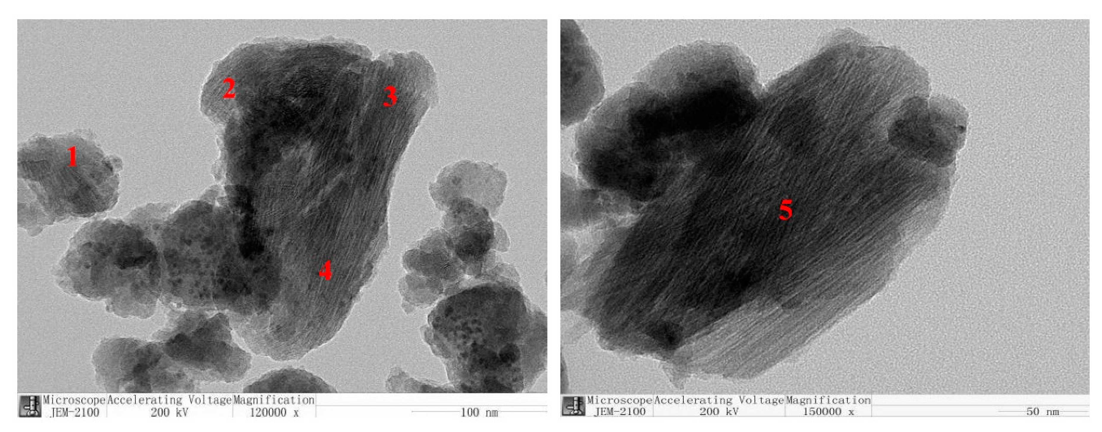
When using analytical equipment for N2 adsorption/desorption experiments, false peaks may be formed, leading to analytical errors. Therefore, based on the BET results, we conducted a more intuitive TEM measurement of the chrysocolla particles, the results of which are shown in Figure 3. As shown in Figure 3, many pores were observed within the chrysocolla at sites 1, 2, 3, 4, and 5. As a result, the BET results for chrysocolla were corrected, proving that chrysocolla is a porous mineral with a large specific surface area.
3.2. Effect of EDP on Sulfidization Flotation of Chrysocolla
The flotation behavior of chrysocolla sulfidized with Na2S·9H2O solution in the absence and presence of EDP is plotted as a function of the NaIX concentrations in Figure 4. Sodium sulfide plays an important role in the flotation of oxidized ores by forming a sulfide metal film on the mineral’s surface and enhancing the binding ability between the mineral’s surface and the xanthate collectors, increasing the mineral surface’s hydrophobicity and increasing flotation recovery. This is the same sulfidizing mechanism used for chrysocolla. In Figure 4, the flotation recovery of chrysocolla increased with increasing NaIX concentrations in the absence of EDP. However, whether chrysocolla was sulfidized using high concentration of Na2S or low concentration of Na2S solution the flotation recovery are very low, even with enormous amounts of xanthate. This may be attributed to the lower surface activity of chrysocolla. The sulfide ions do not have enough energy to force the chemical bonds of the Cu-SiO3 in the chrysocolla to break, making it difficult to form a sufficiently stable copper sulfide film, i.e., xanthate cannot adsorb well on the chrysocolla surfaces. Therefore, EDP was added to modify the properties of the chrysocolla surfaces.
The flotation recovery of chrysocolla was increased dramatically at the same concentration of Na2S and NaIX when EDP was added, indicating that the addition of EDP prior to sulfidization was beneficial to the floatability of chrysocolla. Under these experimental conditions, the flotation recovery of chrysocolla increased considerably with increasing Na2S·9H2O concentration. Although larger quantities of regulators and collectors were used, this may be an effective means of flotation recovery of chrysocolla using the conventional sulfidization-xanthate method. Treating chrysocolla with EDP prior to sulfidization may result in excellent flotation for two reasons. The first reason is that the EDP has enough energy to force the chemical bonds in the chrysocolla to break and the Cu(II) in the chrysocolla to dissolve into the solution as copper-ammonia species ([Cu(en)2]2+), creating a large number of highly active copper sites exposed on the mineral surfaces, which has been confirmed by Xiaojun Xu [23]. This facilitates the adsorption of the sulfide ion species onto the chrysocolla’s surface, which leads to the formation of more copper sulfide species. The second reason that this method improves the flotation behavior may be that the [Cu(en)2]2+ in the solution counter-adsorbs onto the chrysocolla’s surface when Na2S is added, with a strong chemical reaction taking place during this process. This result was confirmed by XPS and ICP-AES analyses.
3.3. XPS Analysis
Our flotation experiments demonstrate that chrysocolla does not float in the absence of EDP, regardless of the amount of Na2S and xanthate used, while the excellent flotation recovery of chrysocolla was observed in the presence of EDP. This result suggests that a greater change in the surface properties of chrysocolla occurred when it was activated with EDP, which is supported by the XPS analyses.
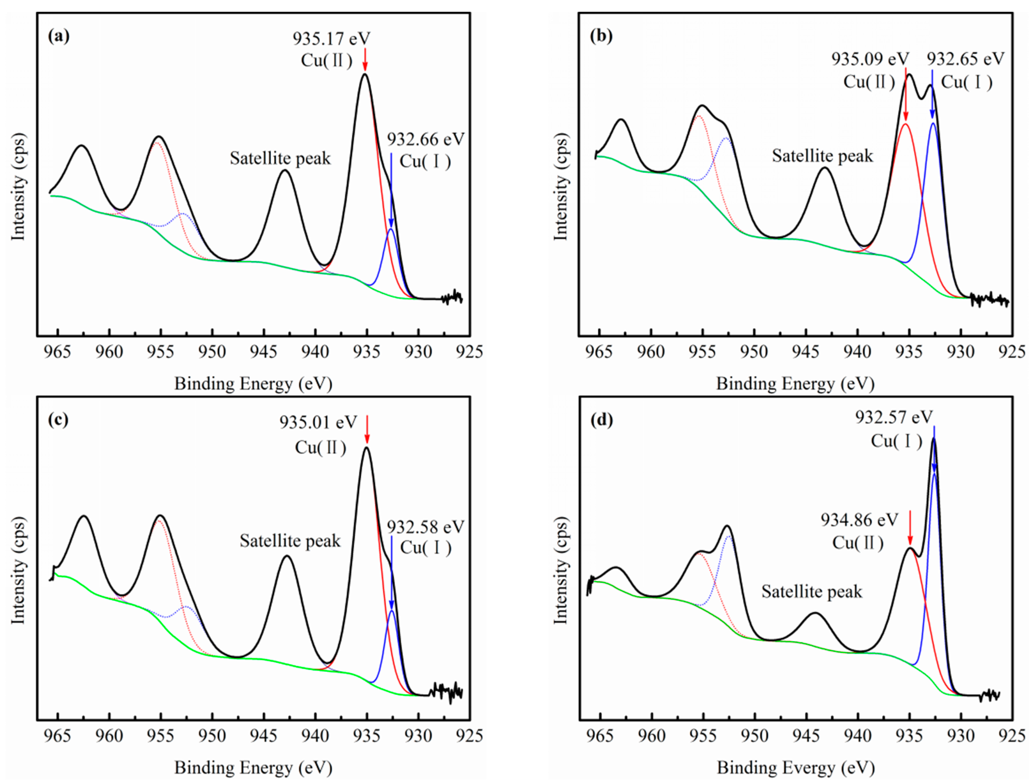
Figure 5 shows the Cu2p spectra of four kinds of chrysocolla samples treated with different conditions. Except for the satellite peaks in the Cu2p spectrum, two pairs of spin–orbit split peaks made up of a Cu2p3/2 and Cu2p1/2 doublet were fitted. The binding energies at 935.17–934.86 eV (cupric) and 932.66–932.57 eV (cuprous) are attributed to Cu2p3/2 [21,24,25,26].
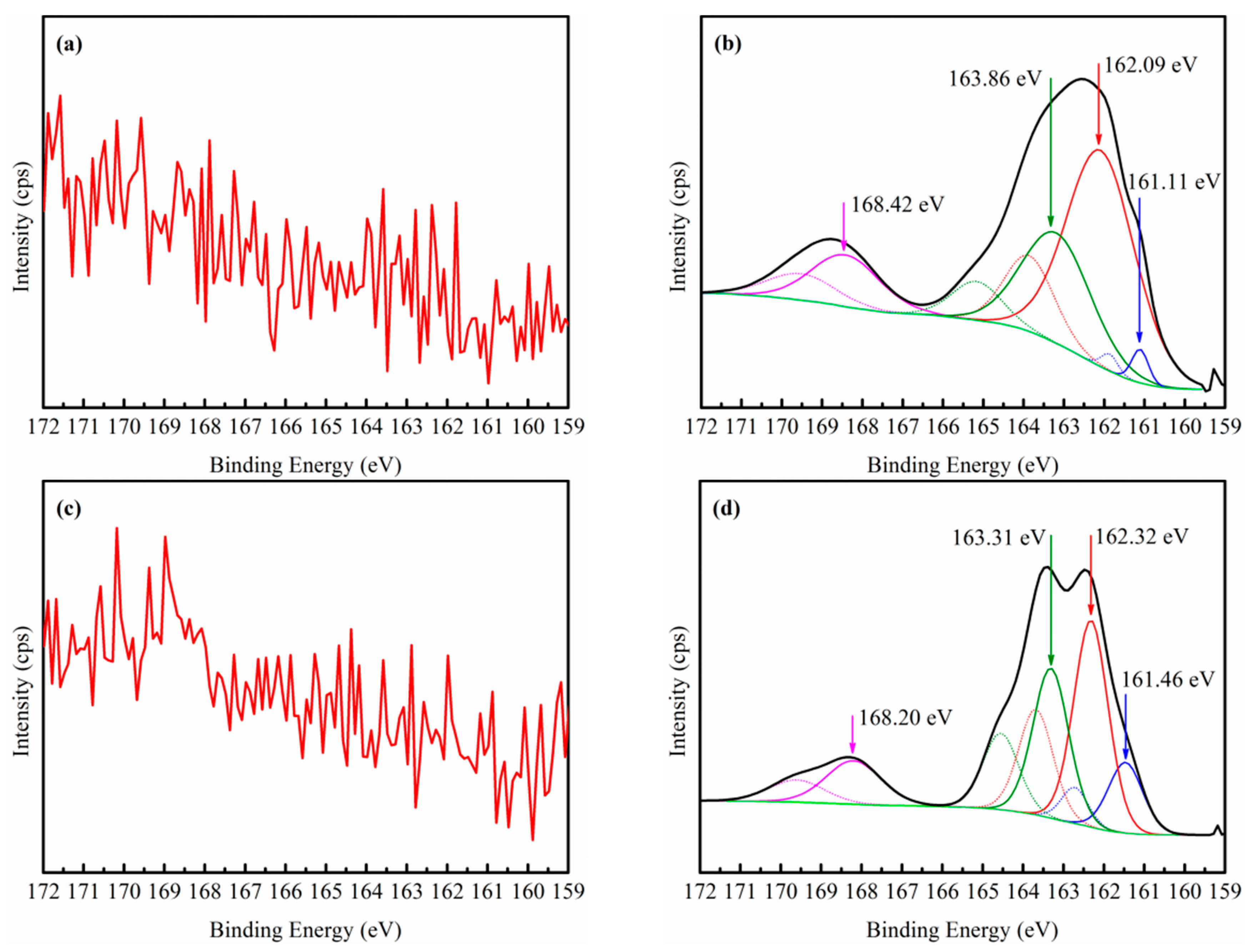
Figure 5a,c show the Cu2p spectra of chrysocolla sulfidized with low and high concentrations of Na2S solution in the absence of EDP. After the chrysocolla was treated with 4 × 10⁻³ M Na2S solution, the binding energies of the Cu(II) and Cu(I) species shifted from 935.17 to 935.01 eV and from 932.66 to 932.58 eV, respectively. This shift to lower binding energies indicates differences in the electronic environment of the two chrysocolla surfaces. As we known, chrysocolla contains only divalent copper ions, while the peak of monovalent copper ions can be seen to occur in Figure 5a,c. These results may be attributed to two reasons. First, the x-ray irradiation the chrysocolla from the analyzing chamber was inevitable, and due to this irradiation, Cu(I) is produced very quickly (few minutes) from Cu(II) [27,28]. This may be the main reason for the appearance of the Cu(I) peaks. Another reason may be the occurrence of a redox reaction between S2− and Cu2+, which would cause the Cu2+ to be reduced to Cu1+. However, the probability of this is very low since the presence of sulfur was not detected on the chrysocolla’s surface (as shown in Figure 6a,c). Therefore, we conclude that the sulfidization of Na2S on the chrysocolla’s surface was very weak in the absence of the EDP solution, which is consistent with the results of the flotation tests.
The Cu2p spectra of chrysocolla treated with EDP prior to sulfidization are shown in Figure 5b,d. As can be seen, the area inside the curve and the peak intensity of the Cu(I) species are obviously larger than those that occur in the absence of EDP. The atomic concentration of Cu(I) was 4.15% and that of Cu(II) was 5.62% (as shown in Table 3b). The atomic concentration of Cu(I) was 7.28% and that of Cu(II) was 6.16% (as shown in Table 3d). Thus, it can be seen that the atomic concentration of Cu(I) increases gradually as the amount of sodium sulfide increases. One possible reason for this is that a strong redox reaction occurred between the [Cu(en)2]2+ and S2− in the solution, causing the Cu(II) in the complex to be reduced to Cu(I), which was then counter-adsorbed onto the chrysocolla’s surface in the form of a new copper-ammonia complex (mainly cuprous), which formed more dense and stable hydrophobic films. The S2p and N1s spectra and the ICP-AES analyses (presented in Section 3.4) also confirm this conclusion. Figure 6b,d show that the two chrysocolla samples were divided into four pairs of spin-orbit split peaks, which were composed of an S2p3/2 and S2p1/2 doublet. The binding energies of the four peaks in Figure 6b,d are 161.11–161.46 eV, 162.09–162.32 eV, 163.86–163.31 eV, and 168.42–168.20 eV and correspond to S2−, S22−, Sn2−, and SO42−, respectively [22,29,30,31]. The change in the valence state of sulfur and the presence of SO42− suggest that a strong redox reaction occurred throughout the pulp system. Therefore, we can infer that the S2− in the solution had two main effects. The first effect is that the S2− underwent a redox reaction with the [Cu(en)2]2+, and the other effect is that it directly bonded to the fresh copper ions on the chrysocolla’s surface to form copper sulfide, and the S2− detected by the XPS is caused by this effect.
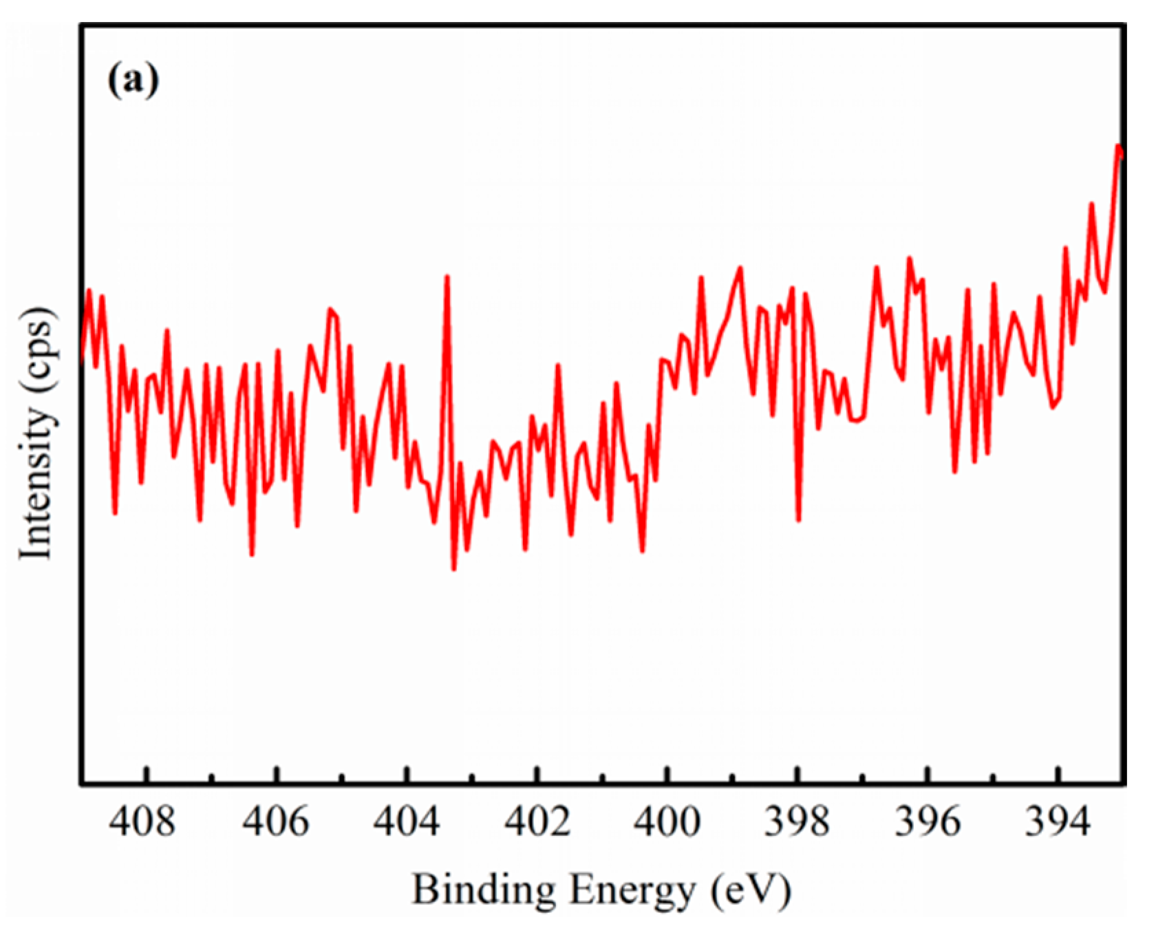
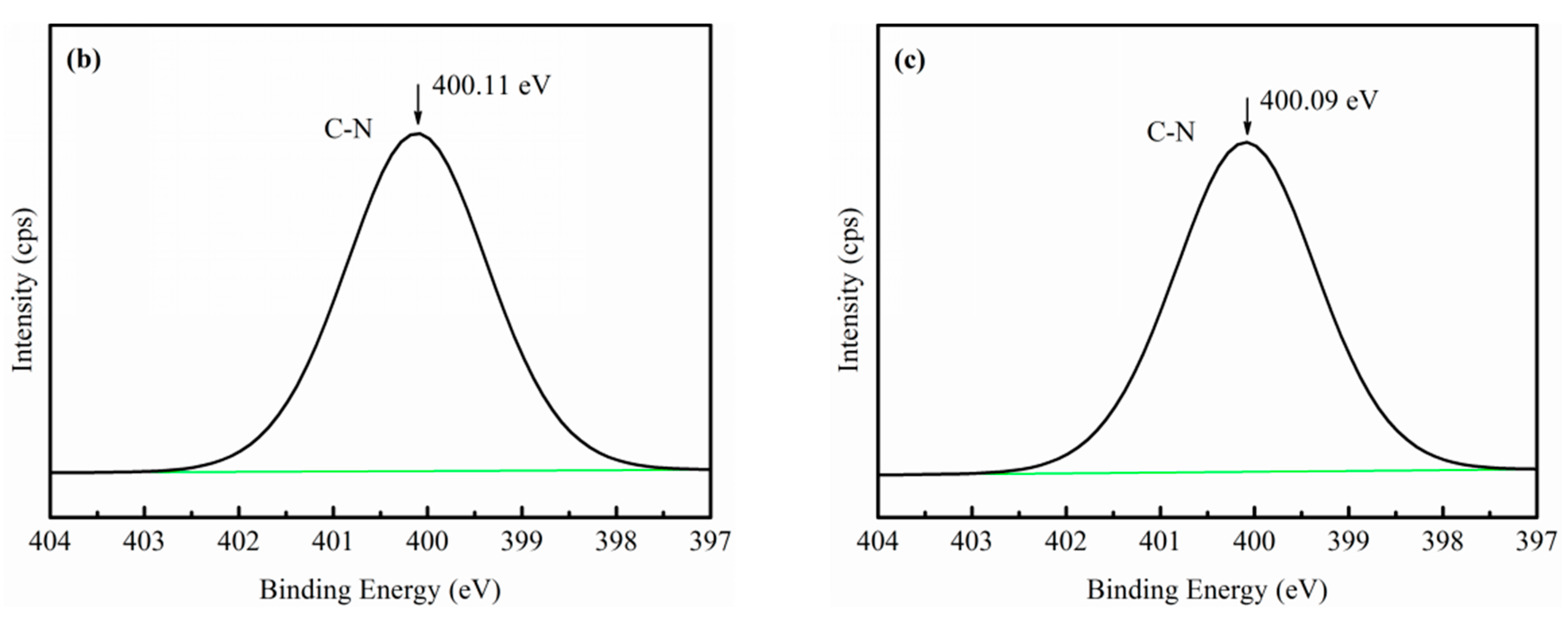
Figure 7 is further interpreted as the effect of EDP on the adsorption of sulfide ion species onto the chrysocolla’s surface. As shown in Figure 7, the N1s spectra of the three chrysocolla samples treated with different conditions. Figure 7a shows that N is not present on the chrysocolla’s surface when the chrysocolla is only activated with the EDP solution, indicating that the EDP has only a micro-dissolution effect on the chrysocolla’s surface and has not been adsorbed onto its surface in any form (shown by the ICP-AES analysis). While Figure 7b,c show the presence of N (C–N) on the chrysocolla’s surface, indicating that the EDP bonded to the copper ions adsorbed onto the chrysocolla’s surface when the Na2S and EDP solutions were both present, which the evidences for [Cu(en)2]2+ counter-adsorbed onto chrysocolla’s surface in the form of new complex.
3.4. ICP-AES Analysis
The flotation of chrysocolla was completed in a complex slurry system. Changes in the surface properties of the minerals and changes in various ions in the solution are important factors affecting the flotation of chrysocolla. The XPS analysis described in the previous section indicates that the presence of EDP increases the adsorption of sulfur and Cu(I) onto the chrysocolla’s surface and forms a dense hydrophobic film. This section focuses on variations in the concentrations of the ions in the solution and shows that EDP activates chrysocolla effectively.
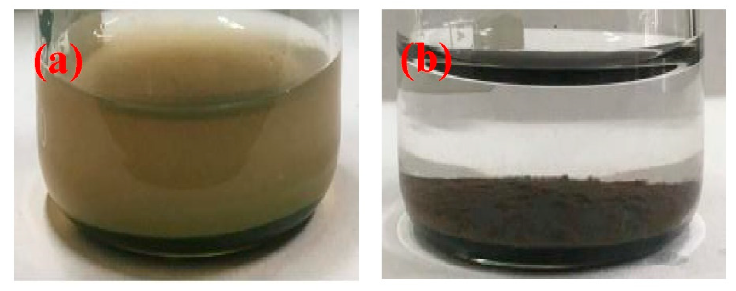
In this section, four conditions (shown in Table 4) were selected for study. As shown in Table 4, after the chrysocolla is conditioned with deionized water and the EDP solution for 13 min, only copper ions exist in the solution and the copper ion concentration of the solution is about three times that of the deionized water, indicating that the addition of EDP promoted the dissolution of the copper ions on the chrysocolla’s surface. This is the strong evidence for the fact that the EDP will react with the copper ions and enter the solution. When only 4 × 10⁻³ M Na2S·9H2O was added to the solution, a large amount (93.33 mg/L) of black copper sulfide colloids (as shown in Figure 8a) appeared and was suspended in the pulp. Some ionic sulfur (25.42 mg/L) was still present in the solution. This may be due to the excessive amounts of sodium sulfide added, resulting in excess sulfide ions remaining. When both 2 × 10⁻³ M EDP and 4 × 10⁻³ M Na2S·9H2O were added, after conditioning, the pulp solution is clear and the black copper sulfide colloid disappears (as shown in Figure 8b). The colloid in the solution was not observed, the remaining sulfide ion and copper ion concentrations were 9.63 mg/L and 0.38 mg/L, respectively, indicating that both the dissolved copper ions and the added sulfide ions were transferred to the chrysocolla’s surface. Therefore, both the intuitive representation of the experimental phenomena and the analysis of the experimental data indicate that the results of the XPS analysis are correct, i.e., the EDP added first reacts with the copper ions on the chrysocolla’s surface, and then, it enters the solution as copper ammine complex ions, causing the fresh chrysocolla surface was exposed. After the Na2S·9H2O was added, some of the S2− directly bonded to the copper ions on the fresh surface of the chrysocolla to form a sulfide metal film, while the rest of the S2− participated in a redox reaction with the copper ammonium complex ions in the solution causing the copper ions in the solution to counter-adsorb onto the chrysocolla’s surface in the form of a new complex (mainly monovalent copper), which facilitated the adsorption of the xanthate and enhanced the hydrophobic properties of the chrysocolla’s surface.
3.5. Zeta Potential Measurements
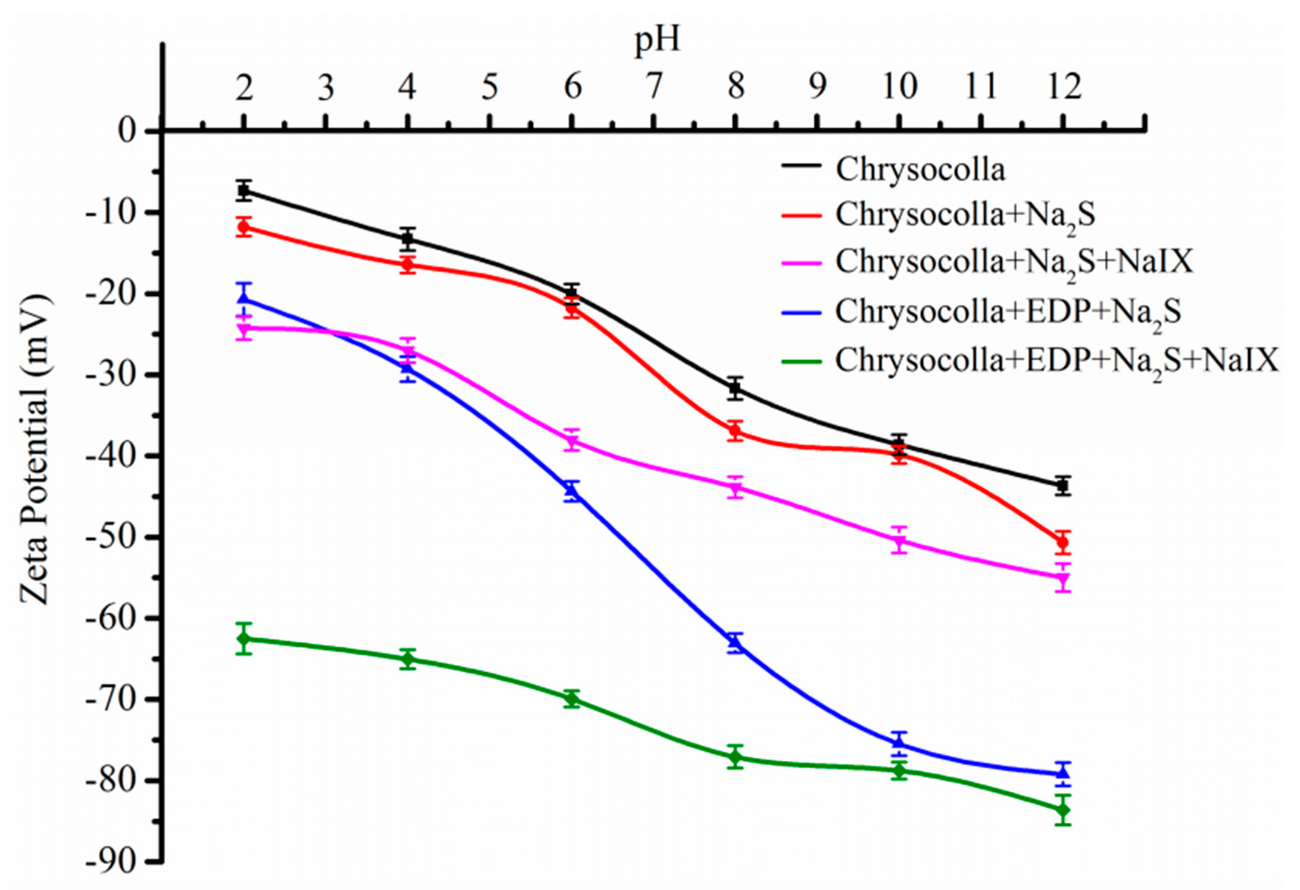
The adsorption of sulfide ion and xanthate onto the mineral’s surface may influence the zeta potential of the dispersed chrysocolla particles. The zeta potential measurements were performed to estimate the interaction between the mineral particles and the reagents in the flotation experiments. This study investigated the effects of flotation regents on changes in the chrysocolla’s surface, the results of which are presented in Figure 9. As shown in Figure 9, the zeta potential decrease with increasing pH for pH of 2 to 12, i.e., chrysocolla in water has no isoelectric point (IEP) across the pH range investigated. This result is consistent with the study of Gonzalez and Soto [19]; however, other authors have reported an IEP for chrysocolla at a pH of 2 and at a pH of 6.5 [32,33]. These different results may be due to excessive conditioning prior to analysis, which would lead to more copper ions being dissolved into the pulp solution, while the surface of the chrysocolla became more negatively charged. Another possible explanation for the different IEP results may be differences in the compositions of minerals from different areas. Chrysocolla has a greater specific surface area, higher solubility, and no fixed composition, which has a significant effect on the zeta potential. Therefore, it is normal for different IEPs to be measured by different authors. For the chrysocolla’s surface was treated with the same concentration of Na2S·9H2O in the absence and presence of EDP, it can be seen that only the addition of sodium sulfide had even a small effect on the zeta potential of the chrysocolla. This suggests that the sulfide ions species were not adsorbing onto the surface of the chrysocolla, while the negative charge of the chrysocolla’s surface decreased dramatically in the presence of EDP, and the zeta potential of the EDP-activated chrysocolla was more negative than that of the original chrysocolla. This result indicates that in the presence of EDP, larger amounts of sulfide ion species in the pulp solution were adsorbed onto the chrysocolla’s surface forming copper sulfide species than in the absence of it.
The adsorption of a collector onto the chrysocolla’s surface ultimately depends on the higher activity Cu sites on the mineral’s surface. Thus, the zeta potentials of chrysocolla in the absence and presence of EDP were used to determine the adsorption states of the xanthate species onto the sulfidized chrysocolla surfaces. The data presented in Figure 9 shows that the addition of NaIX caused the zeta potential of the sulfidized chrysocolla to decrease regardless of the presence of EDP, suggesting that the negatively charged xanthate species were adsorbed onto the sulfidized chrysocolla surfaces. Compared to the shift in the zeta potential that occurred in the presence of EDP, the shift in the zeta potential observed for sulfidized chrysocolla without EDP was minor. This indicates that EDP enhances the ability of sodium sulfide to adsorb onto the chrysocolla surface, which leads to an increase in the number of active Cu sites on the chrysocolla’s surface. It also indicates that chrysocolla is not easily sulfidized without EDP.
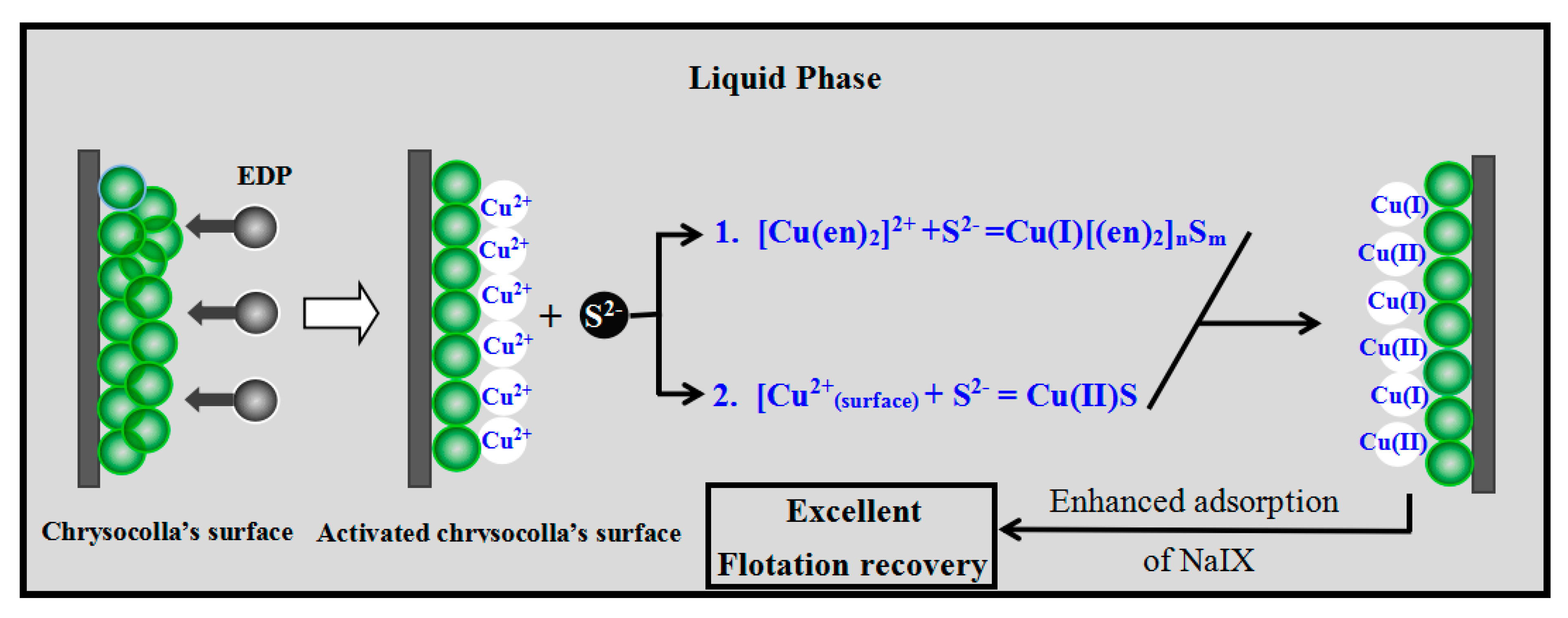
Chrysocolla was activated after modification with EDP, which facilitated the adsorption of sulfide ions and xanthate species onto the mineral’s surface, thereby improving the flotation recovery of chrysocolla. Based on these findings, a speculative model (Figure 10) is presented to describe the activation process and flotation behavior.
4. Conclusions
This study investigated the flotation behavior of chrysocolla in the absence and presence of EDP during sulfidization-xanthate flotation. The primary conclusions of the study are as follows:
- (1)
- TEM and BET analyses indicate that the chrysocolla’s surface is porous and has a large specific surface area, which may be an important reason large amounts of reagents are consumed when better flotation recovery is obtained for chrysocolla.
- (2)
- EDP has a positive effect on the sulfidization flotation of chrysocolla. Chrysocolla was not floated with Na2S and xanthate in the absence of EDP, while excellent flotation recovery was achieved when chrysocolla was activated with EDP prior to sulfidization using xanthate as a collector.
- (3)
- EDP enhanced the sulfidization-flotation of chrysocolla for two reasons. First, the low-activity Cu sites on the chrysocolla’s surface were dissolved into the pulp solution as copper-ammonia complex ions after the amount of EDP was increased, resulting in more high-activity Cu sites being exposed on the chrysocolla’s surface, which improved the sulfidization reaction on its surface. Second, a redox reaction occurred between the S2− and [Cu(en)2]2+ ions, causing the copper ions in the solution to counter-adsorb onto the chrysocolla’s surface as a new complex, and during this process, the Cu(II) was reduced to Cu(I) and the main sulfidization products were S22−, Sn2−, and SO42−. In conclusion, our results indicate that a denser hydrophobic film were formed on the chrysocolla’s surface, improving the flotation behavior of chrysocolla.
Author Contributions
P.S. and D.L. conceived and designed the experiments; P.S. and X.X. performed the experiments; X.J., X.Z., D.L. and R.L. analyzed the data; P.S. wrote this paper and D.L. corrected it.
Acknowledgments
The authors would like to sincerely thank the National Natural Science Foundation of China (Grant No. 51504107 and Grant No. 51264019), National Basic Research Program of China (973 Program, Grant No. 2014CB643404), and the key Project of the National twelfth-five Year Research Program of China (Grant No. 2012BAB10B09) for financial support.
Conflicts of Interest
The authors declare no conflict of interest.
References
- Raghavan, S.; Fuerstenau, D.W. Characterization and pore structure analysis of a copper ore containing chrysocolla. Int. J. Miner. Process. 1997, 4, 381–394. [Google Scholar] [CrossRef]
- Frost, R.L.; Xi, Y.F. Is chrysocolla (Cu, Al)2H2Si2O5(OH)4·nH2O related to spertiniite Cu(OH)2?—Avibrational spectroscopic study. Vib. Spectrosc. 2013, 64, 33–38. [Google Scholar] [CrossRef] [Green Version]
- Bideaux, A.; Nichols, B. Handbook of Mineralogy, Part 1; Mineral Data Publishing: Tucson, AZ, USA, 1995. [Google Scholar]
- Frost, R.L.; Xi, Y.F.; Wood, B.J. Thermogravimetric analysis, PXRD, EDX and XPS study of chrysocolla (Cu, Al)2H2Si2O5(OH)4·nH2O-structural implications. Thermochim. Acta 2012, 545, 157–162. [Google Scholar] [CrossRef] [Green Version]
- Feng, Q.C.; Wen, S.M.; Zhao, W.J.; Lu, C.; Bai, X. Leaching of copper from malachite with methane-sulfonic acid. Solvent. Extr. Res. Dev. 2015, 22, 159–168. [Google Scholar] [CrossRef]
- Habashi, F.; Dugdale, R. Leaching studies on chrysocolla. Trans. AIME 1973, 254, 98–102. [Google Scholar]
- Tanda, B.C.; Eksteen, J.J.; Oraby, E.A. An investigation into the leaching behaviour of copper oxide minerals in aqueous alkaline glycine solutions. Hydrometallurgy 2017, 167, 153–162. [Google Scholar] [CrossRef]
- Jain, N.; Sharma, D. Biohydrometallurgy for nonsulfidic minerals—A review. Geomicrobiol. J. 2004, 21, 135–144. [Google Scholar] [CrossRef]
- Lambert, F.; Gaydardzhiev, S.; Léonard, G.; Lewis, G.; Bareel, P.F.; Bastin, D. Copper leaching from waste electric cables by biohydrometallurgy. Miner. Eng. 2015, 76, 38–46. [Google Scholar] [CrossRef]
- Hu, K.J.; Wu, A.X.; Wang, H.J.; Wang, S.Y. A new heterotrophic strain for bioleaching of low grade complex copper ore. Minerals 2016, 6, 12. [Google Scholar] [CrossRef]
- Gonzalez, A.C.; Gonzalez, G.; Loskowski, J. The effect of ageing on the flotation of chrysocolla. Miner. Process. Extr. M. 1975, 84, C154. [Google Scholar]
- Aplan, F.F.; Fuerstenau, D.W. The flotation of chrysocolla by mercaptan. Int. J. Miner. Process. 1984, 13, 105–115. [Google Scholar] [CrossRef]
- Fuerstenau, D.W.; Herrera-Urbina, R.; McGlashan, D.W. Studies on the applicability of chelating agents as universal collectors for copper minerals. Int. J. Miner. Process. 2000, 58, 15–33. [Google Scholar] [CrossRef]
- Hope, G.A.; Numprasanthai, A.; Buckley, A.N.; Parker, G.K.; Sheldon, G. Bench-scale flotation of chrysocolla with n-octanohydroxamate. Miner. Eng. 2012, 36–38, 12–20. [Google Scholar] [CrossRef]
- Hope, G.A.; Buckley, A.N.; Parker, G.K.; Numprasanthai, A.; Woods, R.; McLean, J. The interaction of n-octanohydroxamate with chrysocolla and oxide copper surfaces. Miner. Eng. 2012, 36–38, 2–11. [Google Scholar] [CrossRef]
- Castro, S.; Soto, H.; Goldfarb, J.; Laskowski, J. Sulfidizing reactions in the flotation of oxidized copper minerals, II. Role of the adsorption and oxidation of sodium sulphide in the flotation of chrysocolla and malachite. Int. J. Miner. Process. 1974, 1, 151–161. [Google Scholar] [CrossRef]
- Castro, S.; Gaytan, H.; Goldfarb, J. The stabilizing effect of Na2S on the collector coating of chrysocolla. Int. J. Miner. Process. 1976, 3, 71–82. [Google Scholar] [CrossRef]
- Parks, G.A.; Kovacs, C. Thermal activation of chrysocolla for xanthate flotation. Trans. Soc. Min. Eng. 1966, 235, 349–354. [Google Scholar]
- Gonzalez, G.; Soto, H. The effect of thermal treatment on the flotation of chrysocolla. Int. J. Miner. Process. 1978, 5, 153–162. [Google Scholar] [CrossRef]
- Peng, W.S.; Liu, G.K. Infrared Spectrum of Minerals; Science Press: Beijing, China, 1982. [Google Scholar]
- Feng, Q.C.; Zhao, W.J.; Wen, S.M.; Cao, Q.B. Copper sulfide species formed on malachite surfaces in relation to flotation. Ind. Eng. Chem. 2017, 48, 125–132. [Google Scholar] [CrossRef]
- Feng, Q.C.; Zhao, W.J.; Wen, S.M. Surface modification of malachite with ethanediamine and its effect on sulfidization flotation. Appl. Surf. Sci. 2018, 436, 823–831. [Google Scholar] [CrossRef]
- Xu, X.J. The Theory of Oxidized Mineral Flotation Using Organic Chelating Agents as Activators; Yunnan Science & Technology Press: Kunming, China, 2000. [Google Scholar]
- Chen, X.M.; Peng, Y.J.; Bradshaw, D. The separation of chalcopyrite and chalcocite from pyrite in cleaner flotation after regrinding. Int. J. Miner. Process. 2014, 58, 64–72. [Google Scholar] [CrossRef]
- Li, F.X.; Zhong, H.; Xu, H.F.; Jia, H.; Liu, G.Y. Flotation behavior and adsorption mechanism of a-hydroxyoctyl phosphinic acid to malachite. Miner. Eng. 2015, 71, 188–193. [Google Scholar] [CrossRef]
- Kartio, I.J.; Basilio, C.I.; Yoon, R.H. An XPS study of sphalerite activation by copper. Langmuir 1998, 14, 5274–5278. [Google Scholar] [CrossRef]
- Kartio, I.; Laajalehto, K.; Suoninen, E.; Karthe, S.; Szargan, R. Technique for XPS measurements of volatile adsorbed layers: Application to studies of sulphide flotation. Surf. Interface Anal. 1992, 18, 807–810. [Google Scholar] [CrossRef]
- Skinner, W.M.; Prestidge, C.A.; Smart, R.S.C. Irradiation effects during XPS studies of Cu (II) activation of zinc sulphide. Surf. Interface Anal. 1996, 24, 620–626. [Google Scholar] [CrossRef]
- Smart, R.S.C.; Skinner, W.M.; Gerson, A.R. XPS of sulphide mineral surfaces: Metal-deficient, polysulphides, defects and elemental sulphur. Surf. Interface Anal. 1999, 28, 101–105. [Google Scholar] [CrossRef]
- Buckley, A.; Woods, R. An X-ray photoelectron spectroscopic study of the oxidation of chalcopyrite. Aust. J. Chem. 1984, 37, 2403–2413. [Google Scholar] [CrossRef]
- Feng, Q.C.; Wen, S.M.; Deng, J.S.; Zhao, W.J. Combined DFT and XPS investigation of enhanced adsorption of sulfide species onto cerussite by surface modification with chloride. Appl. Surf. Sci. 2017, 425, 8–15. [Google Scholar] [CrossRef]
- Xie, G.Y.; Zhang, M.X.; Bian, B.X.; Fan, M.Q. Xuankuangxue; China University of Mining and Technology Press: Xuzhou, China, 2012. [Google Scholar]
- Gonzalez, G.; Laskowski, J. The point of zero charge of oxidized copper minerals: Tenorite, malachite and chrysocolla. Electroanal. Chem. 1974, 53, 452–456. [Google Scholar] [CrossRef]
Figure 1.
FTIR pattern of pure chrysocolla.

Figure 2.
N2 adsorption/desorption isotherms and pore size distribution of chrysocolla.

Figure 3.
TEM images showing the porous structure of chrysocolla.

Figure 4.
The flotation behavior of chrysocolla sulfidized with Na2S·9H2O solution in the absence and presence of EDP.
Figure 4.
The flotation behavior of chrysocolla sulfidized with Na2S·9H2O solution in the absence and presence of EDP.
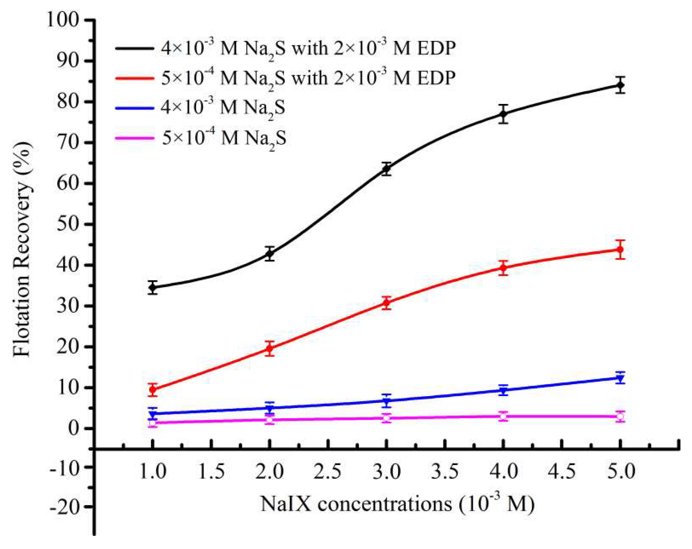
Figure 5.
Cu2p XPS spectra of (a) chrysocolla sulfidized with 5 × 10⁻⁴ M Na2S, (b) EDP-activated chrysocolla sulfidized with 5 × 10⁻⁴ M Na2S, (c) chrysocolla sulfidized with 4 × 10⁻³ M Na2S, and (d) EDP-activated chrysocolla sulfidized with 4 × 10⁻³ M Na2S.
Figure 5.
Cu2p XPS spectra of (a) chrysocolla sulfidized with 5 × 10⁻⁴ M Na2S, (b) EDP-activated chrysocolla sulfidized with 5 × 10⁻⁴ M Na2S, (c) chrysocolla sulfidized with 4 × 10⁻³ M Na2S, and (d) EDP-activated chrysocolla sulfidized with 4 × 10⁻³ M Na2S.

Figure 6.
S2p XPS spectra of (a) chrysocolla sulfidized with 5 × 10⁻⁴ M Na2S, (b) EDP-activated chrysocolla sulfidized with 5 × 10⁻⁴ M Na2S, (c) chrysocolla sulfidized with 4 × 10⁻³ M Na2S, and (d) EDP-activated chrysocolla sulfidized with 4 × 10⁻³ M Na2S.
Figure 6.
S2p XPS spectra of (a) chrysocolla sulfidized with 5 × 10⁻⁴ M Na2S, (b) EDP-activated chrysocolla sulfidized with 5 × 10⁻⁴ M Na2S, (c) chrysocolla sulfidized with 4 × 10⁻³ M Na2S, and (d) EDP-activated chrysocolla sulfidized with 4 × 10⁻³ M Na2S.

Figure 7.
The N1s XPS spectra of (a) chrysocolla activated with 2 × 10⁻³ M EDP, (b) EDP-activated chrysocolla sulfidized with 5 × 10⁻⁴ M Na2S, and (c) EDP-activated chrysocolla sulfidized with 4 × 10⁻³ M Na2S.
Figure 7.
The N1s XPS spectra of (a) chrysocolla activated with 2 × 10⁻³ M EDP, (b) EDP-activated chrysocolla sulfidized with 5 × 10⁻⁴ M Na2S, and (c) EDP-activated chrysocolla sulfidized with 4 × 10⁻³ M Na2S.


Figure 8.
Chrysocolla treated with (a) 4 × 10⁻³ M Na2S and (b) 2 × 10⁻³ M EDP + 4 × 10⁻³ M Na2S.

Figure 9.
Zeta potential of chrysocolla was treated with multiple reagents.

Figure 10.
The proposed activation model of EDP on the sulfidization xanthate flotation of chrysocolla.
Figure 10.
The proposed activation model of EDP on the sulfidization xanthate flotation of chrysocolla.


Table 1.
The chemical composition of chrysocolla.
| Element/Oxide | Cu | Fe | Mn | Al2O3 | MgO | CaO | SiO2 |
|---|---|---|---|---|---|---|---|
| wt (%) | 31.02 | 0.25 | <0.005 | 5.77 | 0.17 | 0.25 | 40.57 |
Table 2.
Surface parameters of pure chrysocolla and malachite from BET analysis.
| Minerals | Pore Size (nm) | Pore Volume (cm3/g) | Specific Surface Area (m2/g) | Particle Size (μm) |
|---|---|---|---|---|
| Chrysocolla | 2.26 | 0.1751 | 242.51 | 45–74 |
| Malachite | – | – | 0.363 | 45–74 |
Table 3.
Atomic concentration of the Cu, S, and N species (%) on the chrysocolla’s surface.
| Element | Atomic Concentration of Cu, S, and N Species (%) | |||
|---|---|---|---|---|
| a | b | c | d | |
| Cu(II) | 7.05 | 5.62 | 7.52 | 6.16 |
| Cu(I) | 1.26 | 4.15 | 1.43 | 7.28 |
| S | 0.00 | 6.31 | 0.00 | 8.65 |
| N | – | 1.65 | – | 2.46 |
Table 4.
The colloids, Cu ion concentrations, and S ion concentrations (mg/L) of the solutions.
| Conditions | Colloid | Cu Ions Concentrations | S Ions Concentrations |
|---|---|---|---|
| Chrysocolla + aqueous solution | – | 26.43 | – |
| Chrysocolla + 2 × 10⁻³ M EDP | 0.00 | 75.38 | – |
| Chrysocolla + 4 × 10⁻³ M Na2S | 93.33 | 0.54 | 25.42 |
| Chrysocolla + 2 × 10⁻³ M EDP + 4 × 10⁻³ M Na2S | <0.000 | 0.38 | 9.63 |
© 2018 by the authors. Licensee MDPI, Basel, Switzerland. This article is an open access article distributed under the terms and conditions of the Creative Commons Attribution (CC BY) license (http://creativecommons.org/licenses/by/4.0/).
Share and Cite
MDPI and ACS Style
Shen, P.; Liu, D.; Xu, X.; Jia, X.; Zhang, X.; Liu, D.; Liu, R. Effect of Ethylene Diamine Phosphate on the Sulfidization Flotation of Chrysocolla. Minerals 2018, 8, 216. https://doi.org/10.3390/min8050216
AMA Style
Shen P, Liu D, Xu X, Jia X, Zhang X, Liu D, Liu R. Effect of Ethylene Diamine Phosphate on the Sulfidization Flotation of Chrysocolla. Minerals. 2018; 8(5):216. https://doi.org/10.3390/min8050216
Chicago/Turabian StyleShen, Peilun, Dianwen Liu, Xiaohui Xu, Xiaodong Jia, Xiaolin Zhang, Dan Liu, and Ruizeng Liu. 2018. "Effect of Ethylene Diamine Phosphate on the Sulfidization Flotation of Chrysocolla" Minerals 8, no. 5: 216. https://doi.org/10.3390/min8050216
Note that from the first issue of 2016, this journal uses article numbers instead of page numbers. See further details here.