Article

Fossilization History of Fossil Resin from Jambi Province (Sumatra, Indonesia) Based on Physico-Chemical Studies

by
Beata Naglik
1,
Barbara Kosmowska-Ceranowicz
2,
Lucyna Natkaniec-Nowak
3,
Przemysław Drzewicz
4,
Magdalena Dumańska-Słowik
3,*,
Jakub Matusik
3,
Marian Wagner
3,
Rastislav Milovsky
5,
Paweł Stach
3 and
Arkadiusz Szyszka
3
1
10 Beskidzka Str., 34-312 Międzybrodzie Żywieckie, Poland
2
The Museum of the Earth of the Polish Academy of Sciences in Warsaw, 20/26 Na Skarpie Str., 00-488 Warsaw, Poland
3
Department of Mineralogy, Petrography and Geochemistry, AGH University of Science and Technology, Av. Mickiewicza 30, 30-059 Krakow, Poland
4
The Polish Geological Institute—National Research Institute, 4 Rakowiecka Str., 00-975 Warsaw, Poland
5
Earth Science Institute, Slovak Academy of Sciences, 1 Ďumbierska Str., 974-11 Banská Bystrica, Slovakia
*
Author to whom correspondence should be addressed.
Minerals 2018, 8(3), 95; https://doi.org/10.3390/min8030095
Submission received: 27 December 2017 / Revised: 23 February 2018 / Accepted: 27 February 2018 / Published: 2 March 2018

Abstract

:
A unique specimen of fossil resin originating from the Dipterocarpaceae tree family found in Miocene brown coal deposits in Jambi Province (Sumatra, Indonesia) was investigated via microscopic observations, microhardness testing and infrared and Raman spectroscopic methods. Its form is rare in nature, being an aggregate of three varieties of resin differing in colour, transparency and internal structure. This suggests the formation of the resins at different stages. Further alteration processes, including fossilization and maturation of the resin in a swamp environment resulted in stepwise aromatization of the cyclohexane ring in steroids and cross-linking through formation of ester bonds as well as carbon–carbon bonds between steroid molecules. The various environmental and geological conditions affecting the formation processes of the resins were recorded in their physico-chemical properties. Additionally, heating conditions accelerated by volcanism were proposed as a factor determining the maturation grade of the resin.

1. Introduction

The natural resins (fossil or modern) are caustobioliths, genetically belonging to the group of liptobiolites [1]. Regardless of their provenance or their geological environment and age, they are products of the defence mechanisms of both coniferous and angiosperm trees and were formed millions of years ago. They are found in sedimentary rocks from the Devonian to the Cenozoic; however, they are mostly accumulated within the Palaeogene to Neogene sediments [2]. Resins were very often transported from primary deposits to younger geological layers. In these cases, resin dating is disputable and uncertain. The accumulation of amber in deposits of various ages is related to a period of increased resin production caused by various environmental and geological conditions, for example volcanic activity [3]. Forest fires and pest infestation caused by climate change may also increase resin production. The intense resin exudation from trees was usually part of the self-protection mechanisms of various plants. The chemical and physical properties of fossil resins depend primarily on their botanical source; nevertheless, various environmental and geological conditions occurring during fossilization may also affect the resins [2,4,5,6]. As an example, one can cite redox conditions after burial, which influence the alteration of organic matter [5]. Later transformations are accelerated by temperature and pressure conditions during diagenesis and/or catagenesis; however, the duration of heating is also important [5]. Knowledge of the composition and physical properties of resins allows their botanical affinity to be determined and their evolutionary history to be reconstructed from the time of their formation through their transportation and final fossilization processes.
Preservation of fossil resins depends on how fast they were buried and isolated from oxygen, sunlight and biological conditions, together with other factors causing their biodegradation [2]. For example, sandy sediments allowing water migration are not suitable for organic matter preservation [5]. Therefore, many fossil resins are found within coal deposits formed under reducing, anaerobic conditions, such as the world’s largest fossil resin deposit located on the island of Borneo—in both the Malaysian and Indonesian parts [7]. This deposit is found within coal beds of Miocene age. Similar conditions occurred in coal deposits located in South Sumatra.
Fossil resins from Sumatra were formed from exudates of the Dipterocarpaceae family, belonging to the angiosperm group of trees, particularly from the genera Shorea and Hopea [8,9,10]. According to [11], Sumatran fossil resins may be distinguished from resins from other localities worldwide (Baltic, Dominican and Myanmar resins) based on distinctive inclusion characteristics (numerous round and oval-shaped droplets) and a diagnostic mid-IR spectrum. However, it must be noted that the presence of droplets is not characteristic only of Sumatran resin. They have also been described in Triassic fossil resins from Italy [12] and French Cretaceous resins [13]. The presence of fossil insects and plants in the Sumatran resins has not been detected in previous studies [11]. A detailed description of the internal structures and forms of Sumatran fossils was given in [3]. The FT-IR characteristic of the specimens, as discussed in [14], revealed their similarity to resins of the glessite type, found in the Bitterfeld deposit (Saxony–Anhalt, Eastern Germany) (vide [15,16,17,18]).
The aim of the study was to reconstruct the fossilization history of a fossil resin specimen from the island of Sumatra (Jambi Province) via microscopic observations, microhardness testing and infrared (FT-IR) and Raman spectroscopy (RS). The specimen is a unique aggregate of three different resin varieties distinguishable on the basis of their colour, transparency and internal structure. This allowed us to examine the alteration processes of those resins from the time of their formation to their present-day form.

2. Materials and Methods

2.1. Sampling

The samples of fossil resin originate from the south part of the island of Sumatra, 15 km south of Jambi city (Sarolangun mine). The size of Sumatran resins varies from very small, with a diameter of a few mm, to very large, weighing up to several hundred grams. Larger specimens of Sumatran resin are usually aggregates of three different types of resin (Figure 1). These three varieties of Sumatran resin were individually analysed with stereoscopic and polarizing microscopes, microhardness testing and finally, spectroscopic analysis (FT-IR and RS). Microscopic observations, microhardness measurements and FT-IR analyses were conducted at laboratories at the Faculty of Geology, Geophysics and Environmental Protection, AGH University of Science and Technology, Krakow (Poland) and Slovak Academy of Sciences, Banská Bystrica (Slovakia). Raman spectra were recorded at Tusnovics Instruments SP, Krakow (Poland).

2.2. Microscopic Methods

The internal structures of resins were observed using a stereoscopic microscope SNZ-168, coupled with a digital camera (with 0.75×, 1×, 2×, 3×, 4× and 5× objectives), in conjunction with the Panasis computer program. Standard optical examinations were carried out with an Olympus BX 51 polarizing microscope (Tokyo, Japan). Organic inclusions were observed using a polarizing microscope (Axioplan, Zeiss-Opton, Oberkochen, Germany) in the reflected light mode. For the microscopic observations thin sections were made.

2.3. Microhardness Measurements

Microhardness measurements were performed using the Russian microhardness tester PMT-3. The Vickers hardness test was performed using the mode in which the microhardness HV (kG/mm2 = 9,806,650 Pa) is determined by the ratio F/A, where F is the force applied to the sample by a square-based diamond pyramid of dihedral angle 136° in kilograms-force and A is the lateral area of the resulting indentation in square millimetres. A 250 mg load was used for the measurements. The duration of the pressure of the diamond pyramid on the sample was 10 s. The test was conducted on the smooth surface of all the varieties of the samples, repeating the measurement 30 times in different places on the samples. Microhardness was calculated based on the arithmetic average of the results.

2.4. Spectroscopic Methods

FT-IR analyses were performed using a Thermo Scientific Nicolet 6700 spectrometer (Madison, WI, USA). Spectra were recorded at room temperature in the range 400–4000 cm−1, with a resolution of 4 cm−1. A sample/KBr mixture (3 wt %) was analysed using the DRIFT (Diffuse Reflectance Infrared Fourier Transform) technique with a Praying Mantis diffuse reflection accessory (Harrick, Pleasantville, NY, USA). The DRIFT technique gives a higher signal-to-noise ratio compared to the standard KBr pellet procedure. Obtained spectra were transformed using the Kubelka-Munk function and signal intensity is given in Kubelka-Munk units.
Raman spectra of fossil resins in the range 2000–200 cm−1 were recorded using a Rigaku Progeny ResQ spectrometer with a laser of wavelength 1064 nm. The laser focus diameter was 1–2 μm.

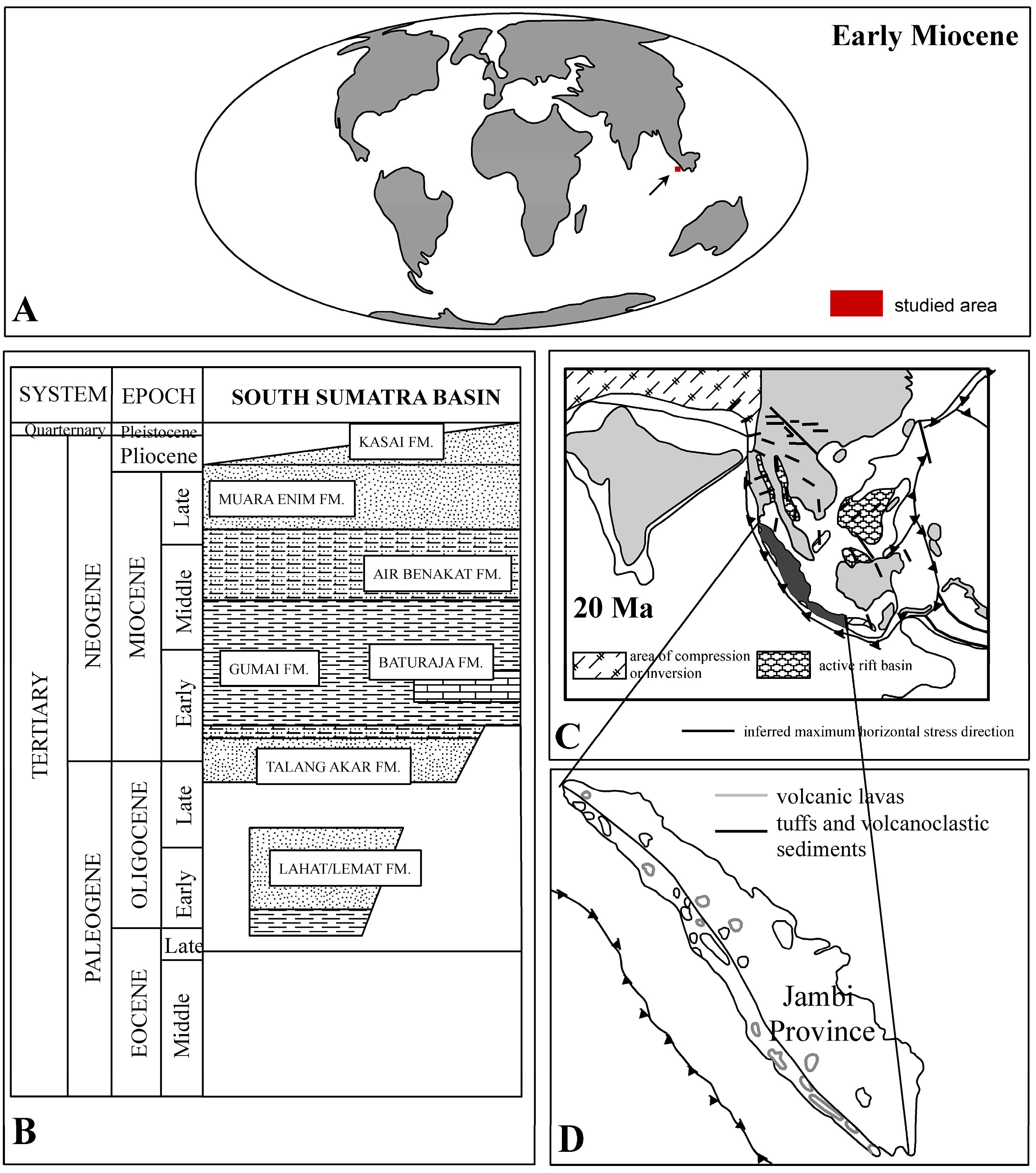
3. Geological Settings

The samples of resins from the dumps of the Sarolangun mine (Figure 2A) were found within coal and lignite beds dating to the early Miocene (20–23 Ma) and belonging to numerous levels of the South Sumatra Basin, including the Talang Akar formation ([19]—vide Figure 2B).
The Talang Akar formation (TAF) represents the sedimentary cycle extending from the late Oligocene to the early Miocene [20]. The TAF succession represents a regressive unit, with shales and sandstones deposited in a fluvio-deltaic environment building the upper part of the formation profile [21]. This unit is overlain by the early and middle Miocene sediments representing transgressive depositional systems. All these formations were uplifted, folded and faulted due to the Pliocene–Pleistocene orogeny [20] associated with volcanic activity (Figure 2C,D). Fossil resins from Jambi Province occur in immature coal layers (lignite beds) [22].
The palaeoenvironment of Sumatra during the Tertiary period had a warm and humid climate. It was a part of southern Sundaland located within ±10° of the equator, with a palaeoclimate favourable for peat formation (Figure 2A) [22].

4. Results

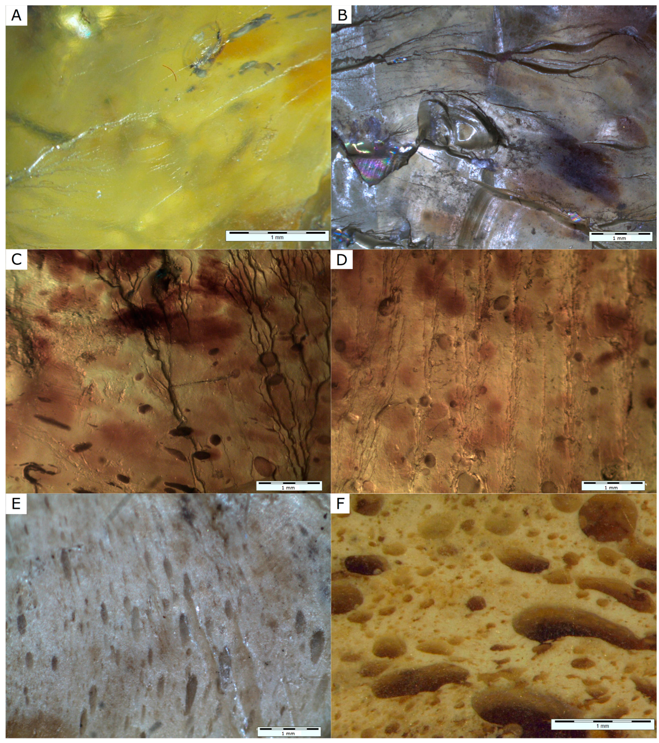
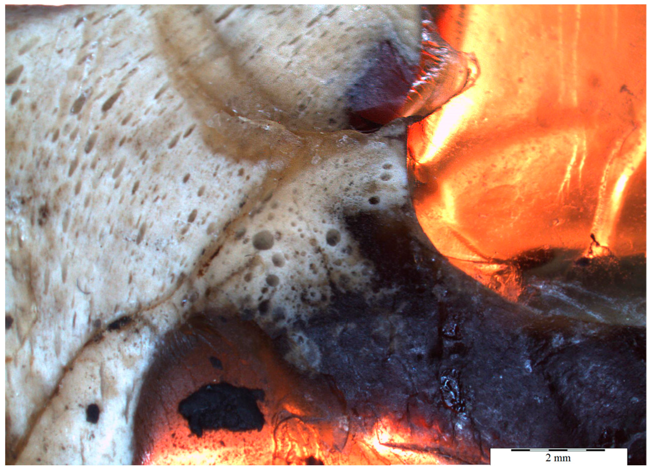
4.1. Physical Investigations

The three varieties within the sample of Sumatran resin (Figure 1, Table 1) were distinguished according to their physical properties, such as colour and transparency.
Under microscopic observation, the three resin varieties differ from each other with regard to their internal structures (Figure 3). Variety I is bright yellow, transparent and relatively clear, with only small amounts of inclusions (Figure 3A,B). Variety II is a dark, translucent resin. Small microdrops with sizes up to 150 μm were observed. Small dark inclusions were probably particles of coalified plant material (Figure 3C,D). Variety III is an opaque resin with a solid foam-like structure containing droplets filled with variety I resin, previously described in [3] as a relic of volcanism (Figure 3E,F). In one sample, all three resin varieties were mixed together; however, the borders between them were distinctly marked (Figure 4).
Under transmitted light, resin variety I showed the presence of coalified plant material with specific morphological shape and opacity. Spherical accumulations of organic material inclusions with preserved envelopes were observed. The inclusions resembled plant microspores or fragments of channels of strongly macerated wood. Resin variety II contained evenly distributed opaque detritus (Figure 5), probably fungus spores or plant pollen, both of which are relatively common in brown coal. This pigment is probably responsible for the darker coloration of this variety. Resin variety III contained evenly distributed microdroplets showing yellow colours with crossed polarizers, probably mineral substances of a lipid type (Figure 6). The presence of numerous elongated, oval microdroplets is responsible for the fluid microtexture of this variety (Figure 3E and Figure 4).
In reflected light, all resin varieties show bright colour with low contrast. Furthermore, the resin varieties differ in their reflectivity. Microscopic observations confirmed the presence of coalified organic material with variable but generally low reflectivity.
In UV light (blue, unpolarized) Sumatran resins emitted variable fluorescence with the intensity depending on the variety. The strongest blue fluorescence was observed for resin variety III (Figure 7A,B), which was due to a sheath of droplets of variety I.
The calculated microhardness values were: 26.297 ± 3.662 (variety I), 24.304 ± 4.780 (variety II) and 24.232 ± 4.631 (variety III) kG/mm2. There was no statistically significant difference between the microhardness measurements of all three specimens; however, variety I showed a slightly higher value than the other varieties.

4.2. Spectroscopic Studies

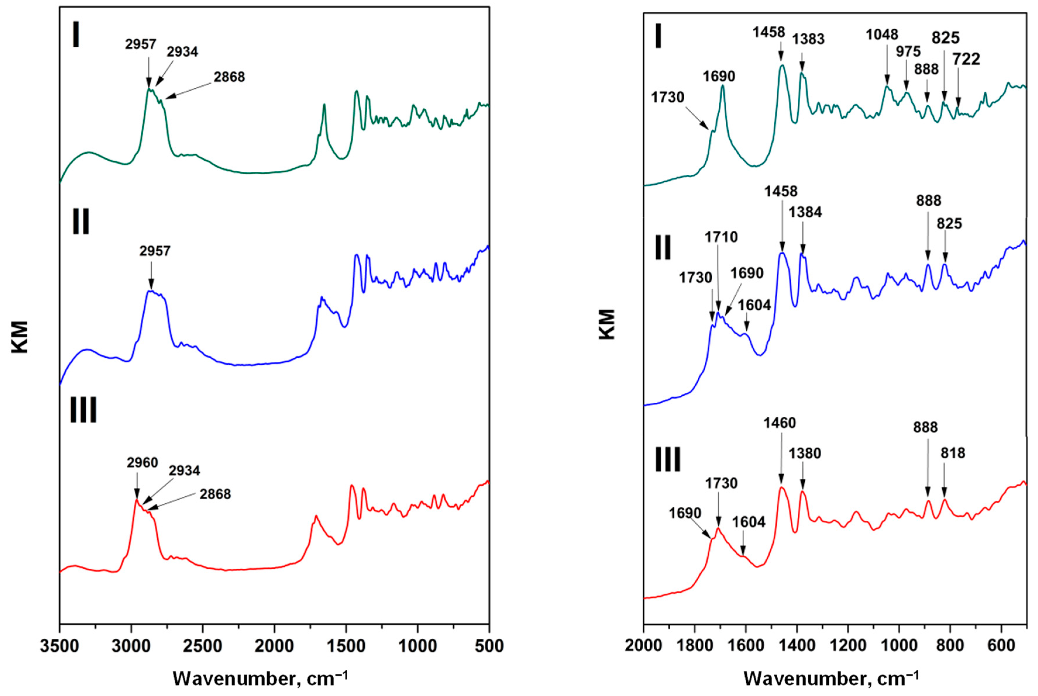
4.2.1. FT-IR Analysis
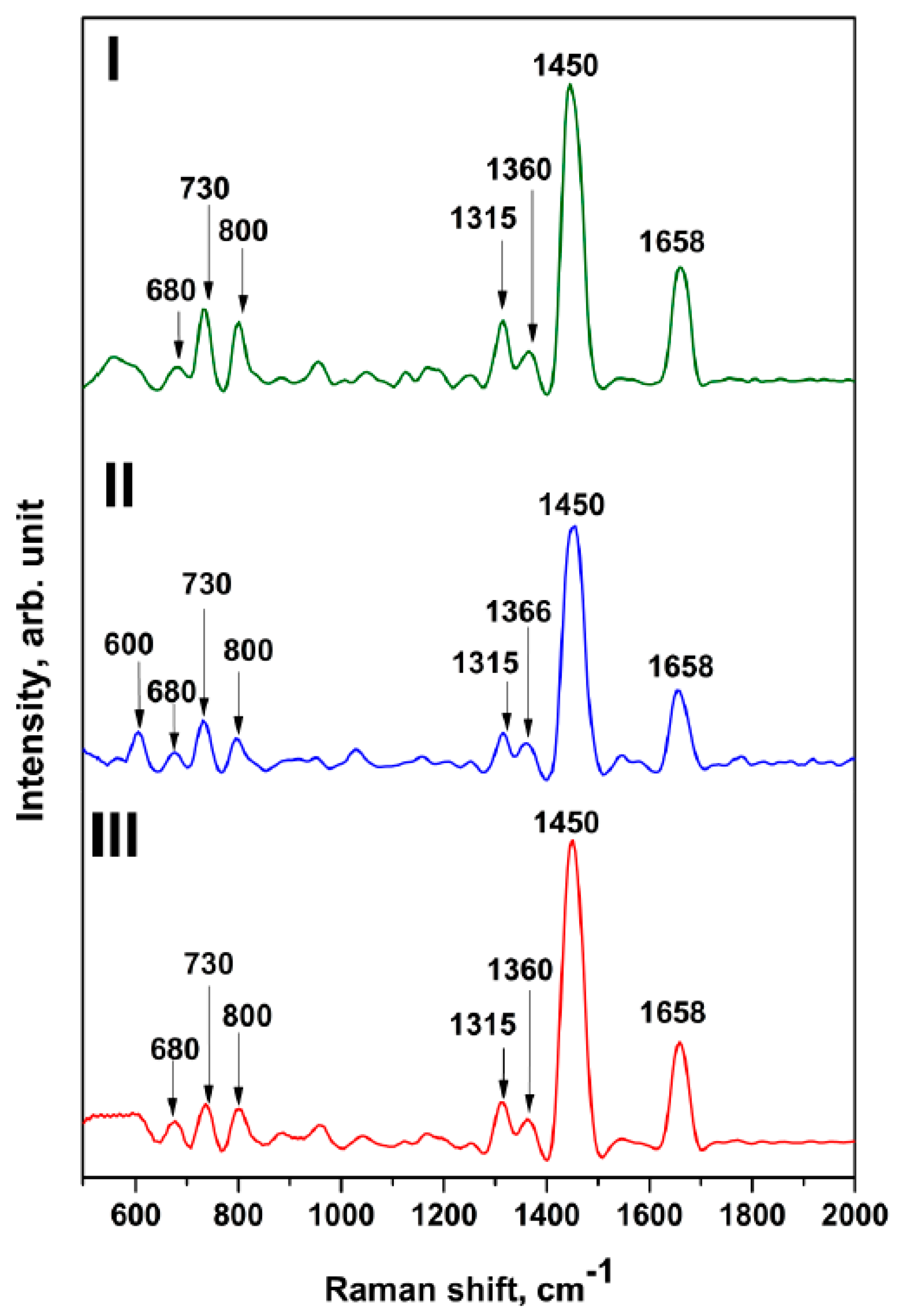
Resins are plant exudates; therefore, adsorption bands characteristic of hydrocarbons are present in their FT-IR spectra (Figure 8, Table 2). In the wavenumber region 3600–3200 cm−1, all spectra show a broad band with a maximum at ~3400 cm−1 corresponding to the OH stretching vibrations in alcohols and carboxylic acids [20,21]. The most intense bands are attributed to C–H stretching and bending vibrations observed in the 3000–2800 and 1500–1350 cm−1 regions respectively. In the region 3000–2800 cm−1, three distinct bands are observed, probably corresponding to the alkyl stretching mode [24,25]; however, their shape differs in the spectra of the investigated samples (Figure 6). The latter region contains bands with maxima at ~1456, ~1384 and ~1367 cm−1 which are exclusively related to CH2 and CH3 bending vibrations [24,25,26]. An important feature of the analysed resins is the presence of absorption bands in the 1800–1600 cm−1 region, corresponding to carbonyl group (C=O) stretching vibrations [24,25,26]. Resin variety I has the most symmetrically shaped band with a maximum at ~1690 cm−1 (Figure 6). The presence of more than one band in this region suggests that the C=O group can be attributed to esters, ketones, carboxylic acids or even lactones. This is clearly observed in the spectra of varieties II and III. A band at ~1730 cm−1 may be assigned to esters [24,25]. An important feature which differentiates variety I from the others is the lack of the ~1600 cm−1 band which is probably related to aromatic compounds. There were no significant differences in the 1350–800 cm−1 region corresponding to vibrations of C–C, C–H and C–O (in esters) bonds. In variety I, the band at ~1050 cm−1 is more visible.

4.2.2. RS Analysis

The Raman spectra of fossil resins from Sumatra show characteristic bands at ~1650 and ~1450 cm−1 (Table 3, Figure 9) which are considered diagnostic for the interpretation of the maturation processes of fossil resins [5,28,29,30]. The ~1650 cm−1 band is related to C=C bonds (unsaturated hydrocarbons), while the ~1450 cm−1 band can probably be assigned to an asymmetric deformation of CH3 and CH2 groups [5]. The band at 1450 cm−1 is higher than that at 1650 cm−1 and the intensity ratio of the 1650/1450 cm−1 bands ranges from 0.39 (variety I) to 0.43 (variety III) and 0.44 (variety II). Additional bands at ~680 cm−1 and ~730 cm−1 are here assigned to aromatic out-of-plane deformation [29]. The bands between 500 and 600 cm−1 can probably be assigned to ring deformation vibration of the alicyclic ring [30].

5. Discussion

The evolutionary history of fossil resins begins at the time of their exudation. Thus, the physical and chemical properties of the resins are shaped in their primary environment, depending on the biocoenosis of the amber-producing forests and on climatic conditions, including humidity and insolation [2]. Further alterations of the fossil resins take place in geological systems, where such factors as pressure, temperature and time influence their diagenetic pathways [2]. The results of microscopic and spectroscopic studies may be helpful for elucidation of a resin’s origin, the steps in fossil resin formation processes and the subsequent alterations in the geological environment.
The factors which induced trees to produce resins in the Sundaland area during the Miocene epoch have not yet been elucidated. According to [32,33], resin exudation was the trees’ response to beetle infestations enhanced by increased temperature. Nevertheless, the presence of volcanic activity as a geological factor responsible for Sumatran resin formation has been already proven in [3] by the presence of numerous mineral and rock inclusions in their structures, e.g., pyrogenic quartz, fragments of tonsteins and volcanic glass. In addition, the internal structure of resin variety III, especially the presence of droplets filled with resin variety I, was previously interpreted as resulting from boiling conditions during volcanic activity [3]. According to these authors, resin production was a tree’s response to unfavourable climatic conditions influenced by volcanic explosions.
The resins were formed from different tree exudate outflows. The Sumatran resin variety I (transparent) is relatively poor in inclusions, and therefore it could be formed inside the trunk or within a specific part of the tree, e.g., branch junctions. Resin variety III (opaque) with a solid foam-like structure was probably formed subsequently outside the trunk, and therefore it contains many organic inclusions which may have been wind-transported from the surrounding ecosystem. The linear distribution of droplets suggests that the resin was formed as a result of the slow flow of resinous material with low viscosity. Therefore, this resin variety was the most affected by environmental factors (biological, climatic). Resin variety II seems to be the youngest—this resin seems to have become a binder for varieties I and III. It was formed as the tree’s response to progressive deterioration in environmental conditions probably due to the increasing volcanism. It contains a great deal of dispersed coalified detritus material in its structure. Hence, it is assumed this resin could have lain on the surface layer of soil formed by decayed plant detritus, just under the resin-exuding trees.
Varieties II and III of the Sumatran resin exhibit similar FT-IR spectra in the fingerprint region (below 1800 cm−1), while variety I shows some differences. Strong differences are observed in the region of carbonyl and alcohol groups and unsaturated hydrocarbons. This may be caused by oxidation processes leading to the formation of oxygen-containing organic compounds. The oxidation processes may be induced by environmental (sunlight), biological and even geological (iron minerals) factors; however, it is still unknown whether the oxidation process occurred before burial or after formation of the fossil resin.
After deposition in the sedimentary basin, fossil resins underwent further alterations controlled by such factors as temperature, pressure, permeating fluids and time [2]. The proposed fossilization mechanism of the Sumatran specimens is polymerization through formation of carbon–carbon bonds between terpenoid molecules and elimination of water molecules from oxygen-containing compounds [8]. FT-IR and RS spectra show that the investigated resin specimens are mainly mixtures of various terpenoids. The infrared spectra show the presence of a cyclohexane ring (the band at 1043 cm−1, which is characteristic of ring deformation vibration). Moreover, Raman and infrared spectra show the presence of an aromatic ring (1600 cm−1 FT-IR band; ~730 cm−1 and ~680 cm−1 RS bands). This suggests that aromatization of the cyclohexane ring was the part of the fossilization process. The presence of carboxylic, ester and hydroxyl groups, as well as a terminal methylene group =CH2 suggests that fossilization occurred through formation of unsaturated C=C bonds and their subsequent oxidation to form carboxylic and hydroxyl groups. Following this, the process was probably the cross-linking of compounds through formation of ester bonds. Plant exudates contain various organic compounds, such as phenols, alcohols and carboxylic acids, that may form polymer-like structures via esterification reactions induced by fossilization processes. The band characteristic of alcohol and large-molecule skeletal vibration overlaps with bands that indicate presence of epoxides or peroxides (865, 1165 and 1265 cm−1) or peroxides (830–890 cm−1).
The degree of a resin’s maturation depends not only on the age of the sample but also on its geological history, as recorded in its physical and chemical properties [5,6]. The microhardness of all the resin specimens was very low in comparison to other Tertiary resins, e.g., Bitterfeld glessite (vide [34]). In contrast, the Raman band intensity ratio 1650/1450 cm−1 is very low, and comparable to older resins, e.g., Baltic amber (succinite) [6,30] which suggests loss of a double bond in polymerization. However, a higher degree of polymerization should have resulted in hardening of the resin. Therefore, the loss of the double bond may be a result of an oxidation process induced, for example, by volcanic activity in the region where resin was formed [3,8]. Volcanism as a source of heating is also suggested by the fact that Sumatran resins exhibit a relatively high maturation grade according to the Raman spectroscopy, which was not expected for resins of Miocene age. Therefore, time could not have been the only factor determining the fossilization mechanisms.
Additionally, the FT-IR spectra of resin varieties II and III revealed the presence of the ~1600 cm−1 band usually assigned to fluorescent aromatic compounds. UV-microscopic imaging shows a higher intensity of blue light in varieties III and II compared to variety I. Fluorescence analysis of Sumatran fossil resins will be a subject of further study, together with their chemical composition. The occurrence of aromatic hydrocarbons in fossil resins from swamp depositional systems is very probable, due to the reducing environment, which is favourable for aromatization of compounds contained in plant exudates.

6. Conclusions

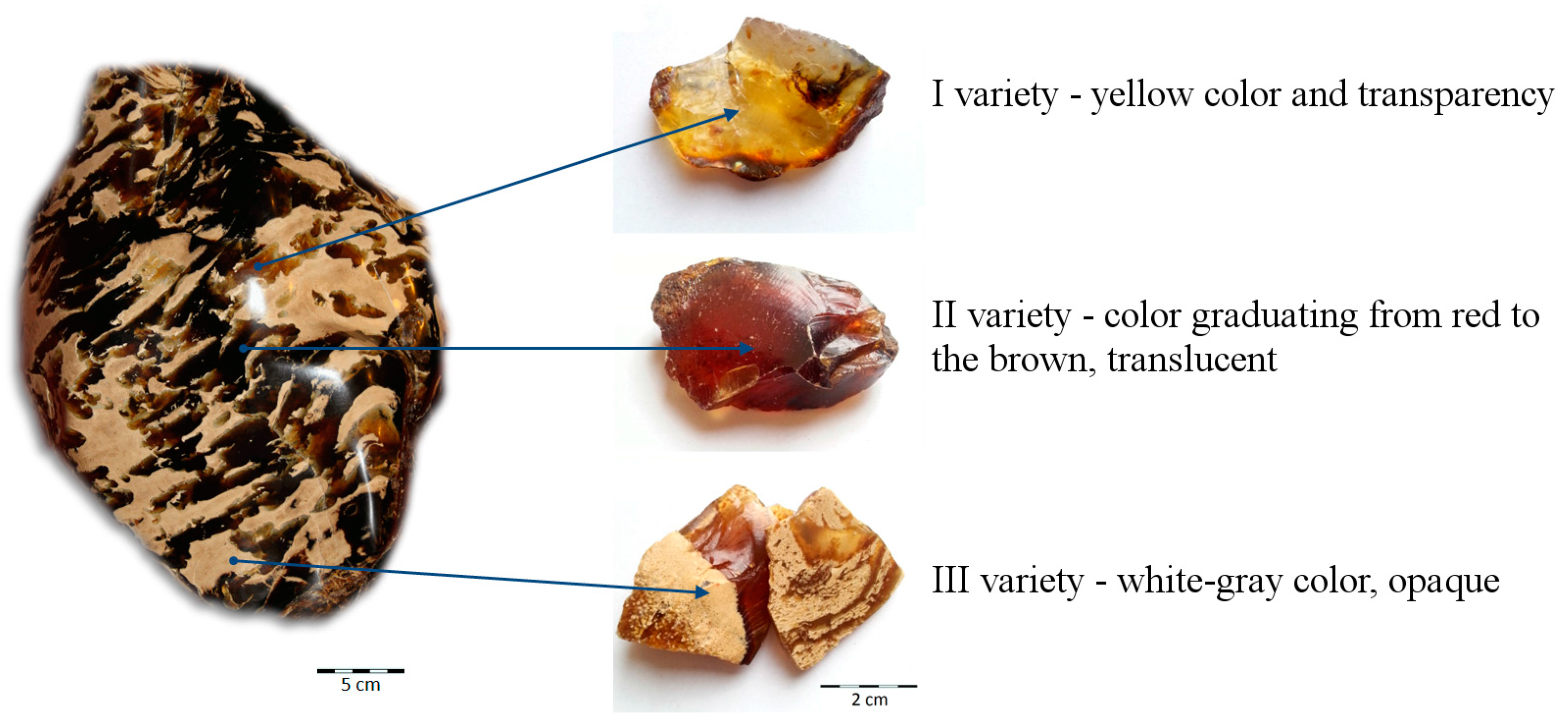
  • Fossil resin from the island of Sumatra (Jambi Province, Sarolangun mine) occurs in three varieties, which differ macroscopically in their colour and transparency.
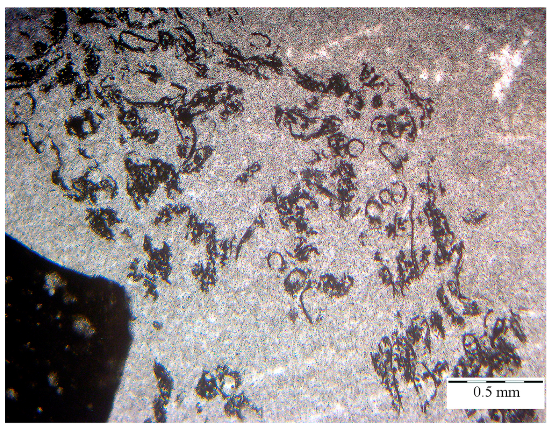
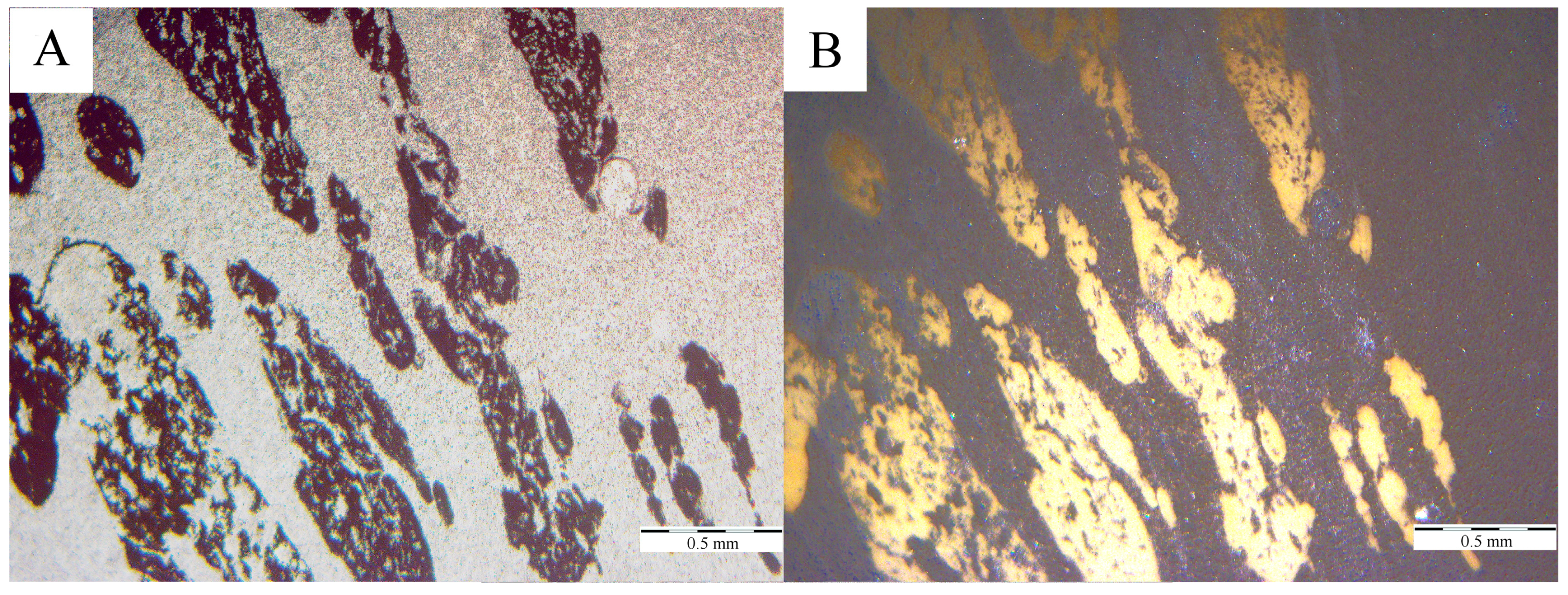
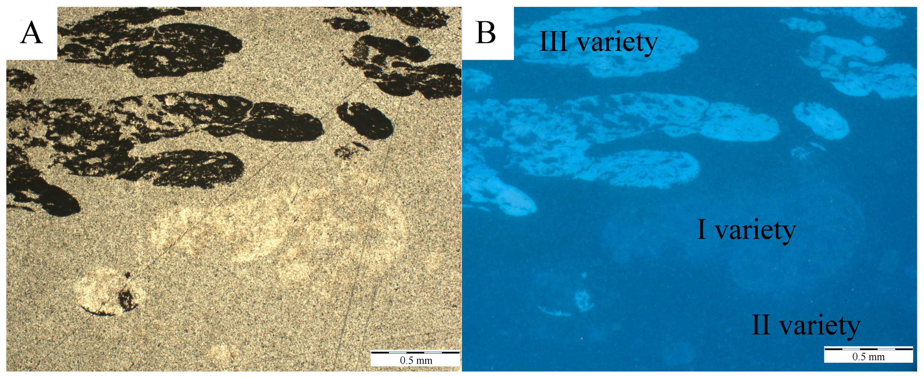
  • Resin specimens were formed from exudates from different times and locations, and thus they varied in clarity, inclusion content, maturation degree and fluorescence properties.
  • Aromatization of the cyclohexane ring was found to be part of the fossilization mechanism. This is supported by consideration of the reducing environment of the swamp where the resins were deposited and buried.
  • The maturation grade of fossil resins assessed via Raman spectroscopy was higher than expected for resins of Miocene age.
  • Volcanism, which was previously documented as a factor determining the increased resin production during the Miocene epoch, was also responsible for the relatively high maturation grade of the fossil resins.

Acknowledgments

The authors are grateful to Jacek Szczerba and Robert Girulski for providing specimens of Sumatran resin for the research. The Reviewers are greatly acknowledged for their friendly and helpful comments on the manuscript. This study was supported by statutory grant (AGH University of Science and Technology) and research project No. 61.2801.1502.000 (The Polish Geological Institute—National Research Institute).

Author Contributions

Beata Naglik, Barbara Kosmowska-Ceranowicz, Przemysław Drzewicz, Magdalena Dumańska-Słowik and Lucyna Natkaniec-Nowak designed the experiments, interpreted the data and wrote the paper. Jakub Matusik, Marian Wagner, Rastislav Milovsky, Paweł Stach and Arkadiusz Szyszka performed the experiments and prepared figures and photographs.

Conflicts of Interest

The authors declare no conflict of interest.

References

  1. Tomkeieff, S.I. Coals and Bitumens and Related Fossil Carbonaceous Substances: Nomenclature and Classification; Pergamon Press: Oxford, UK, 1954. [Google Scholar]
  2. Ragazzi, E.; Alexander, R.S. (Eds.) Amber. In Encyclopedia of Geobiology; Springer: Dordrecht, The Netherlands, 2011; pp. 24–36. [Google Scholar]
  3. Kosmowska-Ceranowicz, B.; Sachanbiński, M.; Łydżba-Kopczyńska, B. Analytical characterization of “Indonesian amber” deposits: Evidence of formation from volcanic activity. Baltica 2017, 30, 55–60. [Google Scholar] [CrossRef]
  4. Kosmowska-Ceranowicz, B. Bursztyn Bałtycki i Inne Żywice Kopalne [Baltic Amber and Other Fossil Resins]; The Museum of the Earth of the Polish Academy of Sciences: Warsaw, Poland, 1993; 141p. (In Polish) [Google Scholar]
  5. Jehlička, J.; Villar, S.E.J.; Edwards, H.G.M. Fourier transform Raman spectra of Czech and Moravian fossil resins from freshwater sediments. J. Raman Spectrosc. 2004, 35, 761–767. [Google Scholar] [CrossRef]
  6. Winkler, W.A. Raman spectroscopic approach to the maturation process of fossil resins. J. Raman Spectrosc. 2001, 32, 59–63. [Google Scholar] [CrossRef]
  7. Kosmowska-Ceranowicz, B. Bursztyn z Borneo—największe na świecie złoże żywicy kopalnej (The greatest fossil resin deposit in the world). Prz. Geol. 1994, 7, 576–578, (In Polish with English Summary). [Google Scholar]
  8. Brackman, W.; Spaargaren, K.; Van Dongen, J.P.C.M.; Couperus, P.A.; Bakker, F. Origin and structure of the fossil resin from an Indonesian Miocene coal. Geochim. Cosmochim. Acta 1984, 48, 2483–2487. [Google Scholar] [CrossRef]
  9. Langenheim, J.H. Higher plant terpenoids: A phytocentric overview of their ecological roles. J. Chem. Ecol. 1994, 20, 1223–1280. [Google Scholar] [CrossRef] [PubMed]
  10. Lambert, J.B.; Levy, A.J.; Santiago-Blay, J.A.; Wu, Y. Nuclear Magnetic Resonance Characterization of Indonesian Amber. Life: Excitement Biol. 2013, 1, 136–155. [Google Scholar] [CrossRef]
  11. Leelawatanasuk, T.; Wathanakul, P.; Paramita, S.; Sutthirat, C.; Sriprasert, B.; Bupparenoo, P. The characteristic of amber from Indonesia. Aust. Gemmol. 2013, 25, 142–145. [Google Scholar]
  12. Schmidt, A.R.; Jancke, S.; Lindquist, E.E.; Ragazzi, E.; Roghi, G.; Nascimbene, P.C.; Schmidt, K.; Wappler, T.; Grimaldi, D.A. Arthropods in amber from the Triassic Period. Proc. Nat. Acad. Sci. 2012, 109, 14796–14801. [Google Scholar] [CrossRef] [PubMed]
  13. Saint Martin, S.; Saint Martin, J.P.; Girard, V.; Grosheny, D.; Néraudeau, D. Filamentous micro-organisms in Upper Cretaceous amber (Martigues, France). Cretac. Res. 2012, 35, 217–229. [Google Scholar] [CrossRef]
  14. Kosmowska-Ceranowicz, B.; Vávra, N. Infrared Spectra of Fossil Resins, Subfossil Resins and Selected Imitation of Amber; Polish Academy of Science Museum of Earth: Warsaw, Poland, 2015. [Google Scholar]
  15. Kosmowska-Ceranowicz, B. Glessite from Malesia and Indonesia. In Bursztyn w Polsce i na Świecie (Amber in Poland and in the World), 2nd ed.; Wydawnictwo Uniwersytetu Warszawskiego: Warsaw, Poland, 2017; pp. 1–310. (In Polish) [Google Scholar]
  16. Krumbiegiel, G.; Kosmowska-Ceranowicz, B. Vorkommen von Glessit, Siegburgit (?) und Krantzit in Tertiär Mitteldeutschlands (Bitterfeld, Niederlausitz). Fundgrube 1990, 26, 78–81. (In German) [Google Scholar]
  17. Krumbiegiel, G. Glessit, ein tertiäres Harz von Bedecktsamern. Fossilien 1993, 10, 83–90. (In German) [Google Scholar]
  18. Kosmowska-Ceranowicz, B.; Krumbiegel, G.; Vávra, N. Glessit, ein tertiäres Harz von Angiospermen der Familie Burseraceae. N. Jb. Geol. Paläont. Abh. 1993, 187, 299–324. (In German) [Google Scholar]
  19. Lambert, J.B.; Johnson, S.C.; Shawl, C.E.; Poinar, G.O. Fossil resin from Asia. Anc. Biomol. 1999, 3, 29–35. [Google Scholar]
  20. Adiwidjaja, P.; De Coster, G.L. Pre-Tertiary paleotopography and related sedimentation in South Sumatra. In 2nd Annual Convention Proceedings; Indonesian Petroleum Association: Jakarta, Indonesia, 1973; pp. 89–103. [Google Scholar]
  21. Young, R.; Atkinson, C.D. A review of Talang Akar Formation (Oligo-Miocene) reservoirs in the offshore areas of Southeast Sumatra and Northwest Java. In Clastic Rocks and Reservoirs of Indonesia: A Core Workshop; Indonesian Petroleum Association: Jakarta, Indonesia, 1993; pp. 177–210. [Google Scholar]
  22. Friederich, M.C.; Moore, T.A.; Flores, R.M. A regional review and new insights into SE Asian Cenozoic coal-bearing sediments: Why does Indonesia have such extensive coal deposits? Int. J. Coal Geol. 2016, 166, 2–35. [Google Scholar] [CrossRef]
  23. Barber, A.J.; Crow, M.J.; Milsom, J.S. (Eds.) Sumatra: Geology, Resources and Tectonic Evolution; Geological Society of London: London, UK, 2005; Memoirs No. 31. [Google Scholar]
  24. Guiliano, M.; Asia, L.; Onoratini, G.; Mille, G. Applications of diamond crystal ATR FTIR spectroscopy to the characterization of ambers. Spectrochim. Acta Part A Mol. Biomol. Spectrosc. 2007, 67, 1407–1411. [Google Scholar] [CrossRef] [PubMed]
  25. Truică, G.I.; Teodor, E.D.; Litescu, S.C.; Radu, G.L. LC-MS and FT-IR characterization of amber artefacts. Cent. Eur. J. Chem. 2012, 10, 1882–1889. [Google Scholar]
  26. Beck, C.; Wilbur, E.; Meret, S.; Kossove, D.; Kermani, K. The infrared spectra of amber and the identification of Baltic amber. Archaeometry 1965, 8, 96–109. [Google Scholar] [CrossRef]
  27. Coates, J. Interpretation of infrared spectra, a practical approach. Encycl. Anal. Chem. 2000. [Google Scholar] [CrossRef]
  28. Edwards, H.G.M.; Farwell, D.W. Fourier transform-Raman spectroscopy of amber. Spectrochim. Acta Part A Mol. Biomol. Spectrosc. 1996, 52, 1119–1125. [Google Scholar] [CrossRef]
  29. Moreno, Y.M.; Christensen, D.H.; Nielsen, O.F. An NIR-FT Raman spectroscopic study of amber. Asian J. Spectrosc. 2000, 4, 49–56. [Google Scholar]
  30. Brody, R.H.; Edwards, H.G.M.; Pollard, A.M. A study of amber and copal samples using FT-Raman spectroscopy. Spectrochim. Acta Part A Mol. Biomol. Spectrosc. 2001, 57, 1325–1338. [Google Scholar] [CrossRef]
  31. Adar, F. Introduction to Interpretation of Raman Spectra Using Database Searching and Functional Group Detection and Identification. Spectroscopy 2016, 31, 16–23. [Google Scholar]
  32. Bąk, M.; Natkaniec-Nowak, L.; Drzewicz, P.; Czapla, D.; Bogdasarov, М. Ambrosia-like fungi in fossil resin from Jambi Province in Sumatra Island—Possible phoretic organisms interacted with invaded insects. In Proceedings of the 17th Czech-Slovak-Polish Palaeontological Conference, Krakow, Poland, 20–21 October 2016; Abstract Volume 23. [Google Scholar]
  33. Natkaniec-Nowak, L.; Drzewicz, P.; Bąk, M.; Matusik, J.; Kowalczyk, J.; Bogdasarov, M.; Ivanina, A.V. Fossil resins from Jambi province (Sumatra, Indonesia)—Proxy indicators of palaeonvironment and palaeoclimate of Sundaland. In Proceedings of the Third International Conference Viet-Pol, Hanoi, Vietnam, 12–15 November 2016; pp. 230–231. [Google Scholar]
  34. Matuszewska, A.; Gołąb, A. Próba wykorzystania parametru mikrotwardości żywic kopalnych i sztucznych, jako cechy klasyfikacyjnej (An attempt at using the parameter of the microhardness of fossil and artificial resins as a classification feature). Bursztynisko 2008, 31, 56–61. [Google Scholar]
Figure 1. Samples of three varieties of Sumatran resin taken from its aggregate.
Minerals 08 00095 g001
Figure 2. (A) tectonic map of Miocene world; (B) geological profile of South Sumatra Basin (on the basis of [23]); (C) Indonesia during Miocene epoch (modified after [22]); (D) volcanic units of the island of Sumatra (according to [23]).
Minerals 08 00095 g002
Figure 3. Stereoscopic photographs of samples; from the top: (A,B) bright yellow (variety I); (C,D) dark (variety II) and (E,F) opaque (variety III) of resins.
Minerals 08 00095 g003
Figure 4. Stereoscopic photo of the contact zone between resin varieties II and III.
Minerals 08 00095 g004
Figure 5. Microphotograph of organic detritus in resin variety II, polarized light; 1 N.
Minerals 08 00095 g005
Figure 6. Microphotograph of resin variety III surrounded by variety II, polarized light; 1 N (A); NX (B).
Minerals 08 00095 g006
Figure 7. Microscopic image of fossil resins from the island of Sumatra, 1N (A); UV-epifluorescence (B).
Minerals 08 00095 g007
Figure 8. Kubelka–Munk-transformed diffuse reflectance spectra of resin varieties I, II, III from the island of Sumatra. The graph on the right depicts fingerprint region of the spectra (2000–500 cm−1).
Minerals 08 00095 g008
Figure 9. Raman spectra of resins varieties I, II and III from the island of Sumatra.
Minerals 08 00095 g009
Table 1. Macro- and microscopic characteristics of Sumatran fossil resins.
Physical PropertyVariety IVariety IIVariety III
Coloryellowred to brownwhite-gray
Transparencytransparenttranslucentopaque
Structurehomogenous non-homogenous with single dropletsfoamy-like with numerous droplets filled with resin of I variety
Inclusionsonly locally—spherical plant microspores or fragments of channels of strongly macerated wooddispersed pigment of fungus spores or pollen plantscoalified plant remains
Fluorescencelowlowintensive
Table 2. FT-IR bands assignment.
FT-IR Wave Numbers (cm−1)Assignment
I VarietyII VarietyIII Variety
825825825Aromatic ring C–H—1,4-Disubstitution (para)
888888888CH ethylenic bending [24]
975975975C–O bonds [24]
103310281027C–O bonds [24]; cyclohexane ring vibrations (1000–1055 cm−1) [27]
104810481048Cyclohexane ring vibrations (1000–1055 cm−1); primary alcohol, C–O stretching [27]
116711731174C–O simple bond stretching of esters [26]; tertiary alcohol, C–O stretching [27]
119912001200Phenol, C–O stretching [27]
125512551255Primary or secondary, OH in-plane bending [27]
131513151317Primary or secondary, OH in-plane bending [27]
-13321339Methane C–H bending (1330–1350 cm−1); primary or secondary, OH in-plane bending [27]
136713671367Due to CH3 bending [24]
137713781377Due to CH3 bending [24]
138313841380Due to CH3 bending [24]
145614551456Involves CH2 and CH3 bending [24]
145814581460Involves CH2 and CH3 bending [24]
-16041614Conjugated C=C (1600 cm−1); aryl-substituted C=C (1625 cm−1); alkenyl C=C stretching (1620–1680 cm−1) [27]
165016591649C=C double bond stretching [22]; alkenyl C=C stretching (1620–1680 cm−1) [27]
-16751674Alkenyl C=C stretch (1620–1680 cm−1) [27]
169016901690Due to carbonyl group (C=O) stretching vibrations and, respectively, to carboxylic acid group [24]
-1710-Due to carbonyl group (C=O) stretching vibrations and, respectively, to ketone and/or carboxylic acid group [24]
173017291730Due to carbonyl group (C=O) stretching vibrations and, respectively, to ester [24]
2868-2868Alkyl stretching and an important level of methyl groups [24]
2934-2934Alkyl stretching; for the CH groups; an important level of methyl groups [24]
295729572960Methyl C–H asymmetric/symmetric stretching [27]; corresponding to the alkyl stretching; for the methyl groups; an important level of methyl groups [24]
341034043410OH stretching of alcohols and/or carboxylic acids [24]
--3544Hydroxy group, H-bonded OH stretch; internally bonded OH stretching [27]
--3566Hydroxy group, H-bonded OH stretch; internally bonded OH stretching [27]
Table 3. FT-Raman band assignment.
Raman Shift (cm−1)Proposed Assignment
Variety IVariety IIVariety III
-600-(?) C–C modes of the alicyclic rings [30]
680680680aromatic out-of-plane deformation [29]
730730730aromatic out-of-plane deformation; bands related to the degree of maturation (600–800 cm−1) [5]
800800800C–C functional group—highly mixed in complex molecule (700–1260 cm−1) [31], bands related to the degree of maturation (600–800 cm−1) [5]
136013661360CH2 deformation [28]
131513151315CH3 umbrella mode (~1375 cm−1) [28]; δCH2, δCH3 [30]
145014501450an asymmetric deformation of CH3 and CH2 groups [6,31]
165816581658C=C bonds (unsaturated hydrocarbons) [6,31]

