Article

Dissolution and Sorption Processes on the Surface of Calcite in the Presence of High Co2+ Concentration

by
Jorge González-López
1,*,
Ángeles Fernández-González
1,
Amalia Jiménez
1,
Athanasios Godelitsas
2,
Spyridon Ladas
3,
Georgios Provatas
4,
Anastasios Lagogiannis
4,
Ioannis N. Pasias
5,
Nikolaos S. Thomaidis
5 and
Manuel Prieto
1
1
Department of Geology, University of Oviedo, Calle Arias de Velasco s/n, Oviedo 33005, Spain
2
Department of Geology & Geoenvironment, National and Kapodistrian University of Athens, Zographou Campus, 15784 Athens, Greece
3
Surface Science Laboratory, Department of Chemical Engineering, University of Patras, 26504 Patras, Greece
4
Tandem Accelerator Laboratory, Institute of Nuclear Physics, NCSR ‘‘DEMOKRITOS”, GR-15310 Attiki, Greece
5
Department of Chemistry, National and Kapodistrian University of Athens, Zografou Campus, 15771 Athens, Greece
*
Author to whom correspondence should be addressed.
Minerals 2017, 7(2), 23; https://doi.org/10.3390/min7020023
Submission received: 31 October 2016 / Revised: 26 January 2017 / Accepted: 10 February 2017 / Published: 15 February 2017
(This article belongs to the Special Issue Mineral Surface Science and Nanogeoscience)

Abstract

:
The interaction of the calcite surface with Co2+-rich aqueous solutions ([Co2+aq]initial = 1000 ppm, i.e., ca. 17 mM) was investigated by means of macroscopic experiments and surface spectroscopic techniques. In the case of the macroscopic experiments, calcite powder and monocrystals were immersed into solutions for different time periods (from 1 min to one month). The Ca concentrations in the filtrates was measured by means of atomic absorption spectrometry (AAS) while the interacted solids were studied using a combination of X-ray photoelectron spectroscopy (XPS) and 12C-rutherford backscattering spectrometry (12C-RBS). The macroscopic data showed a characteristic surface dissolution process, in parallel to the surface sorption processes. Adsorption and co-precipitation were seen for almost the entire immersion period for both calcite powder and monocrystals. The surface study by XPS (analyzed at a depth of approximately 12 nm) suggested that adsorption takes place in the first hour of the interaction, followed by incorporation of Co2+ into calcite surface layers, leading to the formation of a Co2+-bearing surface (co)precipitate, which occurs over a period of hours and days. The 12C-RBS measurements on calcite { 10 1 ¯ 4 } indicated that the thickness of this surface co-precipitate was 270 nm after one day and then stabilized at 320 nm after more than a week.

Graphical Abstract

1. Introduction

Cobalt makes up only about 0.0025 wt % of the Earth’s crust [1], but is essential for our health as it forms the active center of cobalamins. Cobalamins are a group of coenzymes, the most important of which is vitamin B12. Cobalt can also be toxic at high concentrations. The presence of cobalt in the environment is related mainly to anthropogenic activity resulting from mining, the pigment industry and the dumping of Co-Li batteries. The maximum recommended exposure is 1 × 10⁻⁴ mg·cobalt/m3 and 1 × 10⁻² mg/Kg·day for inhalation and oral exposure, respectively [2]. The agency for toxic substances and disease registry (ATSDR) report also takes into account the hazards involved with the artificial radioactive 60Co isotope, which is used in many applications (e.g., medicine, sterilization and industrial radiography). The release of this radiogenic isotope into the environment is harmful to humans, animals and marine life. It is also the most stable artificial isotope with a half-life of 5.27 years, leading to its accumulation in the environment. Since Co2+ is soluble, its mobility in water is high which can lead to the contamination of soils and groundwater and the incorporation of the metal into the trophic chain [3].
Certain mineral structures can remove heavy metals from the environment. Sorption processes onto mineral phases, and particularly onto carbonates [4], frequently control the immobilization of contaminants and govern their dispersion and accumulation. The main mechanisms involved in metal sorption processes are absorption (including ion exchange), adsorption and co-precipitation, finally leading to surface crystal growth [5] (Figure 1). The adsorption only involves the formation of chemical bonds onto the mineral surface sites. Absorption implies the incorporation of dissolved metal ions onto mineral surface layers. On the other hand, co-precipitation fundamentally leads to the formation of a new phase, which is not necessarily related to the bulk mineral composition and structure. Many studies have focused on remediation technologies with natural minerals such as zeolites, sulphates and carbonates in the last two decades [6,7,8]. Among these minerals, carbonates are the most widely used, particularly calcite, which has been employed due to its widespread availability and reactivity [9].
Kornicker et al. [10] measured the sorption process of an aqueous solution of Co2+ ([Co2+aq]initial < 60 ppm) onto calcite and concluded that only adsorption occurs. No CoCO3 was found to be precipitated onto the calcite or aragonite surfaces. Xu et al. [11] investigated the interaction of Co2+ ([Co2+aq]initial < 6 ppm) with both calcite-cleaved monocrystals and powder by means of X-ray photoelectron spectroscopy (XPS). They reported that initially the incorporation of Co into calcite occurred at a fast rate (up to 7.2 nmol/min·mg) and is attributed to adsorption. This was followed by a co-precipitation mechanism and the formation of a (Co,Ca)CO3 solid solution with the Co content related to the initial pH and the calcite saturation index. They also indicated that no oxidation of Co2+ takes place during the interaction. Furthemore, Cheng et al. [12] studied the interaction of the calcite surface with Co ([Co2+aq]initial = 29.47 ppm) using surface-extended X-ray absorption fine structure (SEXAFS) and X-ray standing wave (XSW). Their results indicated that Co replaces Ca atoms in the M–O octahedron at the surface and also that at the top of the surface, the Co2+ ions are bounded to OH groups in order to maintain the octahedral coordination observed in XSW. In addition, Braybrook et al. [13] carried out both experimental and computational work based on calcite growth in the presence of aqueous Co2+. Both their simulations and experiments provided support for the conclusion that synthetic calcite growth occurs. Their simulations showed interference between the { 10 1 ¯ 4 } and { 01 1 ¯ 1 } planes, thus suggesting Co incorporation on the < 44 8 ¯ 1 > acute steps. However, they claimed that both experimental and computational results indicated that Co accumulation only occurs on the calcite mineral surface. Freij et al. [14] performed a series of experiments ([Co2+aq]initial < 600 ppm) using atomic force microscopy (AFM) and concluded that a CoxCa1−xCO3 micro-layer is formed, covering the entire calcite surface. Further, Katsikopoulos et al. [15] studied the crystal growth from a (Co,Ca)CO3 solid solution using silica gel double diffusion and direct precipitation experiments. They demonstrated that a large amount of Co (up to 33 mol % of Co) entered the CaCO3 structure, but experiments were conducted far from equilibrium due to supersaturation; thus, the Co incorporation into calcite could occur in a metastable state. Moreover, Lee et al. [16], in a paper significant for medical applications, investigated the influence of citrate and phthalate on the growth of calcite in the presence of Co2+ and found that citrate and phthalate inhibit the precipitation of calcite. Recently, González-López et al. [17] reported the thermochemistry of (Ca,Co)CO3 solid solutions from density functional theory simulations and concluded that many Ca1−xCoxCO3 mineral solid solutions (with observed compositions of up to x = 0.027, and above x = 0.93) are metastable with respect to phase separation. Finally, Xu et al. [18] studied the interaction of calcite with solutions with a very low Co2+ concentration ([Coaq]initial = 1–10 ppm) using AFM and XPS. They suggested that that the lattice misfit between CoCO3 and CaCO3 is too large to allow heteroepitaxial growth of a pure cobalt carbonate phase in ambient conditions.
This present work attempts to understand the interaction between the calcite surface and aqueous solutions with a high Co2+ concentration ([Co2+aq]initial = 1000 ppm, ~17 mM). For this purpose, different sets of experiments were designed. Macroscopic experiments of calcite powder and monocrystals were immersed into solutions for varying time periods (from 1 min to one month), with the aim of observing surface dissolution processes (analysis of the filtrates for Ca by atomic absorption spectrometry (AAS)). Surface spectroscopic experiments were conducted to obtain a novel spectroscopic “3D image” of the interactions using a combination of surface-sensitive techniques (XPS and 12C-rutherford backscattering spectrometry (12C-RBS)) and also to analyze the distribution of Co onto the calcite surface and resolve the potential sorption mechanisms.

2. Materials and Methods

2.1. Macroscopic Experiments

Natural pure calcite (transparent Iceland spar) was held in solution with the salt Co(NO3)2·6H2O at room temperature. The initial concentration of aqueous cobalt was kept high at 1000 ppm ([Co2+aq]initial = 1000 ppm). The calcite mineral was tested in two different forms: as a fine powder obtained using an agate mortar, and as freshly cleaved { 10 1 ¯ 4 } monocrystals with a base size of ~0.5 × 0.5 and a height of 0.1 cm. In the powder experiments, 50 mg of calcite were mixed with 50 mL (Solid/Liquid ratio = 1 mg/mL) of the initial Co solution ([Co2+aq]initial = 1000 ppm, ~17 mM) and placed into 30 mm × 115 mm polypropylene ( PPN) tubes with a screw tap (Greiner Bio-One). The mixture was stirred vigorously at the start. The solutions were keep stable for different reaction times (1 and 12 h, and 1, 7, 14, 21, 30 and 60 days). The solids were separated from the aqueous solution by filtration using a Nalgene® dispositive filter (Thermo Fisher Scientific, Cambridge, UK) and Millipore® 0.45 µm filter (Millipore Corp., Bedford, MA, USA) which were carefully rinsed with distilled water. The filtrates from each experiment were analyzed for their Ca content using AAS (Perkin-Elmer 2380 Ca lamp with a wavelength of 422.7 nm, Perkin Elmer, Wellesley, MA, USA). For the monocrystals 20 mL of the initial solution ([Co2+aq]initial = 1000 ppm, ~17 mM) was added to a 50 mL glass beaker where the single crystals were immersed. As the crystals were immersed into the solution, they were agitated for a few seconds using Teflon® tweezers to ensure the interaction between crystals and solution. Then, the vessels were covered to prevent contamination from external sources. Experiments with monocrystals were carried out for shorter interaction times, compared to powder experiments. They were immersed for 1, 5, 15 and 30 min, 1 and 12 h, one and two days, one week, and one month (Table 1). The surface area was obtained geometrically, by measuring the height and width of each rhombohedron. After the interaction the crystals were carefully removed and rinsed with distilled water before being dried in air and then characterized by XPS (Leybold-SPECS, Berlin/Cologne, Germany) and 12C-RBS.

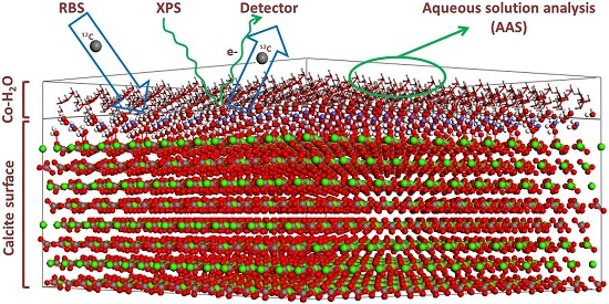
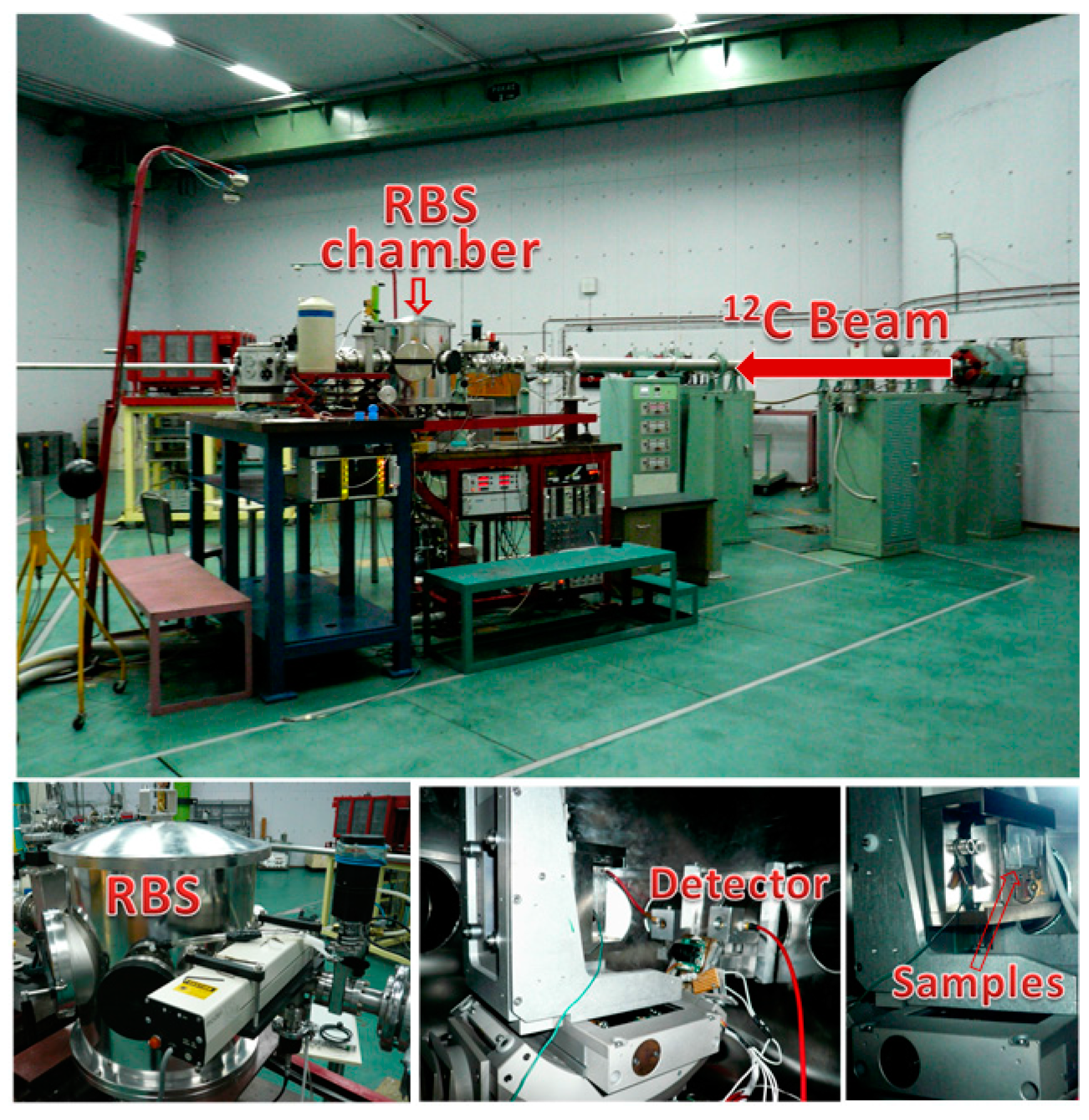
2.2. Surface Spectroscopic Characterization of the Interacted Solids (XPS and 12C-RBS)

XPS measurements were carried out on monocrystals interacted for 1 h, one day and one week. They were placed into the chamber of a MAX200 system (Leybold, Cologne, Germany), at room temperature and ~10−8 mbar pressure. Non-monochromatic Mg Kα X-rays and a Hemispherical Electron Energy Analyser (EA200 detector, SPECS, Berlin, Germany) with Multi-Channel Detection and were calibration certified (ISO15472 and ISO24237 [19,20]). The analyser operated under conditions optimized for the optimal signal intensity (constant pass energy of 100 eV, maximum lens aperture, analysis along the specimen surface normal). The analysed depth of the sample, was about 12 nm and signal intensity decreased roughly exponentially with increasing depth. A surface of 10 mm × 10 mm which was to be analyzed was first subjected to light argon-ion sputtering (1.5 keV ion beam energy, ~0.9 μA sample current and x-y rastering), with the aim to remove some of the superficial ‘carbon’ contamination layer. The 12C-RBS measurements were carried out, in the case of monocrystals which had been immersed for 1 h, one day, one week and one month, using the 5.5 MV Tandem Accelerator at NCSR “DEMOKRITOS” located in Athens (Greece) with an 8 MeV 12C-beam (Figure 2). RBS can be used to obtain composition profiles of the lower surface layers of the calcite crystals. The use of heavier ions (12C), rather than typical protons, enhances the analytical depth resolution.

3. Results and Discussion

3.1. Macroscopic Data—Dissolution of Calcite in the Presence of Co2+

The release of Ca2+ (ppm of Ca per g of the solid), when powdered calcite was immersed in the initial Co2+ aquous solution as a function of time, is shown in Figure 3. The results clearly indicate a progressive surface dissolution process [21,22], running in parallel with the metal sorption processes (which were confirmed by XPS and 12C-RBS and are described in the next section). It is obvious that the dissolution of calcite depends on the S/L ratio (1 mg/mL in our case). The process starts from the surface, where Co2+ also interacts, and theoretically may alter the entire crystal structure. In the case of the experiments with calcite monocrystals it was feasible to correlate—in the presence of [Co2+aq]initial = 1000 ppm in the solution—the release of Ca2+ per geometric specific surface area as a function of time.
It is also evident that dissolution depends significantly on the specific surface area of the carbonate mineral particles. The smaller the crystal size, the higher the dissolution rate. This is clearly seen when the high dissolution rates for the powdered experiments are compared with the single-crystal experiments. There was a rapid increase of the Ca concentration in the solution in short time steps after immersion. This was eventually stabilized after one week, reaching a value of 3.8 × 10⁻³ ppm/g·cm−2. The equivalent total Ca2+ release in ppm for the monocrystals after one week of interaction was 3.2 ppm. Ca2+ release in the powder experiments differed greatly from the monocrystal experiments due to the differences in the surface reactivity. In the powder experiments, Ca2+ liberation never reached an asymptotic maximum value (see Figure 3). This suggests that calcite powder is dissolved during the entire interaction time, together with surface replacement of Ca2+ by Co2+. The cause of this behavior is the fact that not only one monocristal is in place, but there is an accumulation of microcrystals which significantly increase the global surface reactivity. In this way, the total Ca2+ released after immersion for one week with approximately 0.5 g of calcite powder was 33.8 ppm, and assuming the average mass of all monocrystal experiments, the calculated calcium concentration was 6.6 ppm, rather higher than the previous Ca2+ total. Therefore, for the same mass of calcite powder, dissolution was faster. Indeed, it is suggested that the initial fast increase of Ca2+ in the aqueous solution was driven by the coupled dissolution and sorption process; this is supported by the modeling experiments using the PHREEQC code [23] (the total Ca2+ released into the solution was only 23.21 ppm, and thus the approximately 35 ppm of Ca2+ at the end of the first rise exceeded the calcite dissolution equilibrium). On the contrary, the last linear trend in Ca2+ release was only presumably caused by sorption which involves the surface replacement of Ca2+ by Co2+ towards the formation of a Cobalt-bearing co-precipitate.

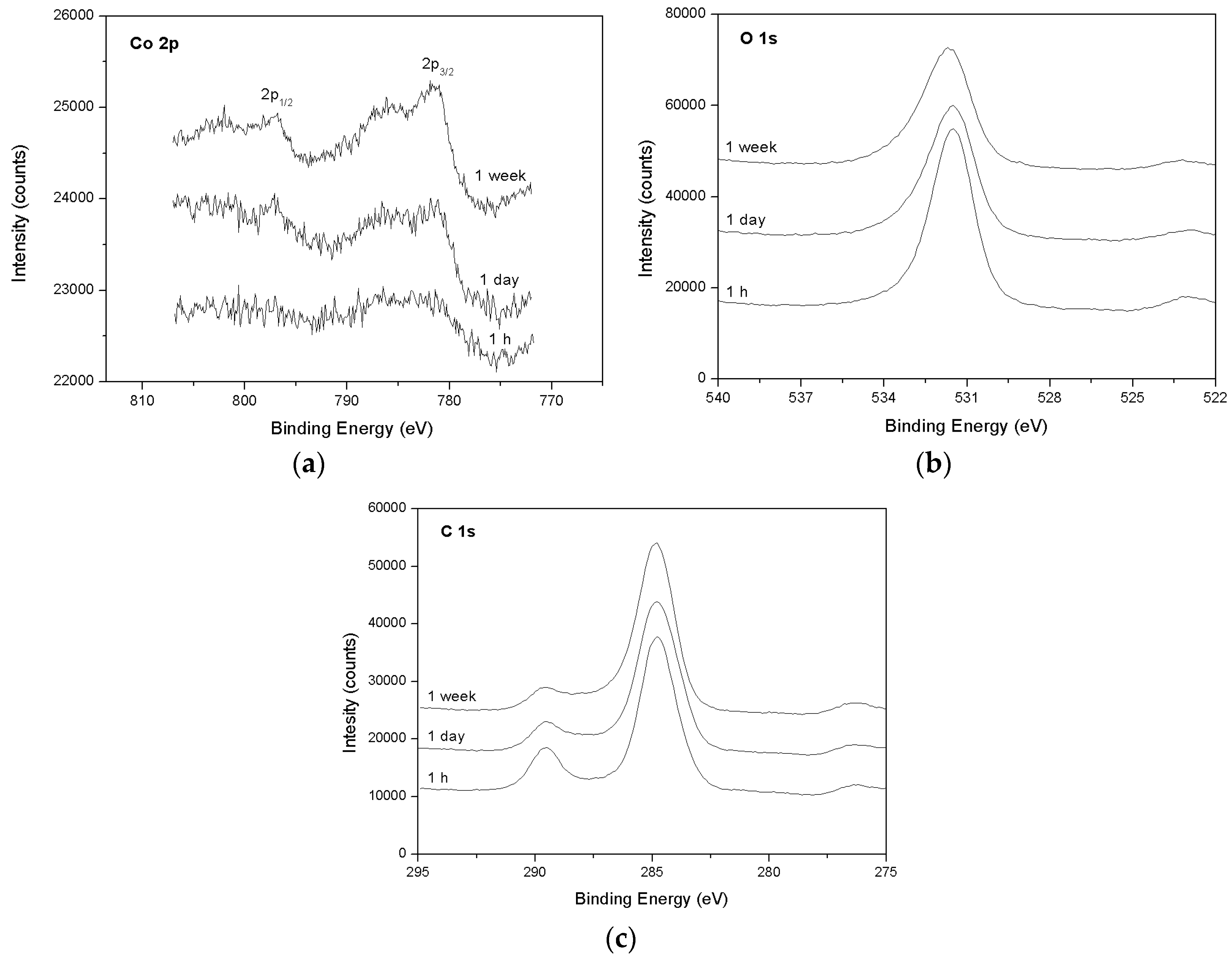
3.2. Spectroscopic Data—Sorption of Co2+ on the Surface of Calcite

The XPS spectra for interacted calcite monocrystals (after 1 h, one day and one week of interaction) are shown in Figure 4. The Co 2p3/2 and 2p1/2 photoelectron peaks correspond to binding energies (BE) of 781.4 and 796.9 eV, respectively. These are characteristic of many Co compounds [11]. However, Co3+ or mixed Co2+ and Co3+ compounds seem not to be present due to the strong satellites of both Co 2p peaks. It is well known that these satellites are characteristic for octahedrally coordinated Co2+ phases [24]. Moreover, the intensity of the peaks reveals that, for the samples immersed for one day and one week, there was evidence of Co incorporation into the calcite near-surface layers. The peaks indicate that there was a correlation between the intensity and the interaction time. Although the peak for 1 h is small, we cannot ignore the possibility that almost all adsorption of Co on the calcite surface was reached. In fact, since the O 1s photoelectron peak (531.6 eV) was assigned to both CaCO3 and surface OH groups, and furthermore there was an excess of oxygen over the 1:3 stoichiometry for CaCO3 (1:3.3), the existence of Co–OH bonds has been suggested as cobalt is adsorbed onto the surface after 1 h of interaction. O 1s peaks show a large tail because of the presence of some organic oxygen together with carbon. Given that XPS is a highly sensitive surface technique, organic carbon and oxygen, ubiquitous at any conditions, were detected. In addition, the decrease of the intensity of the O 1s peaks with time suggests that fewer Co–OH bonds are formed at the calcite surface and, instead, a surface layer of a mixed Co2+ compound could been have been precipitated after one day of interaction. However, the analyzed depth was ~12 nm and therefore deeper incorporation of Co cannot be excluded (for this purpose 12C-RBS was conducted, as discussed below). The C 1s photoelectron peak at 289.6 eV has a distinct hydrocarbon signal (284.8 eV), and is recognized as characteristic of CaCO3 for all samples. The peak’s intensity decreases with time, similar to that of O 1s, revealing again a Co-bearing surface layer replacing the calcite surface. On the other hand, the one week sample which had been slightly sputtered with light argon ions allowed the removal of the first atomic layers, and we found that, in addition to hydrocarbon surface reduction, the Co/Ca ratio was reduced considerably (from 0.33 to 0.09). This is in agreement with the over-layer formation proposed for interaction times longer than 1 h.
As mentioned above, XPS gives information about Co speciation and distribution at near-surface layers (up to 12 nm) of the immersed calcite crystals. It is unclear whether Co2+ ions, except for adsorption, have been incorporated into deeper layers of the dissolved calcite surface structure, forming a real co-precipitate. The complementary use of RBS (e.g., [25]), particularly of 12C-RBS having a higher resolution, may yield a surface spectroscopic “3D image” of the metal. The same methodology has been previously applied in the case of the interaction of calcite with Pb in aqueous solutions [26]. It is known that the shapes of the RBS peaks are related to the metal distribution at varying depths. Sharp peaks reveal the formation of a metal-rich thin layer on the calcite surface (i.e., adsorption), while asymmetrical peaks and plateaus indicate metal incorporation onto calcite bulk (i.e., absorption) [25]. The 12C-RBS spectra, showing Co distributions on surfaces at varying depths for the samples immersed for one day and one week, are shown in Figure 5 and Figure 6, respectively. The RBS from calcite { 10 1 ¯ 4 } interacted for 1 h, and gave no evidence of Co due to detection limits, in contrast to XPS which is more sensitive for near-surface layers.
According to the 12C-RBS data, the surface layer of calcite was affected by Co2+ after an interaction of one day, reaching a depth of 270 nm. That proves that after the initial adsorption, which occurs on a short timescale as indicated by XPS, metal incorporation takes place and a true co-precipitate is formed. Furthermore, after one week of interaction, the depth of this co-precipitate was apparently stabilized, reaching a thickness of 320 nm (almost the same thickness was recorded in the case of calcite immersed for one month). It is therefore argued that Co is removed from the aqueous medium, in the beginning via adsorption onto the dissolving surface, and later on by co-precipitation. A similar behavior has been observed in the case of the interaction of Co2+ with the surface of dolomitic marble, also studied by XPS and RBS [27].

4. Conclusions

The main findings of this work can be summarized in the following points:
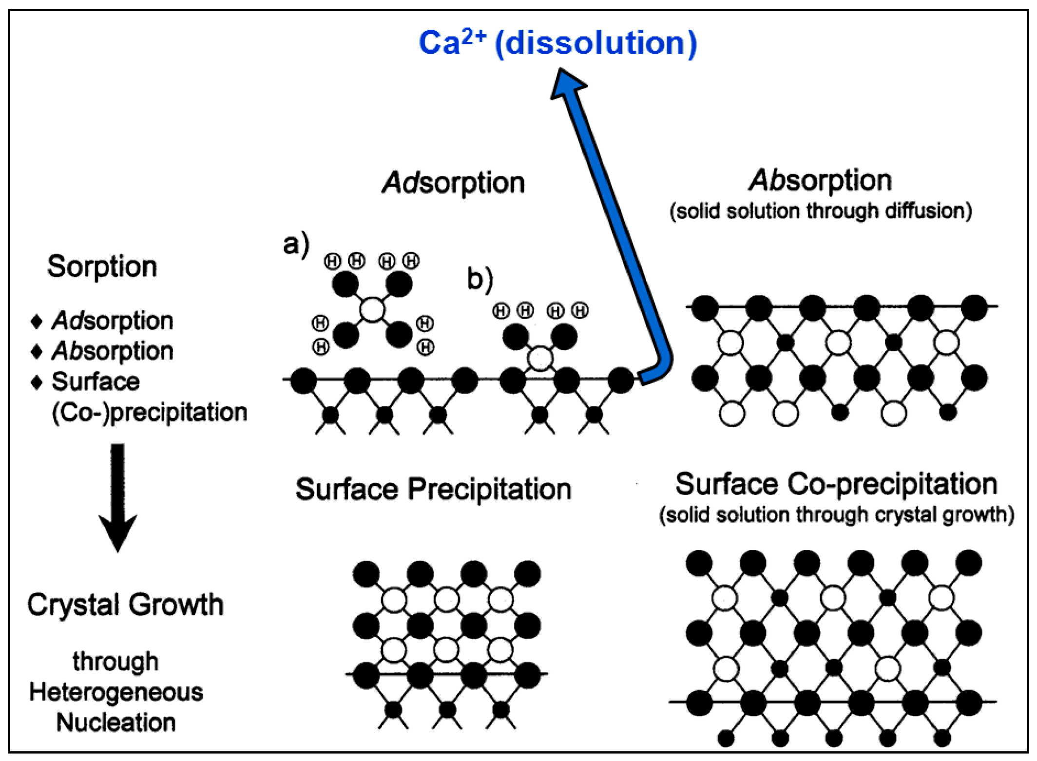
  • According to macroscopic results, for both calcite powder and monocrystals interacted in an aqueous solution with a high Co2+ concentration ([Coaq]initial = 1000 ppm, ~17 mM), dissolution occurs on the surface, causing systematic release of Ca2+ into solution. This runs in parallel to surface Co2+ sorption processes.
  • The XPS surface study (analyzed at a depth of around 12 nm) confirmed that, indeed, sorption occurs at near-surface layers of calcite, initially by adsorption of Co–OH units and later by surface (co-)precipitation.
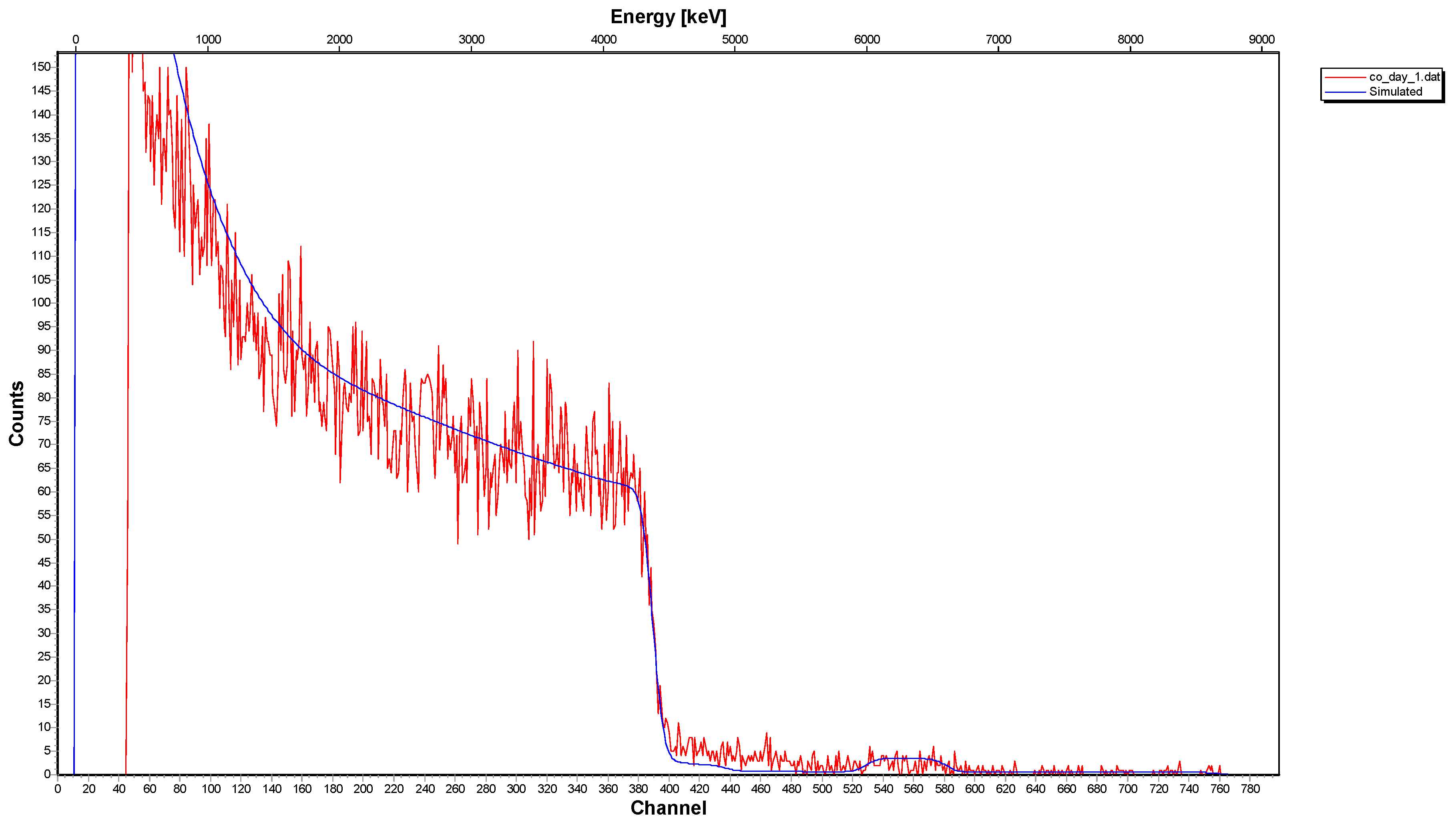
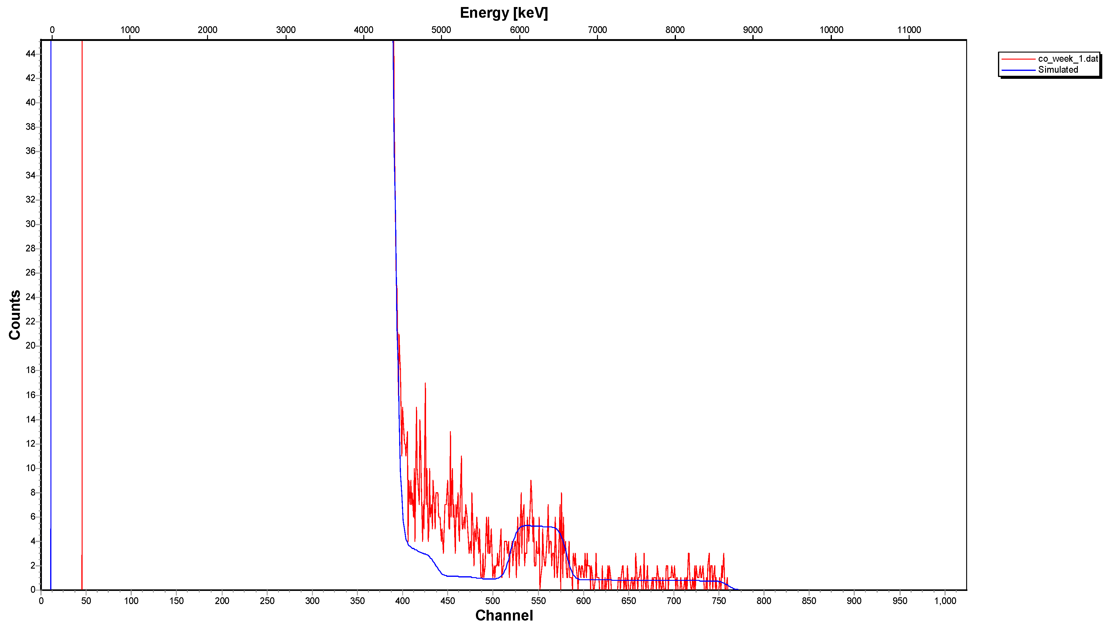
  • The 12C-RBS measurements on calcite { 10 1 ¯ 4 } indicated that co-precipitation is related to a Co2+-bearing surface layer under the calcite surface, showing a thickness of 270 nm after one day of interaction and reaching 320 nm after one week and/or month.

Acknowledgments

This work was supported by the Formación de Personal Investigador (FPI) program of the Spanish Ministry of Economy and Competitiveness and Projects (CGL2013-47988-C2-2-P and CGL2016-77138-C2-2-P). We thank Alex Aziz for useful discussions and English editing.

Author Contributions

Jorge González-López, Ángeles Fernández-González, Amalia Jiménez, Athanasios Godelitsas and Manuel Prieto designed the research; Ioannis N. Pasias and Nikolaos S. Thomaidis contributed to the AAS measurements; Spyridon Ladas contributed to the XPS study; Georgios Provatas and Anastasios Lagogiannis contributed to the 12C-RBS study; Jorge González-López and Athanasios Godelitsas wrote the paper.

Conflicts of Interest

The authors declare no conflict of interest.

Abbreviations

The following abbreviations are used in this manuscript:
AASAtomic absorption spectrometry
XPSX-ray photoelectron spectroscopy
RBSRutherford backscattering spectrometry
SEXAFSSurface-extended X-ray absorption fine structure
XSWX-ray standing wave
AFMAtomic force microscopy

References

  1. Smith, I.; Carson, B.L. Trace Metals in the Environment Vol 6. Cobalt an Apraisal of Environmental Exposure; Ann Arbor Science Publishers Inc.: Ann Arbor, MI, USA, 1981; p. 1202. [Google Scholar]
  2. Agency for Toxic Substances and Disease Registry. Toxicological Profile for Cobalt; U.S. Department of Health and Human Services, Public Health Service: Atlanta, GA, USA, 2004.
  3. Kim, J.H.; Gibb, H.J.; Howe, P.D.; Sheffer, M. Cobalt and Inorganic Cobalt Compounds; Concise International Chemical Assessment Document; World Health Organization: Geneva, Switzerland, 2006; Volume 69. [Google Scholar]
  4. Stipp, S.L.; Hochella, M.F.; Parks, G.A.; Leckie, J.O. Cd2+ uptake by calcite, solid-state diffusion, and the formation of solid-solution: Interface processes observed with near-surface sensitive techniques (XPS, LEED, and AES). Geochim. Cosmochim. Acta 1992, 56, 1941–1954. [Google Scholar] [CrossRef]
  5. Godelitsas, A.; Astilleros, J.M. Dissolution, sorption/(re)precipitation, formation of solid solutions and crystal growth phenomena on mineral surfaces: implications for the removal of toxic metals from the environment. In EMU Notes in Mineralogy—Volume 10: Ion Partitioning in Ambient-Temperature Aqueous Systems; Prieto, M., Stoll, H., Eds.; European Mineralogical Union: Chantilly, VA, USA, 2010. [Google Scholar]
  6. Godelitsas, A.; Charistos, D.; Dwyer, J.; Tsipis, C.; Filippidis, A.; Hatzidimitriou, A.; Pavlidou, E. Copper (II)-loaded heu-type zeolite crystals: Characterization and evidence of surface complexation with N,N-diethyldithiocarbamate anions. Microporous Mesoporous Mater. 1999, 33, 77–87. [Google Scholar] [CrossRef]
  7. Morales, J.; Astilleros, J.M.; Jiménez, A.; Göttlicher, J.; Steininger, R.; Fernández-Díaz, L. Uptake of dissolved lead by anhydrite surfaces. Appl. Geochem. 2014, 40, 89–96. [Google Scholar] [CrossRef]
  8. Prieto, M.; Cubillas, P.; Fernández-González, Á. Uptake of dissolved Cd by biogenic and abiogenic aragonite: A comparison with sorption onto calcite. Geochim. Cosmochim. Acta 2003, 67, 3859–3869. [Google Scholar] [CrossRef]
  9. Astilleros, J.M.; Fernandez-Diaz, L.; Putnis, A. The role of magnesium in the growth of calcite: An AFM study. Chem. Geol. 2010, 271, 52–58. [Google Scholar] [CrossRef] [Green Version]
  10. Kornicker, W.A.; Morse, J.W.; Damasceno, R.N. The chemistry of Co2+ interaction with calcite and aragonite surfaces. Chem. Geol. 1985, 53, 229–236. [Google Scholar] [CrossRef]
  11. Xu, N.; Hochella, M.F.; Brown, G.E., Jr.; Parks, G.A. Co(II) sorption at the calcite-water interface: I. X-ray photoelectron spectroscopic study. Geochim. Cosmochim. Acta 1996, 60, 2801–2815. [Google Scholar] [CrossRef]
  12. Cheng, L.; Sturchio, N.C.; Bedzyk, M.J. Local structure of Co2+ incorporated at the calcite surface: An X-ray standing wave and sexafs study. Phys. Rev. B 2000, 61, 4877–4883. [Google Scholar] [CrossRef]
  13. Braybrook, A.L.; Heywood, B.R.; Jackson, R.A.; Pitt, K. Parallel computational and experimental studies of the morphological modification of calcium carbonate by cobalt. J. Cryst. Growth 2002, 243, 336–344. [Google Scholar] [CrossRef]
  14. Freij, S.J.; Putnis, A.; Astilleros, J. Nanoscale observations of the effect of cobalt on calcite growth and dissolution. J. Cryst. Growth 2004, 267, 288–300. [Google Scholar] [CrossRef] [Green Version]
  15. Katsikopoulos, D.; Fernández-González, Á.; Prieto, A.C.; Prieto, M. Co-crystallization of Co (II) with calcite: Implications for the mobility of cobalt in aqueous environments. Chem. Geol. 2008, 254, 87–100. [Google Scholar] [CrossRef]
  16. Lee, Y.J.; Reeder, R.J. The role of citrate and phthalate during Co (II) coprecipitation with calcite. Geochim. Cosmochim. Acta 2006, 70, 2253–2263. [Google Scholar] [CrossRef]
  17. González-López, J.; Ruiz-Hernández, S.; Fernández-González, Á.; Jiménez, A.; de Leeuw, N.; Grau-Crespo, R. Cobalt incorporation in calcite: Thermochemistry of (Ca,Co)CO3 solid solutions from density functional theory simulations. Geochim. Cosmochim. Acta 2014, 142, 205–216. [Google Scholar] [CrossRef]
  18. Xu, M.; Ilton, E.; Engelhard, M.; Qafokua, O.; Felmy, A.R.; Rosso, K.M.; Kerisit, S. Heterogeneous growth of cadmium and cobalt carbonate phases at the ( 10 1 ¯ 4 ) calcite surface. Chem. Geol. 2015, 397, 24–36. [Google Scholar] [CrossRef]
  19. International Organization for Standardization. Surface Chemical Analysis—X-ray Photoelectron Spectrometers—Calibration of Energy Scales; ISO 15472:2001; International Organization for Standardization: Geneva, Switzerland, 2001. [Google Scholar]
  20. International Organization for Standardization. Surface Chemical Analysis—X-ray Photoelectron Spectrometers—Repeatability and Constancy of Intensity Scale; ISO 24237:2005; International Organization for Standardization: Geneva, Switzerland, 2005. [Google Scholar]
  21. Cubillas, P.; Kohler, S.; Prieto, M.; Chairat, C.; Oelkers, E.H. Experimental determination of the dissolution rates of calcite, aragonite, and bivalves. Chem. Geol. 2005, 216, 59–77. [Google Scholar] [CrossRef]
  22. Morse, J.W. The kinetics of calcium carbonate dissolution and precipitation. Rev. Mineral. Geochem. 1983, 11, 227–264. [Google Scholar]
  23. Parkhurst, D.L.; Appelo, C. User’s Guide to PHREEQC (Version 2): A Computer Program for Speciation, Batch-Reaction, One-Dimensional Transport, and Inverse Geochemical Calculations; U.S. Geological Survey: Reston, VA, USA, 1999.
  24. Petitto, S.C.; Marsh, E.M.; Carson, G.A.; Langell, M.A. Cobalt oxide surface chemistry: The interaction of CoO(100), Co3O4(110) and Co3O4(111) with oxygen and water. J. Mol. Catal. A Chem. 2008, 281, 49–58. [Google Scholar] [CrossRef]
  25. Carroll, S.A.; Bruno, J.; Petit, J.-C.; Dran, J.-C. Interactions of U (VI), Nd, and Th (IV) at the calcite-solution interface. Radiochim. Acta 1992, 58, 245–252. [Google Scholar] [CrossRef]
  26. Godelitsas, A.; Astilleros, J.M.; Hallam, K.; Harissopoulos, S.; Putnis, A. Interaction of calcium carbonates with lead in aqueous solutions. Environ. Sci. Technol. 2003, 37, 3351–3360. [Google Scholar] [CrossRef] [PubMed]
  27. Godelitsas, A.; Kokkoris, M.; Misaelides, P. Investigation of the interaction of Greek dolomitic marble with metal aqueous solutions using Rutherford backscattering and X-ray photoelectron spectroscopy. J. Radioanal. Nucl. Chem. 2007, 272, 339–344. [Google Scholar] [CrossRef]
Figure 1. Metal (e.g., Co) sorption and dissolution processes on carbonate mineral surfaces (modified after [5]).
Minerals 07 00023 g001
Figure 2. Experimental set-up of the 12C-rutherford backscattering spectrometry (12C-RBS) measurements.
Minerals 07 00023 g002
Figure 3. Release of Ca2+ (ppm of Ca per g of the solid) upon interaction of the powdered calcite with [Co2+aq]initial = 1000 ppm as a function of time.
Minerals 07 00023 g003
Figure 4. X-ray photoelectron spectra (XPS) spectra of calcite surfaces immersed in an aqueous solutions with a high Co2+ concentration ([Coaq]initial = 1000 ppm) for 1 h, one day and one week. (a) Co 2p (b) O 1s (c) C 1s.
Minerals 07 00023 g004
Figure 5. 12C-RBS spectrum of calcite { 10 1 ¯ 4 } interacted in an aqueous solution with a high Co2+ concentration ([Coaq]initial = 1000 ppm) over one day.
Minerals 07 00023 g005
Figure 6. 12C-RBS spectrum of calcite { 10 1 ¯ 4 } interacted in an aqueous solution with a high Co2+ concentration ([Coaq]initial = 1000 ppm) over one week.
Minerals 07 00023 g006
Table 1. Data of the macroscopic experiments of the immersed calcite monocrystals.
Monocrystal Experiments
Interaction TimeSurface Area (cm2)Weigh (g)Geometric Specific Surface Area (cm2/g)
1 min75.900.085897.15
5 min45.470.0401142.44
15min81.330.105774.58
30 min67.620.0681000.22
1 h73.930.106697.44
12 h78.920.115668.06
1 day85.530.099861.34
2 days111.130.181613.64
1 week56.230.064878.66
1 month66.560.0501344.63

