Review

Coliphages as Model Organisms in the Characterization and Management of Water Resources

Department of Microbiology, University of Barcelona, Diagonal 643, 08028 Barcelona, Spain
*
Author to whom correspondence should be addressed.
Water 2016, 8(5), 199; https://doi.org/10.3390/w8050199
Submission received: 1 April 2016 / Revised: 25 April 2016 / Accepted: 5 May 2016 / Published: 12 May 2016
(This article belongs to the Special Issue Pathogens in Water)

Abstract

:
Two groups of bacteriophages that infect Escherichia coli, somatic and F-specific coliphages, have been used in academia as both fecal and viral indicators for many years. Regulatory authorities in different parts of the world are beginning to consider coliphages as indicators of water quality in a range of settings. However, issues such as their potential replication in natural water environments, the cumbersome detection and enumeration methods, a lack of definition concerning which of the two groups should be included in future regulations, and the lack of a clear correlation between coliphages and human viruses and health risks in different water settings remain controversial. This review attempts to shed some light on these contentious issues. The conclusions are that: 1) supposing that they can replicate in some natural water settings, the contribution of coliphages replicated outside the gut will not affect the numbers contributed by fecal pollution and detected by strains recommended for standardized methods; 2) there are easy, fast, and cost-effective methods that can be used in routine laboratories after a little training; 3) perhaps the best option is to determine both groups in a single step; and 4) the low correlation of coliphages with human viruses and health risks is no worse than the correlation between different human viruses.

1. Introduction

Bacteriophages or phages are viruses that infect bacteria. They consist of at least a nucleic acid molecule, the genome, surrounded by a protein coat called capsid. Many phages also contain additional structures such as tails and spikes. Much less frequently, they contain lipids. The sizes of phages are in the range of viral dimensions. These features imply that in terms of composition, structure, morphology, and capsid size, phages share many properties with animal, and hence human, viruses. In fact, there is an elevated degree of consensus that the persistence in the environment and resistance to treatment of bacteriophages resemble those of viruses. Bacteriophages only can replicate inside susceptible and metabolizing host bacteria. A given bacteriophage can only infect certain bacteria and therefore they are considered as host specific. The degree of host specificity is wide-ranging and depends on the bacteriophage. Receptor molecules on the surface of the bacteria mainly determine the host-specificity of phages. Phage receptors have been described in different parts of bacteria (capsule, cell wall, flagella, and pili). Phages attached to the receptors located in the cell wall are the most common, normally known as somatic phages.
Two groups of bacteriophages that infect E. coli, somatic and F-specific coliphages, have been used as both fecal and viral indicators in academia for many years [1,2,3,4]. The high concentration of coliphages found in raw wastewater and many other matrices contaminated with fecal remains, the easy, fast, and cost-effective detection and enumeration methods, their persistence in the water environment, and their resistance to treatment used for viruses make indicator bacteriophages good surrogate indicators for a range of applications. Additional benefits are that samples can be kept at 4 °C for at least 48 hours without any significant change in the numbers of infectious bacteriophages [5] and that small volume samples can keep their phage densities for months when stored at −20 °C or −80 °C after the addition of 10% v/v glycerol [6]. Finally, phenomena such as “stress,” “injury,” and “reactivation,” which frequently lead to the misinterpretation of environmental data on bacterial indicators, do not, as far as it is known, apply to bacteriophages.
Furthermore, regulatory authorities in different parts of the world are beginning to consider coliphages as indicators of water quality. Examples of regulatory guidelines that involve bacteriophages include those concerning water reclamation [7,8], groundwater [9], and biosolids used in agriculture [10,11]. In addition, U.S. regulatory authorities [12] are currently studying the possibility of using coliphages in the quality control of surface water used for bathing and recreation.
However, the use of coliphages as indicators in water quality control is not free from controversy. In this review, we will attempt to address the contentious issues.

2. Coliphages

Coliphages are bacteriophages that infect E. coli. In fact, the name is very vague, since it can refer to phages that infect any strain of E. coli,and the susceptibility of host strains to different sets of bacteriophages is known to vary. From the point of view of using phages as indicators in the water environment, there is an unofficial consensus that the term applies to phages that infect certain host strains. This will be addressed in the Methods section below. Again, in the field of water quality, the term “coliphage” refers to two groups: somatic coliphages and F-specific coliphages.
Somatic coliphages are a heterogeneous group of bacteriophages that infect Escherichia coli through the cell wall (Figure 1). Most known somatic coliphages detected by the host strains recommended in the standardized methods and found in municipal wastewater belong to the Myoviridae, Siphoviridae, Podoviridae, and Microviridae families [13,14].
F-specific bacteriophages, also known as sexual coliphages or male-specific bacteriophages, infect bacteria through the sex pili, which are coded by the F plasmid that was first detected in E. coli K-12. F-specific RNA bacteriophages, a subgroup of F-specific bacteriophages, consist of a simple capsid with cubic symmetry and a diameter of between 21 nm and 30 nm. They contain a single-stranded RNA molecule as the genome. The F-specific RNA bacteriophage group (Leviviridae family) contains two genera (Levivirus and Allolevirus) and three minor unclassified groups [15]. The genus Levivirus contains subgroups I and II, whereas the genus Allolevirus contains subgroups III and IV. These four groups coincide with the serotypes first described by Furuse in 1987 [16]. Subsequent genomic characterization has established that genogroups match the serotypes [17,18], at least for practical purposes. Other F-specific bacteriophages are the rod-shaped DNA bacteriophages of the Inoviridae family [15].
As described in further detail below, both coliphage groups are found in significant numbers in raw municipal and animal wastewater as well as animal slurries and manure around the world. It is also worth noting that neither somatic nor F-specific phages show seasonal variation in either pollution sources or surface waters [19,20,21,22,23].

3. Replication of Coliphages Outside the Gut

Host strains of somatic coliphages include E. coli and related species, primarily Shigella spp. and Klebsiella spp. Some Klebsiella species may live outside the gut, albeit not in large quantities [24]. E. coli has also been reported to replicate in certain water environments [25]. Therefore, somatic coliphages can occasionally find hosts in these environments, including E. coli that has replicated in certain settings. Furthermore, somatic coliphages have been reported to replicate in E. coli under environmental conditions [26,27]. Some researchers argue that the potential replication of somatic coliphages outside the gut invalidates them as indicators.
It cannot be ruled out that a particular coliphage found in a non-gut sample has not replicated outside the gut and that coliphages replicate in water environments. However, there are numerous reasons to believe that, if they do replicate in water environments, the numbers of coliphages originating from replication outside the gut and detected by the host strains recommended by the standardized methods will not constitute a detectable increase in the numbers of somatic coliphages originating from the fecal pollution measured.
A small number of scientific publications support the statement that somatic coliphages replicate in water environments [26,27,28,29]. A meticulous review of the scientific basis of this assumption led us to question it. Vaughn and Metcalf (1975) first reported phage replication in estuarine waters at densities of 1 CFU of E. coli host cells and 100 PFU of somatic coliphages per mL [26]. These densities are much lower than those required for replication to occur, according to theoretical models and experimental findings that have emerged since then and are reviewed in detail below. Moreover, with such low numbers, the uncertainty of the enumeration method is enough to explain the increase in the numbers of coliphages reported by the authors. Borrego et al. reported replication in seawater after seeding it with selected coliphages and E. coli as the host [29]. Replication failed at 18 °C. In contrast, replication occurred at 36 °C, but under experimental conditions with densities of E. coli cells and phages that were much higher than the values that naturally occur in contaminated seawater samples. In addition, these experiments were performed with E. coli host cells in physiological conditions that were much more appropriate for phage replication than those expected in a water environment. Seeley and Primrose supported their hypothesis of environmental coliphage replication with indirect evidence by isolating phages replicating at low temperatures from river water, which the authors considered to be an indication of replication in the environment [27]. A third study [28] reported that somatic coliphage numbers increased in both sand and biologically active carbon filters in a water reclamation plant. One potential reason proposed to explain the increase in the average coliphage counts in these treatment units was replication. However, such phage replication was never confirmed, and the increase in average numbers may have been due to accumulation and breakthrough at times.
By contrast, a number of the studies addressed below reveal many factors that reduce the probability of somatic coliphage replication in water environments. The joint replication threshold densities of non-replicating E. coli and their phages expressed in log10 units account for at least 7.0 log10 units per mL [30,31]. This means that combinations of threshold densities of phages and host cells of 4 log10 units vs. 3 log10 units, 3 log10 units vs. 4 log10 units, 5 log10 units vs. 2 log10 units or vice versa, etc., would be required to allow host–phage encounters. Additionally, less than 3% of naturally occurring hosts (E. coli) have been reported to support the replication of phages that infect the host strains used in the standardized methods [32]. This low percentage reduces the densities of the hosts available for replication in nature and makes it very unlikely that water environments containing the joint replication threshold density of 7.0 log10 units per mL would be found. The presence in water of numerous background bacterial flora and particulate materials that interfere with coliphage replication further increases the value of the joint replication threshold densities [30,31]. The metabolic activity of the host needed for phage replication is too low in the water environment to support phage replication [33]. The envelope stress response that affects E. coli when it is released into the water environment triggers responses that have been reported to reduce phage infection [34,35]. The contribution of lysogenic induction to the presence of free coliphages detectable by the standard methods is not noticeable [36]. Moreover, Hernandez-Delgado and Toranzos showed that neither sewage isolates nor laboratory phage strains replicate in pristine river water in a tropical area [37]. These and other data reviewed [38] strongly suggest that the contribution of somatic coliphages replicated outside human and animal guts to the numbers of somatic coliphages detected by host strains recommended by the standardized methods is negligible when analyzing water, food, or biosolid samples. The paradigm of environmental coliphage replication as an impediment to the application of coliphages as fecal indicators in water should therefore be revisited.
With regard to F-specific bacteriophages, in spite of the temperature-conditioned (32 °C) production of sexual pili necessary for phage replication, there are conflicting reports on the replication of F-specific bacteriophages in wastewater and groundwater [39]. Nevertheless, Woody and Cliver reviewed the conditions that affect F-specific RNA phage replication in the host bacteria, other than pili formation [40]. They concluded that replication outside the gut cannot be ruled out, but that it is highly improbable and that the influence of replication outside the gut on the numbers of these phages in the water environment can be considered as insignificant compared to those contributed by feces.

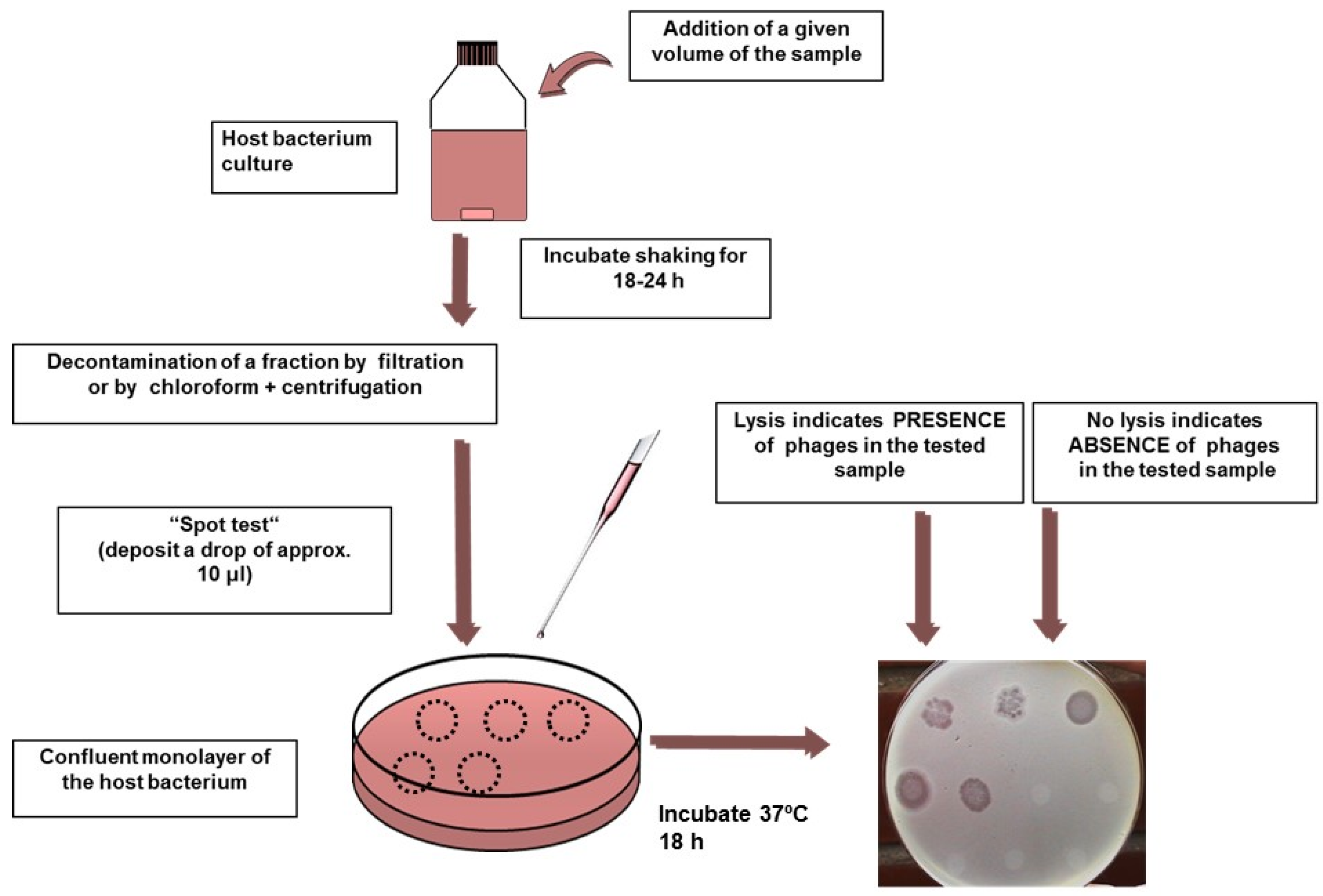
4. Are the Methods Cumbersome?

Some people argue that the methods for detecting bacteriophages are cumbersome, but there are many reasons to disagree with this viewpoint. As with all bacteriophages, coliphages can only replicate in metabolizing host cells. Infectious bacteriophages are typically detected by the effects they have on the host bacteria they infect, especially lysis. The most important factor in defining a method for the detection of a given phage or group of bacteriophages is the bacterial host strain. Phages are enumerated by direct quantitative plaque assays [41] (Figure 2); the plaque assay provides the results in plaque-forming units (PFUs). A plaque-forming unit is an entity, usually a single virion, but it may also be a clump of virions that gives rise to a single plaque of lysis in a monolayer of the host strain. PFUs are also known as plaque-forming particles (PFPs) in the ISO standards. The presence of phages in a given volume of sample can also be detected by the qualitative presence/absence enrichment test [41] (Figure 3). The enrichment of multiple tube serial dilutions allows the numbers of phages to be estimated using “quantal” methods such as the most probable number procedure.
Standardized methods are available for both somatic and F-specific coliphages. Mooijman and collaborators [6] and USEPA [42,43] have carried out interlaboratory validation tests of the ISO and USEPA methods described below. In addition, Mooijman et al. showed that the implementation of the ISO standard method is feasible in routine microbiology laboratories without any prior experience of bacteriophage handling [6].

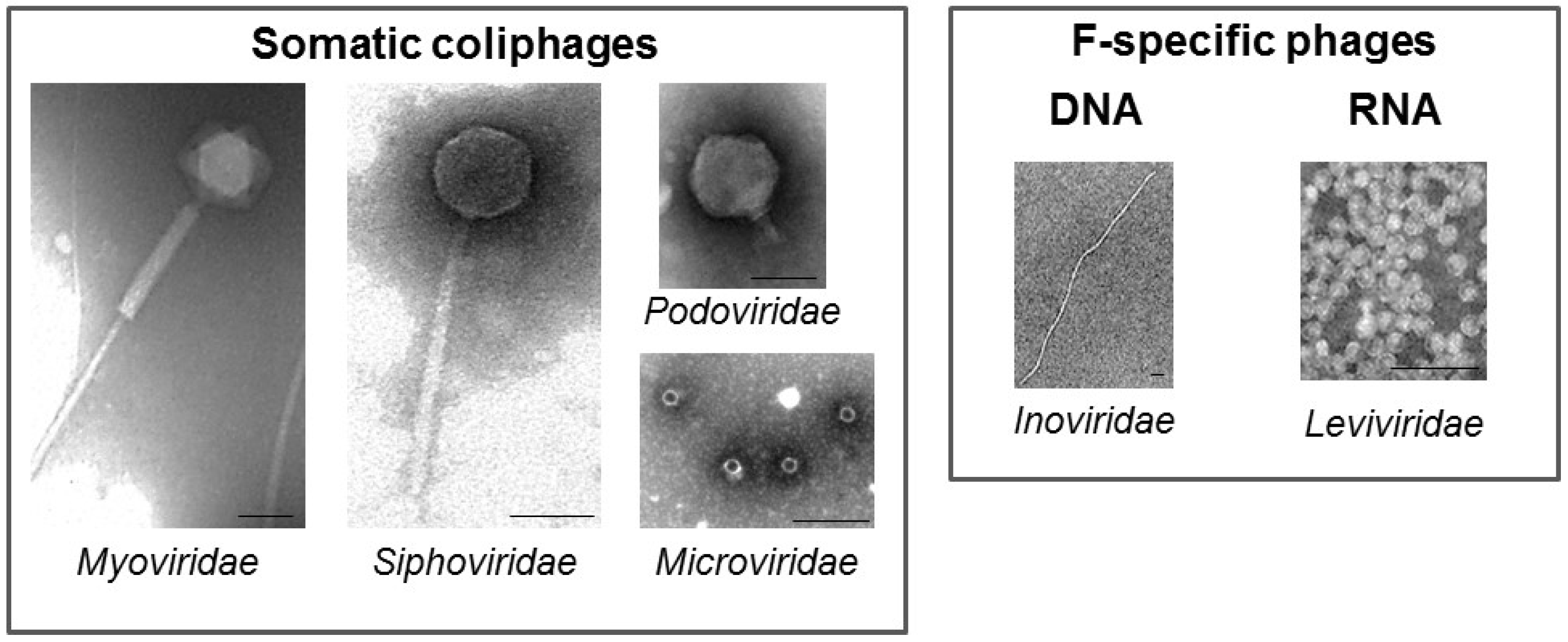
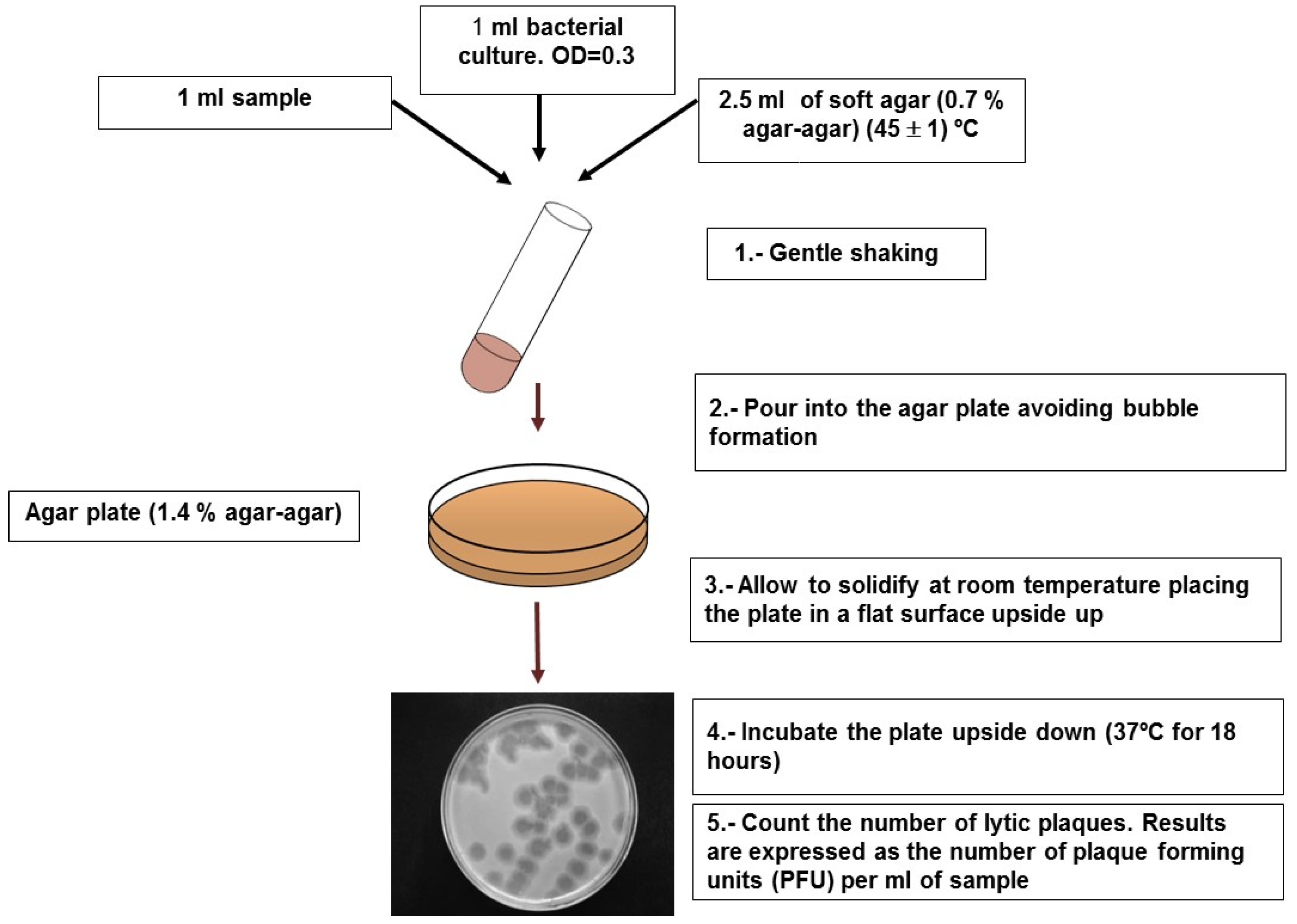
4.1. Somatic Coliphages

ISO [44], USEPA [45,46], and Standard Methods [47] have standardized procedures for the detection of somatic coliphages. The three methods use E. coli C (ATCC 13706) as the host strain and differ only in minor details relating to the media and assay conditions. The E. coli used is either E. coli ATCC 13706 (Standard Methods procedure) or its nalidixic acid-resistant clones E. coli CN13 (ATCC 700609), used in the USEPA 1601 and 1602 methods [45,46]; or E. coli CN13, more frequently referred to as WG5 (ATCC 700078), used in the ISO 10705-2 method [44]. Hosts resistant to nalidixic acid were introduced to minimize the growth of background bacteria that frequently interfere with the correct visualization of plaques in the plaque assay test. Strains CN13 and WG5 detect similar phage numbers in water matrices when the standardized method is used [48,49]. In addition, the present Standard Methods procedure theoretically provides similar counts to ISO and USEPA. By contrast, the Standard Methods procedure described in previous editions has been reported to perform poorly [50]. ISO-10705-2 [44] includes both the double agar layer (DAL) plaque assay method for the quantification of PFU (Figure 2) and the presence/absence test (Figure 3). A simplified version of these methods can be found at Havelaar and Hogeboom [51]. The USEPA Method 1601 [45] uses the presence/absence procedure and EPA Method 1602 [46] uses the single agar layer (SAL) plaque assay. A simplified version of these methods can be consulted on the USEPA website [52]. Results of quantification by PFU can be obtained within six hours.

4.2. F-Specific Coliphages

Host strains used to detect sexual coliphages must produce sexual pili encoded by the F-plasmid. Strains expressing pili and detecting minimal amounts of somatic coliphages have been tailored. These are strains E. coli HS (E. coli Famp, ATCC 700891) [53] and Salmonella enterica serovar Typhimurium (most frequently reported as Salmonella typhimurium) WG49 (NCTC 12484) [54]. The phages detected by these strains are counted as F-specific coliphages or male-specific coliphages or sexual coliphages. The number of F-specific RNA bacteriophages is the difference between the numbers of phages counted in the absence and in the presence of RNase in the assay medium, since this enzyme interferes with the infection of F-specific RNA phages. The counts achieved with strains HS and WG49 are similar [48,55,56]. Because of the genetic markers (resistance to ampicillin and capacity to use lactose), the stability (and re-selection) of strain WG49 is easier to verify than that of strain HS, which has no explicit genetic markers. However, strain HS seems more stable than strain WG49. The ISO-10705-1 standard method [57] for the detection and enumeration of F-specific bacteriophages includes a step with RNase in the assay medium. Consequently, it detects both F-specific and ARN F-specific bacteriophages. The ISO standard includes both the double layer plaque assay method and the presence/absence method. A simplified version can be found at Havelaar and Pot Hogeboom [54] and Standard Methods [47]. The EPA Method 1601 standard procedure [45] uses the presence/absence method and EPA method 1602 [46] uses the single-layer plaque assay (SAL) for the detection of F-specific bacteriophages that use E.coli Famp as the host strain. A simplified version can be found at USEPA [52] and Standard Methods [47]. Results using both plaque assay methods can be obtained within 18 hours.

4.3. Molecular and Fast Methods

No procedures based on serologic detection or polymerase chain reaction (PCR) that are applicable to the detection of the full somatic coliphage group in water have been described. The diversity of the bacteriophages in this group makes this approach complex, though not impossible.
For F-specific coliphages, both serological [58] and PCR-based methods are available to quantify F-specific RNA phages [59,60,61,62] and F-specific DNA phages [63]. However, these methods are used for a specific phage or subgroup, not for the full group of F-specific bacteriophages. The sum of values obtained for the different subgroups accounts for all F-specific bacteriophages. These methods can be applied directly to the sample, in which case they do not reveal the infectivity status of the detected genomes or genome fragments, or after the enrichment of multiple tube serial dilutions through the standardized methods, which allows phage numbers to be estimated by “quantal” methods such as the most probable number procedure.
Methods based on phage-induced host lysis through the detection of a molecule released by the lysed cell are also available and provide results for infectious bacteriophages. These methods are relatively fast because of the short time needed for a replication cycle and subsequent replication cycles, which lead to a high number of lysed cells in just a few hours in E. coli. Thus, Izjerman et al. described a method based on a rapid extracellular β-galactosidase enzyme release and detection during coliphage-induced lysis [64]. Later, a method based on the detection of somatic coliphages through a bioluminescence assay that measured the phage-mediated release of adenylate kinase and detected adenosine 5’-triphosphate was described [65]. In order to obtain results within a day, these methods were improved and adapted to include the enrichment of multiple tube serial dilutions based on the presence/absence test in the USEPA and ISO standards by Salter and Durbin and Salter et al., respectively [66,67]. Ongoing procedures to generate tailored host strains that can detect phage-induced cell lysis would reduce the time required to obtain results to less than four hours, even at low concentrations of somatic coliphages (Muniesa, personal communication). It should be relatively easy to adapt these methods based on the detection of molecules released by host lysis into the medium to user-friendly commercial kits.

4.4. Concentration Methods

Though the presence/absence method allows relatively large volumes to be tested (up to 1 L is quite feasible), concentration may sometimes be required, either because greater volumes need to be tested or because quantification is required. Most methods described for concentrating animal viruses are not suitable for concentrating bacteriophages [3], although ongoing projects may help change this situation. For volumes ranging from 10 to 1000 mL, two methods are recommended. For water samples with low turbidity, a simple, inexpensive, and practical procedure for the recovery and detection of F-specific phages using mixed cellulose and acetate membrane filters with a diameter of 47 mm and a pore size of 0.45 μm after addition of salts and pH adjustment was developed [68]. This method was modified slightly [69] and performed extremely well when concentrating somatic coliphages and F-specific RNA phages from samples of up to one liter. For samples with high turbidity, flocculation with magnesium hydroxide [70] is suitable for the two bacteriophage groups [71].
With respect to somatic coliphages, we cannot imagine many scenarios in which it would be necessary to concentrate them from water samples of more than one liter. The ratios of culturable viruses to somatic coliphages (mainly with reference to enteroviruses detected by cell culture) are 10:104 or 10:105 in the scenarios for which data on both are available. The claim is true for raw sewage, secondary and tertiary effluents [72,73,74,75,76,77], surface waters [73,78,79,80], and sludge [81,82]. The same ratio is true for infectious rotaviruses detected by cell culture followed by qRT-PCR [83]. No comparative data are available for other infectious viruses vs. coliphages in the same sample. However, some data that compared culturable viruses in the same water samples indicate that adenoviruses and enteroviruses [84,85] and rotavirus and enteroviruses [83,86] are found in concentrations of the same order of magnitude, and therefore the same ratio vs. somatic coliphages will be applicable. Moreover, the concentration of volumes greater than 1000 L is very seldom recommended and feasible, because higher volumes increase the uncertainty of the concentration methods [87], and if volumes greater than 1000 L are desired, several aliquots of 1000 L are recommended [88].

4.5. Additional Methodological Issues

4.5.1. Scale-Up

For all these standardized methods, the volumes of the samples tested, as described in the Methods section, can be scaled up by maintaining the proportions of the medium mixtures, host suspension, and sample, and, for the agar layer methods, by using larger Petri dishes.

4.5.2. Reference Material

Reference suspensions of model bacteriophages [6] and naturally occurring somatic and F-specific coliphages needed for quality assurance are easily prepared and conserved [5].

4.5.3. Costs

Detection and enumeration of somatic coliphages and F-specific phages by the standardized methods is relatively cost-effective. The costs of the material, media, reagents, and labor are similar to those required for the detection of fecal coliforms or E. coli. The cost of detecting F-specific RNA bacteriophages is 10–15% higher because of the need for RNase and double the number of plates. All these methods can be applied in routine microbiology laboratories without the need for extra equipment.

5. Which Group Should Be Used? Somatic vs. F-Specific Coliphages

There is some debate regarding which of the two groups of phages should be addressed by the regulations or whether both should be included. Information about the relative abundance of both phage groups and their relative persistence and resistance would help clarify the issue. Although there is a great deal of literature on these issues, this section only considers reports in which both phage groups have been analyzed by the standardized methods in the same samples.

5.1. Abundance in Pollution Sources

The primary origin of coliphages found in the water environment is human and animal feces. If we consider the percentage of human and animal individuals excreting phages and the amounts of phages excreted, the inputs of somatic coliphages [89,90,91] in the water environment directly from feces are greater than those of F-specific coliphages [90,91,92,93].
Coliphages reach water for human use by either point or diffuse pollution. Point pollution is contributed mostly by raw or treated human and animal (i.e., from abattoirs) wastewater, whereas diffuse pollution is mostly contributed by septic tank overflow, sewer leakage, and the spread of solid waste such as raw and treated sewage sludge, slurry, and manure, and the feces of pets, farmed animals, and wild animals. Other than feces, the main contributors, i.e., raw wastewater, secondary effluents, sludge, and slurry, will be reviewed below as polluting sources, in which somatic coliphages clearly outnumber F-specific phages when both groups of phages are counted using the standardized methods (USEPA or ISO) in the same samples, with a few exceptions.
Thus, somatic coliphages are detected in raw municipal wastewater throughout the world in 100% of the samples tested and values range from 5 × 10⁶–107 PFUs per 100 mL, with those of F-specific phages ranging from 104 to 106 PFUs per 100 mL [20,48,71,94,95]. With respect to community septic tanks, somatic and F-specific coliphages were detected in all samples tested, with values ranging from 106 to 107 PFU per 100 mL and 105 to 106 PFU per 100 mL, respectively [94].
Regarding animal contamination sources, the reported somatic coliphage values range from 104–105 PFU per 100 mL in slurry and manure to 4 × 10⁸ in abattoir wastewater, whereas the reported values for F-specific bacteriophages range from 104 PFU per 100 mL in animal waste slurries to 2 × 10⁸ in abattoir wastewaters [48,96,97].
Primary sedimentation, flocculation-aided sedimentation, activated sludge digestion, activated sludge digestion followed by precipitation, and trickling filters remove somatic coliphages and F-specific bacteriophages in numbers ranging from 50% to 99.9%. The most frequent somatic coliphage values in secondary effluents therefore normally range from 103 to 105 PFU per 100 mL and those of F-specific and RNA F-specific phages from 102 to 5 × 10⁴ PFU per 100 mL [20,48,74,76,95,98]. The relative proportions of somatic coliphages and F-specific and RNA F-specific bacteriophages are similar to those found in untreated wastewater.
The concentrations of somatic and F-specific phages detected in effluents released by lagooning are relatively variable depending on the extent of the treatment. In climates with considerable seasonal variation, the quality of the effluents in terms of pathogens and indicators numbers may vary from summer to winter in the same treatment facility [74,96,99,100]. In any case, the relative proportion of somatic coliphages to F-specific RNA phages remains the same or increases during warm periods.
The same trend, i.e., somatic coliphages outnumbering F-specific phages, is true for primary and raw sludge. Concentrations above 107 PFU for somatic coliphages and well over 106 PFU for F-specific bacteriophages per gram of dry weight of primary and raw sludge have been reported [81,101].

5.2. Persistence in Water Environments and Resistance to Treatment

Numerous data are available on persistence experiments conducted with laboratory-grown model somatic coliphages (ϕX174, PDR1, T2, and T7) and F-specific coliphages (f2, MS2, Qβ). Nevertheless, we will limit the discussion to naturally occurring coliphages. Concentrations of these phages in raw sewage are high enough to carry out model inactivation experiments after diluting sewage in fresh, brackish, or marine water.
Sinton et al. performed in situ inactivation experiments with sewage and/or waste stabilization pond effluent mixed with river or seawater in a proportion of 10% and placed at a depth of 560 mm (300 L) in open-top chambers [102,103]. Experiments were performed in winter (approx. 12 °C) and summer (approx. 16 °C), and in the dark and with sunlight. The calculated T90 for somatic coliphages ranged from seven hours in seawater, summer, and sunlight to 2303 hours in river water, winter, and darkness; the calculated T90 for F-specific RNA bacteriophages ranged from 4.8 hours in seawater, summer, and sunlight to >2303 hours in river water, winter, and darkness.
Durán et al. and Mocé-Llivina et al. mixed raw municipal wastewater at a proportion of 2% with either river or seawater and placed the mixture in a dialysis tube with a cutoff of 14,000 Daltons that was sealed and placed at a depth of 20 cm to 25 cm in the same site where the river and seawater were collected [104,105]. These sites were in the shadow for part of the day. With respect to somatic coliphages, T90 ranged from 53 hours in seawater and summer (>25 °C) to 385 hours in winter (6–10 °C) and river water, whereas F-specific RNA T90 ranged from 14 hours in seawater and summer to 323 hours in river water and winter.
Studies of the abatement of both phage groups after releasing manure, slurry, and sludge into soil are scarce. Following the land application of liquid pig manure, Gessel et al. found that the numbers of somatic coliphages after application were always higher than the values of F-specific phages, and that the proportion of somatic coliphages to F-specific phages increased moderately with time [106].
At this point in the review, it is worth mentioning that the great majority of data regarding the persistence and presence of F-specific RNA bacteriophages in receiving waters have been obtained in cold and temperate climates. However, some evidence indicates that in areas or periods with surface water temperatures higher than 25 °C the picture might be different. Thus, in the experiments performed in the summer [104,105], the T90 of E. coli and F-specific RNA bacteriophages were close; this situation was not observed for either somatic coliphages or phages infecting Bacteroides that show more persistence than E. coli. Also, with respect to oxidation ponds, which can be used for in situ inactivation experiments, it was reported that, in a series of stabilization ponds in the dry and warm desert of Judea (Israel), F-specific bacteriophages were removed more efficiently than fecal coliforms and somatic coliphages, primarily during the summer [100]. Moreover, F-specific RNA bacteriophages occurring in secondary effluents are significantly more sensitive to temperatures ranging from 25 °C to 40 °C than E. coli and somatic coliphages [107]. Cole et al. and Rahman et al. determined the numerical ratios between F-DNA phages and F-RNA phages in surface waters and observed that in summer F-specific RNA phages were in the minority, whereas they accounted for more than 90% during the other seasons [108,109]. The abatement of F-specific RNA phages in sludge mesophilic (30–35 °C) anaerobic digestion is faster (approximately two log10 units versus one log10 unit) than that of somatic coliphages or Bacteroides phages [81,101]. This acceleration in the inactivation kinetics of F-specific bacteriophages with increasing temperatures is also observed in experiments with laboratory-grown bacteriophages. Thus, McLaughlin and Rose described very high decay rates for MS2, an F-specific bacteriophage, in seawater at 30 °C [110], and Allwood et al. revealed that the decay rate of MS2 at 37 °C is even greater than that of E. coli [111]. All these results suggest that the persistence of F-specific RNA phages might be rated as low in waters with temperatures greater than 25 °C. Since geographic areas with surface seawater and fresh water temperatures exceeding 25 °C either all year round (between 40° latitude north and 40° latitude south) or during the warm seasons (many areas of Europe and the USA) are fairly extensive, the persistence of F-specific and RNA F-specific bacteriophages requires additional verification before their persistence in warm areas can be rated.

5.3. Resistance to Treatment

It has been mentioned previously that all indicators in general and somatic coliphages and F-specific bacteriophages are eliminated in a similar way in primary and secondary wastewater treatment. However, the situation changes frequently when additional treatments such as filtration through depth filters (sand, anthracite, etc.) or membrane filtration and either chemical or physical disinfection are used.
There is a great deal of literature on the effects of these treatments in certain specific somatic (ϕX174, PDR1, T2, and T7) and F-specific bacteriophages (f2, MS2, Qβ). However, as indicated in a previous section, both groups are heterogeneous and what happens to a particular bacteriophage may not reflect what happens to the whole group. The significant amounts of somatic and F-specific coliphages in some matrices (secondary effluents, some contaminated rivers, raw sludge, etc.) make it possible to reveal the effect of the treatments on both groups of bacteriophages as a whole. A certain amount of information is available from pilot or full-scale tertiary treatments used for water reclamation, sludge treatment, and filtration through soil. As discussed below, the two groups respond differently depending on the treatment.
Depth filtration (sand, anthracite, etc.), precipitation/flocculation plus filtration [96,112,113,114], and even membrane filtration reactors [20,114,115,116] remove somatic and specific coliphages found in secondary effluent in equal measures. Consequently, the proportions of the two groups of bacteriophages are maintained after filtration and somatic coliphages normally outnumber F-specific bacteriophages in the permeated waters.
Chlorination causes a greater effect on somatic coliphages than on F-specific phages, but this difference is not great enough to change the relative proportions, and somatic coliphages outnumber F-specific phages in most chlorinated secondary effluents [76,113,117,118].
With respect to radiation, UV irradiation has a significantly greater effect on somatic coliphages than on F-specific phages, and after strong UV treatment F-specific numbers surpass those of somatic coliphages in many settings [76,117].
Photo-oxidation (H2O2, TiO2) used to be a highly effective treatment for bacteriophages, as described for F-specific bacteriophages, but when photo-oxidizing a secondary effluent, Agulló et al. detected significantly greater elimination of F-specific bacteriophages and thus concluded that effluents harbor greater numbers of somatic coliphages [107].
Most sludge treatments required prior to its release in soil are based on heat. Major F-specific phage inactivation is already observed in mesophilic digestion (30–35 °C) and all treatments with higher temperatures increase the difference [81,101,119], so the great majority of biosolids released into the environment contain higher numbers of somatic coliphages than F-specific phages.
Regarding the effect of soil filtration on the abatement of both phage groups, studies that compare both groups are once again in short supply. Sinton et al. reported that F-specific RNA phages showed greater attenuation than somatic coliphages in an alluvial gravel aquifer [120].

5.4. Abundance of Bacteriophages in Surface Water and Groundwater

Due to the discharge of raw or treated contaminants and their inactivation once in nature, the gap between the numbers of somatic and F-specific phages in receiving surface water and groundwater is equal to or greater than the gap at the source, with a few exceptions.
Reports concerning concentrations of both phage groups measured by either the USEPA or ISO methods in fresh and marine surface waters indicate that the numbers of somatic coliphages are greater than the numbers of F-specific phages (Table 1).
Most reports on phages in groundwater only account for their presence or absence in a given volume. A few examples of the values found are reported in Table 2. Somatic coliphages predominate overall, but in this case somatic coliphages prevail in some aquifers and F-specific phages in others. This is very likely to depend on the characteristics of the soil, since both phage groups have been reported to adsorb differently to different minerals [121].

5.5. General Conclusions for Practical Uses

The general trend is that somatic coliphages outnumber F-specific coliphages. However, there are some situations, such as reclaimed water, in which UV plays the main role in inactivation [117], clayey sediments [121], and groundwater from certain aquifers [128], in which F-specific phages have been observed to predominate.
Therefore, detecting both bacteriophage groups simultaneously would appear to be a good option. Strain E. coli C3000 (ATCC 15597) has been used extensively for this purpose, mainly in the USA, but it detects lower numbers of somatic coliphages than strains CN13 and WG5. More recently, Guzmán et al. tailored a strain [49], CB390, which detects both phage groups, and reported numbers in Spain similar to the sum of those detected by WG5 or CN13 and those detected by strains WG49 or F (amp) HS. Recently, this strain has been validated in the USA [132] and has proved to be just as efficient as in Spain.

6. Relationship of Coliphages to the Occurrence of Human Viruses and to Health Risk

6.1. Relationship to Human Viruses

In addition to their value as indicators of fecal contamination, indicator bacteriophages have been viewed as potential surrogates of human viruses. Consequently, the possible relationship between the presence and levels of indicator phages and human viruses in water has been studied, though with disparate results.
Thus, many studies have failed to show any correlation in fresh surface waters [133,134,135,136,137], marine surface water [136,137,138], and groundwater [131,139]. In contrast, other studies have shown some correlation between human viruses and F-specific phages [23,123,126,140,141] and between human viruses and somatic coliphages in surface waters [104,140,142], between enteroviruses and somatic coliphages in sludge [82], and between human viruses and both somatic and F-specific phages in surface water [78]. Thus, even if the results far from indicate a clear correlation between indicator coliphage densities and human viruses in water, there is evidence that somatic and F-specific coliphages are more strongly associated with pathogenic viruses than the traditional bacterial indicators and even a particular pathogenic virus. Accordingly, low rates of co-occurrence or an absence of correlation among different human viruses have been reported in different settings. In groundwater these failures have been identified for enteroviruses, rotaviruses, hepatitis-A viruses, and noroviruses [131,139]; noroviruses, enteroviruses, rotaviruses, adenoviruses, and hepatitis-A viruses [129]; enteroviruses and noroviruses [127]; and enteroviruses, adenoviruses, and reoviruses [143]. In surface waters, a lack of co-occurrence or correlation has been reported for adenoviruses and enteroviruses [136]; enteroviruses and adenoviruses [142]; enteroviruses and adenoviruses [124]; adenoviruses and noroviruses [144]; and enteroviruses and polyomaviruses [145]. In contrast, some studies on surface waters have shown correlations among different pathogenic viruses, such as astroviruses, noroviruses, rotaviruses, and adenoviruses [23]; enteroviruses, reoviruses, noroviruses, and rotaviruses [78]; and adenoviruses and noroviruses [141]. However, these three studies also report correlations between human viruses and coliphages.
Potential theories have been proposed to explain these unequal results. One is that somatic and F-specific coliphages are released into the environment by humans and animals, whereas human viruses are not. For this purpose, the use of bacteriophages found mostly in humans, such as those detected by some specific strains of Bacteroides or some genogroups (II and III) of F-specific phages [146], would probably offer more congruent results. Other reasons are strictly methodological, since these studies vary somewhat in terms of the parameters assessed and the concentration and detection procedures used. Bacteriophages have usually been evaluated by plaque assay using standardized (or equivalent) methods in small volumes, and without a concentration procedure. Human viruses have been evaluated after the concentration of great volumes and the subsequent detection using very different methods, such as plaque assay on cell culture and different PCR approaches. Most of these counts, which have mostly been in matrices where the numbers of phages and viruses are low, are subject to a great amount of uncertainty, which partly explains the considerable heterogeneity of the results. Moreover, in our opinion, the statistical methods used to establish associations are not always suited to the characteristics of the data, which are frequently characterized by predominance in non-detected values, with detection limits for the different parameters analyzed not always equivalent.

6.2. Relationship to Health Risk

With respect to the relationship to health risk, seven epidemiological studies conducted to evaluate the relationships between the presence of indicator bacteriophages in surface waters and swimming illnesses have been carried out with disparate outcomes. Three of them only studied F-specific phages, and while Lee et al. and Wade et al. found some correlation [147,148], Van Asperen et al. did not [149]. Wiedenmann et al. only studied somatic coliphages and found some correlation [150]. In the studies where both phages were tested, the results are incongruent, since Von Schirnding et al. failed to show any correlation with either somatic or F-specific phages [151], Colford et al. found a correlation with F-specific phages [152], and Abdelzaher et al. found a correlation with somatic coliphages [145]. No epidemiological studies to correlate phages and disease in drinking water have been performed. However, some overlap between a jaundice outbreak and a high incidence of somatic coliphages in drinking water in a municipality of West Bengala, India was reported in 2014 [153].
Epidemiological studies on the relationship between the presence of human viruses and disease associated with water are scarce and those available fail to show any association between pathogenic viruses in surface waters and infections associated with bathing [145,152].

7. Conclusions

The joint threshold concentrations of the bacterial hosts required to guarantee phage replication and the known concentrations of coliphages detected by the host strains of standardized methods limit the sites where these numbers are reached. Moreover, some basic aspects of phage biology indicate that the replication of coliphages outside the gut is highly inefficient. Therefore, coliphage replication in the water environment cannot be ruled out, but if it does occur the resulting numbers will be lower than those contributed by fecal waste.
There are easy, rapid, and cost-effective standardized methods for detecting and enumerating coliphages. Implementing these methods without the need for extra equipment in routine laboratories is feasible after a little training. It should be relatively easy to adapt recent developments to very fast methods (three to four hours) based on the detection of molecules released into the culture medium by phage-induced host lysis to user-friendly commercial kits.
In fecal pollution sources and most water environments with varying degrees of pollution, numbers of somatic coliphages exceed those of F-specific coliphages. Persistence in the environment is similar, with the exception of warm water, where the survival of F-specific RNA phages is relatively low. With respect to resistance, this depends on the treatment; thus, while F-specific phages survive better after chlorination and UV irradiation used in tertiary treatments, somatic coliphages are more resistant to the thermal treatments used in sludge hygienization. With a few exceptions, the difference in resistance is not enough to make F-specific phages more abundant than somatic coliphages. The general trend is that somatic coliphages outnumber F-specific coliphages in a wide range of samples. However, there are some situations, such as reclaimed water, in which UV plays the main role in inactivation, clayey sediments, and groundwater from certain aquifers, in which F-specific phages have been observed to predominate. Therefore, detecting both bacteriophage groups simultaneously would appear to be a good option. For this purpose, strain CB 390 of E. coli, which detects both groups in numbers similar to those detected by somatic and F-specific standardized methods, is available.
Possible correlations between the presence and levels of coliphages and human viruses in water have been studied, though with disparate results. However, the correlations between the presence and levels of different human viruses are no higher.
The epidemiological evidence of the correlation between coliphage concentrations in water and health risks suggests a likely relationship, although no sound statistical correlations have been found. The same statement is valid for human viruses.
As a general conclusion, coliphages that can be detected by easy, rapid, and cost-effective methods are likely to be a better indicator of viruses in fecal contamination than the current FIB (i.e., enterococci and E. coli).

Acknowledgments

This study was supported by Generalitat de Catalunya regional authorities via the Recognized Research Group: SGR-2014-007, and the Water Research Institute (University of Barcelona).

Author Contributions

The authors have similarly contributed in the preparation of this review article.

Conflicts of Interest

The authors declare no conflict of interest.

References

  1. AWPRC Study Group on Health Related Water Microbiology. Bacteriophages as model viruses in water quality controlag. Water Res. 1991, 25, 529–545. [Google Scholar]
  2. Armon, R.; Kott, Y. Bacteriophages as indicators of pollution. Crit. Rev. Environ. Sci. Technol. 1996, 26, 299–335. [Google Scholar] [CrossRef]
  3. Grabow, W. Bacteriophages: update on application as models for viruses in water. Water SA 2001, 27, 251–268. [Google Scholar] [CrossRef]
  4. Jofre, J. Indicators of waterborne viruses. In Human Viruses in Water; Bosch, A., Ed.; Perspectives in Medical Virology Volume 17; Elsevier: Amsterdam, The Netherlands, 2007. [Google Scholar]
  5. Mendez, J.; Jofre, J.; Lucena, F.; Contreras, N.; Mooijman, K.; Araujo, R. Conservation of phage reference materials and water samples containing bacteriophages of enteric bacteria. J. Virol. Methods 2002, 106, 215–224. [Google Scholar] [CrossRef]
  6. Mooijman, K.A.; Ghameshlou, Z.; Bahar, M.; Jofre, J.; Havelaar, A.H. Enumeration of bacteriophages in water by different laboratories of the European Union in two interlaboratory comparison studies. J. Virol. Methods 2005, 127, 60–68. [Google Scholar] [CrossRef] [PubMed]
  7. Queensland-Government. Queensland Water Recycling Guidelines; WaterWise; Queensland Environmental Protection Agency: Brisbane, Australia, 2005. [Google Scholar]
  8. North Carolina Environmental Quality. North Carolina Adm. Code 15A NCAC 2U Reclaimed Water; North Carolina Department of Environment and Natural Resources: Raleigh, NC, USA, 2011. [Google Scholar]
  9. USEPA. National Primary Drinking Water Regulations: Groundwater rule. Final Rule; 40 CFR Parts 9, 141 and 142, Federal Register, Vol. 71, n. 216; USEPA: Washington, DC, USA, 2006. [Google Scholar]
  10. Department of Environment and Conservation. Western Australian Guidelines for Biosolids Management; Department of Environment and Conservation: Perth, Australia, 2012. [Google Scholar]
  11. Criterios para el uso de biosólidos generados en plantas de tratamiento de aguas residuales municipales; Decreto Número 1287; Ministerio de Vivienda, Ciudad y Territorio de Colombia: Bogot, Colombia, 2014.
  12. USEPA Office of Water. Review of Coliphages as Possible Indicators of Fecal Contamination for Ambient Water Quality. EPA 820-R-15-098; USEPA Office of Water: Washington, DC, USA, 2015. [Google Scholar]
  13. Rajala-Mustonen, R.L.; Heinonen-Tanski, H. Sensitivity of host strains and host range of coliphages isolated from Finnish and Nicaraguan wastewater. Water Res. 1994, 28, 1811–1815. [Google Scholar] [CrossRef]
  14. Muniesa, M.; Lucena, F.; Jofre, J. Study of the potential relationship between the morphology of infectious somatic coliphages and their persistence in the environment. J. Appl. Microbiol. 1999, 87, 402–409. [Google Scholar] [CrossRef] [PubMed]
  15. Fauquet, C.; Mayo, M.A.; Maniloff, J.; Desselberger, U.; Ball, L. Virus Taxonomy: Eighth Report of the International Committee on Taxonomy of Viruses; Elsevier/Academic Press: London, UK, 2005. [Google Scholar]
  16. Furuse, K. Distribution of coliphages in the environment: general considerations. In Phage Ecology; Goyal, S.M., Gerba, C.P., Bitton, G., Eds.; John Wiley & Sons, Inc.: New York, NY, USA, 1987; pp. 87–124. [Google Scholar]
  17. Beekwilder, J.; Nieuwenhuizen, R.; Havelaar, A.H.; van Duin Leiden, J. An oligonucleotide hybridization assay for the identification and enumeration of F-specific RNA phages in surface water. J. Appl. Bacteriol. 1996, 80, 179–186. [Google Scholar] [CrossRef] [PubMed]
  18. Hsu, F.C.; Shieh, Y.S.; van Duin, J.; Beekwilder, M.J.; Sobsey, M.D. Genotyping male-specific RNA coliphages by hybridization with oligonucleotide probes. Appl. Environ. Microbiol. 1995, 61, 3960–3966. [Google Scholar] [PubMed]
  19. Muniesa, M.; Lucena, F.; Blanch, A.R.; Payán, A.; Jofre, J. Use of abundance ratios of somatic coliphages and bacteriophages of Bacteroides thetaiotaomicron GA17 for microbial source identification. Water Res. 2012, 46, 6410–6418. [Google Scholar] [CrossRef] [PubMed]
  20. Zhang, K.; Farahbakhsh, K. Removal of native coliphages and coliform bacteria from municipal wastewater by various wastewater treatment processes: Implications to water reuse. Water Res. 2007, 41, 2816–2824. [Google Scholar] [CrossRef] [PubMed]
  21. Haramoto, E.; Fujino, S.; Otagiri, M. Distinct behaviors of infectious F-specific RNA coliphage genogroups at a wastewater treatment plant. Sci. Total Environ. 2015, 520, 32–38. [Google Scholar] [CrossRef] [PubMed]
  22. Ferrer, O.; Casas, S.; Galvañ, C.; Lucena, F.; Bosch, A.; Galofré, B.; Mesa, J.; Jofre, J.; Bernat, X. Direct ultrafiltration performance and membrane integrity monitoring by microbiological analysis. Water Res. 2015, 83, 121–131. [Google Scholar] [CrossRef] [PubMed]
  23. Rezaeinejad, S.; Vergara, G.G.; Woo, C.H.; Lim, T.T.; Sobsey, M.D.; Gin, K.Y. Surveillance of enteric viruses and coliphages in a tropical urban catchment. Water Res. 2014, 58, 122–131. [Google Scholar] [CrossRef] [PubMed]
  24. Imhoff, J.F. Enterobacteriales. In Bergey’s Manual® of Systematic Bacteriology. Volume 2: The Proteobacteria, Part B: The Gammaproteobacteria; Garrity, G., Brenner, D.J., Krieg, N.R., Staley, J.R., Eds.; Springer US: Boston, MA, USA, 2005; pp. 587–850. [Google Scholar]
  25. Ishii, S.; Sadowsky, M.J. Escherichia coli in the Environment: Implications for Water Quality and Human Health. Microbes Environ. 2008, 23, 101–108. [Google Scholar] [CrossRef] [PubMed]
  26. Vaughn, J.M.; Metcalf, T.G. Coliphages as indicators of enteric viruses in shellfish and shellfish raising estuarine waters. Water Res. 1975, 9, 613–616. [Google Scholar] [CrossRef]
  27. Seeley, N.D.; Primrose, S.B. The effect of temperature on the ecology of aquatic bacteriophages. J. Gen. Virol. 1980, 46, 87–95. [Google Scholar] [CrossRef]
  28. Grabow, W.O.K.; Burger, J.S.; Nupen, E.P. Evaluation of acid-fast bacteria, Candida albicans, enteric viruses and conventional indicators for monitoring wastewater reclamation systems. Prog. Water Technol 1980, 12, 803–817. [Google Scholar]
  29. Borrego, J.J.; Córnax, R.; Moriñigo, M.A.; Martínez-Manzanares, E.; Romero, P. Coliphages as an indicator of faecal pollution in water. their survival and productive infectivity in natural aquatic environments. Water Res. 1990, 24, 111–116. [Google Scholar] [CrossRef]
  30. Wiggins, B.A.; Alexander, M. Minimum bacterial density for bacteriophage replication: Implications for significance of bacteriophages in natural ecosystems. Appl. Environ. Microbiol. 1985, 49, 19–23. [Google Scholar] [PubMed]
  31. Muniesa, M.; Jofre, J. Factors influencing the replication of somatic coliphages in the water environment. Antonie Van Leeuwenhoek 2004, 86, 65–76. [Google Scholar] [CrossRef] [PubMed]
  32. Muniesa, M.; Mocé-Llivina, L.; Katayama, H.; Jofre, J. Bacterial host strains that support replication of somatic coliphages. Antonie Van Leeuwenhoek 2003, 83, 305–315. [Google Scholar] [CrossRef] [PubMed]
  33. Cornax, R.; Moriñigo, M.A.; Balebona, M.C.; Castro, D.; Borrego, J.J. Significance of several bacteriophage groups as indicators of sewage pollution in marine waters. Water Res. 1991, 25, 673–678. [Google Scholar] [CrossRef]
  34. Raivio, T. Identifying your enemies—Could envelope stress trigger microbial immunity? Mol. Microbiol. 2011, 79, 557–561. [Google Scholar] [CrossRef] [PubMed]
  35. Perez-Rodriguez, R.; Haitjema, C.; Huang, Q.; Nam, K.H.; Bernardis, S.; Ke, A.; DeLisa, M.P. Envelope stress is a trigger of CRISPR RNA-mediated DNA silencing in Escherichia coli. Mol. Microbiol. 2011, 79, 584–599. [Google Scholar] [CrossRef] [PubMed]
  36. Muniesa, M.; Jofre, J. The contribution of induction of temperate phages to the numbers of free somatic coliphages in waters is not significant. FEMS Microbiol. Lett. 2007, 270, 272–276. [Google Scholar] [CrossRef] [PubMed]
  37. Hernández-Delgado, E.A.; Toranzos, G.A. In situ replication studies of somatic and male-specific coliphages in a tropical pristine river. Water Sci. Technol. 1995, 31, 247–250. [Google Scholar] [CrossRef]
  38. Jofre, J. Is the replication of somatic coliphages in water environments significant? J. Appl. Microbiol. 2009, 106, 1059–1069. [Google Scholar] [CrossRef] [PubMed]
  39. Havelaar, A.H.; Pot-Hogeboom, W.M. F-specific RNA-Bacteriophages as model viruses in water hygiene: Ecological aspects. Water Sci. Technol. 1988, 20, 399–407. [Google Scholar]
  40. Woody, M.A.; Cliver, D.O. Replication of coliphage Q beta as affected by host cell number, nutrition, competition from insusceptible cells and non-FRNA coliphages. J. Appl. Microbiol. 1997, 82, 431–440. [Google Scholar] [CrossRef] [PubMed]
  41. Adams, M.H. Bacteriophages; Interscience Publishers, Inc.: New York, NY, USA, 1959. [Google Scholar]
  42. USEPA Office of Water. Results of the Interlaboratory Validation of EPA Method 1601 for Presence/Absence of Male-specific (F+) and Somatic Coliphages in Water by Two-Step Enrichment; EPA 821-R-03-015; USEPA Office of Water: Washington, DC, USA, 2003. [Google Scholar]
  43. USEPA Office of Water. Results of the Interlaboratory Validation of EPA Method 1602 for Enumeration of Male-specific (F+) and Somatic Coliphage in Water by Single Agar Layer (SAL); EPA 821-R-03-016; USEPA Office of Water: Washington, DC, USA, 2003. [Google Scholar]
  44. International Standardization Organization. Water Quality—Detection and Enumeration of Bacteriophages—Part 2: Enumeration of Somatic Coliphages; ISO-10705-2; International Standardization Organization: Geneva, Switzerland, 2000. [Google Scholar]
  45. USEPA Office of Water. Method 1601: Male-specific (F+) and Somatic Coliphage in Water by Two-step Enrichment Procedure; EPA 821-R-01-030; USEPA Office of Water: Washington, DC, USA, 2001. [Google Scholar]
  46. USEPA Office of Water. Method 1602: Male-specific (F+) and Somatic Coliphage in Water by Single Agar Layer (SAL) Procedure; EPA 821-R-01-029; USEPA Office of Water: Washington, DC, USA, 2001. [Google Scholar]
  47. Rice, E.W.; Baird, R.B.; Eaton, A.D.; Clesceri, L.S. Standard Methods for the Examination of Water and Wastewater; American Public Health Association, American Water Works Association, Water Environment Federation: Washington, DC, USA, 2012. [Google Scholar]
  48. Grabow, W.O.K.; Holtzhausen, C.S.; de Villiers, J.C. Research on Bacteriophages as Indicators of Water Quality; WRC Report No. 321/1/93; Water Research Commission: Pretoria, South Africa, 1993. [Google Scholar]
  49. Guzmán, C.; Mocé-Llivina, L.; Lucena, F.; Jofre, J. Evaluation of Escherichia coli host strain CB390 for simultaneous detection of somatic and F-specific coliphages. Appl. Environ. Microbiol. 2008, 74, 531–534. [Google Scholar] [CrossRef] [PubMed]
  50. Green, J. The APHA standard method for the enumeration of somatic coliphages in water has low efficiency of plating. Water Res. 2000, 34, 759–762. [Google Scholar] [CrossRef]
  51. Havelaar, A.H.; Hogeboom, W.M. Factors affecting the enumeration of coliphages in sewage and sewage-polluted waters. Antonie Van Leeuwenhoek 1983, 49, 387–397. [Google Scholar] [PubMed]
  52. USEPA Office of Research and Development. USEPA Manual of Methods for Virology—Chapter 16; EPA 600/4-84/013 (N16); USEPA Office of Research and Development: Washington, DC, USA, 2001. [Google Scholar]
  53. Debartolomeis, J.; Cabelli, V.J. Evaluation of an Escherichia coli host strain for enumeration of F male-specific bacteriophages. Appl. Environ. Microbiol. 1991, 57, 1301–1305. [Google Scholar] [PubMed]
  54. Havelaar, A.H.; Hogeboom, W.M. A method for the enumeration of male-specific bacteriophages in sewage. J. Appl. Bacteriol. 1984, 56, 439–447. [Google Scholar] [CrossRef] [PubMed]
  55. Chung, H.; Sobsey, M.D. Comparative survival of indicator viruses and enteric viruses in seawater and sediment. Water Sci. Technol. 1993, 27, 425–428. [Google Scholar]
  56. Schaper, M.; Jofre, J. Comparison of methods for detecting genotypes of F-specific RNA bacteriophages and fingerprinting the origin of faecal pollution in water samples. J. Virol. Methods 2000, 89, 1–10. [Google Scholar] [CrossRef]
  57. International Standardization Organization. Water Quality—Detection and Enumeration of Bacteriophages—Part 1: Enumeration of F-specific RNA Bacteriophages; ISO-10705-1; International Standardization Organization: Geneva, Switzerland, 1995; p. 11. [Google Scholar]
  58. Love, D.C.; Sobsey, M.D. Simple and rapid F+ coliphage culture, latex agglutination, and typing assay to detect and source track fecal contamination. Appl. Environ. Microbiol. 2007, 73, 4110–4118. [Google Scholar] [CrossRef] [PubMed]
  59. Ogorzaly, L.; Gantzer, C. Development of real-time RT-PCR methods for specific detection of F-specific RNA bacteriophage genogroups: Application to urban raw wastewater. J. Virol. Methods 2006, 138, 131–139. [Google Scholar] [CrossRef] [PubMed]
  60. Kirs, M.; Smith, D.C. Multiplex quantitative real-time reverse transcriptase PCR for F+-specific RNA coliphages: a method for use in microbial source tracking. Appl Env. Microbiol 2007, 73, 808–814. [Google Scholar] [CrossRef] [PubMed]
  61. Friedman, S.D.; Cooper, E.M.; Casanova, L.; Sobsey, M.D.; Genthner, F.J. A reverse transcription-PCR assay to distinguish the four genogroups of male-specific (F+) RNA coliphages. J. Virol. Methods 2009, 159, 47–52. [Google Scholar] [CrossRef] [PubMed]
  62. Wolf, S.; Hewitt, J.; Greening, G.E. Viral multiplex quantitative PCR assays for tracking sources of fecal contamination. Appl. Environ. Microbiol. 2010, 76, 1388–1394. [Google Scholar] [CrossRef] [PubMed]
  63. Long, S.C.; El-Khoury, S.S.; Oudejans, S.J.G.; Sobsey, M.D.; Vinjé, J. Assessment of sources and diversity of male-specific coliphages for source tracking. Environ. Eng. Sci. 2005, 22, 367–377. [Google Scholar] [CrossRef]
  64. Ijzerman, M.M.; Falkinham, J.O.; Hagedorn, C. A liquid, colorimetric presence-absence coliphage detection method. J. Virol. Methods 1993, 45, 229–234. [Google Scholar] [CrossRef]
  65. Guzmán Luna, C.; Costán-Longares, A.; Lucena, F.; Jofre, J. Detection of somatic coliphages through a bioluminescence assay measuring phage mediated release of adenylate kinase and adenosine 5’-triphosphate. J. Virol. Methods 2009, 161, 107–113. [Google Scholar] [CrossRef] [PubMed]
  66. Salter, R.S.; Durbin, G.W. Modified USEPA method 1601 to indicate viral contamination of groundwater. J. Am. Water Works Assoc. 2012, 104, 480–488. [Google Scholar] [CrossRef]
  67. Salter, R.S.; Durbin, G.W.; Conklin, E.; Rosen, J.; Clancy, J. Proposed modifications of Environmental Protection Agency Method 1601 for detection of coliphages in drinking water, with same-day fluorescence-based detection and evaluation by the performance-based measurement system and alternative test protocol valida. Appl. Environ. Microbiol. 2010, 76, 7803–7810. [Google Scholar] [CrossRef] [PubMed]
  68. Sobsey, M.D.; Schwab, K.J.; Handzel, T.R. A simple membrane filter method to concentrate and enumerate male-specific RNA coliphages. J. Am. Water Work. Assoc. 1990, 82, 52–59. [Google Scholar]
  69. Méndez, J.; Audicana, A.; Isern, A.; Llaneza, J.; Moreno, B.; Tarancón, M. L.; Jofre, J.; Lucena, F. Standardised evaluation of the performance of a simple membrane filtration-elution method to concentrate bacteriophages from drinking water. J. Virol. Methods 2004, 117, 19–25. [Google Scholar] [CrossRef] [PubMed]
  70. Schulze, E.; Lenk, J. Concentration of coliphages from drinking water by Mg(OH)2 flocculation. Naturwissenschaften 1983, 70, 612–613. [Google Scholar] [CrossRef] [PubMed]
  71. Contreras-Coll, N.; Lucena, F.; Mooijman, K.; Havelaar, A.; Pierz, V.; Boque, M.; Gawler, A.; Höller, C.; Lambiri, M.; Mirolo, G.; et al. Occurrence and levels of indicator bacteriophages in bathing waters throughout Europe. Water Res. 2002, 36, 4963–74. [Google Scholar] [CrossRef]
  72. Gerba, C.P.; Pepper, I.L.; Whitehead, L.F. A risk assessment of emerging pathogens of concern in the land application of biosolids. Water Sci. Technol. 2002, 46, 225–230. [Google Scholar] [PubMed]
  73. Lodder, W.J.; de Roda Husman, A.M. Presence of noroviruses and other enteric viruses in sewage and surface waters in The Netherlands. Appl. Environ. Microbiol. 2005, 71, 1453–1461. [Google Scholar] [CrossRef] [PubMed]
  74. Gomila, M.; Solis, J.J.; David, Z.; Ramon, C.; Lalucat, J. Comparative reductions of bacterial indicators, bacteriophage-infecting enteric bacteria and enteroviruses in wastewater tertiary treatments by lagooning and UV-radiation. Water Sci. Technol. 2008, 58, 2223–2233. [Google Scholar] [CrossRef] [PubMed]
  75. Gantzer, C.; Maul, A.; Audic, J.M.; Schwartzbrod, L. Detection of infectious enteroviruses, enterovirus genomes, somatic coliphages, and Bacteroides fragilis phages in treated wastewater. Appl. Environ. Microbiol. 1998, 64, 4307–4312. [Google Scholar] [PubMed]
  76. Costán-Longares, A.; Montemayor, M.; Payán, A.; Méndez, J.; Jofre, J.; Mujeriego, R.; Lucena, F. Microbial indicators and pathogens: Removal, relationships and predictive capabilities in water reclamation facilities. Water Res. 2008, 42, 4439–4448. [Google Scholar] [CrossRef] [PubMed]
  77. Harwood, V.J.; Levine, A.D.; Scott, T.M.; Chivukula, V.; Lukasik, J.; Farrah, S.R.; Rose, J.B. Validity of the indicator organism paradigm for pathogen reduction in reclaimed water and public health protection. Appl. Environ. Microbiol. 2005, 71, 3163–3170. [Google Scholar] [CrossRef] [PubMed]
  78. Lodder, W.J.; van den Berg, H.H.J.L.; Rutjes, S.A.; de Roda Husman, A.M. Presence of enteric viruses in source waters for drinking water production in The Netherlands. Appl. Environ. Microbiol. 2010, 76, 5965–5971. [Google Scholar] [CrossRef] [PubMed]
  79. Rubiano, M.-E.; Agulló-Barceló, M.; Casas-Mangas, R.; Jofre, J.; Lucena, F. Assessing the effects of tertiary treated wastewater reuse on a Mediterranean river (Llobregat, NE Spain): Pathogens and indicators. Environ. Sci. Pollut. Res. Int. 2012, 19, 1026–1032. [Google Scholar] [CrossRef] [PubMed]
  80. Lipp, E.K.; Farrah, S.A.; Rose, J.B. Assessment and impact of microbial fecal pollution and human enteric pathogens in a coastal community. Mar. Pollut. Bull. 2001, 42, 286–293. [Google Scholar] [CrossRef]
  81. Guzmán, C.; Jofre, J.; Montemayor, M.; Lucena, F. Occurrence and levels of indicators and selected pathogens in different sludges and biosolids. J. Appl. Microbiol. 2007, 103, 2420–2429. [Google Scholar] [CrossRef] [PubMed]
  82. Martin-Diaz, J.; Casas-Mangas, R.; Garcia-Aljaro, C.; Blanch, A.R.; Lucena, F. Somatic coliphages as surrogates for enteroviruses in sludge hygienization treatments. Water Sci. Technol. 2016, 73, wst2016066. [Google Scholar] [CrossRef] [PubMed]
  83. Rutgers, S.A.; Rutjes, S.A.; Lodder, W.J.; Van Leeuwen, A.D.; de Roda Husman, A.M. Detection of infectious rotavirus in naturally contaminated source waters for drinking water production. J. Appl. Microbiol. 2009, 107, 97–105. [Google Scholar]
  84. Tani, N.; Shimamoto, K.; Ichimura, K.; Nishii, Y.; Tomita, S.; Oda, Y. Enteric virus levels in river water. Water Res. 1992, 26, 45–48. [Google Scholar] [CrossRef]
  85. Lee, S.-H.; Kim, S.-J. Detection of infectious enteroviruses and adenoviruses in tap water in urban areas in Korea. Water Res. 2002, 36, 248–256. [Google Scholar] [CrossRef]
  86. Bosch, A.; Pintó, R.M.; Blanch, A.; Jofre, J. Detection of human rotavirus in sewage through two concentration procedures. Water Res. 1988, 22, 343–348. [Google Scholar] [CrossRef]
  87. Gerba, C.P.; Rose, J.B.; Haas, C.N.; Crabtree, K.D. Waterborne rotavirus: A risk assessment. Water Res. 1996, 30, 2929–2940. [Google Scholar] [CrossRef]
  88. USEPA Office of Research and Development. Method 1615. Measurement of Enterovirus and Norovirus Occurrence in Water by Culture and RT-qPCR; Version 1.1; National Exposure Research Laboratory, USEPA Office of Research and Development: Cincinnati, OH, USA, 2012. [Google Scholar]
  89. Havelaar, A.H.; Furuse, K.; Hogeboom, W.M. Bacteriophages and indicator bacteria in human and animal faeces. J. Appl. Bacteriol. 1986, 60, 255–262. [Google Scholar] [CrossRef] [PubMed]
  90. Grabow, W.O.K.; Neubrech, T.E.; Holtzhausen, C.S.; Jofre, J. Bacteroides fragilis and Escherichia coli bacteriophages: excretion by humans and animals. Water Sci. Technol. 1995, 31, 223–230. [Google Scholar] [CrossRef]
  91. Lee, J.E.; Lim, M.Y.; Kim, S.Y.; Lee, S.; Lee, H.; Oh, H.-M.; Hur, H.-G.; Ko, G. Molecular characterization of bacteriophages for microbial source tracking in Korea. Appl. Environ. Microbiol. 2009, 75, 7107–7114. [Google Scholar] [CrossRef] [PubMed]
  92. Havelaar, A.H.; Pot-Hogeboom, W.M.; Furuse, K.; Pot, R.; Hormann, M.P. F-specific RNA bacteriophages and sensitive host strains in faeces and wastewater of human and animal origin. J. Appl. Bacteriol. 1990, 69, 30–37. [Google Scholar] [CrossRef] [PubMed]
  93. Calci, K.R.; Burkhardt, W.; Watkins, W.D.; Rippey, S.R. Occurrence of male-specific bacteriophage in feral and domestic animal wastes, human feces, and human-associated wastewaters. Appl. Environ. Microbiol. 1998, 64, 5027–5029. [Google Scholar] [PubMed]
  94. Lucena, F.; Mendez, X.; Moron, A.; Calderon, E.; Campos, C.; Guerrero, A.; Cardenas, M.; Gantzer, C.; Shwartzbrood, L.; Skraber, S.; et al. Occurrence and densities of bacteriophages proposed as indicators and bacterial indicators in river waters from Europe and South America. J. Appl. Microbiol. 2003, 94, 808–815. [Google Scholar] [CrossRef] [PubMed]
  95. Yahya, M.; Hmaied, F.; Jebri, S.; Jofre, J.; Hamdi, M. Bacteriophages as indicators of human and animal faecal contamination in raw and treated wastewaters from Tunisia. J. Appl. Microbiol. 2015, 118, 1217–1225. [Google Scholar] [CrossRef] [PubMed]
  96. Hill, V.; Sobsey, M. Microbial indicator reductions in alternative treatment systems for swine wastewater. Water Sci. Technol. 1998, 38, 119–122. [Google Scholar] [CrossRef]
  97. Blanch, A.R.; Belanche-Muñoz, L.; Bonjoch, X.; Ebdon, J.; Gantzer, C.; Lucena, F.; Ottoson, J.; Kourtis, C.; Iversen, A.; Kühn, I.; et al. Integrated analysis of established and novel microbial and chemical methods for microbial source tracking. Appl. Environ. Microbiol. 2006, 72, 5915–5926. [Google Scholar] [CrossRef] [PubMed]
  98. Aw, T.G.; Gin, K.Y.-H. Environmental surveillance and molecular characterization of human enteric viruses in tropical urban wastewaters. J. Appl. Microbiol. 2010, 109, 716–730. [Google Scholar] [CrossRef] [PubMed]
  99. Lucena, F.; Duran, A.E.; Morón, A.; Calderón, E.; Campos, C.; Gantzer, C.; Skraber, S.; Jofre, J.; Moron, A.; Calderon, E.; et al. Reduction of bacterial indicators and bacteriophages infecting faecal bacteria in primary and secondary wastewater treatments. J. Appl. Microbiol. 2004, 97, 1069–1076. [Google Scholar] [CrossRef] [PubMed]
  100. Alcalde, L.; Oron, G.; Gillerman, L.; Salgot, M.; Manor, Y. Removal of fecal coliforms, somatic coliphages and F-specific bacteriophages in a stabilization pond and reservoir system in arid regions. Water Sci. Technol. Water Supply 2003, 3, 177–184. [Google Scholar]
  101. Mandilara, G.; Mavridou, A.; Lambiri, M.; Vatopoulos, A.; Rigas, F. The use of bacteriophages for monitoring the microbiological quality of sewage sludge. Environ. Technol. 2006, 27, 367–375. [Google Scholar] [CrossRef] [PubMed]
  102. Sinton, L.W.; Finlay, R.K.; Lynch, P.A. Sunlight inactivation of fecal bacteriophages and bacteria in sewage-polluted seawater. Appl. Environ. Microbiol. 1999, 65, 3605–3613. [Google Scholar] [PubMed]
  103. Sinton, L.W.; Hall, C.H.; Lynch, P.A.; Davies-Colley, R.J. Sunlight inactivation of fecal indicator bacteria and bacteriophages from waste stabilization pond effluent in fresh and saline waters. Appl. Environ. Microbiol. 2002, 68, 1122–1131. [Google Scholar] [CrossRef] [PubMed]
  104. Mocé-Llivina, L.; Lucena, F.; Jofre, J. Enteroviruses and bacteriophages in bathing waters. Appl. Environ. Microbiol. 2005, 71, 6838–6844. [Google Scholar] [CrossRef] [PubMed]
  105. Durán, A.E.; Muniesa, M.; Méndez, X.; Valero, F.; Lucena, F.; Jofre, J. Removal and inactivation of indicator bacteriophages in fresh waters. J. Appl. Microbiol. 2002, 92, 338–347. [Google Scholar] [CrossRef] [PubMed]
  106. Gessel, P.D.; Hansen, N.C.; Goyal, S.M.; Johnston, L.J.; Webb, J. Persistence of zoonotic pathogens in surface soil treated with different rates of liquid pig manure. Appl. Soil Ecol. 2004, 25, 237–243. [Google Scholar] [CrossRef]
  107. Agulló-Barceló, M.; Polo-López, M.I.; Lucena, F.; Jofre, J.; Fernández-Ibáñez, P. Solar advanced oxidation processes as disinfection tertiary treatments for real wastewater: Implications for water reclamation. Appl. Catal. B Environ. 2013, 136–137, 341–350. [Google Scholar] [CrossRef]
  108. Cole, D.; Long, S.C.; Sobsey, M.D. Evaluation of F+ RNA and DNA coliphages as source-specific indicators of fecal contamination in surface waters. Appl. Environ. Microbiol. 2003, 69, 6507–6514. [Google Scholar] [CrossRef] [PubMed]
  109. Rahman, R.; Alum, A.; Ryu, H.; Abbaszadegan, M. Identification of microbial faecal sources in the New River in the United States-Mexican border region. J. Water Health 2009, 7, 267–275. [Google Scholar] [CrossRef] [PubMed]
  110. McLaughlin, M.R.; Rose, J.B. Application of Bacteroides fragilis phage as an alternative indicator of sewage pollution in Tampa Bay, Florida. Estuaries Coasts 2006, 29, 246–256. [Google Scholar] [CrossRef]
  111. Allwood, P.B.; Malik, Y.S.; Hedberg, C.W.; Goyal, S.M. Survival of F-specific RNA coliphage, feline calicivirus, and Escherichia coli in water: A comparative study. Appl. Environ. Microbiol. 2003, 69, 5707–5710. [Google Scholar] [CrossRef] [PubMed]
  112. Nieuwstad, T.J.; Mulder, E.P.; Havelaar, A.H.; Van Olphen, M. Elimination of micro-organisms from wastewater by tertiary precipitation and simultaneous precipitation followed by filtration. Water Res. 1988, 22, 1389–1397. [Google Scholar] [CrossRef]
  113. Rose, J.B.; Farrah, S.R.; Harwood, V. Reduction of Pathogens, Indicator Bacteria, and Alternative Indicators by Wastewater Treatment and Reclamation Processes; IWA Publishing: Lodon, UK; Water Environment Research Foundation: Alexandria, VA, USA, 2005. [Google Scholar]
  114. Ottoson, J.; Hansen, A.; Westrell, T.; Johansen, K.; Norder, H.; Stenström, T.A. Removal of noro- and enteroviruses, Giardia cysts, Cryptosporidium oocysts, and fecal indicators at four secondary wastewater treatment plants in Sweden. Water Environ. Res. 2006, 78, 828–834. [Google Scholar] [CrossRef] [PubMed]
  115. De Luca, G.; Sacchetti, R.; Leoni, E.; Zanetti, F. Removal of indicator bacteriophages from municipal wastewater by a full-scale membrane bioreactor and a conventional activated sludge process: Implications to water reuse. Bioresour. Technol. 2013, 129, 526–531. [Google Scholar] [CrossRef] [PubMed]
  116. Marti, E.; Monclús, H.; Jofre, J.; Rodriguez-Roda, I.; Comas, J.; Balcázar, J.L. Removal of microbial indicators from municipal wastewater by a membrane bioreactor (MBR). Bioresour. Technol. 2011, 102, 5004–5009. [Google Scholar] [CrossRef] [PubMed]
  117. Montemayor, M.; Costan, A.; Lucena, F.; Jofre, J.; Muñoz, J.; Dalmau, E.; Mujeriego, R.; Sala, L. The combined performance of UV light and chlorine during reclaimed water disinfection. Water Sci. Technol. 2008, 57, 935–940. [Google Scholar] [CrossRef] [PubMed]
  118. Mandilara, G.D.; Smeti, E.M.; Mavridou, A.T.; Lambiri, M.P.; Vatopoulos, A.C.; Rigas, F.P. Correlation between bacterial indicators and bacteriophages in sewage and sludge. FEMS Microbiol. Lett. 2006, 263, 119–126. [Google Scholar] [CrossRef] [PubMed] [Green Version]
  119. Mignotte-Cadiergues, B.; Gantzer, C.; Schwartzbrod, L. Evaluation of bacteriophages during the treatment of sludge. Water Sci. Technol. 2002, 46, 189–194. [Google Scholar] [PubMed]
  120. Sinton, L.W.; Finlay, R.K.; Pang, L.; Scott, D.M. Transport of bacteria and bacteriophages in irrigated effluent into and through an alluvial gravel aquifer. Water. Air. Soil Pollut. 1996, 98, 17–42. [Google Scholar] [CrossRef]
  121. Skraber, S.; Schijven, J.; Italiaander, R.; de Roda Husman, A.M. Accumulation of enteric bacteriophage in fresh water sediments. J. Water Health 2009, 7, 372–379. [Google Scholar] [CrossRef] [PubMed]
  122. Ibarluzea, J.M.; Santa Marina, L.; Moreno, B.; Serrano, E.; Larburu, K.; Maiztegi, M.J.; Yarzabal, A. Somatic coliphages and bacterial indicators of bathing water quality in the beaches of Gipuzkoa, Spain. J. Water Health 2007, 5, 417–426. [Google Scholar] [CrossRef] [PubMed]
  123. Jiang, S.; Noble, R.; Chu, W. Human adenoviruses and coliphages in urban runoff-impacted coastal waters of Southern California. Appl. Environ. Microbiol. 2001, 67, 179–184. [Google Scholar] [CrossRef] [PubMed]
  124. Choi, S.; Jiang, S.C. Real-time PCR quantification of human adenoviruses in urban rivers indicates genome prevalence but low infectivity. Appl. Environ. Microbiol. 2005, 71, 7426–7433. [Google Scholar] [CrossRef] [PubMed]
  125. Skraber, S.; Gantzer, C.; Maul, A.; Schwartzbrod, L. Fates of bacteriophages and bacterial indicators in the Moselle river (France). Water Res. 2002, 36, 3629–3637. [Google Scholar] [CrossRef]
  126. Love, D.C.; Rodriguez, R.A.; Gibbons, C.D.; Griffith, J.F.; Yu, Q.; Stewart, J.R.; Sobsey, M.D. Human viruses and viral indicators in marine water at two recreational beaches in Southern California, USA. J. Water Health 2014, 12, 136–150. [Google Scholar] [CrossRef] [PubMed]
  127. Locas, A.; Barthe, C.; Barbeau, B.; Carrière, A.; Payment, P. Virus occurrence in municipal groundwater sources in Quebec, Canada. Can. J. Microbiol. 2007, 53, 688–694. [Google Scholar] [CrossRef] [PubMed]
  128. Lucena, F.; Ribas, F.; Duran, A.E.; Skraber, S.; Gantzer, C.; Campos, C.; Morón, A.; Calderón, E.; Jofre, J. Occurrence of bacterial indicators and bacteriophages infecting enteric bacteria in groundwater in different geographical areas. J. Appl. Microbiol. 2006, 101, 96–102. [Google Scholar] [CrossRef] [PubMed]
  129. Jung, J.H.; Yoo, C.H.; Koo, E.S.; Kim, H.M.; Na, Y.; Jheong, W.H.; Jeong, Y.S. Occurrence of norovirus and other enteric viruses in untreated groundwaters of Korea. J. Water Health 2011, 9, 544–555. [Google Scholar] [CrossRef] [PubMed]
  130. Méndez, J.; Audicana, A.; Cancer, M.; Isern, A.; Llaneza, J.; Moreno, B.; Navarro, M.; Tarancón, M.L.; Valero, F.; Ribas, F. Assessment of drinking water quality using indicator bacteria and bacteriophages. J. Water Health 2004, 2, 201–214. [Google Scholar] [PubMed]
  131. Abbaszadegan, M.; LeChevallier, M.; Gerba, C. Occurrence of viruses in U.S. groundwaters. J. Am. Water Work. Assoc. 2003, 95, 107–120. [Google Scholar]
  132. Bailey, E. Evaluation of a candidate bacteria host for simultaneous detection and quantification of somatic and male-specific/F+ coliphages in reclaimed water. In Proceedings of the Water Microbiology Conference, University of North Carolina at Chapel Hill, NC, USA, 18–21 May 2015; pp. 75–77.
  133. Hot, D.; Legeay, O.; Jacques, J.; Gantzer, C.; Caudrelier, Y.; Guyard, K.; Lange, M.; Andréoletti, L. Detection of somatic phages, infectious enteroviruses and enterovirus genomes as indicators of human enteric viral pollution in surface water. Water Res. 2003, 37, 4703–4710. [Google Scholar] [CrossRef]
  134. Westrell, T.; Teunis, P.; van den Berg, H.; Lodder, W.; Ketelaars, H.; Stenström, T.A.; de Roda Husman, A.M. Short- and long-term variations of norovirus concentrations in the Meuse river during a 2-year study period. Water Res. 2006, 40, 2613–2620. [Google Scholar] [CrossRef] [PubMed]
  135. Viau, E.J.; Lee, D.; Boehm, A.B. Swimmer risk of gastrointestinal illness from exposure to tropical coastal waters impacted by terrestrial dry-weather runoff. Environ. Sci. Technol. 2011, 45, 7158–7165. [Google Scholar] [CrossRef] [PubMed]
  136. Jiang, S.C.; Chu, W.; He, J.-W. Seasonal detection of human viruses and coliphage in Newport Bay, California. Appl. Environ. Microbiol. 2007, 73, 6468–6474. [Google Scholar] [CrossRef] [PubMed]
  137. Wyer, M.D.; Wyn-Jones, A.P.; Kay, D.; Au-Yeung, H.-K.C.; Gironés, R.; López-Pila, J.; de Roda Husman, A.M.; Rutjes, S.; Schneider, O. Relationships between human adenoviruses and faecal indicator organisms in European recreational waters. Water Res. 2012, 46, 4130–4141. [Google Scholar] [CrossRef] [PubMed]
  138. Boehm, A.B.; Yamahara, K.M.; Love, D.C.; Peterson, B.M.; McNeill, K.; Nelson, K.L. Covariation and photoinactivation of traditional and novel indicator organisms and human viruses at a sewage-impacted marine beach. Environ. Sci. Technol. 2009, 43, 8046–8052. [Google Scholar] [CrossRef] [PubMed]
  139. Borchardt, M.A.; Haas, N.L.; Hunt, R.J. Vulnerability of drinking-water wells in La Crosse, Wisconsin, to enteric-virus contamination from surface water contributions. Appl. Environ. Microbiol. 2004, 70, 5937–5946. [Google Scholar] [CrossRef] [PubMed]
  140. Ballester, N.A.; Fontaine, J.H.; Margolin, A.B. Occurrence and correlations between coliphages and anthropogenic viruses in the Massachusetts Bay using enrichment and ICC-nPCR. J. Water Health 2005, 3, 59–68. [Google Scholar] [PubMed]
  141. Haramoto, E.; Katayama, H.; Oguma, K.; Ohgaki, S. Application of cation-coated filter method to detection of noroviruses, enteroviruses, adenoviruses, and torque teno viruses in the Tamagawa River in Japan. Appl. Environ. Microbiol. 2005, 71, 2403–2411. [Google Scholar] [CrossRef] [PubMed]
  142. Skraber, S.; Gassilloud, B.; Gantzer, C. Comparison of coliforms and coliphages as tools for assessment of viral contamination in river water. Appl. Environ. Microbiol. 2004, 70, 3644–3649. [Google Scholar] [CrossRef] [PubMed]
  143. Lee, H.K.; Jeong, Y.S. Comparison of total culturable virus assay and multiplex integrated cell culture-PCR for reliability of waterborne virus detection. Appl. Environ. Microbiol. 2004, 70, 3632–3636. [Google Scholar] [CrossRef] [PubMed]
  144. Wyn-Jones, A.P.; Carducci, A.; Cook, N.; D’Agostino, M.; Divizia, M.; Fleischer, J.; Gantzer, C.; Gawler, A.; Girones, R.; Höller, C.; et al. Surveillance of adenoviruses and noroviruses in European recreational waters. Water Res. 2011, 45, 1025–1038. [Google Scholar] [CrossRef] [PubMed] [Green Version]
  145. Abdelzaher, A.M.; Wright, M.E.; Ortega, C.; Hasan, A.R.; Shibata, T.; Solo-Gabriele, H.M.; Kish, J.; Withum, K.; He, G.; Elmir, S.M.; et al. Daily measures of microbes and human health at a non-point source marine beach. J. Water Health 2011, 9, 443–457. [Google Scholar] [CrossRef] [PubMed]
  146. Jofre, J.; Stewart, J.R.; Grabow, W. Phage Methods. In Microbial Source Tracking: Methods, Applications, and Case Studies; Hagedorn, C., Blanch, A.R., Harwood, V.J., Eds.; Springer New York: New York, NY, USA, 2011; pp. 137–156. [Google Scholar]
  147. Wade, T.J.; Sams, E.; Brenner, K.P.; Haugland, R.; Chern, E.; Beach, M.; Wymer, L.; Rankin, C.C.; Love, D.; Li, Q.; et al. Rapidly measured indicators of recreational water quality and swimming-associated illness at marine beaches: A prospective cohort study. Environ. Health 2010, 9, 66. [Google Scholar] [CrossRef] [PubMed]
  148. Lee, J.V.; Dawson, S.R.; Ward, S.; Surman, S.B.; Neal, K. Bacteriophages are a better indicator of illness rates than bacteria amongst users of a white water course fed by a lowland river. Water Sci. Technol. 1997, 35, 165–170. [Google Scholar] [CrossRef]
  149. van Asperen, I. Risk of gastroenteritis among triathletes in relation to faecal pollution of fresh waters. Int. J. Epidemiol. 1998, 27, 309–315. [Google Scholar] [CrossRef] [PubMed]
  150. Wiedenmann, A.; Krüger, P.; Dietz, K.; López-Pila, J.M.; Szewzyk, R.; Botzenhart, K. A randomized controlled trial assessing infectious disease risks from bathing in fresh recreational waters in relation to the concentration of Escherichia coli, intestinal enterococci, Clostridium perfringens, and somatic coliphages. Environ. Health Perspect. 2006, 114, 228–236. [Google Scholar] [CrossRef] [PubMed]
  151. von Schirnding, Y.E.; Kfir, R.; Cabelli, V.; Franklin, L.; Joubert, G. Morbidity among bathers exposed to polluted seawater. A prospective epidemiological study. S. Afr. Med. J. 1992, 81, 543–546. [Google Scholar] [PubMed]
  152. Colford, J.M.; Wade, T.J.; Schiff, K.C.; Wright, C.C.; Griffith, J.F.; Sandhu, S.K.; Burns, S.; Sobsey, M.; Lovelace, G.; Weisberg, S.B. Water quality indicators and the risk of illness at beaches with nonpoint sources of fecal contamination. Epidemiology 2007, 18, 27–35. [Google Scholar] [CrossRef] [PubMed]
  153. Mookerjee, S.; Batabyal, P.; Halder, M.; Palit, A. Specificity of coliphages in evaluating marker efficacy: A new insight for water quality indicators. J. Virol. Methods 2014, 208, 115–118. [Google Scholar] [CrossRef] [PubMed]
Figure 1. The most common morphological types in somatic coliphages and F-specific phages. Bar 50 nm.
Water 08 00199 g001
Figure 2. Double agar layer method for phage enumeration.
Water 08 00199 g002
Figure 3. Detection of bacteriophages by the Spot Test method.
Water 08 00199 g003
Table 1. Number of bacteriophages (PFU / 100mL) in surface water. * Average per site.
SamplesMethodsNumber of SamplesGeographical LocationSomatic Coliphages(% +)F-Specific(% +)RNA F-Specific(% +)Reference
River waterISO392Spain, France, Colombia, Argentina6.2 × 103 (?)5 × 10² (?)[94]
Freshwater reservoirUSEPA65Singapore2.2 × 102 (98)1.1 × 10² (98)[23]
Sea waterISO806 for somatic and 427 for RNA F-specific phagesSpain32.8 (72.6)8 (25.5)[122]
Fresh and sea waterISO1399 European countries, 13 sampling sites1.7 × 102 (92)12 (50)[71]
Fresh and sea waterUSEPA12California2.0–3.3 × 10² (100)<0.02–30 (25)[123]
River waterUSEPA120California6.0–103 (?)5.0–1.1 × 10² (?)[124]
River waterISO96France1.7 × 103 (100)2.0 × 10² (92)[125]
Sea waterISO20Spain<10–1.2 × 10⁴ (95)0–84 (15)[104]
River waterISO75 (10 sites)The Netherlands*8.8–4.3 × 10² (100)0.04–93.6 (?)[78]
Sea waterUSEPA436California3.1–4.9 (median)0.3 (median)[126]
Table 2. Bacteriophages in groundwater (% of positive samples).
Sampling siteMethodNumber of SamplesGeographical LocationSomatic ColiphagesF-Specific PhagesRNA F-Specific PhagesReference
Wells of varied characteristicsUSEPA160Canada8.71.8[127]
Variety of wells and springsISO197Argentina, Colombia, France, Spain41.728.8[128]
Variety of wellsUSEPA39Korea12.57.5[129]
Wells and springsISO125Spain53.636.0[130]
Variety of wellsF+ ISO, somatic coliphages C3000444USA10.89.5[131]

