Article

Effects of Commercially Available Ultrasound on the Zooplankton Grazer Daphnia and Consequent Water Greening in Laboratory Experiments

1
Aquatic Ecology & Water Quality Management Group, Department of Environmental Sciences, Wageningen University, P.O. Box 47, Wageningen 6700 AA, The Netherlands
2
Department of Aquatic Ecology, Netherlands Institute of Ecology (NIOO-KNAW), P.O. Box 50, Wageningen 6700 AB, The Netherlands
3
Regional Water Authority Delfland, P.O. Box 3061, Delft 2061 DB, The Netherlands
*
Author to whom correspondence should be addressed.
Water 2014, 6(11), 3247-3263; https://doi.org/10.3390/w6113247
Submission received: 17 July 2014 / Revised: 9 October 2014 / Accepted: 11 October 2014 / Published: 28 October 2014

Abstract

:
We tested the hypothesis that ultrasound in controlling cyanobacteria and algal blooms is “environmental friendly” by exposing the non-target zooplankton grazer Daphnia magna to ultrasound produced by commercially available ultrasound transducers. In populations of 15 Daphnia (~2 mm body size) exposed in 800 mL of water to ultrasound supplied at 20 kHz, 28 kHz, 36 kHz or 44 kHz, all animals were killed between 10 min (44 kHz) and 135 min (20 kHz). Differently sized Daphnia (0.7–3.2 mm) were all killed between 4 and 30 min when exposed to 44 kHz. Increasing water volumes up to 3.2 L and thus lowering the ultrasound intensity did not markedly increase survival of Daphnia exposed to 44 kHz ultrasound. A tank experiment with six 85 L tanks containing a mixture of green algae, cyanobacteria and D. magna was performed to study the effect of ultrasound over a longer time period (25 d). In controls, when Daphnia flourished, algal biomass dropped and the water became clear. In contrast, in ultrasound treatments, Daphnia abundance was extremely low releasing phytoplankton from grazing control, which resulted in high phytoplankton biomass. Hence, we conclude that ultrasound from commercially available transducers sold to clear ponds, aquaria and small reservoirs, should not be considered environmentally friendly and cannot be viewed as efficient in controlling phytoplankton.

1. Introduction

Eutrophication of surface waters may lead to cyanobacterial proliferation and formation of surface scum [1,2]. Such cyanobacteria blooms might be a nuisance because of bad smell, nocturnal oxygen deficiency leading to fish kills and recreational restrictions. Moreover, they can be a threat to the health of humans and animals, as cyanobacteria might produce very potent toxins [3,4]. Hence, controlling eutrophication and mitigating cyanobacteria nuisance are key challenges to water quality managers. The implementation of the European Water Framework Directive [5] and the EU Bathing Water Directive [6] gave further impulse for the attainment of good water quality. Therefore, water authorities display great interests in products and measures that can control eutrophication and its symptoms.
Blooms of cyanobacteria are a wide spread phenomenon during summer in Dutch recreational waters [7]. Following the 2006 heat wave in The Netherlands and associated media attention, Dutch water authorities were confronted with a number of (commercial) parties offering “the solution” for the cyanobacteria-related problems. Among the most intensely promoted solution was ultrasound as an “effective measure to control cyanobacteria”. The effectiveness of ultrasound in controlling cyanobacteria has been shown in several studies, where effects of ultrasound on cyanobacterial growth, the collapse of gas vesicles, cell wall disruption and disturbance of the photosynthetic activity have been proposed as underlying modes of action (reviewed in [8,9]). However, those studies have used relatively high ultrasound intensities (0.018 to 0.32 W mL−1 [10]), which made [9] conclude “These intensities cannot be implemented practically in larger lakes or ponds as there is significantly less power transmitted in larger volumes and thus relatively less impact on the cyanobacteria”.
Because of such uncertainties on the efficacy of commercially available ultrasound devices, we have performed controlled laboratory experiments in a previous study testing the hypothesis that commercially available ultrasound transducers strongly reduce cyanobacteria biomass [10]. Although the manufacturer of the ultrasound transducers we’ve used stated that “phytoplankton would be killed within one week” [11], we found no clearing of cyanobacteria cultures during seven, nine or 19 days of exposure to ultrasound in 800 mL of water test units. However, in one experiment where we had added the zooplankton grazers Daphnia, we observed rapid death of Daphnia only in the ultrasound treatments [10]. That observation is in direct conflict with the supposed “environmental friendliness” of ultrasound [8,9]. Therefore, we further examined the deleterious effect of ultrasound on the zooplankton grazer Daphnia by using different frequencies that were detected in the devices, experimental vessel size and animal size. Furthermore, we tested the hypothesis that the emitted ultrasound might have an opposite effect than claimed, i.e., cause phytoplankton dominated water through a detrimental impact on Daphnia and thus releasing phytoplankton from grazing control.
Our results confirm that ultrasound from commercially available transducers sold to clear ponds, aquaria and small reservoirs, should not be considered environmentally friendly and that they seem inefficient in controlling phytoplankton.

2. Materials and Methods

2.1. Ultrasound

Three ultrasound devices (Flexidal AL-05, Flexidal BVBA, Aalter, Belgium) were purchased commercially. According to the manufacturer these transducers are applied commonly in small ponds, aquaria and small water reservoirs, where phytoplankton will be tackled resolutely and killed within one week [11]. The transducers were analyzed in the laboratory on the produced electronic frequencies using an Agilent 54622D Mixed Signal Oscilloscope (Agilent Technologies Netherlands B.V., Amstelveen, The Netherlands) that revealed block or square waves at frequencies of ~20 kHz, ~28 kHz and ~44 kHz. The transducers have a diameter of 5 cm.
The acoustic power (P) of the transducers was determined following standard calorimetric procedure by measuring the increase in water temperature (ΔT) of 800 mL demineralized water over exposure time (Δt) using the equation (e.g., [12,13]): P = cwater × Mwater × ∆T/∆t, in which cwater is the heat capacity of water (4.18 J·g–1·K−1) and Mwater is the mass of the water (800 g). The power of the transducers was 0.63 (±0.05) W (n = 3).

2.2. Effect on Daphnia Magna

The zooplankton grazer Daphnia magna Straus 1820 was isolated from Lake Zwemlust (The Netherlands) in 1999 and has been maintained in our laboratory since then. Daphnia was cultured at 20 °C in 1 L jars containing 800 mL artificial RT-medium [14]. Three times a week Daphnia cultures received about 4 mg C·L−1 of the green alga Scenedesmus obliquus (Turpin) Kützing 1833 strain SAG276/3a collected from the overflow vessels of two continuous cultures. These cultures were grown at 20 °C in continuous light of about 100 µmol photons m−2·s−1 and with a dilution rate of 1.0 d−1 on modified WC (Woods Hole modified CHU10) -medium [15]. Each three-four weeks about 20 adult specimens from stock cultures were transferred to clean jars with fresh medium. In this study, animals born on the same day from several stock cultures were transferred to different jars to obtain cohorts of similarly sized specimens to be used in experiments.
In January 2008, three short-term experiments were conducted in which the effect of ultrasound on survival of the zooplankton grazer D. magna was tested. In the first two experiments (Section 2.2.1 and Section 2.2.2) 15 animals from cohorts were placed in experimental units containing 800 mL RT-medium. In the third experiment (Section 2.2.3), 10 animals were placed in diverse volumes of RT medium. At different time intervals the number of moving animals were counted. Non-moving animals lying on the bottom of the jars were considered dead. When all animals had stopped moving the treatment was stopped and non-moving animals were inspected under a dissecting microscope for presence of heartbeat. In each experiment the mean survival time (min) for Daphnia was determined running Kaplan-Meier Survival Analysis in the tool pack SigmaPlot (version 12.3; Systat Software Inc., San Jose, CA, USA).

2.2.1. Effect of Different Frequencies

The effect of different frequencies was tested as the transducers produced different frequencies simultaneously (i.e., of ~20 kHz, ~28 kHz and ~44 kHz). For this purpose, a controllable ultrasound unit was used, which consisted of an AL-05 ultrasound transducer coupled with an M&R Systems Waveform-Generator WG-810 frequencies regulator (Mair & Rohner OEG, Vienna, Austria). Fifteen D. magna from the same cohort were added to 1 L jars that were filled with 800 mL RT-medium. These animals had a body size of 1.97 (±0.29) mm (n = 25), which was measured from just above the eye until the base of the tail spine. One jar remained untreated while the other was exposed to either 20, 28, 36 or 44 kHz ultrasound. At different time intervals varying between five and 30 min the amount of surviving animals was counted. The mortality rates of animals exposed to different ultrasound frequencies was analyzed by running Parallel Lines Analysis in SigmaPlot (version 12.5)—testing for equality of slopes and intercepts—on subsequent recordings of immobilized animals from just prior to first immobilization (last time of 0% mortality) until all animals were dead (100% mortality).

2.2.2. Effect on Animals of Different Size

Differently sized D. magna were exposed to 44 kHz ultrasound testing the hypothesis that juveniles are more susceptible than adults. Six different cohorts were used with animals having body sizes of 0.69 (±0.04) mm, 0.97 (±0.07) mm, 1.11 (±0.07) mm, 1.68 (±0.05) mm, 2.05 (±0.07) mm and 3.16 (±0.11) mm, respectively for each cohort (n = 15). For each cohort 30 animals were evenly distributed over two jars with 800 mL RT-medium. One jar was exposed to 44 kHz, while the other jar was left untreated (control). Survival was checked every minute over the first 5 min, then every one or two minutes over the next 7 min and every 2 to 5 min thereafter until all animals had died.

2.2.3. Effect of Size of Experimental Unit

Animals were exposed to 44 kHz ultrasound in different water volumes testing the hypothesis that survival time will increase with larger volumes, as less power will be transmitted in larger volumes. Ten animals from the same cohort, body size 1.94 (±0.12) mm (n = 18), were placed in 100 mL, 200 mL, 400 mL, 800 mL, 1600 mL and 3200 mL RT-medium in which they were exposed to 44 kHz ultrasound, while simultaneously 10 animals in similar volumes were left untreated (controls). The experiment was run in triplicate. Every minute the number of moving animals were counted. When animals no longer moved the treatment was stopped and non-moving animals were inspected under a dissecting microscope for presence of heartbeat. The mean survival time (min) for Daphnia exposed in different water volumes to 44 kHz was determined running Kaplan-Meier Survival Analysis in the tool pack SigmaPlot (version 12.3).

2.3. Tank Experiment

The efficacy of the AL-05 transducers was studied in Perspex tanks with a diameter of 60 cm and a height of 45 cm. Six tanks were filled with 85 L algal suspension made from groundwater to which nutrients were added as in WC-medium and a phytoplankton mixture comprised of cyanobacteria and a green alga. The initial density was 19 µg·L−1 cyanobacteria chlorophyll-a and 70 µg·L−1 green algae chlorophyll-a. The cyanobacteria were Anabaena sp. Lemmermann 1896 strain PCC7122 (originating from the Pasteur Culture Collection, Paris, France), Cylindrospermopsis raciborskii (Woloszýnska) Seenayya et Subba Raju 1972 strain LETC CIRF-01 (obtained from the Laboratory of Ecophysiology and Toxicology of Cyanobacteria, Universidade Federal do Rio de Janeiro, Rio de Janeiro, Brazil) and Microcystis aeruginosa (Kützing) Kützing 1846 strain NIVA-CYA43 (obtained from the Norwegian Water Institute NIVA, Oslo, Norway) that were inoculated from stock cultures grown in separate 2 L Erlenmeyer flasks containing modified WC-medium. Also overflow from the chemostats with the green alga Scenedesmus obliquus were transferred to three 2 L Erlenmeyer flasks. These flasks were placed at 25 °C in 40 µmol quanta m−2·s−1 provided in a 14:10 h light-dark cycle to provide inoculum for the tank experiment. Each tank was also inoculated with 50 non-egg-bearing D. magna. In three tanks a Flexidal AL-05 transducer was positioned at the bottom directing diagonally upward (ultrasound treatment), while three other tanks had no transducer (control). The tanks were placed in a temperature controlled water bath at 20 °C and illuminated from above by Philips 400 W lamps providing 75 µmol quanta m−2·s−1 to each tank in a 14:10 h light-dark cycle.
At the start, after 4 h, and after one, four, six, eight, 11, 14 and 18 days standard water quality variables temperature, pH, oxygen concentration and electric conductivity were measured in each tank. Water samples were analyzed on cyanobacterial- and total chlorophyll-a concentrations (µg·L−1) using a PHYTO-PAM phytoplankton analyzer (Heinz Walz GmbH Effeltrich, Germany), biovolume concentration (µm3·mL−1), particle concentration (# mL−1) and mean particle volume (µm3) using a cell-counter system (Innovatis Casy® model TT, Roche Applied Science, Indianapolis, IN, USA). The number of Daphnia was counted in subsamples of 250 to 1000 mL by sieving the water over a 250 µm, collecting the animals in a beaker after which they were pipetted off, counted and transferred back into the tanks.
The water quality variables, chlorophyll-a concentrations, biovolume concentrations, particle concentrations, mean particle volumes and Daphnia densities were statistically evaluated running repeated measure ANOVAs in the tool pack SPSS (version 19.0, IBM statistics, Armonk, NY, USA).

3. Results and Discussion

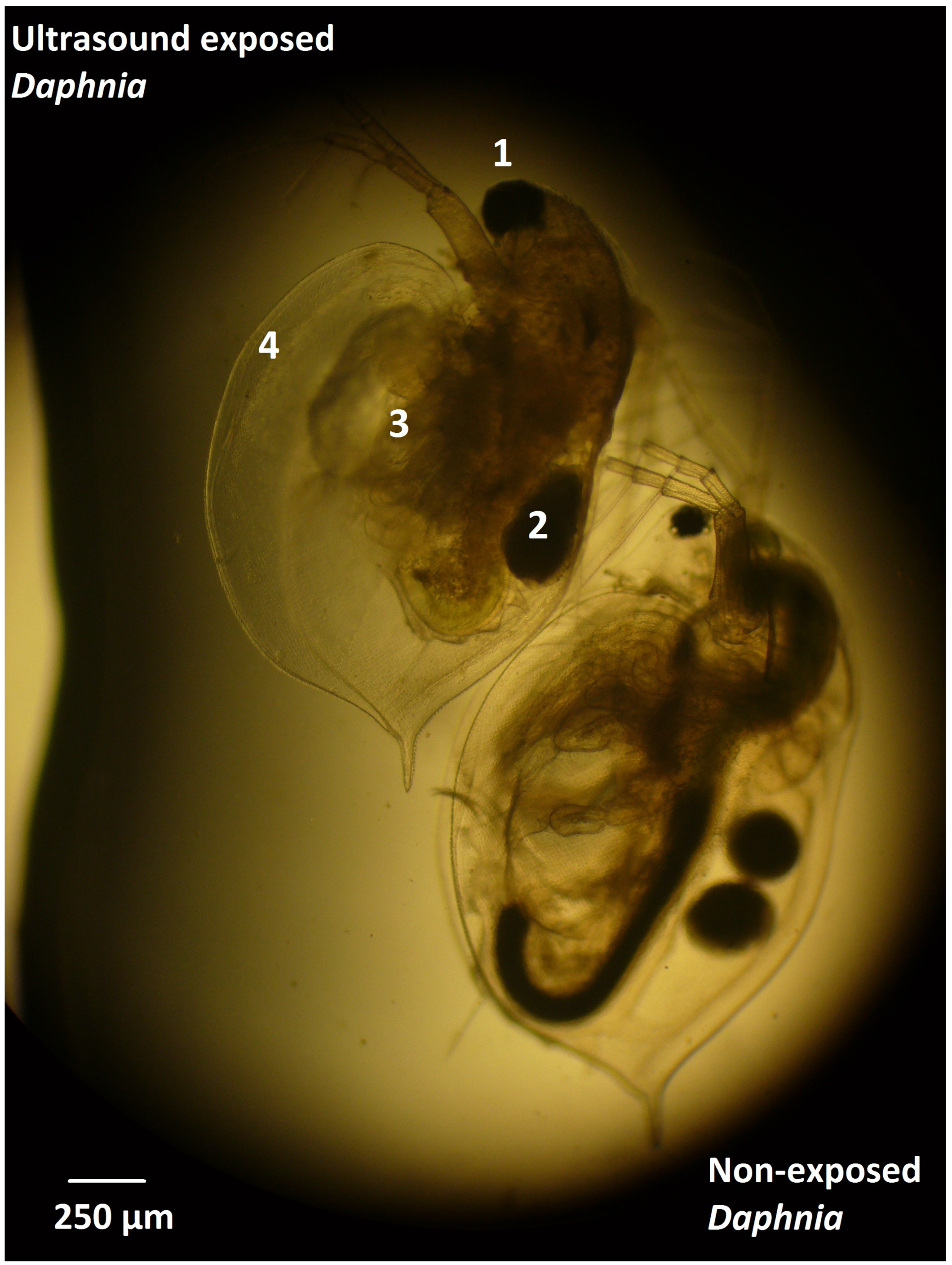
3.1. Effect of Ultrasound on Daphnia

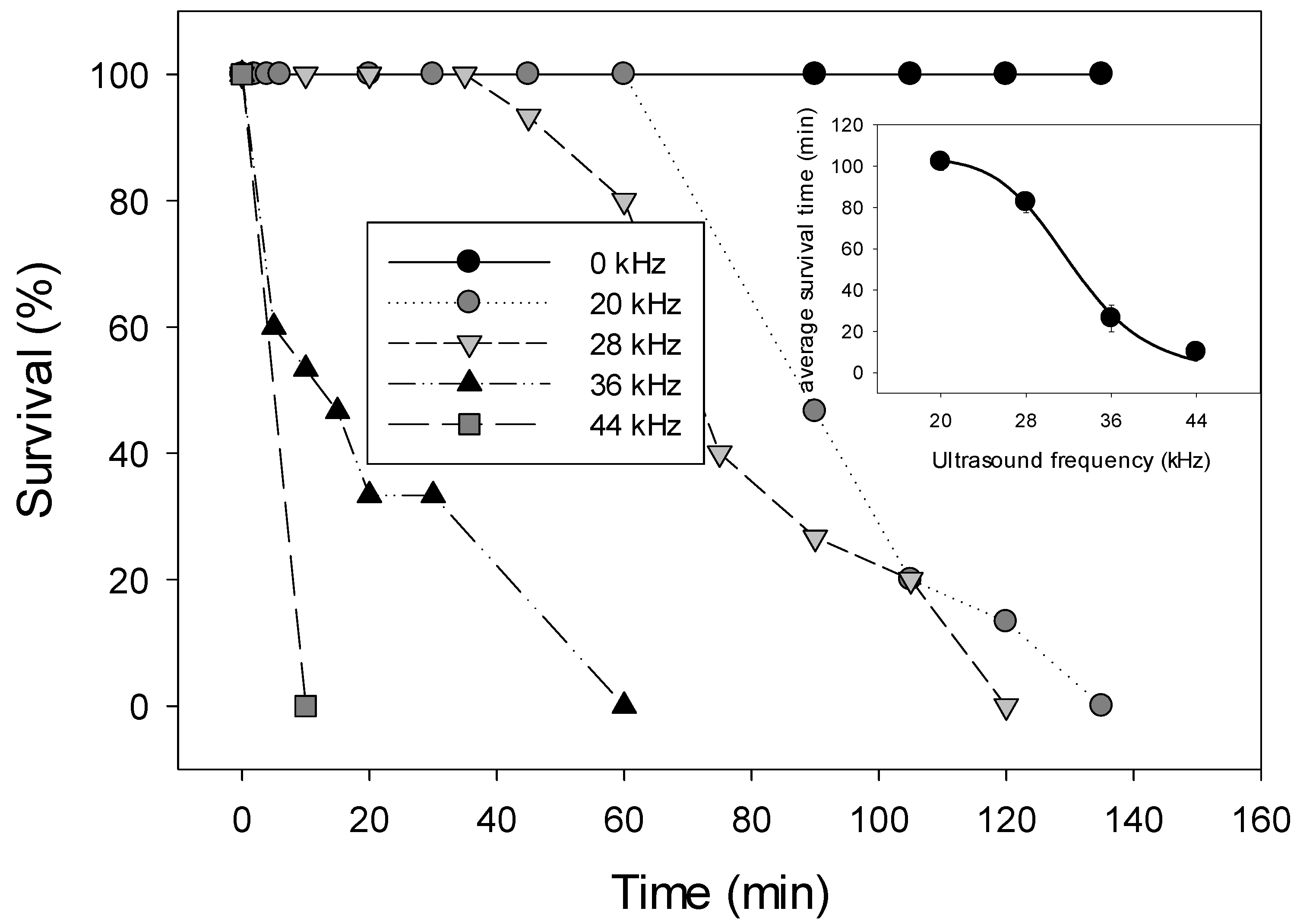
3.1.1. Effect of Different Frequencies

Ultrasound had an acute lethal effect on D. magna as in all treatments the experimental animals died rapidly contrasting the 100% survival in all controls (Figure 1). D. magna died within 2½ h depending on the exposure frequency with higher frequencies causing much faster death than lower frequencies (Figure 1). Kaplan-Meier Survival Analysis revealed that Daphnia survival time was 102 min in 20 kHz ultrasound, 83 min when exposed to 28 kHz, 26 min in 36 kHz and 10 min in 44 kHz (Figure 1). Parallel Lines Analysis revealed that from the moment animals started to die they did so at similar speed (F2,13 = 0.92; p = 0.423) in the 20 kHz, 28 kHz and 36 kHz treatments (the 44 kHz treatment was excluded because of very fast death of the animals). However, the intercepts were significantly different (F2,15 = 153.2; p < 0.001) meaning that Daphnia started to die after different exposure durations; after 60 min, 45 min and 5 min in the 20 kHz, 28 kHz and 36 kHz treatments, respectively (Figure 1).
Figure 1. Survival (%) of Daphnia magna in populations not-exposed to ultrasound (0 kHz; controls) and in populations exposed to ultrasound varying in frequency (20, 28, 36 and 44 kHz). The inserted graph gives the mean survival time (min) for Daphnia exposed to each frequency as determined with Kaplan-Meier Survival Analysis (error bars indicate the standard error derived from the Kaplan-Meier Survival Analysis).
Water 06 03247 g001

3.1.2. Effect on Animals of Different Size

D. magna exposed to ultrasound of 44 kHz started to die within 1 to 3 min and all animals were dead between 4 and 45 min exposure. The survival time in organisms of different body-size seemed lowest in animals between 1.1 and 1.7 mm and larger in the smallest and largest animals tested (Table 1). Survival time in ultrasound-exposed animals was very low and varied between 2.3 and 17 min, while in controls all animals survived (Table 1).
Table 1. Mean survival time (min) of Daphnia magna in six cohorts varying in size (0.7–3.2 mm, ±1 SD) exposed to ultrasound (44 kHz) or not exposed (Control). Survival time in treatments (44 kHz) was determined with Kaplan-Meier Survival Analysis.
D. magnaSurvival Time (min)
Body-length (mm)Control44 kHz
0.69 (0.04)>6010.6
0.97 (0.07)>604.9
1.11 (0.07)>602.3
1.68 (0.05)>602.4
2.05 (0.07)>607.6
3.16 (0.11)>6017.1

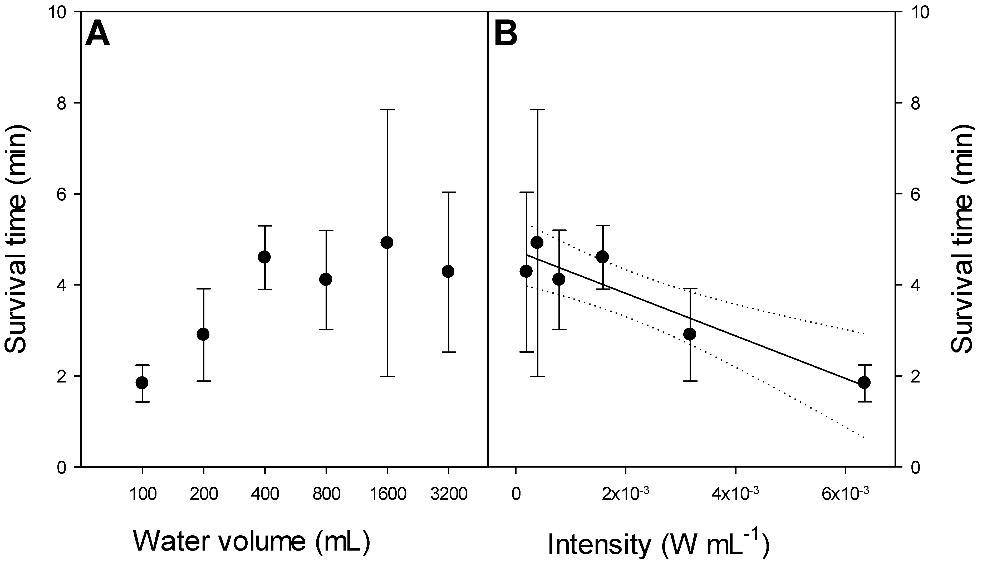
3.1.3. Effect of Size of Experimental Unit

Survival time of experimental animals with a body-size of 1.94 (±0.12) mm that were exposed to 44 kHz in volumes ranging from 100 mL to 3200 mL was lowest in the smallest volumes and higher in larger volumes (Figure 2A). Although there was a linear relation between survival time (ST) and ultrasound intensity (IUS) (ST = 4.747 − 467.8 × IUS; r2 = 0.880), a one-way ANOVA indicated that ST was not different between populations exposed in different volumes (F5,12 = 1.70; p = 0.208). Ultrasound remained acutely lethal with mean survival time ranging from 1.8 to 4.9 min (Figure 2B).
Figure 2. Left panel A: Survival time (min) of D. magna exposed to 44 kHz ultrasound in volumes ranging from 100 mL to 3200 mL. Error bars indicate one standard deviation (n = 3); Right panel B: Relationship between mean survival time (ST) of Daphnia magna (body-size 1.94 ± 0.12 mm) and ultrasound intensity (IUS, in W·mL−1); ST = 4.747 − 467.8 × IUS (r2 = 0.880). Solid line represents linear regression, the dotted lines indicate the 95% confidence and the error bars indicate one standard deviation (n = 3).
Water 06 03247 g002
All ultrasound exposed animals that were inspected microscopically showed some distinct alterations when compared with non-exposed specimens: (1) the eye was much enlarged in ultrasound exposed animals as if exploded; (2) eggs, if present in brood pouch, were visually damaged; (3) intestines were clearly damaged and (4) the carapax gape was enlarged (Figure 3).

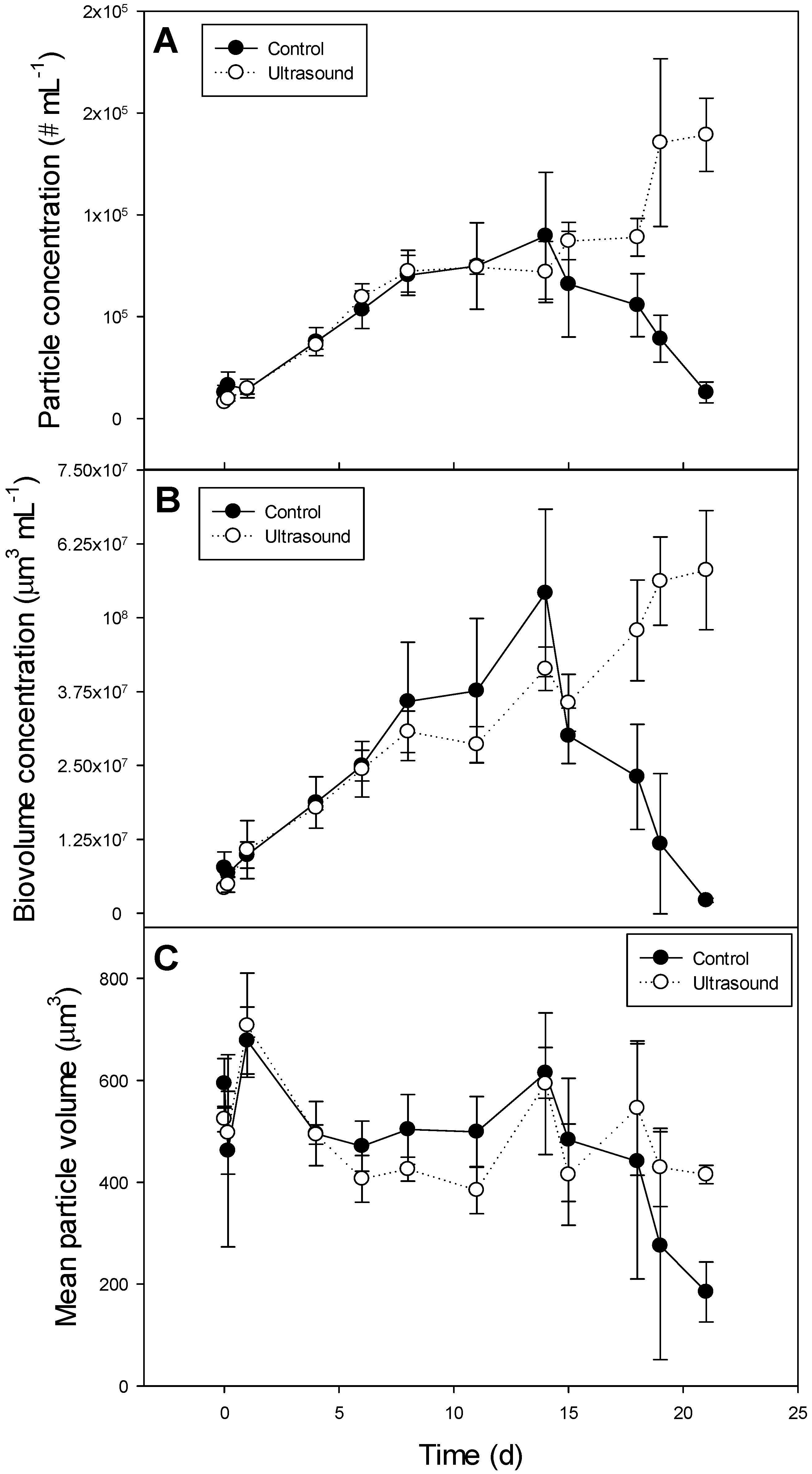
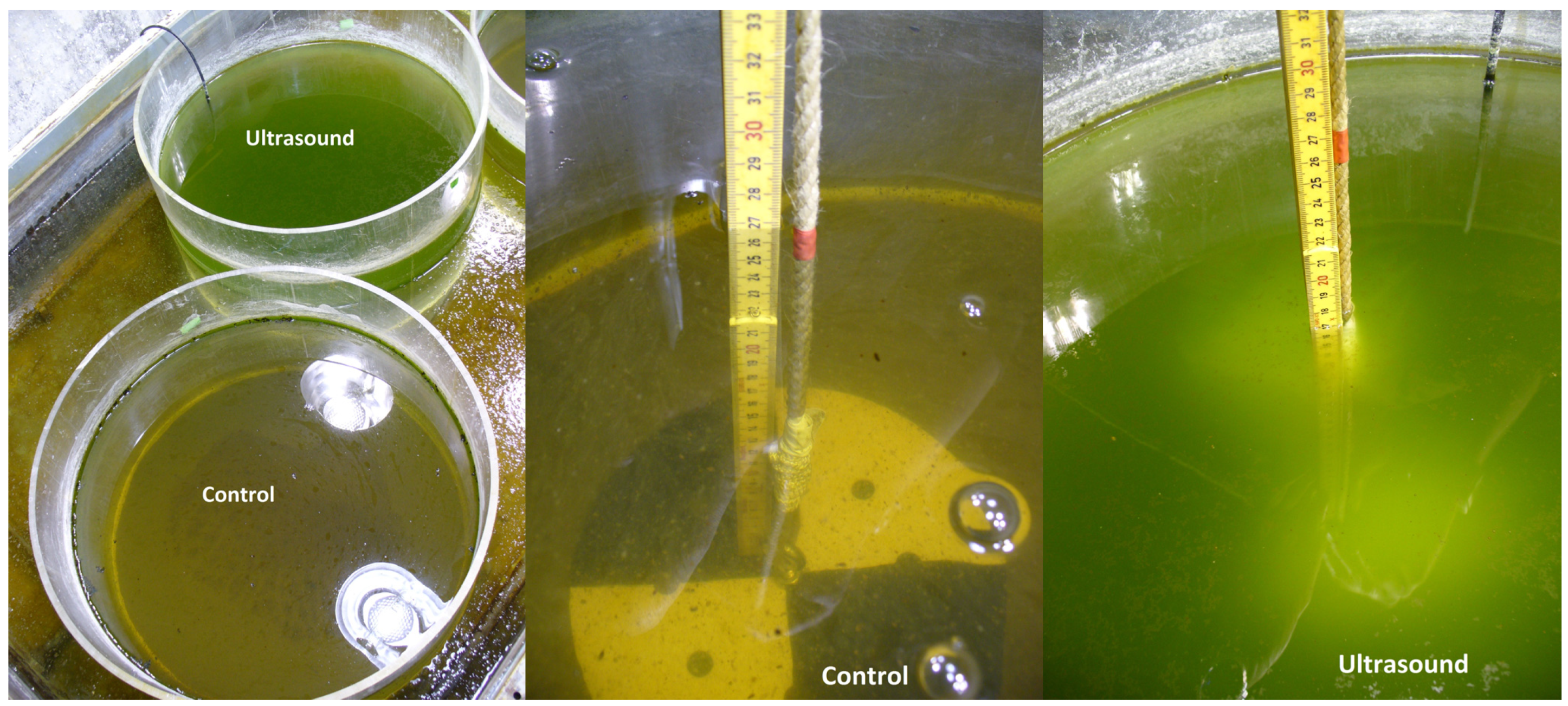
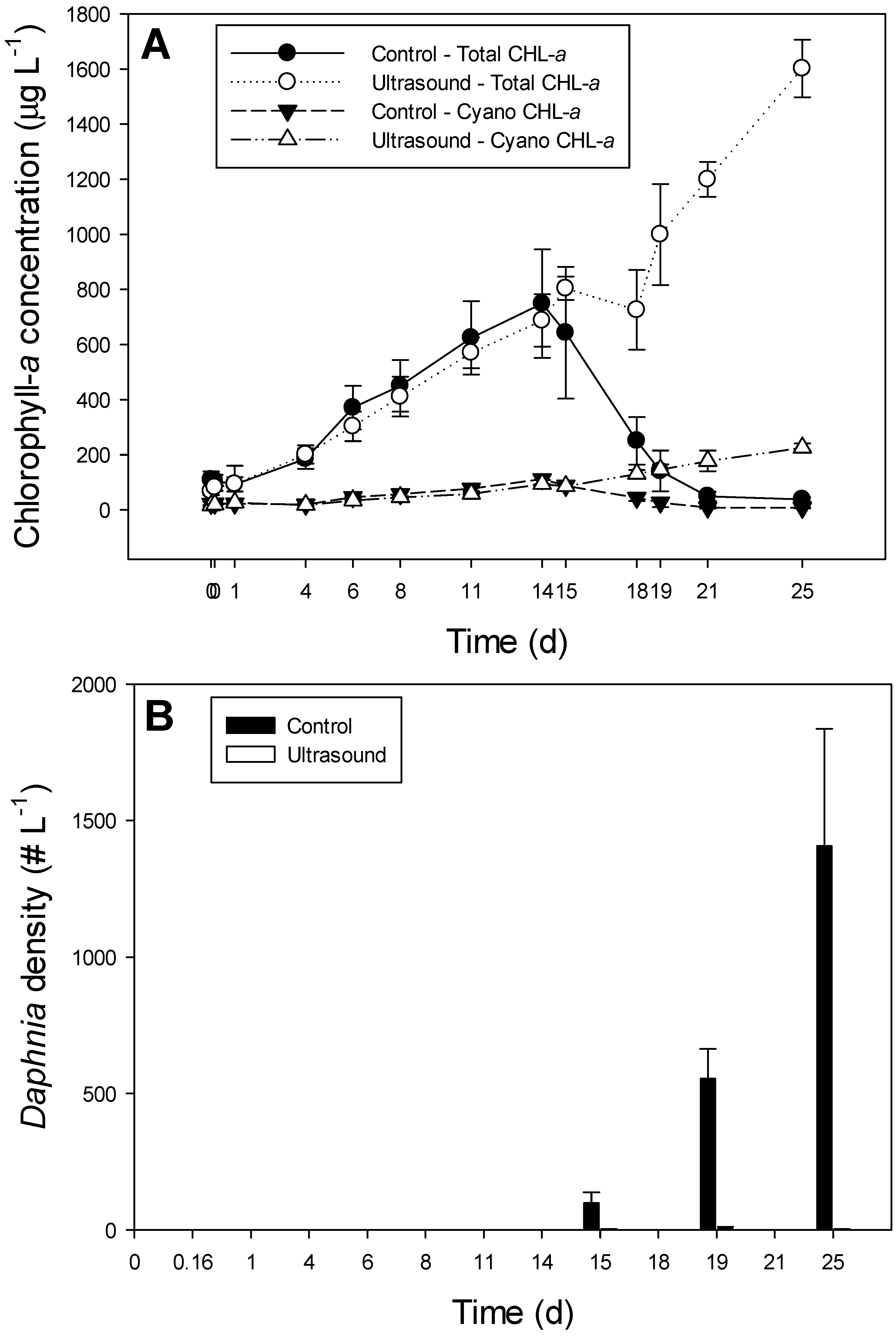
3.2. Effect of Ultrasound on a Plankton Community in 85 L Tanks

During the first two weeks of the experiment, the course of the total chlorophyll-a concentrations was similar in controls and ultrasound treatments (rmANOVA total chlorophyll-a: F1,4 = 0.06; p = 0.832) and the same was observed for cyanobacterial chlorophyll-a concentrations (rmANOVA: F1,4 = 4.52; p = 0.101). However, hereafter chlorophyll-a concentrations started to decline in controls, whereas they remained to increase in the ultrasound treatments (Figure 4A), causing significant differences in total-, cyanobacterial- and green algal chlorophyll-a concentrations, as well as significant time x treatment interaction effects (Table 2). The decline in the chlorophyll-a concentrations in controls coincided with strong increase in the Daphnia densities (Figure 4B). The number of Daphnia per L was significantly higher in the controls than in the ultrasound treatments (rmANOVA F1,4 = 50.0; p = 0.002); at the end of the experiment controls contained on average 1400 Daphnia L−1, while ultrasound treatments had only 1 Daphnia L−1 (Figure 4B).
Figure 3. Photograph of an ultrasound (44 kHz) killed Daphnia magna (animal upper left) and a non-exposed specimen from the same cohort (animal right under). 1 = eye, 2 = egg, 3 = intestine, 4 = carapax.
Water 06 03247 g003
Figure 4. Panel A: Course of total chlorophyll-a concentrations (µg·L1) in non-exposed plankton communities in 85 L tanks (control; solid circles) and ultrasound treatments (open circles) as well as cyanobacteria chlorophyll-a concentrations in controls (filled triangles) and ultrasound treatments (open triangles). Error bars indicate one standard deviation (n = 3); Panel B: Course of the density of Daphnia magna (# L−1) in non-exposed 85 L tanks (control; filled bars) and ultrasound treatments (open bars). Error bars indicate one standard deviation (n = 3).
Water 06 03247 g004
Table 2. Summary of repeated measures ANOVAs for total chlorophyll-a concentration, biovolume of phytoplankton, particle numbers, mean particle volume, cyanobacteria chlorophyll-a and green algae chlorophyll-a concentrations in 85 L tanks without and with exposure to ultrasound (treatment) as the fixed factor.
Chlorophyll-a ConcentrationBiovolume Concentration
Tests of within-subjects Effects
SourcedfFpdfFp
Time1263.5<0.0011124.8<0.001
Time × treatment1269.8<0.0011117.0<0.001
Error48 44
Tests of between-subjects Effects
SourcedfFpdfFp
Treatment140.20.003117.10.014
Error4 4
Particle concentrationMean Particle Volume
Tests of within-subjects Effects
SourcedfFpdfFp
Time1129.4<0.001117.06<0.001
Time × treatments1117.3<0.001111.780.087
Error44 44
Tests of between-subjects Effects
SourcedfFpdfFp
Treatments110.10.03410.090.784
Error4 4
Cyanobacteria chlorophyll-aGreen algae chlorophyll-a
Tests of within-subjects Effects
SourcedfFpdfFp
Time1262.4<0.0011256.1<0.001
Time × treatment1282.1<0.0011259.1<0.001
Error48 48
Tests of between-subjects Effects
SourcedfFpdfFp
Treatment153.90.002131.90.005
Error4 4
The biovolume and particle concentrations showed similar patterns as the chlorophyll-a concentrations, i.e., equal increase in controls and ultrasound treatments during the first two weeks where after concentrations dropped in controls and kept increasing in the ultrasound treatments (Figure 5A and Figure 5B). Likewise, significant treatment and significant time × treatment interaction effects were found (Table 2). The mean particle volume was fairly similar in controls and ultrasound exposed phytoplankton communities (Figure 5C; Table 2).
Figure 5. Course of particle concentrations (# mL−1; panel A), biovolume concentration (µm3·mL1; panel B) and mean particle volume (µm3; panel C) in plankton communities in 85 L tanks that were either non-exposed (control; solid circles) or exposed to ultrasound (open circles). Error bars indicate one standard deviation (n = 3).
Water 06 03247 g005
The relative share of cyanobacteria in the phytoplankton community was not changed over time or ultrasound treatment. Parallel line analysis revealed no differences in the slopes of the share of cyanobacteria- and green algae chlorophyll-a concentrations (F3,44 = 1.95; p = 0.136) and the mean slope was almost zero (i.e., 1.45 × 10⁻¹⁷). On average cyanobacteria comprised 16.7 (±4.4) % of the chlorophyll-a concentration in the controls and 15.5 (±5.9) % in the ultrasound treatments.
There were no differences in the water quality variables temperature, pH, oxygen concentration and electric conductivity between controls and ultrasound treatments (Table 3).
Table 3. Mean values (±1 SD) of water quality variables in 85 L plankton communities kept in the absence (Control) or presence of ultrasound (Ultrasound). Also included are the results (F- and p values) of the between subject effects from repeated measure ANOVAs.
VariableControlUltrasoundF- and p values
pH8.2 (1.4)8.4 (1.4)F1,4 = 1.97; p = 0.233
Conductivity (µS·cm−1)236 (8)230 (4)F1,4 = 0.05; p = 0.839
Oxygen (mg·L−1)13.9 (5.1)14.8 (4.4)F1,4 = 0.78; p = 0.426
Temperature (°C)21.5 (2.1)21.3 (2.1)F1,4 = 0.87; p = 0.404

4. Discussion

The results of this study confirm our previous findings that ultrasound from commercially available ultrasound transducers can be acutely lethal to the non-target zooplankton grazer Daphnia. This mortality cannot be explained from ultrasound induced warming of the water in the experimental units of 800 mL, as the increase in water temperature was only about 1 °C in 70 min. A similar observation was made in our previous experiment, where included temperature controls clearly showed that elevated temperature had no effect on Daphnia survival [10]. Furthermore, in the 85 L tanks no difference in water temperature was detected between controls and ultrasound treatments, while animal densities were extremely low in treatments compared to controls.
Higher frequencies exerted a stronger effect on D. magna than lower frequencies, but all should be considered acutely lethal at the tested conditions, because animals had low survival time varying from 10 to about 100 min. Animals between 1 and 2 mm seemed stronger affected by 44 kHz ultrasound than smaller and larger specimens, but again survival times were very short (about 2 to 17 min only). Increasing water volume and thereby lowering the intensity did not elevate the survival time significantly. However, further increase in volume as in the 85 L, which meant a 26× more reduction in intensity compared to the largest volume tested in the experiment (3.2 L), kept some specimens alive, albeit in very low numbers. Hence, even in small ponds and aquaria the tested transducers are expected to exert an effect on non-target organisms such as Daphnia. This finding is supported in a field study conducted in The Netherlands in 2007 [16]. These authors [16] described an experiment in two identical ponds that were interconnected and received the same water. One of the ponds was treated with ultrasound produced by a Flexidal AL-50 transducer, while the other served as control. These authors reported an almost complete disappearance of Daphnia from the ultrasound treated pond, while Daphnia remained abundant in the non-treated pond. Such field observations are correlative, but our controlled experiments clearly provide proof of causality: ultrasound is lethal to Daphnia.
There are only a few reports on the effect of ultrasound on zooplankton. Ultrasound is used for disinfection of ballast water or raw water for drinking water preparation, where it may inactivate motile plankton [17] or kill zooplankton, especially larger cladocerans [18]. Short exposure of D. magna to 3 MHz was lethal to the animals [19], while a Russian study reported on immediate death of Daphnia in 50, 500 and 1000 kHz [20]. In the macrophyte Elodea gas body activation generated intracellular micro-streaming that disrupted the cells, which was also the probable mechanism of death in ultrasound exposed fruit flies [21]. This means that potentially also freshwater insects with air bubbles, such as notonectids and Chaoborus [22,23] could be influenced by ultrasound. Insofar as our studies found, a clear and fast lethal impact of ultrasound on Daphnia produced by commercially available transducers, which seems supported by a field trial [16], ultrasound should not be considered “environmentally friendly” [9] or a “green solution” for controlling phytoplankton [8].
Our tank experiment provided strong support for the hypothesis that the emitted ultrasound might have the opposite effect to the claim on water clearing, i.e., cause phytoplankton dominated water through a detrimental impact on Daphnia and thus releasing phytoplankton from grazing control (Figure 6). In the ultrasound treatment the share of cyanobacteria remained constant, which means they grew as fast as the green algae. In fact, cyanobacteria chlorophyll-a had increased from 17 µg·L−1 to 225 µg·L−1 in the ultrasound treatments, while it was reduced to 7 µg·L−1 in controls at the end of the experiment. Hence, ultrasound from the tested transducers was not capable of removing cyanobacteria and was not able to clear the tanks from phytoplankton refuting the manufacturers claim that the AL-05 transducers kill phytoplankton within one week [11]. This result is fully in line with those we have obtained in controlled experiments with different cyanobacteria cultures that could not be wiped out by the ultrasound, not even in small experimental units of 800 mL [10]. Our results contrast the numerous positive reports on highly effective control of cyanobacteria by ultrasound as reviewed in [8,9]. However, it should be noted that the vast majority of those studies applied other devices—using intensities dozens to hundreds times higher—than the commercial ones for clearing ponds [10]. Remarkably, none of the studies reviewed in [8,9] included controlled experiments to examine the effect of ultrasound on non-targeted organisms such as Daphnia.
Figure 6. Pictures of a control tank and ultrasound treated tank (85 L) taken after 21 days in which Daphnia magna had cleared the water in the control, while strong Daphnia suppression in the ultrasound treatment released phytoplankton from grazing control causing a green soup.
Water 06 03247 g006
Daphnia obviously was of great importance in filtering the phytoplankton out of the water in the control tanks. It took about two weeks before Daphnia densities were high enough to reduce phytoplankton biomass rapidly, which is comparable to what was observed in an enclosure study [24]. The share of cyanobacteria had dropped slightly in the first few days of our experiment from 27% to about 16%, where after it remained constant. These relatively low proportions of cyanobacteria in the diet will not hamper growth of D. magna [25,26]. After two weeks cyanobacteria chlorophyll-a concentrations in the control tanks were above 100 µg·L−1, which apparently did not impair Daphnia’s ability for strong top-down control. Another study found that even in 150 µg·L−1 of cyanobacteria dominated phytoplankton, Daphnia were able to suppress total phytoplankton biomass by 74% relative to no Daphnia controls [24]. Therefore, Daphnia might be able to greatly reduce cyanobacteria, unless their abundance is controlled by planktivorous fishes [24] or ultrasound.
The ultrasound transducers we’ve tested in our experiments are for use in small ponds, aquaria and small water reservoirs, according to the manufacturer [11]. However, despite several studies suggest ultrasound might be applicable in situ (e.g., [8,9]), field trials are far from conclusive. Three field trials with ultrasound in the UK were reviewed [27]; no effect on total algal concentration in all three were reported, a significant lower chlorophyll-a concentration in one experiment, but no selective inhibition of cyanobacteria [27]. In contrast, a strong decrease of cyanobacteria by ultrasound as supportive for the selective effect of ultrasound on cyanobacteria was claimed [28]. However, as the control pond in [28] was also dominated by diatoms and the treated pond already at start had significantly lower chlorophyll-a concentration than the control, such conclusions should be met critically. Likewise, [29,30] described a field study and reported that chlorophyll-a concentrations were lower in the two years of ultrasound treatment, but this is not supported by the data as chlorophyll-a concentrations digitally extracted from Figure 4 in [30], which corresponds to Figure 3 in [29], yields 81 (±56) µg·L−1 before and 74 (±42) µg·L−1 during ultrasound. The somewhat lower peak biomass in 1997 could also be explained from hydrology [29,30].
Field trials that have been conducted in The Netherlands in 2007 did not provide any evidence of an effect of ultrasound on cyanobacteria or phytoplankton [16,31]. As mentioned earlier, the study of [16] was conducted in two identical ponds of which one was treated with ultrasound produced by a Flexidal AL-50 transducer, while the other one served as control. During the four months of operation chlorophyll-a concentrations in the control were around 64 (±13) µg·L−1 and in the ultrasound treatment around 69 (±26) µg·L−1 (data digitally extracted from Figure 2 in [16]). Moreover, no difference in phytoplankton composition was found [16]. Two other field trials in The Netherlands were described in [31]; one in the Southwest of the Netherlands in a harbor area near Tholen and the other one in a bay of recreational area De Gouden Ham near the river Maas. Surface scums and high Microcystis densities were observed on both sites despite the ultrasound treatment and the authors concluded that ultrasound was not effective in reducing cyanobacteria [31].
Based on our experiments that revealed detrimental effects of ultrasound on Daphnia without any control of phytoplankton, even in relatively small water volume, we conclude that there is no music in fighting cyanobacteria and algal blooms with commercially available ultrasound transducers. We recommend water authorities, regardless the stringent time lines of achieving European Water Framework Directive demands and meeting EU Bathing Water Directive targets, to keep focus on the primary cause of cyanobacterial and algal blooms: i.e., an over-enrichment of the water with nutrients [2]. Source-oriented measures principally targeting the phosphorus inflow and internal loading remain essential in reducing eutrophication [32]. However, in situations where source-oriented measures are not easy achievable, cost-effective end-of-pipe solutions might be an alternative, but these should have proven efficacy in situ.

5. Conclusions

We tested the hypothesis that ultrasound in controlling cyanobacteria and algal blooms is “environmentally friendly” by exposing the non-target zooplankton grazer Daphnia magna to ultrasound produced by commercially available ultrasound transducers. Based on the results of this study it can be concluded that:
  • Ultrasound had an acute lethal effect on D. magna;
  • Higher ultrasound frequencies caused faster death than lower frequencies;
  • Survival time of differently sized Daphnia exposed to ultrasound in different volumes varied only slightly;
  • Ultrasound strongly suppressed Daphnia in 85 L tanks and freed phytoplankton from grazing control causing a turbid phytoplankton dominated water, whereas phytoplankton in the controls was under strong Daphnia control;
  • The commercial available ultrasound transducers for clearing lakes and ponds are not environmentally friendly and are ineffective in controlling phytoplankton.

Acknowledgments

We thank Wendy Beekman for assistance during the experiments and VHL University of Applied Sciences (Leeuwarden, The Netherlands) for lending an AL-05 ultrasound transducer coupled with an M&R Systems Waveform-Generator WG-810 frequencies regulator. This study was supported by the Ministry of Transport, Public Works and Water Management, Directorate—General for Public Works and Water Management (The Netherlands).

Author Contributions

Conceived and designed the experiment: Miquel Lürling, Yora Tolman. Performed the experiment: Miquel Lürling, Yora Tolman. Analyzed the data: Miquel Lürling, Yora Tolman. Wrote the paper: Miquel Lürling.

Conflicts of Interest

The authors declare no conflict of interest.

References

  1. Smith, V.H.; Tilman, G.D.; Nekola, J.C. Eutrophication: Impacts of excess nutrient inputs on freshwater, marine, and terrestrial ecosystems. Environ. Pollut. 1999, 100, 179–196. [Google Scholar] [PubMed]
  2. Smith, V.H. Eutrophication of freshwater and coastal marine ecosystems a global problem. Environ. Sci. Pollut. Res. 2003, 10, 126–139. [Google Scholar] [CrossRef]
  3. Codd, G.A. Cyanobacterial toxins, the perception of water quality, and the prioritisation of eutrophication control. Ecol. Eng. 2000, 16, 51–60. [Google Scholar] [CrossRef]
  4. Dittmann, E.; Wiegand, C. Cyanobacterial toxins—Occurrence, biosynthesis and impact on human affairs. Mol. Nutr. Food Res. 2006, 50, 7–17. [Google Scholar] [CrossRef] [PubMed]
  5. Directive 2000/60/EG of the European Parliament and of the Council Establishing a Framework for the Community Action in the Field of Water Policy of. 23 October; European Union: Brussels, Belgium, 2000; pp. 1–72.
  6. Directive 2006/7/EC of the European Parliament and of the Council of 15 February 2006 Concerning the Management of Bathing Water Quality and Repealing Directive 76/160/EEC; European Union: Brussels, Belgium, 2006; pp. 37–51.
  7. Ibelings, B.W.; Stroom, J.M.; Lürling, M.F.L.L.W.; Kardinaal, W.E.A. Netherlands: Risks of toxic cyanobacterial blooms in recreational waters and guidelines. In Current Approaches to Cyanotoxin Risk Management, Risk Management and Regulations in Different Countries; Chorus, I., Ed.; Federal Environment Agency: Dessau Roβlau, Germany, 2012; pp. 82–96. [Google Scholar]
  8. Wu, X.; Joyce, E.M.; Mason, T.J. The effects of ultrasound on cyanobacteria. Harmful Algae 2011, 10, 738–743. [Google Scholar] [CrossRef]
  9. Rajasekhar, P.; Fan, L.; Nguyen, T.; Roddick, F.A. A review of the use of sonication to control cyanobacterial blooms. Water Res. 2013, 46, 4319–4329. [Google Scholar] [CrossRef]
  10. Lürling, M.; Tolman, Y. Beating the blues: Is there any music in fighting cyanobacteria with ultrasound? Water Res. 2014, 66, 361–373. [Google Scholar] [CrossRef]
  11. Flexidal Technics. Available online: http://flexidal.be/nl/produktenvanflexidal_algen.asp?rubriek=algen&fotoid=8 (accessed on 12 July 2014).
  12. Kikuchi, T.; Uchida, T. Calorimetric method for measuring high ultrasonic power using water as a heating material. J. Phys. Conf. Ser. 2011, 279. [Google Scholar] [CrossRef]
  13. Wu, X.; Joyce, E.M.; Mason, T.J. Evaluation of the mechanisms of the effect of ultrasound on Microcystis aeruginosa at different ultrasonic frequencies. Water Res. 2012, 46, 2851–2858. [Google Scholar] [CrossRef] [PubMed]
  14. Tollrian, R. Neckteeth formation in Daphnia pulex as example of continuous phenotypic plasticity: Morphological effects of Chaoborus kairomone concentration and their quantification. J. Plankton Res. 1993, 15, 1309–1318. [Google Scholar] [CrossRef]
  15. Lürling, M.; Beekman, W. Palmelloids formation in Chlamydomonas reinhardtii: Defence against rotifer predators? Ann. Limnol. 2006, 42, 65–72. [Google Scholar] [CrossRef]
  16. Govaert, E.; Vanderstukken, M.; Muylaert, K. Evaluatie van Effecten van Ultrasone Straling op Het Ecosysteem; KU Leuven Kortrijk: Kortrijk, Belgium, 2007; p. 20. (In Dutch) [Google Scholar]
  17. Hoyer, O.; Clasen, J. The application of new technologies in the water treatment process of a modern waterworks. Water Sci. Technol. 2002, 2, 63–69. [Google Scholar]
  18. Holm, E.R.; Stamper, D.M.; Brizzolara, R.A.; Barnes, L.; Deamer, N.; Burkholder, J.M. Sonication of bacteria, phytoplankton and zooplankton: Application to treatment of ballast water. Mar. Pollut. Bull. 2008, 56, 1201–1208. [Google Scholar] [CrossRef] [PubMed]
  19. Wells, P.N.T. The effect of ultrasonic irradiation on the survival of Daphnia magna. Exp. Biol. 1968, 49, 61–70. [Google Scholar]
  20. Kamenskii, I.V. Influence of ultrasound on eggs and larvae of some fish trematodes. Byulleten’ Vsesoyuznogo Instituta Gel’mintologii im 1970, 4, 47–50. (In Russian) [Google Scholar]
  21. Miller, D.L. A review of the ultrasonic bioeffects of microsonation, gas-body activation, and related cavitation-like phenomena. Ultrasound Med. Biol. 1987, 13, 443–470. [Google Scholar] [CrossRef]
  22. Wells, R.M.G.; Hudson, M.J.; Brittain, T. Function of the hemoglobin and the gas bubble in the backswimmer Anisops assimilis (Hemiptera: Notonectidae). J. Comp. Physiol. B 1981, 142, 515–522. [Google Scholar] [CrossRef]
  23. Knudsen, F.R.; Larsson, P.; Jakobson, P.J. Acoustic scattering from a larval insect (Chaoborus flavicans) at six echosounder frequencies: Implication for acoustic estimates of fish abundance. Fish. Res. 2006, 79, 84–89. [Google Scholar] [CrossRef]
  24. Chislock, M.F.; Sarnelle, O.; Jernigan, L.M.; Wilson, A.E. Do high concentrations of microcystin prevent Daphnia control of phytoplankton? Water Res. 2013, 47, 1961–1970. [Google Scholar] [CrossRef] [PubMed]
  25. Lürling, M. Effects of microcystin-free and microcystin-containing strains of the cyanobacterium Microcystis aeruginosa on growth of the grazer Daphnia magna. Environ. Toxicol. 2003, 18, 202–210. [Google Scholar] [CrossRef] [PubMed]
  26. Soares, M.C.S.; Lürling, M.; Panosso, R.; Huszar, V. Effects of the cyanobacterium Cylindrospermopsis raciborskii on feeding and life-history characteristics of the grazer Daphnia magna. Ecotoxicol. Environ. Saf. 2009, 72, 1183–1189. [Google Scholar]
  27. Purcell, D.; Parsons, S.A.; Jefferson, B.; Holden, S.; Campbell, A.; Wallen, A.; Chipps, M.; Holden, B.; Ellingham, A. Experiences of algal bloom control using green solutions barley straw and ultrasound, an industry perspective. Water Environ. J. 2013, 27, 148–156. [Google Scholar] [CrossRef]
  28. Ahn, C.-Y.; Joung, S.-H.; Choi, A.; Kim, H.-S.; Jang, K.-Y.; Oh, H.-M. Selective control of cyanobacteria in eutrophic pond by a combined device of ultrasonication and water pumps. Environ. Technol. 2007, 28, 371–379. [Google Scholar] [CrossRef] [PubMed]
  29. Nakano, K.; Lee, T.J.; Matsumara, M. In situ algal bloom control by the integration of ultrasonic radiation and jet circulation to flushing. Environ. Sci. Technol. 2001, 35, 4941–4946. [Google Scholar] [CrossRef] [PubMed]
  30. Lee, T.J.; Nakano, K.; Matsumara, M. A novel strategy for cyanobacterial bloom control by ultrasonic irradiation. Water Sci. Technol. 2002, 46, 207–215. [Google Scholar] [PubMed]
  31. Kardinaal, E.; de Haan, M.; Ruiter, H. Maatregelen ter voorkoming blauwalgen werken onvoldoende. H2O 2008, 7, 4–7. (In Dutch) [Google Scholar]
  32. Mackay, E.B.; Maberly, S.C.; Pan, G.; Reitzel, K.; Bruere, A.; Corker, N.; Douglas, G.; Egemose, S.; Hamilton, D.; Hatton-Ellis, T.; et al. Geoengineering in lakes: Welcome attraction or fatal distraction? Inland Waters 2014, 4, 349–356. [Google Scholar]

