Communication

Biocatalytic Self-Cleaning Polymer Membranes

Leibniz Institute of Surface Modification, Permoserstr. 15, Leipzig D-04318, Germany
*
Author to whom correspondence should be addressed.
Polymers 2015, 7(9), 1837-1849; https://doi.org/10.3390/polym7091485
Submission received: 25 June 2015 / Revised: 26 August 2015 / Accepted: 15 September 2015 / Published: 21 September 2015
(This article belongs to the Special Issue Polymer Thin Films and Membranes 2015)

Abstract

:
Polymer membrane surfaces have been equipped with the digestive enzyme trypsin. Enzyme immobilization was performed by electron beam irradiation in aqueous media within a one-step method. Using this method, trypsin was covalently and side-unspecific attached to the membrane surface. Thus, the use of preceding polymer functionalization and the use of toxic solvents or reagents can be avoided. The resulting membranes showed significantly improved antifouling properties as demonstrated by repeated filtration of protein solutions. Furthermore, the biocatalytic membrane can be simply “switched on” to actively degrade a fouling layer on the membrane surface and regain the initial permeability. The membrane pore structure (pore size and porosity) was neither damaged by the electron beam treatment nor blocked by the enzyme loading, ensuring a stable membrane performance.

Graphical Abstract

1. Introduction

Porous polymer membranes are of increasing importance regarding modern separation technologies such as waste water treatment, sterilization filtration, hemodialysis, production of fine chemicals, dairy industry, etc. [1]. To comply with required process conditions (chemical and physical stability within a broad range) these polymer membranes are predominantly fabricated from synthetic materials such as polyethersulfone (PES), polysulfone (PSf), or polyvinylidene fluoride (PVDF) (see Figure 1) [2]. However, membranes made from these polymers are prone to fouling which is caused by hydrophobic interactions of the membrane surface with biomolecules or colloids in the mixture to be filtered and result in irreversible adsorption, aggregation, and finally in a reduced filtration performance [3,4,5,6].
Figure 1. Typical membrane polymers.
Polymers 07 01485 g001
Therefore, membranes have to be regularly cleaned to remove the fouling layer, depending on the filtration application. This may include a daily chemically enhanced backwash, a weekly maintenance cleaning with a higher chemical concentration, and an intensive chemical cleaning once or twice a year [7]. Commonly used cleaning agents are strong oxidants such as hypochlorite and citric acid.
To reduce the problem of initial fouling different approaches for surface hydrophilization have been investigated such as copolymerization or grafting with hydrophilic monomers [8,9,10,11,12,13,14], blending using hydrophilic polymers [15,16,17,18,19,20], and, finally, chemical modification of the membrane polymer [21].
It has been demonstrated that the resulting membranes show improved antifouling properties. However, the cited methods exhibit serious disadvantages because a substantial number of process steps is required (e.g., activation of the scaffold polymer for subsequent grafting and additional purification steps), as well as the use of toxic reagents and solvents. Furthermore, contamination of the eluent by non-permanently immobilized compounds or by used initiators/catalysts was reported.
Here, a new electron beam (EB) based method for efficient surface modification of polymer membranes has been developed. This technology enables direct immobilization of functional organic molecules [22,23], engineering polymers [24], or even biomolecules [25,26] on a membrane’s surface. The use of low-energy EB radiation combines surface activation (formation of activated species) of the membrane polymer and simultaneous covalent immobilization of the desired compounds from an aqueous system. Because EB is capable of interpenetrating the entire cross-section of polymer materials also the inner surface of porous membranes can be activated for desired modification reactions. No catalysts, organic solvents, or other toxic reagents are required. Furthermore, additional preceding surface functionalization and purification steps are avoided since the synthesis of specific monomers/polymers is not necessary. Since immobilization is covalent, no compound leaching has been observed. Irradiation of polymers such as PES [27,28] and PVDF [29] with electrons and γ‑radiation has been extensively studied and is well understood [30].
In the present paper we discuss the preparation of a self-cleaning antifouling membrane by immobilization of the enzyme trypsin on the membrane’s surface. Trypsin is a well-studied proteolytic enzyme that catalyzes the hydrolysis of peptide bonds at an optimum pH value of 7.5–8.5 [31]. Since free trypsin is stable within a pH range of 3–11 [32,33] this enzyme can be used in common membrane-based water treatment applications, even when pH changes occur in the process. There are only few studies that report on immobilization of trypsin by entrapping within [34] or coating [35] on the membrane polymer. Furthermore, the covalent immobilization of trypsin on polymer membranes was reported [36,37,38]. However, the described covalent immobilization technique requires several synthesis steps including critical reagents. Therefore, we chose to use electron beam-mediated immobilization as a one-step procedure to create a biocatalytic membrane surface aiming to actively degrade a protein fouling layer, and thus, to create a self-cleaning membrane surface (see Figure 2).
Figure 2. Schematic concept of the self-cleaning membrane: the digestive enzyme trypsin was immobilized on the membrane surface to enable the active degradation of a protein fouling layer at an optimum pH range of 7.5–8.5.
Polymers 07 01485 g002

2. Experimental Section

2.1. Chemicals and Materials

Poly(vinylidene fluoride) membranes (A: hydrophobic, Roti, 0.45 µm, Carl Roth; B: hydrophilic, Durapore, 0.22 mm, Millipore; C: hydrophilic, Durapore, 0.45 µm, Millipore) were purchased from Carl Roth GmbH & Co., Karlsruhe, Germany. Trypsin from bovine pancreas (24 kDa), albumin from bovine serum (67 kDa), N-α-benzoyl-l-arginine 4-nitroanilide hydrochloride (BAPNA), ethanol, dimethyl sulfoxide (DMSO), p-nitroaniline (p-NA) as well as calcium chloride dihydrate, hydrochloric acid (HCl, reagent grade), and phosphate buffered saline (PBS, pH 7.4) were obtained from Sigma Aldrich, Hamburg, Germany. Bicinchoninic acid (BCA, Pierce, IL, USA) protein assay reagent A + B was provided by ThermoFisher SCIENTIFIC, Darmstadt, Germany. Deionized water was used for preparing all buffer solutions. Unless otherwise stated, water was used in Millipore® grade. All chemicals were of analytical grade and used without further purification.

2.2. Preparation and Characterization of Biocatalytic Membranes

The enzyme was immobilized via EB radiation on different PVDF microfiltration membranes according to a recently described method [25]. The modifications were performed by immersing the membranes for 5 min into an aqueous buffer solution (HEPES pH 8.0) of trypsin (2 mg/mL) at room temperature followed by electron beam irradiation with an irradiation dose of 150 kGy. Irradiation was performed in an N2 atmosphere with O2 quantities <10 ppm using a home-made electron accelerator. The voltage and the current were set to 160 kV and 10 mA, respectively. The absorbed dose was adjusted by the speed of the sample transporter. The modified membrane was rinsed three times per 10 min with buffer solution before performing subsequent analysis and characterization tests.
The three biocatalytic trypsin/PVDF membranes were analyzed regarding their immobilized enzyme amount using the bicinchoninic acid (BCA) test [39]. Therefore, the samples were washed three times with 1 mL of PBS buffer solution (pH 7.4). Then, the BCA reagent was added to the membrane samples and the plate was incubated for 25 min at 37 °C. The plate was then shaken for 5 min at room temperature, the solution was transferred to a new microtiter plate and light adsorption at 562 nm was measured using a microtiter plate reader (Infinite M200, Tecan, Germany). For calibration, seven trypsin concentrations of 1000, 500, 250, 125, 62.50, 31.25 and 0.00 μg/mL were used.
The residual proteolytic activity of the immobilized trypsin was determined using N-α-benzoyl-l-arginine 4-nitroanilide hydrochloride (BAPNA) as substrate. Testing the activity of immobilized trypsin was performed according to the method of Oliveira et al. [38]. The substrate solution was prepared by mixing 0.3 mM BAPNA, 1 mL DMSO and 9 mL 50 mM TRIS-HCl buffer containing 10 mM CaCl2·2H2O, pH 8.5. Then, 1 mL of the substrate solution was added to the modified and washed membranes. The plate with the samples was shaken for 5 min at room temperature and 200 μL of the supernatant were transferred to a 96 well microtiter plate. Then the optical absorption of the solution at 405 nm was monitored over a period of 130 min using a microtiter plate reader (Infinite M200, Tecan, Männedorf, Switzerland). After every sampling 200 µL of fresh substrate solution were added to the samples. The resulting extinctions from the BAPNA assay were converted (nmol) and displayed in concentrations vs. time diagrams. For calibration, seven standards based on a 0.3 mM p-nitroaniline stock solution with concentrations of 83.00, 41.50, 20.75, 10.38, 5.19, 2.59 and 0.00 µg/mL were used.
The morphology of the membranes was studied by scanning electron microscopy (SEM, Ultra 55, Carl Zeiss SMT, Oberkochen, Germany). In order to prevent charging the sample was sputtered with a thin gold layer. For investigation of pore size and bulk porosity of the membranes the mercury porosimeter PoreMaster PM-60-15 (Quantachrome GmbH & Co. KG, Odelzhausen, Germany) was used. XPS (X-ray photoelectron spectroscopy) analyses were carried out on a Kratos Axis Ultra (Kratos Analytical, Ltd., Manchester, UK) with a monochromatized Al excitation source at 150 W (15 kV, 10 mA, with a pass energy of 40 eV). Surface spectra were collected over a range of 0–1200 eV. The nominal resolutions were 1.0 eV for the survey (pass energy = 160 eV) and 0.1 eV for the high-resolution scans (pass energy = 20/40 eV), respectively. The binding energies were corrected for the static charging of the samples by reference to the C1s peak set at a binding energy of 285.0 eV.
The chosen fouling experiment was performed as outlined in Figure 3 and was interpreted according to the water permeation flux. In general, the permeation flux of the membrane was recorded after every single step to be able to identify exactly the reason for performance changes (fouling, less fouling, and self-cleaning). To simulate a long-time application, a high starting protein concentration was used, and fouling cycles were repeated several times as also reported by others [40,41,42,43,44]. First, the initial pure water permeation flux of a membrane disc sample (Φ = 45 mm) was determined ( symbol) by dead-end filtration. Then, fouling was initiated by filtration of 6 × 800 mL of a protein solution ( symbol) containing 3 g/L BSA (bovine serum albumin) in phosphate buffered saline at pH 7.0. A backwashing step with 120 mL of pure water ( symbol) was conducted after every 1.6 L of protein fouling step simulating real water treatment conditions. Finally, pure water permeation flux ( symbol) was determined again after completion of the fouling cycle. Then, trypsin activation was achieved by immersing the fouled membrane into a buffered solution at 37 °C and pH 8.0 overnight. After self-cleaning the resulting pure water permeation flux ( symbol) was determined before a next fouling cycle was started. This procedure was repeated several times to investigate and compare the modified biocatalytic membranes.
Figure 3. General fouling experiment procedure.
Polymers 07 01485 g003

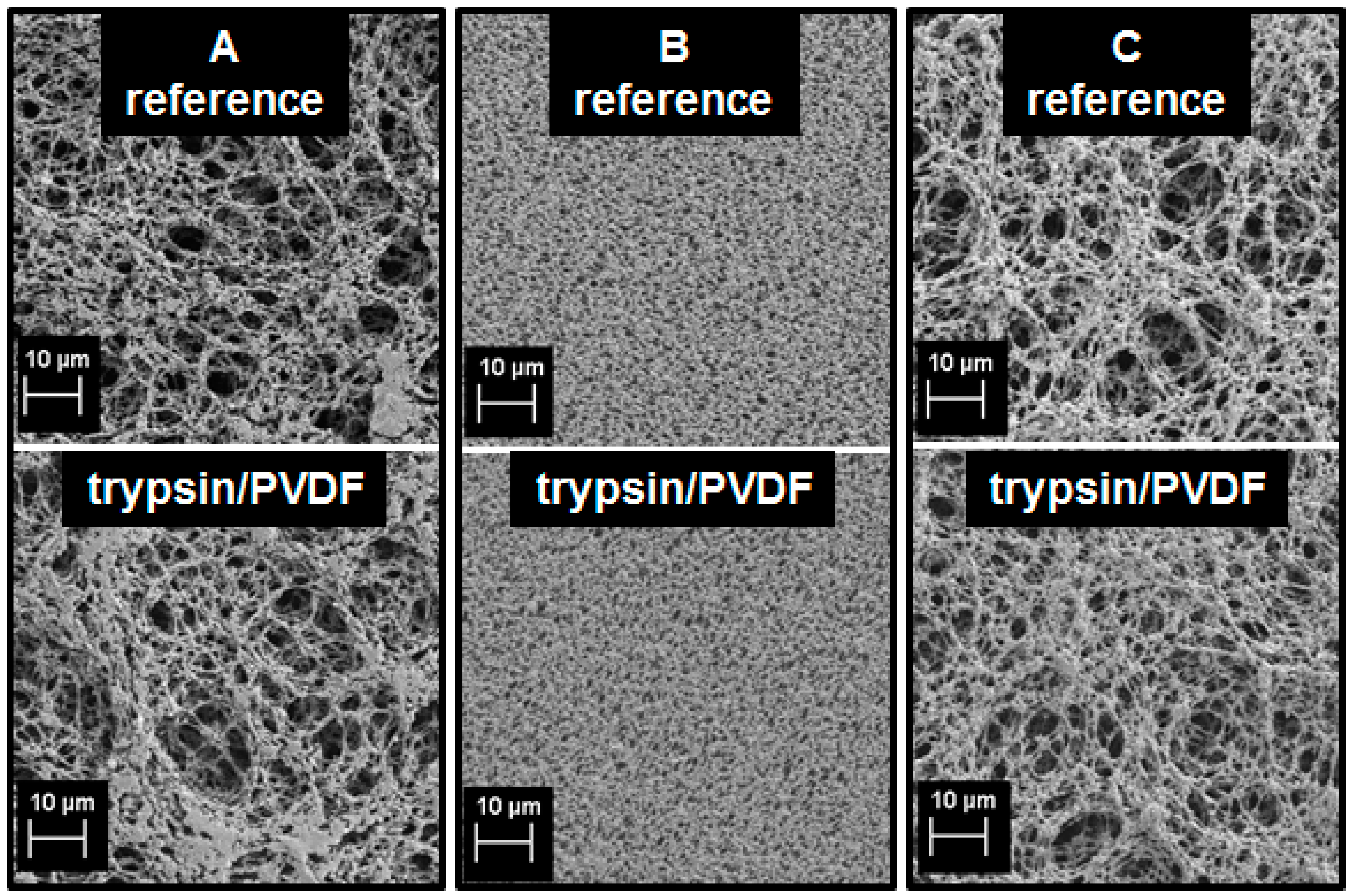
3. Results and Discussion

The EB treatment results in the generation of a mixture of ions, excited molecules and free radicals as described for the radiolysis of water [45] ensuring the activation of both the dissolved trypsin [25,26,46] as well as of the membranes [22,23,24,27,28,29,47]. The formed radicals/activated species can undergo various reactions, such as cross-linking or recombination reactions. This way, links between the polymer matrix and the enzyme can be formed that are assumed to be side-unspecific. Previous work demonstrated the formation of covalent chemical bonds between the membrane polymer and the immobilized molecules by XPS analysis, which was supported by extensive extraction experiments [22,23,24,26,47]. It is worth emphasizing that the use of an aqueous system is a crucial requirement since different results were obtained in the dry state as also reported in the literature [48].
The enzyme trypsin was immobilized via EB radiation on three different PVDF microfiltration membranes (A: hydrophobic, Roti, 0.45 µm, Carl Roth; B: hydrophilic, Durapore, 0.22 mm, Millipore; C: hydrophilic, Durapore, 0.45 µm, Millipore) with different pore sizes and surface energies (see Table 1). While membrane A has a hydrophobic surface (surface energy: 6.4 mN/mm2) with pores of 0.45 μm membranes B and C are hydrophilized by the manufacturer (surface energy: 66.6 and 35.4 mN/mm2, respectively) with pores of 0.22 µm and 0.45 µm, respectively. The thickness of all used membranes is comparable and in the range of 110–130 µm.
Table 1. Characteristics of three different biocatalytic trypsin/polyvinylidene fluoride (PVDF) membranes.
#Surface energy a [mN/mm2]Contribution polar/dispersivePore size b [µm]Thickness c [µm]Trypsin amount [µg/cm2]ktrypsin [−10−5 s−1]
A6.4 ± 1.90.2/6.10.4513030.9 ± 1.2−882
B66.6 ± 3.825.9/40.80.221153.0 ± 0.5−282
C35.4 ± 5.19.6/25.80.451100.6 ± 0.4−216
a Calculated via contact angle determination using water and diiodomethane, respectively (prior to enzyme immobilization); b Manufacturer information; c Determined using a thickness gauge.
Furthermore, the successful enzyme immobilization was demonstrated by XPS analysis (Table 2). According to the structure of PVDF (Figure 1) the theoretically expected elemental composition would be 50% for C and F, respectively. However, the three different PVDF reference membranes consist of different amounts of F and C. Since membranes B and C were hydrophilized by the manufacturer, the high O ratio (membrane A: 0.13%; membrane B: 26.44%; membrane C: 14.58%) can be explained with material modifications during the membrane preparation process which result in the before detected differences in surface energy (Table 1). After EB-mediated trypsin immobilization new signals for N and S occur (N: 5.21%–5.89%; S: 0.73%–1.21%) that indicate a successful enzyme attachment on the membrane. In the case of membrane A also a significant increase of oxygen was detected (reference: 0.13%, after immobilization: 9.73%).
Table 2. XPS (X-ray photoelectron spectroscopy) data of the unmodified and modified PVDF membranes.
#SampleC 1s [mol %]F 1s [mol %]O 1s [mol %]N 1s [mol %]S 2p [mol %]
Areference43.1256.750.13--
trypsin/PVDF51.9731.479.735.890.95
Breference62.3811.1926.44--
trypsin/PVDF62.366.7924.915.210.73
Creference53.2532.1714.58--
trypsin/PVDF59.3112.5921.035.871.21
The trypsin immobilization was most successful on the hydrophobic membrane A with an enzyme loading of 30.9 µg/cm2 and a corresponding reaction rate constant k of −882 × 10−5 s−1 (Table 1). Compared to the hydrophilic membranes this corresponds to a 10 to 50 fold amount of immobilized trypsin. This effect can be explained by hydrophobic interactions between the hydrophobic PVDF membrane A and the protein backbone of the enzyme which enhances surface immobilization.
While these hydrophobic interactions are usually a problem because they promote fouling, here, they are beneficial for the desired enzyme immobilization. Since the immobilization reaction by EB is performed within a very short time interval of 5 min, no pore blocking by the enzyme itself occurred during the process (see Figure 4). Furthermore, the pore structure of the membranes was not damaged by the EB treatment. The surface structure was not altered as demonstrated by the SEM images (Figure 4).
Figure 4. SEM images of membranes A, B, and C prior (reference) and after modification (trypsin/PVDF).
Polymers 07 01485 g004
Regarding the results of pure water permeation flux, pore size, and porosity of the membranes before and after EB treatment (see Table 3) no significant changes were observed. Water permeation flux was not changed by the EB-induced trypsin immobilization. Regarding the median pore size the enzyme immobilization led to slight decrease of the pore size for membranes A and B while membrane C showed a slight increase. When comparing the pore size determined by mercury porosimetry (Table 3) with the pore size information delivered by the manufacturer (Table 1) we can see significant differences, e.g., according to the manufacturer membrane A should possess a pore size of 0.45 µm. Mercury porosimetry revealed a median pore size of 0.95 µm for this membrane. However, there are different methods to determine the pore size of a membrane and no standard method to obtain comparable results. So we explain the differences of the determined pore sizes by a different measurement method used by the manufacturer. The porosity of all modified membranes was slightly reduced. However, the changes are not significant, and we can assume that the membrane structure was not destroyed or altered by the EB treatment nor blocked by the immobilized enzyme. Further investigation on the impact of EB on the membrane material was published before and confirms these results [22,24,25].
Table 3. Membrane properties before (reference) and after electron beam (EB) treatment (EB).
#SampleWater permeation flux [mL/(min·cm2·bar)]Pore size a [µm]Porosity a [%]
Areference29.70.95 ± 0.272.1 ± 2.4
EB30.30.94 ± 0.266.6 ± 1.9
Breference6.10.55 ± 0.159.7 ± 1.4
EB6.10.51 ± 0.152.3 ± 1.5
Creference30.71.16 ± 0.462.7 ± 2.1
EB30.71.25 ± 0.458.8 ± 1.9
a Determined by mercury porosimetry.
Because of the highest enzymatic activity membrane A was chosen for investigating the fouling properties as well as the ability to self-clean its surface. The fouling experiment was conducted by filtration of 4.8 L of protein solution through the membrane and subsequent investigation of the change in pure water flux (for details see Section 2.2). Then, trypsin activation was achieved by immersing the fouled membrane into a buffered solution at 37 °C and pH 8.0. Under these conditions the enzyme catalyzes the hydrolysis of peptide bonds and therefore it shall degrade the protein fouling layer on the membrane surface. After self-cleaning the resulting pure water permeation flux was determined again before a next fouling cycle was started. This procedure was repeated several times to investigate and compare the modified biocatalytic membranes.
Figure 5 summarizes the resulting membrane properties concerning fouling and self-cleaning activity. First, the pristine PVDF membrane A was investigated according to the experimental procedure described in Figure 3. Since no enzyme is immobilized on this reference membrane, no self-cleaning is possible. The membrane fouling after one cycle of the experiment is enormous since the resulting water permeation flux was decreased to 27% compared to the initial flux (see Figure 5).
Figure 5. Fouling and self-cleaning properties were characterized by initial water permation flux, water flux after fouling treatment, and water flux after self-cleaning. Displayed are PVDF reference membrane A (prior to enzyme immobilization, REF), as well as trypsin/PVDF membranes A after repeated fouling cycles (1–5; 1–3) determined directly after enzyme immobilization, and after one week of storage, respectively.
Polymers 07 01485 g005
In contrast, the biocatalytic trypsin/PVDF membrane was less prone to fouling, and the water permeation flux after one cycle was reduced to 50%. Obviously, the presence of the enzyme on the membrane surface partially prevents a direct contact of the fouling protein with the PVDF surface. Therefore, undesired hydrophobic attractions that can result in surface blocking and subsequent reduction of water permeation flux (as experienced in case of the reference membrane) are reduced compared to the reference membrane. Water permeation flux after fouling is significantly higher compared to the reference membrane. Furthermore, the biocatalytic membrane is able to actively self-clean its surface and 90% of the initial water permeation flux could be regained. When the same membrane is exposed to further fouling treatment cycles the fouling effect slightly increases with every additional cycle. The self-cleaning activity of the biocatalytic membrane is effective for the length of four cycles. However, we have chosen to use a highly concentrated protein solution (3 g/L) within the fouling experiment to demonstrate the self-cleaning property of the immobilized trypsin on the PVDF membrane. Thus, it is likely that with repeated fouling cycles (4.8 L protein solution per cycle) at one point there will be adsorption of the protein onto the enzyme of the biocatalytic membrane resulting in decreased water permeation flux, as well as blocking of the active site of the enzyme. After five cycles the water permeation flux after fouling is comparable to that of the reference membrane. The self-cleaning effect was determined to improve the water permeation flux now by only 6% compared to flux after fouling.
The biocatalytic membrane is still active after one week of storage (at 4 °C and pH 7.0). It shows comparable flux recovery as determined for non-stored membranes (Figure 5) within two repeating fouling cycles. However, the efficiency is depleted after that. Probably, the enzyme is less robust after this storage period and tends to higher protein adsorption.
However, to simulate a long time fouling in real systems it is necessary to repeat several fouling cycles (using high protein concentrations) which is also described by many others in the literature [40,41,42,43,44]. The method by which we performed the test aimed for proving that the self-cleaning effect is originated by the enzyme activity, and not by back-washing steps or surface modification effects. However, when this membrane is used in an application, the system would be much easier since realistic system provide much less concentrated protein solutions (less fouling) and the membrane would not have to be removed from the system for self-cleaning. Typically, membrane modules are constructed in a way that regular back-washing steps are performed with the same water that is cleaned at that moment. Then, chemical cleaning is necessary by immersing the entire module for several hours into an extra tank with hypochlorite solution or acidic media [7]. This step could be replaced by immersing the module into a buffer solution as demonstrated by our method which would be beneficial because no toxic or aggressive chemicals would be necessary, the membrane material would not be degraded by the oxidative cleaning, and the disposal of waste that is required after chemical cleaning can be avoided.
In future experiments we aim to immobilize different enzymes for different pH ranges and extend the storage period. Instead of the here chosen model enzyme trypsin more stable enzymes should be used that were designed for industrial applications. Even a combination of different enzymes on the membrane is envisaged to enable an application of one membrane for different pH ranges and/or different target molecules (e.g., proteins, saccharides, lipids) in the fouling system.

4. Conclusions

Trypsin was successfully immobilized via electron beam irradiation on different polymer membranes. The best enzyme loading and highest resulting catalytic activity was obtained using highly hydrophobic membranes for immobilization.
The biocatalytic membranes showed significantly improved antifouling properties compared to the pristine one, and furthermore, by changing the pH trypsin can be “switched on” to actively degrade a fouling layer of proteins. This process leads to a 90% recovery of the initial water permeation flux compared to a reduction to 27% of the non-modified membrane. This new antifouling self-cleaning membrane system shall enable efficient and ecological use in filtration applications avoiding extensive chemical cleaning treatments, and therefore, reduce waste and energy effort.

Acknowledgments

The authors would like to thank the Federal Republic of Germany and the Free State of Saxony for financial support.

Author Contributions

The listed authors contributed to this work as follows: Astrid Stoelzer, Karl Striegler, and Sandra Starke developed and performed the membrane modification reactions as well as the membrane investigation. Andrea Prager conducted SEM and XPS measurements. Agnes Schulze designed the experimental concepts and prepared the manuscript.

Conflicts of Interest

The authors declare no conflict of interest.

References

  1. Marchand-Brynaert, J. Polymer Membranes; CRC Press: Boka Raton, FL, USA, 2012; pp. 4854–4873. [Google Scholar]
  2. Nunes, S.P.; Peinemann, K.-V. Membrane Technology in the Chemical Industry, 2nd ed.; Wiley-VCH Verlag GmbH: Weinheim, Germany, 2006. [Google Scholar]
  3. Hilal, N.; Ogunbiyi, O.O.; Miles, N.J.; Nigmatullin, R. Methods employed for control of fouling in MF and UF membranes: A comprehensive review. Sep. Sci. Technol. 2005, 40, 1957–2005. [Google Scholar] [CrossRef]
  4. Ulbricht, M. Advanced functional polymer membranes. Polymer 2006, 47, 2217–2262. [Google Scholar] [CrossRef]
  5. Fang, Y.; Xu, Z.-K.; Wu, J.; Hoek, E.M.V.; Tarabara, V.V. Surface modification of membranes. In Encyclopedia of Membrane Science and Technology; John Wiley & Sons, Inc.: New York, NY, USA, 2013. [Google Scholar]
  6. Kochkodan, V.; Johnson, D.J.; Hilal, N. Polymeric membranes: Surface modification for minimizing (bio)colloidal fouling. Adv. Colloid Interface Sci. 2014, 206, 116–140. [Google Scholar] [CrossRef] [PubMed]
  7. Le-Clech, P.; Chen, V.; Fane, T.A.G. Fouling in membrane bioreactors used in wastewater treatment. J. Membr. Sci. 2006, 284, 17–53. [Google Scholar] [CrossRef]
  8. Ke, Y.; Zhang, X.; Wu, G.; Ren, L.; Wang, Y. Comparative degradation study of surface-modified polyacrylamide/poly(3-hydroxybutyrate-co-3-hydroxyvalerate) membranes. Polym. Sci. Ser. B 2015. [Google Scholar] [CrossRef]
  9. Wang, J.; Wang, Z.; Wang, J.; Wang, S. Improving the water flux and bio-fouling resistance of reverse osmosis (RO) membrane through surface modification by zwitterionic polymer. J. Membr. Sci. 2015, 493, 188–199. [Google Scholar] [CrossRef]
  10. Nazri, N.; Lau, W.; Ismail, A. Improving water permeability and anti-fouling property of polyacrylonitrile-based hollow fiber ultrafiltration membranes by surface modification with polyacrylonitrile-g-poly(vinyl alcohol) graft copolymer. Korean J. Chem. Eng. 2015, 9, 1853–1863. [Google Scholar] [CrossRef]
  11. Ren, P.-F.; Fang, Y.; Wan, L.-S.; Ye, X.-Y.; Xu, Z.-K. Surface modification of polypropylene microfiltration membrane by grafting poly(sulfobetaine methacrylate) and poly(ethylene glycol): Oxidative stability and antifouling capability. J. Membr. Sci. 2015, 492, 249–256. [Google Scholar] [CrossRef]
  12. Li, F.; Ye, J.; Yang, L.; Deng, C.; Tian, Q.; Yang, B. Surface modification of ultrafiltration membranes by grafting glycine-functionalized pva based on polydopamine coatings. Appl. Surf. Sci. 2015, 345, 301–309. [Google Scholar] [CrossRef]
  13. Cheng, Q.; Zheng, Y.; Yu, S.; Zhu, H.; Peng, X.; Liu, J.; Liu, J.; Liu, M.; Gao, C. Surface modification of a commercial thin-film composite polyamide reverse osmosis membrane through graft polymerization of N-isopropylacrylamide followed by acrylic acid. J. Membr. Sci. 2013, 447, 236–245. [Google Scholar] [CrossRef]
  14. Chung, Y.T.; Ng, L.Y.; Mohammad, A.W. Sulfonated-polysulfone membrane surface modification by employing methacrylic acid through UV-grafting: Optimization through response surface methodology approach. J. Ind. Eng. Chem. 2014, 20, 1549–1557. [Google Scholar] [CrossRef]
  15. Muthumeenal, A.; Neelakandan, S.; Rana, D.; Matsuura, T.; Kanagaraj, P.; Nagendran, A. Sulfonated polyethersulfone (SPES)-charged surface modifying macromolecules (cSMMs) blends as a cation selective membrane for fuel cells. Fuel Cells 2014, 14, 853–861. [Google Scholar] [CrossRef]
  16. Roy, A.; Dadhich, P.; Dhara, S.; De, S. In vitro cytocompatibility and blood compatibility of polysulfone blend, surface-modified polysulfone and polyacrylonitrile membranes for hemodialysis. RSC Adv. 2015, 5, 7023–7034. [Google Scholar] [CrossRef]
  17. Rana, D.; Narbaitz, R.M.; Garand-Sheridan, A.-M.; Westgate, A.; Matsuura, T.; Tabe, S.; Jasim, S.Y. Development of novel charged surface modifying macromolecule blended pes membranes to remove edcs and ppcps from drinking water sources. J. Mater. Chem. A 2014, 2, 10059–10072. [Google Scholar] [CrossRef]
  18. Ouradi, A.; Nguyen, Q.T.; Benaboura, A. Polysulfone–AN69 blend membranes and its surface modification by polyelectrolyte-layer deposit—Preparation and characterization. J. Membr. Sci. 2014, 454, 20–35. [Google Scholar] [CrossRef]
  19. Mehrparvar, A.; Rahimpour, A.; Jahanshahi, M. Modified ultrafiltration membranes for humic acid removal. J. Taiwan Inst. Chem. Eng. 2014, 45, 275–282. [Google Scholar] [CrossRef]
  20. Mahlicli, F.; Altinkaya, S. Surface modification of polysulfone based hemodialysis membranes with layer by layer self assembly of polyethyleneimine/alginate-heparin: A simple polyelectrolyte blend approach for heparin immobilization. J. Mater. Sci. Mater. Med. 2013, 24, 533–546. [Google Scholar] [CrossRef] [PubMed]
  21. Wu, X.-M.; Wang, L.-L.; Wang, Y.; Gu, J.-S.; Yu, H.-Y. Surface modification of polypropylene macroporous membrane by marrying raft polymerization with click chemistry. J. Membr. Sci. 2012, 421, 60–68. [Google Scholar] [CrossRef]
  22. Schulze, A.; Marquardt, B.; Kaczmarek, S.; Schubert, R.; Prager, A.; Buchmeiser, M.R. Electron beam-based functionalization of poly(ethersulfone) membranes. Macromol. Rapid Commun. 2010, 31, 467–472. [Google Scholar] [CrossRef] [PubMed]
  23. Schulze, A.; Marquardt, B.; Went, M.; Prager, A.; Buchmeiser, M.R. Electron beam-based functionalization of polymer membranes. Water Sci. Technol. 2012, 65, 574–580. [Google Scholar] [CrossRef] [PubMed]
  24. Schulze, A.; Maitz, M.F.; Zimmermann, R.; Marquardt, B.; Fischer, M.; Werner, C.; Went, M.; Thomas, I. Permanent surface modification by electron-beam-induced grafting of hydrophilic polymers to PVDF membranes. RSC Adv. 2013, 3, 22518–22526. [Google Scholar] [CrossRef]
  25. Starke, S.; Went, M.; Prager, A.; Schulze, A. A novel electron beam-based method for the immobilization of trypsin on poly(ethersulfone) and poly(vinylidene fluoride) membranes. React. Funct. Polym. 2013, 73, 698–702. [Google Scholar] [CrossRef]
  26. Jahangiri, E.; Reichelt, S.; Thomas, I.; Hausmann, K.; Schlosser, D.; Schulze, A. Electron beam-induced immobilization of laccase on porous supports for waste water treatment applications. Molecules 2014, 19, 11860–11882. [Google Scholar] [CrossRef] [PubMed]
  27. Brown, J.R.; O’Donnell, J.H. Effects of gamma radiation on two aromatic polysulfones. J. Appl. Polym. Sci. 1975, 19, 405–417. [Google Scholar] [CrossRef]
  28. Hill, D.J.T.; Lewis, D.A.; O’Donnell, J.H.; Whittaker, A.K. The crosslinking mechanism in gamma irradiation of polyarylsulfone: Evidence for Y-links. Polym. Adv. Technol. 1998, 9, 45–51. [Google Scholar] [CrossRef]
  29. Klimová, M.; Szöcs, F. ESR and DSC study of the radiation crosslinking effect on macroradical decay in poly(vinylidene fluoride). J. Appl. Polym. Sci. 1989, 37, 3449–3458. [Google Scholar] [CrossRef]
  30. Cleland, M.R.; Parks, L.A.; Cheng, S. Applications for radiation processing of materials. Nucl. Instrum. Methods Phys. Res. Sect. B 2003, 208, 66–73. [Google Scholar] [CrossRef]
  31. Barrett, A.J.; Rawlings, N.D.; Woessner, J.F. Handbook of Proteolytic Enzymes, 2nd ed.; Elsevier Academic Press: London, UK, 2004. [Google Scholar]
  32. Simon, M.L.; László, K.; Kotormán, M.; Szajáni, B. A comparative study of the conformational stabilities of trypsin and alpha-chymotrypsin. Acta Biol. Szeged. 2001, 45, 43–49. [Google Scholar]
  33. Crewther, W.G. The effect of pH and cations on the thermal denaturation of trypsin. Aust. J. Biol. Sci. 1953, 6, 597–616. [Google Scholar] [PubMed]
  34. Pinto, S.C.; Rodrigues, A.R.; Saraiva, J.A.; Lopes-da-Silva, J.A. Catalytic activity of trypsin entrapped in electrospun poly(ε-caprolactone) nanofibers. Enzyme Microb. Technol. 2015, 79, 8–18. [Google Scholar] [CrossRef] [PubMed]
  35. Ahn, H.-K.; Kim, B.C.; Jun, S.-H.; Chang, M.S.; Lopez-Ferrer, D.; Smith, R.D.; Gu, M.B.; Lee, S.-W.; Kim, B.S.; Kim, J. Robust trypsin coating on electrospun polymer nanofibers in rigorous conditions and its uses for protein digestion. Biotechnol. Bioeng. 2010, 107, 917–923. [Google Scholar] [CrossRef] [PubMed]
  36. Shi, Q.; Su, Y.; Ning, X.; Chen, W.; Peng, J.; Jiang, Z. Trypsin-enabled construction of anti-fouling and self-cleaning polyethersulfone membrane. Bioresour. Technol. 2011, 102, 647–651. [Google Scholar] [CrossRef] [PubMed]
  37. Liu, J.; Huang, J.; Wujcik, E.K.; Qiu, B.; Rutman, D.; Zhang, X.; Salazard, E.; Wei, S.; Guo, Z. Hydrophobic electrospun polyimide nanofibers for self-cleaning materials. Macromol. Mater. Eng. 2015, 300, 358–368. [Google Scholar] [CrossRef]
  38. Oliveira, G.B.; Lima Filho, J.L.; Cavalcante Chaves, M.E.; Azevedo, W.M.; Carvalho, L.B., Jr. Enzyme immobilization on anodic aluminum oxide/polyethyleneimine or polyaniline composites. React. Funct. Polym. 2008, 68, 27–32. [Google Scholar] [CrossRef]
  39. Smith, P.K.; Krohn, R.I.; Hermanson, G.T.; Mallia, A.K.; Gartner, F.H.; Provenzano, M.D.; Fukimotot, E.K.; Goeke, N.M.; Olson, B.J.; Klenk, D.C. Measurement of protein using bicinchoninic acid. Anal. Biochem. 1985, 150, 76–85. [Google Scholar] [PubMed]
  40. Smith, A.L.; Skerlos, S.J.; Raskin, L. Membrane biofilm development improves cod removal in anaerobic membrane bioreactor wastewater treatment. Microb. Biotechnol. 2015, 8, 883–894. [Google Scholar] [CrossRef] [PubMed]
  41. Lin, J.; Ye, W.; Huang, J.; Ricard, B.; Baltaru, M.-C.; Greydanus, B.; Balta, S.; Shen, J.; Vlad, M.; Sotto, A.; et al. Toward resource recovery from textile wastewater: Dye extraction, water and base/acid regeneration using a hybrid NF-BMED process. ACS Sustain. Chem. Eng. 2015, 3, 1993–2001. [Google Scholar] [CrossRef]
  42. Zhao, Q.; Hou, J.; Shen, J.; Liu, J.; Zhang, Y. Long-lasting antibacterial behavior of a novel mixed matrix water purification membrane. J. Mater. Chem. A 2015, 3, 18696–18705. [Google Scholar] [CrossRef]
  43. Chu, K.H.; Yoo, S.S.; Yoon, Y.; Ko, K.B. Specific investigation of irreversible membrane fouling in excess of critical flux for irreversibility: A pilot-scale operation for water treatment. Sep. Purif. Technol. 2015, 151, 147–154. [Google Scholar] [CrossRef]
  44. Duan, L.; Li, S.; Han, L.; Song, Y.; Zhou, B.; Zhang, J. Comparison between moving bed-membrane bioreactor and conventional membrane bioreactor systems. Part I: Membrane fouling. Environ. Earth. Sci. 2015, 73, 4881–4890. [Google Scholar] [CrossRef]
  45. Borrely, S.I.; Cruz, A.C.; Mastro, N.L.D.; Sampa, M.H.O.; Somessari, E.S. Radiation processing of sewage and sludge. A review. Prog. Nucl. Energy 1998, 33, 3–21. [Google Scholar] [CrossRef]
  46. Sharpatyi, V.A. Aspects of radiation-chemistry of protein molecules. High Energy Chem. 1995, 29, 77–90. [Google Scholar]
  47. Boulares-Pender, A.; Thomas, I.; Prager, A.; Schulze, A. Surface modification of polyamide and polyvinylidene fluoride membranes. J. Appl. Polym. Sci. 2013, 128, 322–331. [Google Scholar] [CrossRef]
  48. Marletta, G.; Pignataro, S. X-ray, electron, and ion beam induced modifications of poly(ether sulfone). Macromolecules 1991, 24, 99–150. [Google Scholar] [CrossRef]

