Article

Conjugation of ß-Adrenergic Antagonist Alprenolol to Implantable Polymer-Aescin Matrices for Local Delivery

1
Department of Inorganic and Analytical Chemistry, Faculty of Pharmacy with the Laboratory Medicine Division, Medical University of Warsaw, Banacha 1, Warsaw 02-097, Poland
2
Department of Environmental Health Science, Faculty of Pharmacy with the Laboratory Medicine Division, Medical University of Warsaw, Banacha 1, Warsaw 02-097, Poland
*
Author to whom correspondence should be addressed.
Polymers 2015, 7(9), 1820-1836; https://doi.org/10.3390/polym7091484
Submission received: 24 July 2015 / Revised: 8 September 2015 / Accepted: 10 September 2015 / Published: 18 September 2015
(This article belongs to the Special Issue Functional Polymers for Medical Applications)

Abstract

:
The sustained release of alprenolol, a β-adrenergic antagonist, could be beneficial for the treatment of various heart diseases while reducing the side effects resulting from its continuous use. The novel and branched copolymers uniquely composed of biodegradable components (lactide and glycolide) have been synthesized using natural and therapeutically-efficient β-aescin-initiator, and consequently characterized to determine their structures and physicochemical properties. The obtained matrices were not cyto- and genotoxic towards bacterial luminescence, protozoan, and Salmonella typhimurium TA1535. The copolymers release the drug in vitro in a sustained manner and without burst release. The value of the drug released was strongly dependent on the copolymer composition and highly correlated with the hydrolytic matrices’ degradation results.

1. Introduction

Aescin, a natural mixture of triterpenoid saponins isolated from the horse chestnut tree (Aesculus hippocastanum (Hippocastanaceae)), possesses diverse biochemical and pharmacological actions. It exists in two forms: α and β, distinguished by differences in melting point, specific rotation, hemolytic index, and solubility in water [1]. However, the β-aescin form appears as an active component of the mixture and is the molecular form present in the major pharmaceutical products available. In the past few years, the pharmacological profile of β-aescin has received significant attention due to the establishment of its pharmacological basis for the major clinical indication in the treatment of chronic venous insufficiency (CVI) [1]. There are at least three types of pharmacodynamic actions attributed to β-aescin: anti-edematous and venotonic properties, as well as anti-inflammatory activities [2]. It is assumed that these actions are based on a molecular mechanism, identified as a selective vascular permeabilization, allowing a higher sensitivity of, e.g., calcium channels, to molecular ions, resulting in increased venous and arterial tone [3,4]. These sensitizing effects to ions and other molecules, e.g., 5-HT, result in enhanced venous contractile activity and, consequently, in the anti-edematous property of the molecule [1]. Therefore, β-aescin is now widely quoted in the literature as a pharmacological tool for assessing the sensitivity of vascular tissues to different agonists, in order to evaluate the mechanism of, for example, hypertension development in animal models [5]. In addition to pharmacological activity, β-aescin might be used effectively as a macroinitiator of the ring-opening polymerization (ROP) of cyclic esters, due to the presence of hydroxyl groups in the molecule [6].
β-adrenergic blockers (β-blockers) are an important class of drugs for the treatment of various heart diseases, including high blood pressure, insufficiency of blood flow to the heart muscle (angina pectoris), irregular heartbeat (arrhythmias), thickened heart muscle (hypertrophic cardiomyopathy) and decreased ability of the heart to empty or fill normally (heart failure) [7]. To the family of β-adrenergic blockers belongs alprenolol (ALP), a non-cardioselective β-blocker, developed in 1966 [8]. ALP is reported to have intrinsic sympathomimetic activity and some membrane-stabilizing properties. ALP has been given orally, as a benzoate or hydrochloride, in the management of hypertension, angina pectoris, and cardiac arrhythmias. ALP is non-polar and hydrophobic, freely soluble in water and ethanol, with low to moderate lipid solubility, and a half-life of 2–3 h [9]. When ALP is administered orally, its bioavailability is lowered because it is subjected to the first-pass effect of the liver. For example, it has been reported that the bioavailability of ALP is only 15% in oral administration; thus, multiple dosing is required to maintain therapeutic drug blood levels [9]. These factors make ALP a suitable candidate for implantable local delivery, which improves the bioavailability of the drug, may maintain a high drug concentration in the blood circulation or prolonging the duration of action, may enhance its physical stability, increase specificity and reduce systemic adverse effects for targeted drug delivery and reduces the frequency of its dosing. In previous years, a series of o-cyclopropane carboxylic acid ester prodrugs of various β-blocking agents, including ALP, were synthesized. All prodrugs were hydrolyzed to give their parent compounds an aqueous phosphate buffer of pH 7.4 or in 80% human plasma [10]. It has been found that the formation of the o-cyclopropane carboxylic acid ester derivatives significantly increased the lipophilicities of the β-blockers, as measured by the distribution coefficient between n-octanol and an aqueous phosphate buffer of pH 7.4. Prokai et al. studied ocular delivery of ALP by site-specific bioactivation of its methoxime analogue [11]. They demonstrated that the benefit of this chemical delivery system approach includes the facile release of a potential anti-glaucoma agent at the site of the action, only.
Over the past few decades, there has been a rapid growth in the medical use of biomaterials in many fields, e.g., tissue engineering, the implantation of medical devices and artificial organs, prostheses, ophthalmology, dentistry and bone repair [12]. Among all of them, the use of a biodegradable copolymer of poly (lactide-co-glycolide) (PLGA) has shown immense potential as a drug-delivery carrier and scaffold for tissue engineering [13]. PLGA belongs to a family of biodegradable polymers approved by the FDA because of its high biocompatibility, favorable degradation characteristics and possibilities for sustained drug delivery. Its use also has the possibility of tuning the overall physical properties of the PLGA-drug matrix by controlling the relevant parameters, such as polymer molecular weight, the ratio of lactide to glycolide, and drug concentration for achieving a desired dosage and release interval depending upon the drug type [14,15,16].
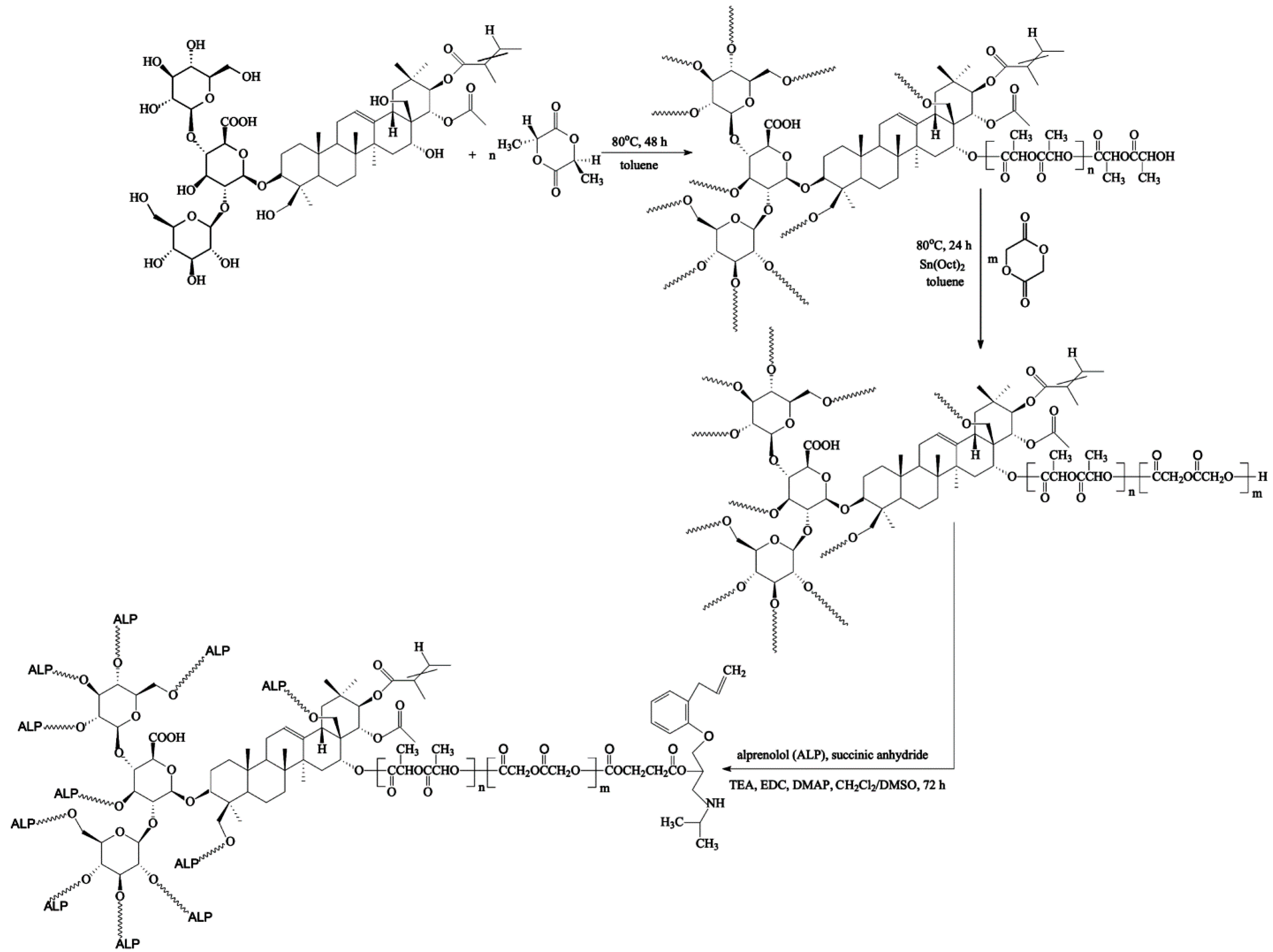
In the present work, we report on the synthesis, characterization and drug-release behavior of a newly formed conjugate of ALP that is bonded to the matrices of the PLGA-aescin for implantable local delivery. To the best of our knowledge, no research group has developed a covalent conjugate of ALP constructed from a biodegradable copolymer with incorporated β-aescin as a central core. To achieve this goal, the copolymers were firstly obtained by the ring-opening polymerization (ROP) of d,l-lactide (LA) and glycolide (GL) using a natural, and therapeutically efficacious, initiator—β-aescin. Different ratios of lactide and glycolide were employed to obtain a suitable copolymer composition for a high drug-release efficiency. The synthesized branched copolymeric matrices have been subjected to cyto- and genotoxicity assays and then bonded to ALP using the EDC (carbodiimide) coupling method. The in vitro release rate of the drug obtained from the synthesized conjugates was analyzed. It was found that the ester-linked ALP conjugate was a promising implantable drug carrier for local delivery, one that might improve the bioavailability and pharmacokinetics of ALP as well as increase its efficiency, resulting from the synergistic action of ALP with β-aescin.

2. Experimental Section

2.1. Materials

Alprenolol hydrochloride (1-(propan-2-ylamino)-3-(2-prop-2-enylphenoxy)propan-2-ol, anhydrous, ≥98%, Sigma Co., Poznan, Poland), succinic anhydride (97%, Aldrich Co. Poznan, Poland), N-(3-dimethylaminopropyl)-N′-ethylcarbodiimide hydrochloride (EDC, 98%, Aldrich Co. Poznan, Poland), triethylamine (TEA, ≥99%, Sigma-Aldrich Co., Poznan, Poland) and 4-(dimethylamino)pyridine (DMAP, ≥99%, Aldrich Co., Poznan, Poland) were used without further purification. d,l-lactide (3,6-dimethyl-1,4-dioxane-2,5-dione, 98.0%, Aldrich Co., Poznan, Poland) was recrystallized from dried ethyl acetate in a dry nitrogen atmosphere and then thoroughly dried in a vacuum before use. Glycolide (GL, ≥99%, Sigma Co., Poznan, Poland) and β-aescin (≥95%, MP Biomedicals, Poznan, Poland) were thoroughly dried in a vacuum before use. Stannous octoate (Sn(Oct)2, tin(II) 2-ethylhexanoate, 2-ethylhexanoic acid tin(II) salt, ~95%, Aldrich Co., Poznan, Poland) was used as received. Dichloromethane (DCM, anhydrous, ≥99.8%, POCh, Gliwice, Poland), dimethyl sulfoxide (DMSO, anhydrous, 99%, Aldrich Co., Poznan, Poland), toluene (anhydrous, 99.8%, POCh, Gliwice, Poland) and diethyl ether (anhydrous, 99.8%, POCh, Gliwice, Poland) were used as received. Phosphate buffer solution (pH 7.4 ± 0.05, 0.1 M, PBS, potassium dihydrogen phosphate/di-sodium hydrogen phosphate, 20 °C, Avantor Performance Materials, Gliwice, Poland) was also used as received.

2.2. Copolymerization Procedure

The branched copolymeric materials were prepared using different molar ratios of initiator (aescin) to monomers (d,l-lactide (LA) and glycolide (GL)) (Table 1). The initiator/monomer (LA or GL) feed ratios for the synthesized copolymers were 1/50/50, 1/30/70, and 1/70/30, denoted as aescin/PLA50/PGL50, aescin/PLA30/PGL70 and aescin/PLA70/PGL30, respectively. For the copolymerization, dry aescin and LA were accurately weighed and introduced into a 50 mL polymerization tube. The tube was then connected to a Schlenk line, where exhausting-refilling processes were repeated three times before toluene was added. The tube was immersed in an oil bath at 80 °C under an argon atmosphere for 48 h. After an appropriate time, the precise weight of GL and a catalytic amount of Sn(Oct)2 (0.02 mol % relative to the total monomer concentration) were added to the mixture and the exhausting-refilling process was carried out again (toluene, 24 h). The resulting products were dissolved in a dry mixture of CH2Cl2/CHCl3, precipitated twice from methanol and dried in a vacuum for 72 h.

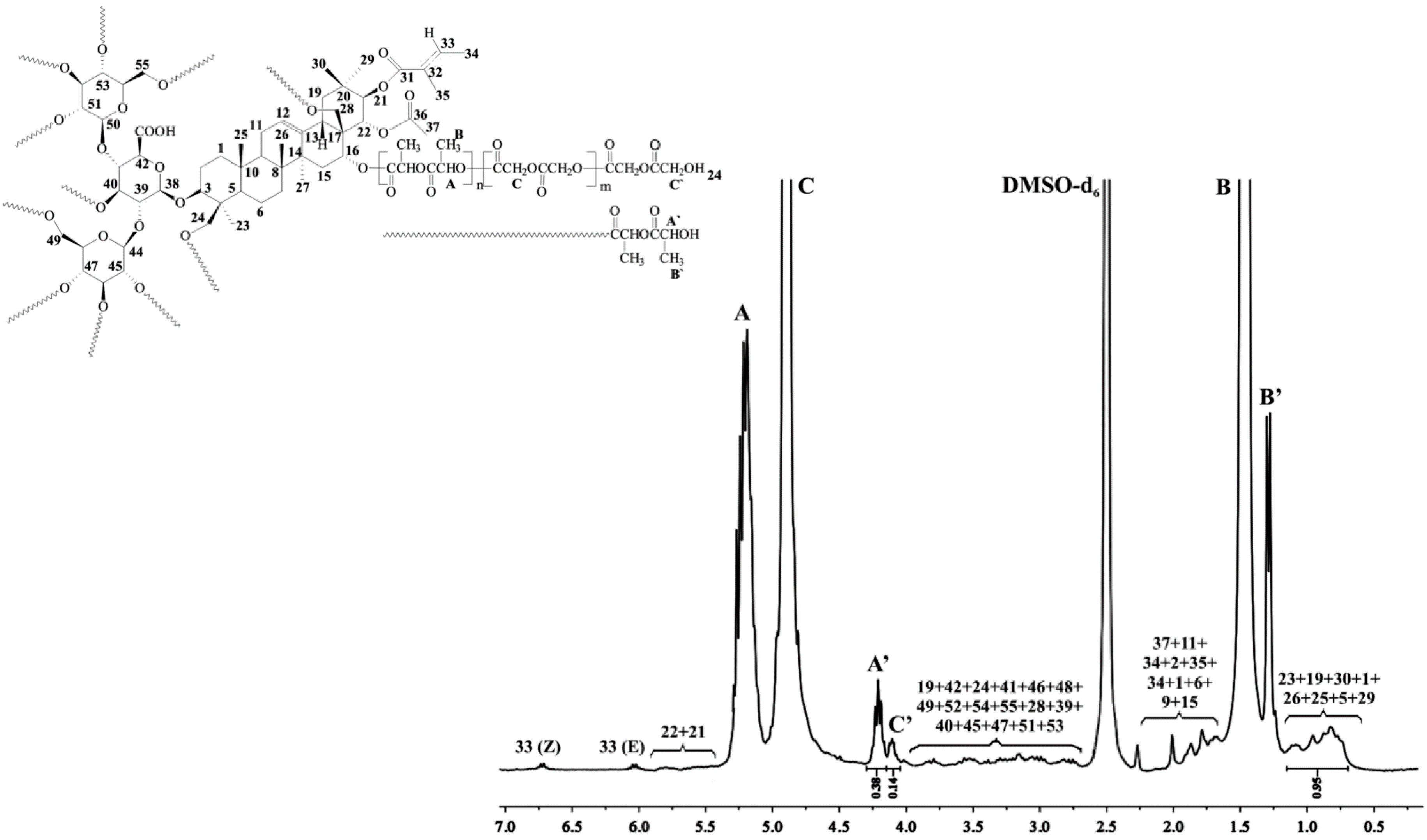
2.2.1. Spectral Characterization of Aescin/PLA50/PGL50

1H NMR (DMSO-d6, 300 MHz, δH, ppm); 0.75–1.09 (18H of aescin, 23, 19, 30, 1, 26, 25, 5, 29), 1.29 (d, –CH3, end group of PLA (B’)), 1.47 (d, –CH3 of PLA (B)), 1.52–2.01 (17H of aescin, 37, 11, 24, 2, 35, 34, 1, 6, 9, 15), 2.85–3.95 (21H of aescin, 19, 42, 24, 41, 46, 48, 49, 52, 54, 55, 28, 39, 40, 45, 47, 51, 53), 4.10 (d, –C(O)CH2OH, end group of PGL (C’)), 4.21 (q, –C(O)CH(CH3)OH, end group of PLA (A’)), 4.90 (d, –C(O)CH2O– of PGL (C)), 5.20 (q, –C(O)CH(CH3)O– of PLA (A)), 5.43–5.74 (2H of aescin, 21, 22), 6.01–6.68 (2H of aescin, 33 (Z, E)) (Figure 1).
13C NMR (DMSO-d6, 300 MHz, δC, ppm): 15.15 (C’), 16.49 (C), 21.9–35.8 (23, 11, 2, 27, 29, 7, 15, 10, 20), 42.9–46.3 (4, 17, 19), 59.96 (E’), 60.76 (E), 67.93 (B’), 68.93 (B), 73.1–76.8 (21, 51, 42, 45, 52, 48, 46, 53, 47), 102.4–103.0 (38, 44, 50), 141.9 (13), 166.76 (D), 169.58 (A), 169.77 (A’) (Figure S1, Supplementary Material).
FTIR (KBr, cm−1): 3520 (υO–H), 2963 (υasCH2), 1750 (υC=O), 1422 (δasCH3), 1167 (υOC–O), 1091 (υC–O–C) (Figure S2, Supplementary Material).

2.2.2. Spectral Characterization of Aescin

1H NMR (DMSO-d6, 300 MHz, δH, ppm); 6.68 (H-33 Z), 6.01 (H-33 E), 5.74 (H-22), 5.43 (H-21), 5.25 (H-12), 4.99 (OH-40, OH-45, OH-51, OH-53), 4.85 (OH-24), 4.78 (OH-16), 4.65 (H-44), 4.56 (OH-47), 4.51 (H-38), 4.48 (OH-46, OH-49, OH-52, OH-55), 4.39 (OH-28), 4.25 (H-50), 3.95 (H-16), 3.91 (H-42), 3.85 (H-24a), 3.69 (H-49a), 3.67 (H-48), 3.62 (H-41), 3.60 (H-46, H-52, H-55a), 3.51 (H-55b), 3.49 (H-54), 3.41 (H-49b), 3.30 (H-3, HDO), 3.24 (H-47), 3.13 (H-24b), 3.03–3.13 (H-39, H-40, H-53) 3.02 (H-28a), 2.99 (H-51), 2.92 (H-45), 2.85 (H-28b), 2.56 (H-18), 2.49 (H-19a), 2.01 (H-37 CH3), 1.88 (H-34 E CH3), 1.83 (H-11a), 1.80 (H-35 E CH3) 1.77 (H-2a, H-11b), 1.74 (H-34 Z CH3) 1.72 (H-35 Z CH3), 1.71 (H-2b), 1.59 (H-15a), 1.55 (H-9), 1.52 (H-1a, H-6a, b), 1.46 (H-7a), 1.39 (H-27 CH3), 1.24 (H-7b), 1.22 (H-15b), 1.09 (H-19b, H-23 CH3), 0.96 (H-1b, H-30 CH3), 0.84 (H-5), 0.81 (H-26 CH3), 0.80 (H-25 CH3), 0.75 (H-29 CH3) (Figure S3a–c, Supplementary Material).
13C NMR (DMSO-d6, 300 MHz, δC, ppm): 169.8 (C43), 169.5 (C36), 166.7 (C31), 141.9 (C13), 136.3 (C33 E+Z), 128.4 (C32 E+Z), 122.8 (C12), 103.0 (C38), 102.4 (C44, C50), 90.1 (C3), 80.0 (C41), 78.0 (C22), 77.7 (C54), 76.8 (C47), 76.3 (C53), 74.5 (C46, C48, C52), 73.9 (C45), 73.3 (C42, C51), 73.1 (C21), 69.9 (C39), 68.7 (C40), 66.9 (C16), 62.9 (C28), 62.1 (C24), 60.9 (C49), 60.3 (C55), 55.4 (C5), 46.3 (C19), 46.0 (C17), 45.9 (C9), 42.9 (C4), 40.7 (C14), 39.8 (C8), 39.6 (C18), 38.2 (C1), 35.8 (C20), 35.5 (C10), 33.4 (C15), 32.5 (C7), 29.0 (CH3 29), 26.9 (C27), 25.6 (C2), 23.2 (C11), 21.9 (CH3 23), 20.8 (CH3 37), 20.3 (CH3 35 E), 19.5 (CH3 30), 18.0 (C6), 16.2 (CH3 26), 15.2 (CH3 25, CH3 34 E), 14.1 (CH3 34 Z), 12.0 (CH3 35 Z) (Figure S4, Supplementary Material)
HSQC (DMSO-d6, 300 MHz, δH, δC, ppm) (Figure S5a–c, Supplementary Material).
FTIR (KBr, cm−1): 3438 (υO–H), 2929 (υasCH2), 1723 (υC=O), 1437 (δsCH3), 1268 (υasCOC), 1161 (υsCOC), 1075 (υC–O–C) (Figure S6, Supplementary Material).

2.3. Synthesis of ALP Conjugated the Synthesized Branched Copolymeric Matrices

The synthesized copolymeric matrices—0.5 g of aescin/PLA50/PGL50, aescin/PLA30/PGL70 or aescin/PLA70/PGL30 (the Mn(GPC) of the copolymers are listed in Table 1)—were first dissolved in 50 mL anhydrous THF under stirring for 30 min at room temperature. Then, succinic anhydride (the molar ratio of the copolymeric matrices to succinic anhydride was 1:1.5 for each hydroxyl group, see Table 1) and 0.8 mL of TEA as the catalyst were added to the reaction flask under equal reaction conditions for 24 h. The crude product with carboxyl terminal groups was then precipitated in the cold diethyl ether twice and dried in a vacuum (the yields ranged from 85% to 88%). Subsequently, to a solution of synthesized copolymeric products with carboxyl terminal groups (0.4 g of aescin/PLA50/PGL50, aescin/PLA30/PGL70 or aescin/PLA70/PGL30) in DMSO (20 mL), EDC, and DMAP were added under nitrogen (the molar ratio of copolymeric products with a carboxyl terminal group to EDC was 1:3 for each carboxyl group, and the molar ratio of EDC to DMAP was 1:1.5). The mixture was stirred at room temperature for 2 h and alprenolol hydrochloride (ALP) was added (the molar ratio of copolymeric matrices with a carboxyl terminal group to ALP was 1:1.5 for each carboxyl group). The reaction was stirred for 48 h at room temperature. The crude conjugation products were precipitated in a cold diethyl ether three times and dried in a vacuum. The yields of the conjugation products were 75%, 70% and 78% for aescin/PLA50/PGL50/ALP, aescin/PLA30/PGL70/ALP, and aescin/PLA70/PGL30/ALP, respectively.
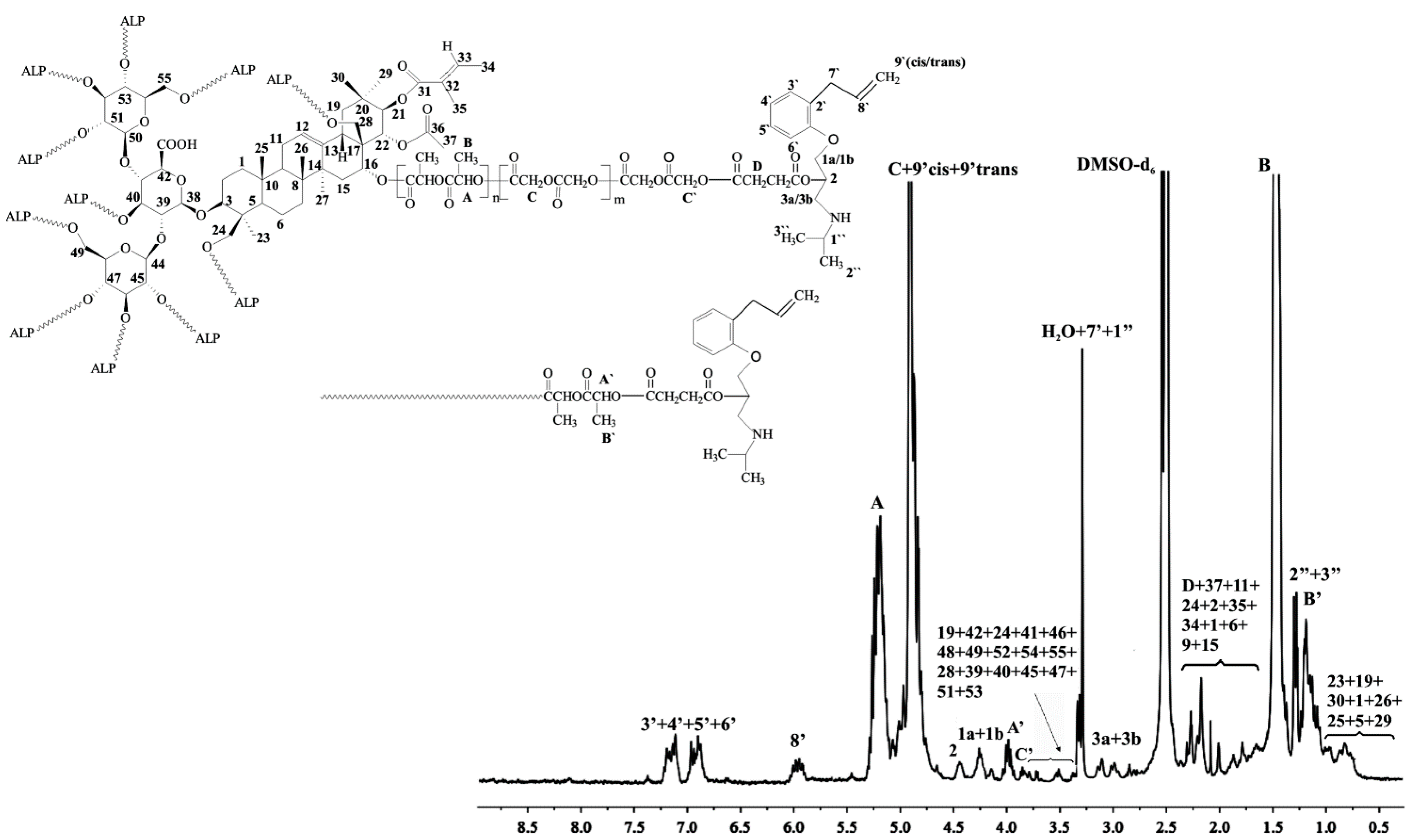
1H NMR of aescin/PLA30/PGL70/ALP (DMSO-d6, 300 MHz, δH, ppm): 0.71–1.05 (18H of aescin, 23, 19, 30, 1, 26, 25, 5, 29), 1.17–1.29 (d, –CH3, end group of PLA (B’) and (6H, two –CH3 of ALP (2”, 3”), 1.46 (d, –CH3 of PLA (B)), 1.68–2.29 (17H of aescin, 37, 11, 24, 2, 35, 34, 1, 6, 9, 15 and (C(O)OCH2CH2– of succinic anhydride (D)), 2.85–3.93 (21H of aescin, 19, 42, 24, 41, 46, 48, 49, 52, 54, 55, 28, 39, 40, 45, 47, 51, 53) and (2H, –CHCH2NH– of ALP (3a, 3b)) and (2H, –CH2=CHCH2–, –NHCH(CH3)2 of ALP (7’, 1”)); 3.96 (2H, –OCH2CH– of ALP (1a, 1b)), 4.19 (–C(O)CH2–, of PGL (C’)), 4.25 (–C(O)CH(CH3)– of PLA (A’)); 4.47 (1H, –CH2CHCH2– of ALP (2)), 4.83–4.91 (d, –C(O)CH2O– of PGL (C)) and (2H, H2C=CH– of ALP (9’cis, 9’trans)), 5.21 (q, –C(O)CH(CH3)O– of PLA (A)), 5.47 (1H, H2C=CH– of ALP (8’)), 6.92–7.16 (4H of benzyl ring of ALP (3’, 4’, 5’, 6’)) (Figure 3).
FTIR (KBr, cm−1): 3531 (υC–Haromatic), 2999–2962 (υasCH2), 1759 (υC=O), 1453 (υC–H), 1396 (δasCH3), 1268 (υasCOC), 1131 (υOC–O), 1091 (υC–O–C) (Figure S7, Supplementary Material).

2.4. Toxicity Assays

2.4.1. Microtox® Assay

Microtox® assay with the luminescent bacteria Vibrio fischeri was performed with the lyophilized bacteria purchased from Modern Water (New Castle, DE, USA). The metabolism of the bacteria was inhibited by toxic substances, resulted in the luminescence of the samples decreasing. The measurement was made in a luminometer Microtox® M500 after 15 and 30 min incubation of bacteria with the sample. On the basis of obtained results (MicrotoxOmni), the PE as the percent of luminescence changing was calculated and compared to control (non-toxic 2% NaCl solution).

2.4.2. Spirotox Test

Spirotox test with the protozoan Spirostomum ambiguum was performed according to the standard protocol [17]. The test was carried out in disposable, polystyrene multiwell plates (24 wells). Ten organisms were added to each well of the multiwell. The samples were incubated in the darkness at 25 °C for 24 h. Afterwards test responses: different deformations such as shortening, bending of the cell, etc., and lethal response were observed with the use of dissection microscope (magnification of 10). As a diluent and a control, Tyrod solution was used.
Prior to the toxicity test, the materials were pulverized. In the direct contact test, three concentrations of the samples were tested in Spirotox test (1.0, 0.5, and 0.25 mg mL−1) and two in Microtox® assay (0.8 and 0.4 mg mL−1). The tested sample was weighted directly to the test containers and poured with 1 mL of diluent. The test organisms were incubated with the suspension of the tested sample. All the samples were run in triplicate for the toxicity measurements.

2.4.3. The Umu-Test

The umu-test is a bioassay for evaluating the genotoxic potential of environmental samples and chemical compounds. The test organism is Salmonella typhimurium TA1535/pSK1002. As a response to different types of DNA damage the umuC gene in bacterial cells is induced which is a part of the SOS system. The test strain is genetically modified—the umuC gene activity is linked to the synthesis of β-galactosidase while other DNA regions responsible for this enzyme synthesis were deleted. The transcription of the umuC gene correlates with the amount of secreted β-galactosidase. The enzyme converts colorless substrate (ortho-nitrophenyl-β-galactoside) into the yellow product which can be quantified colorimetrically at 420 nm. Additionally, the bacteria growth (G) is evaluated by a measurement of an optical density to determine the cytotoxicity of tested samples. The genotoxic potential of the sample is presented as the Induction Ratio (IR)—the β-galactosidase activity ratio of tested sample in comparison to the negative control. Samples with IR ≥ 1.5 are considered as genotoxic [18]. The umu-test was performed in 96-well microplates according to the ISO 13829 protocol [18] with and without metabolic activation (S9 liver fraction from male Sprague–Dawley rats treated five days before the isolation with a single dose of 500 mg kg−1 body weight of Aroclor 1254 in soya oil). Deionized sterile water was used as a negative control, PBS as solvent control, while 2-aminoanthracene and 4-nitroquinoline N-oxide were used as positive controls. All tested copolymeric matrices were incubated in phosphate buffered saline (PBS from Gibco, Carlsbad, CA, USA, 1 mg mL−1) for 24 h, 37 °C with shaking. Before the assay all extracts were sterilized by filtration. All samples were tested in two fold dilution series (six concentrations).

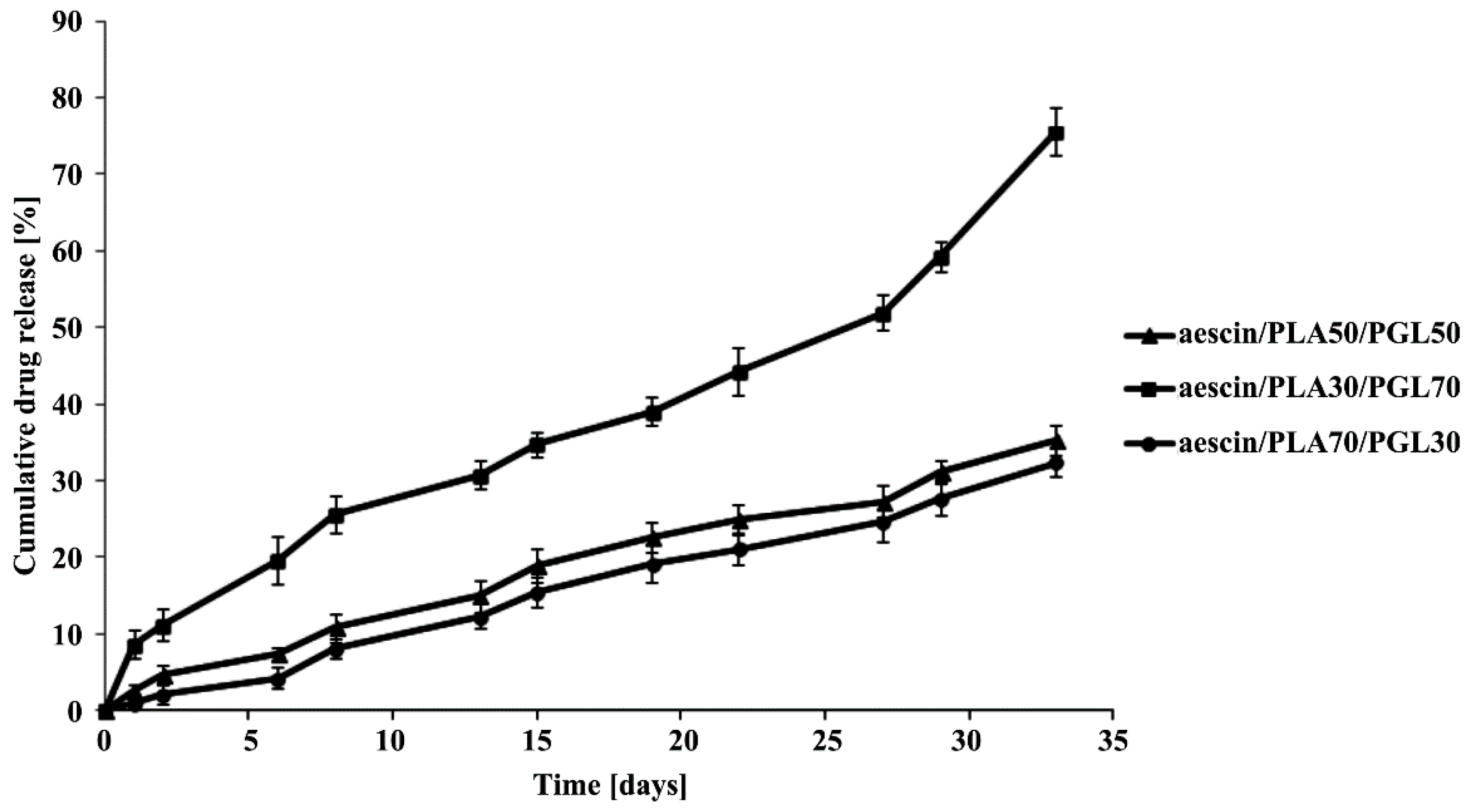
2.5. ALP Release Study from the Copolymeric Conjugates

The in vitro release study of ALP from the synthesized copolymeric conjugates (aescin/PLA50/PGL50/ALP, aescin/PLA30/PGL70/ALP and aescin/PLA70/PGL30/ALP) were investigated by measuring the concentration of ALP released at pH 7.4 ± 0.05. All experiments were carried out in triplicate. 200 milligram of dried copolymeric conjugates, were immersed into 100 mL of buffer solution (pH 7.4 ± 0.05) and incubated at 37 °C, with continuous orbital rotation at 50 [cycles/min]. At predetermined time intervals, 10 mL samples were withdrawn from the release medium using the filter followed by replacing with 10 mL of a fresh buffer solution. The absorption of buffer solution was determined by a UV-VIS spectrophotometer at the absorbance peak with a wavelength at 274 nm [19]. The absorbance peak was correlated very well with the concentration of ALP. A linear calibration curve was obtained by measuring the absorption of solutions with predetermined ALP concentrations. For all the measurements in this study, the absorbance readings were within the calibration range.

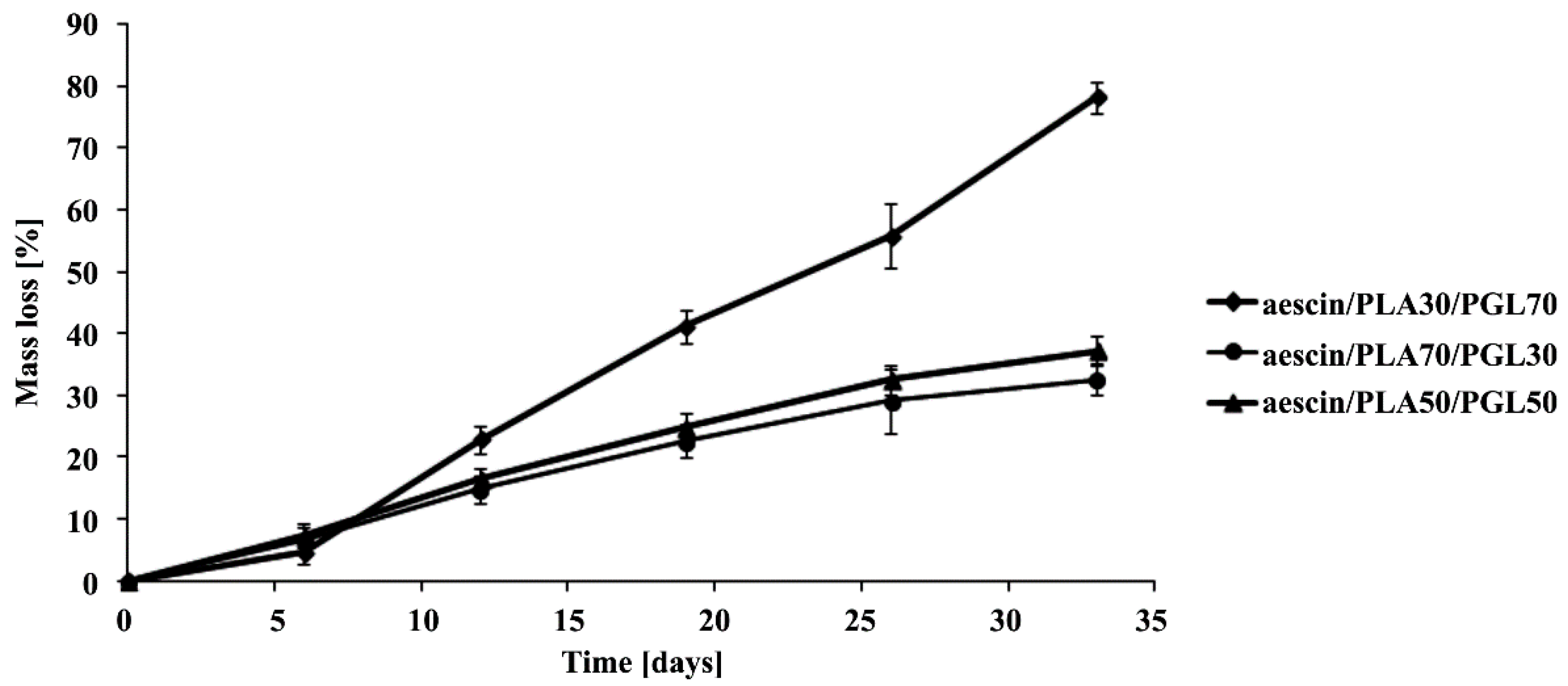
2.6. Hydrolytic Degradation Test

The hydrolytic degradation of the synthesized copolymeric matrices was performed in a 10 mL phosphate buffer solution (pH 7.4 ± 0.05) at 37 °C for 30 days. Following hydrolysis, the polymer samples were washed intensively with distilled water to remove any residual buffer solution, followed by drying under reduced pressure for four days. The degradation rates were estimated by weight loss (WL, %), calculated with the following equation:
WL (%) = 100 × (W0Wt)/W0
where W0 is initial weight and Wt is weight after biodegradation.
Furthermore, there results were evaluated by the ηinh of the copolymeric matrices decreasing. The polymeric matrices viscosity was measured in N,N-dimethylformamide (DMF, at 30 °C) using an Ubbelohde viscometer (on Stabinger Viscometer SVM 3000, Anton Paar’s, Graz, Austria).

2.7. Measurements

The polymerization products were characterized in the DMSO-d6 or CDCl3 solution by means of 1H or 13C NMR (Varian 300 MHz, Palo Alto, CA, USA). The IR spectra were measured from KBr pellets (Perkin–Elmer spectrometer, PerkinElmer, Waltham, MA, USA).
Number-average molecular weights (Mn) and polydispersity indexes (Mw/Mn) were measured using SEC-MALLS instrument (Wyatt Technology Corporation, Santa Barbara, CA, USA) composed of an 1100 Agilent isocratic pump, autosampler, degasser, thermostatic box for columns, a photometer MALLS DAWN EOS (Wyatt Technology Corporation, Santa Barbara, CA, USA) and differential refractometer Optilab Rex (Wyatt Technology Corporation, Santa Barbara, CA, USA). ASTRA 4.90.07 software (Wyatt Technology Corporation, Santa Barbara, CA, USA) was used for data collecting and processing. Two 2× TSKgel MultiporeHXL columns were used for separation. The samples were injected as a solution in methylene chloride. The volume of the injection loop was 100 mL. Methylene chloride was used as a mobile phase at a flow rate of 0.8 mL·min−1.
The amount of the released ALP was quantitatively determined by a UV-VIS spectrophotometry (UV‑1202 Shimadzu, Duisburg, Germany) in aqueous buffered solutions at the adsorption maximum of the free drug (λ = 274 nm) using a 1 cm quartz cell.

3. Results and Discussion

3.1. Structural Characterization of the Copolymeric Matrices

As outlined in Scheme 1, the branched aescin/PLA/PGL copolymers were obtained using the natural and additionally possessing pharmacological activity aescin initiator and Sn(Oct)2, a (co)catalyst of the ROP of LA and GL.
Scheme 1. Schematic outline for the synthesis of the copolymeric matrices and ALP conjugates.
Polymers 07 01484 g006
The copolymers with three different molar ratios were synthesized by the ROP of LA and GL monomers in a two-step polymerization process (Table 1). First, the core was synthesized by adding LA to aescin to form a branched aescin/LA middle core. The block copolymers were completed by the addition of a GL monomer to react further with the aescin/LA core.
Table 1. Copolymerization of LA and GL initiated by aescin. Molecular characterization of the synthesized copolymers.
Entry[I]/[LA]/[GA]Copolymer composition [LA]/[GA] aYield [%]DP bDS cMn(NMR) dMn(SEC-MALLS) eMw/Mn e
aescin/PLA50/PGL501/50/5035/659451.17.8022,10021,3001.3
aescin/PLA30/PGL701/30/7020/808048.210.510,7009,8001.4
aescin/PLA70/PGL301/70/3055/458568.68.5014,60013,5001.4
Reaction conditions: nitrogen atmosphere, reactions temperature, −80 °C (for homo- and copolymerization, toluene), d,l-lactide (LA), glycolide (GL); I, initiator—aescin; a Estimated from the integral height of hydrogen shown in 1H NMR spectrum; b DP, the average degree of polymerization determined by 1H NMR analysis, calculated based on the area ratio of the terminal methine proton of PLA (4.21 ppm) or methylene protons of PGL (4.10 ppm) to the internal methine proton of PLA (5.19 ppm) or methylene proton of PGL (4.91 ppm); c DS, the average degree of substitution determined by 1H NMR analysis; calculated based on the peak areas for the terminal methine proton of PLA (4.19 ppm) or methylene protons of PGL (4.10 ppm) and the aliphatic protons of aescin (18H, area of 0.75–1.09 ppm); d Mn(NMR) determined by 1H NMR spectroscopy; e Mn(SEC-MALLS) and Mw/Mn determined using SEC-MALLS instrument.
Figure 1. 1H NMR spectrum of the aescin/PLA50/PGL50 copolymeric matrix.
Polymers 07 01484 g001
Table 1 presents the SEC-MALLS and NMR results of the branched copolymers obtained. An increase in the Mn values of all the copolymers proved that the copolymers were growing in chain length after the further addition of a GL monomer. In addition, the copolymers exhibited a unimodal distribution with polydispersity ranging from 1.3 to 1.4. In Table 1, the molecular weight determined by 1H NMR spectroscopy (Mn(NMR)) was calculated from the average degree of polymerization (DP) and average degree of substitution (DS) of synthesized copolymers. The result reveals that between eight and 11 copolymer arms can be attached to the surface of aescin initiator. It was noted that there are unreacted hydroxyl groups of aescin in the resulting branched copolymers. This fact may be attributed to the change in the density and distribution of hydroxyl groups on the surface of the initiator and the stearic hindrance of the attached copolymer chains [20]. The resulting branched copolymers were characterized by 1H, 13C NMR and FTIR analysis (Figure 1, as well as Figure S1 and Figure S2, Supplementary Material). Figure 1 shows the 1H NMR spectra of aescin/PLA50/PGL50 copolymeric sample. It was clearly seen that characteristic signals at 1.47 and 5.20 ppm were assigned to the methyl and methine protons in the lactidyl units, as well as to the methylene protons of glycolidyl units (4.90 ppm). Importantly, the existence of the proton signals of ascent was also detected with high resolution, indicating the incorporation of the initiator into a macromolecule (the NMR data are in good agreement with the literature evidence [21,22,23]). Furthermore, the presence of two signals at 4.10 and 4.21 ppm assigned as a methylene terminal group of the glycolidyl unit (C’, Figure 1) and methine terminal group of lactidyl unit (A’, Figure 1) indicated that the synthesized copolymers were not arranged in a block but at random. This occurrence of transesterification is, however, common when Sn(Oct)2 is used as co(catalyst) of the copolymerization of cyclic esters [24]. As was demonstrated by Kasperczyk [25], the microstructure of the synthesized copolymers is influenced by the kind of the applied initiator (catalyst) as well as by the transesterification occurring during the synthesis. This reaction results in the redistribution of the sequences along the polymer chain leading to changes in the chain structure and the lengths of the microblocks [25]. For this reason, the microstructure of the representative aescin/PLA70/PGL30 sample has been examined by 1H NMR spectroscopy (Figure 2).
Figure 2. 1H NMR spectrum of the aescin/PLA70/PGL30 copolymeric matrix; region of glycolide methylene protons.
Polymers 07 01484 g002
The observation of 1H NMR spectra shows the presence of sequences of an LGL-type (Figure 2). They were the result of second mode transesterification of the synthesized copolymeric sample [26]. The quantitative determination of the second mode transesterification was calculated according to the Kasperczyk equation [25]. The yield of transesterification was calculated to be TII = 0.415, and was in a good agreement with the results received by Kasperczyk with the application of an Sn(Oct)2 initiator.
Owing to the toxicity standards for materials applied in biomedical applications, the obtained copolymeric matrices (aescin/PLA50/PGL50, aescin/PLA30/PGL70, and aescin/PLA70/PGL30) have been subjected to cyto- and genotoxicity assays, by using the luminescent bacterium V. Fischeri and the ciliated protozoan S. ambiguum (Table 2), as well as Salmonella typhimurium TA1535. It has been found that the obtained copolymers are not cytotoxic to all test bionts (both bacteria and protozoa) and are, therefore, adequate for biomedical applications [27]. Concerning genotoxicity results (Table 3), there were no significant differences in the intensity of bacteria growth or IR in comparison with the negative control for the copolymeric samples. All tested samples were not cytotoxic for Salmonella typhimurium TA1535 (G > 0.5). Moreover, none of the tested copolymers exhibited genotoxic activity (IR < 1.5).
Table 2. The cytotoxicity results of the synthesized copolymeric matrices.
EntryMicrotox 15 min-PE 1Microtox 30 min-PE 1Spirotox 24h-PE 1, 2
0.8 mg mL−10.4 mg mL−10.8 mg mL−10.4 mg mL−1
aescin/PLA50/PGL5016 ± 22 ± 113 ± 24 ± 30
aescin/PLA30/PGL7018 ± 28 ± 416 ± 410 ± 20
aescin/PLA70/PGL3015 ± 25 ± 416 ± 28 ± 40
1 Percent of toxic effect; 2 The results for three sample concentration (1.0, 0.5 and 0.25 mg mL−1).
Table 3. The results of the umu-test for the highest concentrations of the tested copolymers extracts.
Entry−S9+S9
G ± SDIR ± SDG ± SDIR ± SD
aescin/PLA50/PGL500.89 ± 0.050.85 ± 0.050.86 ± 0.070.93 ± 0.07
aescin/PLA30/PGL700.89 ± 0.050.90 ± 0.150.94 ± 0.060.98 ± 0.15
aescin/PLA70/PGL300.98 ± 0.110.90 ± 0.170.92 ± 0.07.93 ± 0.07

3.2. The Conjugates’ Synthesis and Drug-Release Characteristics

β-adrenergic blockers, such as ALP, are an important class of drugs used for various heart diseases. If they are covalently conjugated onto the surface of the branched copolyester matrix, an efficient local drug-delivery system with high stability could be obtained. The synthesis of such conjugates is illustrated in Scheme 1. First, the carboxyl groups are introduced onto the surface of copolymeric matrices to produce aescin/PLA/PGL-COOH matrices, via the reaction of copolymeric matrices with the succinic anhydride in the presence of TEA as a catalyst. After that, the obtained copolymeric matrices with a carboxyl end group were reacted with a hydroxyl group of ALP in the presence of EDC and DMAP to yield the appropriate conjugation products (Scheme 1). The synthesized conjugates were confirmed by a proton NMR chemical shift (Figure 3, aescin/PLA30/PGL70/ALP as an example) and integration of the aliphatic protons of aescin (18H, the area of 0.75–1.09 ppm), as well as chemical shifts of protons of a benzyl ring of ALP (4H, 6.92–7.16 ppm). This suggested that, on average, six molecules of ALP were conjugated to the polymeric matrices (6.25, 5.90, and 6.78 for aescin/PLA50/PGL50/ALP, aescin/PLA30/PGL70/ALP, and aescin/PLA70/PGL30/ALP, respectively). Furthermore, in the 1H NMR spectra of aescin/PLA30/PGL70/ALP (Figure 3) signals, characteristic hydrogen atoms of PLA and PGL repeating units are present, in addition to signals attributed to aescin initiator and ALP end groups. A considerable 1H NMR resonance shift was observed for the –(CH2)CH(CH2)– of ALP (2, Figure 3) in the final product as compared to that of pure ALP (Figure S8, Supplementary Material). Moreover, the characteristic signal of the hydroxyl group of ALP (6.78 ppm, Figure S8) disappeared, clearly indicating the formation of an aescin/PLA/PGL-ALP conjugate.
The ALP content in the obtained conjugates was calculated by integration of the 1H NMR spectra. The signal intensities of four protons on the benzyl ring of ALP (3’, 4’, 5’, 6’) and the signal intensities of two protons of the glycolidyl units (C) were compared, indicating the following ALP content in the conjugates: 34.8 mol % in the aescin/PLA50/PGL50/ALP, 38.5 mol % in the aescin/PLA30/PGL70/ALP and 32.3 mol % in the aescin/PLA70/PGL30/ALP (calculated as mole of the drug per mole of the whole macromolecular system).
Figure 3. 1H NMR spectrum of the aescin/PLA30/PGL70/ALP conjugate.
Polymers 07 01484 g003
Figure 4 shows the in vitro release of ALP from the synthesized conjugates of different compositions in a pH 7.4 ± 0.05 PBS solution. Meanwhile, the stability of the synthesized copolymeric matrices as a drug-delivery system was investigated (Figure 5). As plotted in Figure 4, there was no initial burst release for studied conjugates. This fact can be explained by the chemical conjugation of ALP molecules to the polymer chain and their release; hence, the physical diffusion was eliminated. However, the total percentage of ALP released was dependent on the copolymeric matrix composition. Take aescin/PLA50/PGL50/ALP, for instance, about 3% of ALP was released after one day of study and reached a value of 35% after 33 days. In the same time period, aescin/PLA30/PGL70/ALP released about 9% of ALP after one day, and almost 76% after 33 days. Contrary to this result, the aescin/PLA70/PGL30/ALP showed a less significant total percentage of the drug released: about 1% of ALP was released after one day and about 32% after 33 days. Thus, it can be clearly seen that the release rate increases with the increase of GL percentage in the copolymer. The above in vitro release results were highly correlated to the hydrolytic degradation results presented in Figure 5, which shows the degradation profile of each copolymeric matrix.
Figure 4. Release profile of ALP from the synthesized conjugates (pH 7.4 ± 0.05).
Polymers 07 01484 g004
Figure 5. Hydrolytic degradation results of the synthesized copolymeric matrices.
Polymers 07 01484 g005
All three copolymeric matrices showed similar and sustained rates of weight loss (WL) without significant loss of weight during the early stages. In the case of aescin/PLA30/PGL70, the WL was about 78% after 33 days of degradation, whereas for aescin/PLA50/PGL50 and aescin/PLA70/PGL30 these values were about 37% and 32%, respectively. Our results show that an increase in the GL percentage in the copolymer accelerates the weight loss of the copolymer; the aescin/PLA50/PGL50 matrix exhibited a faster degradation than the aescin/PLA70/PGL30 because of the preferential degradation of the GL proportion that is assigned by higher hydrophilicity [28]. Therefore, the value of the degradation and the drug-release rate is closely related to the amount of GL in the copolymer. The results were directly compared to the results of the intrinsic viscosity measurements (ηinh), which follow the same trend (Table 4) [29].
Table 4. The hydrolytic degradation results of the synthesized copolymeric matrices.
Time (days)Aescin/PLA50/PGL50Aescin/PLA30/PGL70Aescin/PLA70/PGL30
WL (%)ηinh aΔηinh (%)WL (%)ηinh aΔηinh (%)WL (%)ηinh aΔηinh (%)
67.4 ± 0.50.7984.6 ± 0.40.4646.7 ± 0.10.593
1216.5 ± 0.80.76822.9 ± 0.50.411514.8 ± 0.60.567
1924.8 ± 0.90.701841.2 ± 0.90.343022.6 ± 0.90.5014
2632.5 ± 0.60.652155.8 ± 0.20.274429.1 ± 0.80.4824
3337.2 ± 0.30.612778.2 ± 0.40.196032.6 ± 0.30.4425
a The ηinh was determined by a viscosity method. The ηinh of the synthesized polymeric matrices before degradation was: 0.81 (dL/g) for aescin/PLA50/PGL50; 0.48 (dL/g) for aescin/PLA30/PGL70; 0.61 (dL/g) for aescin/PLA70/PGL30; The Δηinh was decrease of ηinh.

4. Conclusions

In summary, novel copolymeric conjugates of ALP have been synthesized and characterized for a local delivery. The matrices were composed of natural and therapeutically-efficient initiators (β-aescin) and the biodegradable copolymer of PLGA. In order to obtain sustained drug-release efficiency, the branched copolymers with different ratios of lactide and glycolide were synthesized. The resulting matrices were spectrally characterized and then subjected to cyto- and genotoxicity assays using bacterial luminescence, protozoan and Salmonella typhimurium TA1535 tests. The in vitro release studies showed that with these branched matrices the duration of drug release can be prolonged (by more than one month) with no burst release. Furthermore, it is strongly dependent on the matrix composition; the value of the hydrolytic degradation of the synthesized matrices and the drug-release rate is closely related to the amount of GL in the copolymer. The synthesized matrices could be used to deliver ALP in a sustained and prolonged manner; thus, they have the potential to treat various heart diseases. It is also worth emphasizing that the obtained ester-linked ALP conjugates might improve the bioavailability and pharmacokinetics of the ALP as well as increase its efficiency, resulting from the synergistic action of ALP with β-aescin. Our future work includes these studies, as well as in vivo testing.

Supplementary Materials

Supplementary materials can be accessed at: https://www.mdpi.com/2073-4360/7/9/1484/s1.

Acknowledgments

We would like to thank P. Wroczynski, Dean of the Faculty of Pharmacy with the Laboratory Medicine Division, for a financial support.

Author Contributions

The contributions of the respective authors are as follows: Ewa Oledzka gave the concept of work, synthesized and characterized products, interpreted the results and wrote the whole article, made discussion and conclusion. Dagmara Pachowska synthesized and characterized the matrices, conjugates and participated in other steps of the research. Agnieszka Lis-Cieplak interpreted the NMR spectra of aescin and copolymer microstructure. Marcin Sobczak participated in all steps of the research, helped in the writing of the paper. Grzegorz Nalecz-Jawecki performed cytotoxicity assay. Anna Zgadzaj performed genotoxicity assay. Waclaw Kolodziejski helped in NMR spectra interpretation and writing of the paper. All authors have contributed to seen and approved the manuscript.

Conflicts of Interest

The authors declare no conflict of interest.

References

  1. Sirtori, C.R. Aescin: Pharmacology, pharmacokinetics and therapeutic profile. Pharmacol. Res. 2001, 44, 183–193. [Google Scholar] [CrossRef] [PubMed]
  2. Zhang, L.-M.; Wang, T.; Fan, H.-Y.; Yu, X.; Han, B.; Zhu, M.; Fu, F.-H. Escin may exert a synergistic anti-inflammatory effect with glucocorticoids. Health 2010, 2, 79–81. [Google Scholar] [CrossRef]
  3. Mrwa, U.; Guth, K.; Haist, C.; Troschka, M.; Herrmann, R.; Wojciechowski, R.; Gagelmann, M. Calcium-requirements for activation of skinned vascular smooth muscle from spontaneously hypertensive (SHRSP) and normotensive control rats. Life Sci. 1986, 38, 191–196. [Google Scholar] [PubMed]
  4. Pearson, P.J.; Vanhoutte, P.M. Vasodilator and vasoconstrictor substances produced by the endothelium. Rev. Physiol. Biochem. Pharmacol. 1993, 122, 1–68. [Google Scholar] [PubMed]
  5. Satoh, S.; Kreutz, R.; Wilm, C.; Ganten, D.; Pfitzer, G. Augmented agonist-induced Ca2+ sensitization of coronary artery contraction in genetically hypertensive rats: Evidence for altered signal transduction in the coronary smooth muscle cells. J. Clin. Investg. 1994, 94, 1397–403. [Google Scholar] [CrossRef] [PubMed]
  6. Oledzka, E.; Sobczak, M.; Kolakowski, M.; Kraska, B.; Kamysz, W.; Kołodziejski, W. Development of creatine and arginine-6-oligomer for the ring-opening polymerization of cyclic ester. Macromol. Res. 2013, 21, 161–168. [Google Scholar] [CrossRef]
  7. Frishman, W.H. Beta-adrenergic blockers. Circulation 2003, 107, 117–119. [Google Scholar] [CrossRef] [PubMed]
  8. Madill, F.L.; Nyberg, G. Alprenolol in hypertension: An open assessment in general practice. J. Int. Med. Res. 1975, 3, 338–344. [Google Scholar]
  9. Sweetman, S.C. Martindale: The Complete Drug Reference, 36th ed.; The Pharmaceutical Press: London, UK, 2009; p. 1207. [Google Scholar]
  10. Hovgaard, L.; Brøndsted, H.; Buur, A.; Bundgaard, H. Drug delivery studies in Caco-2 monolayers. Synthesis, hydrolysis, and transport of O-cyclopropane carboxylic acid ester prodrugs of various β-blocking agents. Pharm. Res. 1995, 12, 387–392. [Google Scholar] [CrossRef] [PubMed]
  11. Prokai, L.; Wu, W.M.; Somogyi, G.; Bodor, N. Ocular delivery of the beta-adrenergic antagonist alprenolol by sequential bioactivation of its methoxime analogue. J. Med. Chem. 1995, 26, 2018–20. [Google Scholar] [CrossRef]
  12. D’Avila Carvalho Erbetta, C.; Alves, R.J.; Resende, J.M.; de Souza, F.R.F.; de Sousa, R.G. Synthesis and characterization of poly(d,l-lactide-co-glycolide) copolymer. J. Biomater. Nanobiotech. 2012, 3, 208–225. [Google Scholar] [CrossRef]
  13. Jain, R.A. The manufacturing techniques of various drug loaded biodegradable poly(lactide-co-glycolide) (PLGA) devices. Biomaterials 2000, 21, 2475–2490. [Google Scholar] [CrossRef]
  14. Allison, S.D. Effect of structural relaxation on the preparation and drug release behavior of poly(lactic-co-glycolic)acid microparticle drug delivery systems. J. Pharm. Sci. 2008, 97, 2022–2035. [Google Scholar] [CrossRef] [PubMed]
  15. Mohamed, F.; van der Walle, C.F. Engineering biodegradable polyester particles with specific drug targeting and drug release properties. J. Pharm. Sci. 2008, 97, 71–87. [Google Scholar] [CrossRef] [PubMed]
  16. Chen, H.; Li, G.; Chi, H.; Wang, D.; Tu, C.; Pan, L.; Zhu, L.; Qiu, F.; Guo, F.; Zhu, X. Alendronate-conjugated amphiphilic hyperbranched polymer based on boltorn H40 and poly(ethylene glycol) for bone-targeted drug delivery. Bioconjug. Chem. 2012, 23, 1915–1924. [Google Scholar] [CrossRef] [PubMed]
  17. Nalecz-Jawecki, G. Spirotox Test—Spirostomum Ambiguum Acute Toxicity Test. In Small-Scale Freshwater Toxicity Investigations; Blaise, C., Férard, J.-F., Eds.; Springer: The Netherlands, 2005; pp. 299–322. [Google Scholar]
  18. Water Quality—Determination of the Genotoxicity of Water and Waste Water Using the Umu-Test; ISO 13829:2000; International Organization for Standardization: Geneva, Switzerland, 2000.
  19. European Pharmacopoeia. European Directorate for the Quality of Medicines & HealthCare (EDQM); European Pharmacopoeia Commission: Strasbourg, France, 2014. [Google Scholar]
  20. Oledzka, E.; Kaliszewska, D.; Sobczak, M.; Raczak, A.; Nickel, P.; Kolodziejski, W. Synthesis and properties of a star-shaped poly(ε-caprolactone)-ibuprofen conjugate. J. Biomater. Sci. Polym. Ed. 2012, 23, 2039–2054. [Google Scholar] [PubMed]
  21. Gruza, M.M.; Jatczak, K.; Zagrodzki, B.; Laszcz, M.; Koziak, K.; Malińska, M.; Cmoch, P.; Giller, T.; Zegrocka-Stendel, O.; Woźniak, K.; et al. Preparation, purification and regioselective functionalization of protoescigenin-the main aglycone of escin complex. Molecules 2013, 18, 4389–4402. [Google Scholar] [CrossRef] [PubMed]
  22. Agrawal, P.K.; Thakur, R.S.; Shoolery, J.N. Application of 2D NMR spectroscopy to the structural establishment of the major hydrolysis product of aescin. J. Nat. Prod. 1991, 54, 1394–1396. [Google Scholar] [CrossRef]
  23. Zhang, Z.; Koike, K.; Jia, Z.; Nikaido, T.; Guo, D.; Zheng, J. New saponins from the seeds of Aesculus chinensis. Chem. Pharm. Bull. (Tokyo) 1999, 47, 1515–1520. [Google Scholar] [CrossRef] [PubMed]
  24. Bero, M.; Czapla, B.; Dobrzyński, P.; Janeczek, H.; Kasperczyk, J. Copolymerization of glycolide and ε-caprolactone. 2. Random copolymerization in the presence of tin octoate. Macromol. Chem. Phys. 1999, 200, 911–916. [Google Scholar] [CrossRef]
  25. Kasperczyk, J. Microstructural analysis of poly[(l,l-lactide)-co-(glycolide)] by 1H and 13C NMR spectroscopy. Polymer 1996, 37, 201–203. [Google Scholar] [CrossRef]
  26. Dobrzynski, P.; Kasperczyk, J.; Janeczek, H.; Bero, M. Synthesis of biodegradable glycolide/l-lactide copolymers using iron compounds as initiators. Polymer 2002, 43, 2595–2601. [Google Scholar] [CrossRef]
  27. Sobczak, M.; Debek, C.; Oledzka, E.; Nałecz-Jawecki, G.; Kołodziejski, W.; Rajkiewicz, M. Segmented polyurethane elastomers derived from aliphatic polycarbonate and poly(ester-carbonate) soft segments for biomedical applications. J. Polym. Sci. Part A Polym. Chem. 2012, 50, 3904–3913. [Google Scholar] [CrossRef]
  28. Makadia, H.L.; Siegel, S.J. Poly lactic-co-glycolic acid (PLGA) as biodegradable controlled drug delivery carrier. Polymers 2011, 3, 1377–1397. [Google Scholar] [CrossRef] [PubMed]
  29. Sobczak, M.; Kamysz, W.; Tyszkiewicz, W.; Debek, C.; Kozłowski, R.; Oledzka, E.; Piotrowska, U.; Nalecz-Jawecki, G.; Plichta, A.; Grzywacz, D.; et al. Biodegradable macromolecular conjugates of citropin: Synthesis, characterization and in vitro efficiency study. React. Funct. Polym. 2014, 83, 54–61. [Google Scholar] [CrossRef]

