Article

The Syntheses, Crystal Structure and Luminescence Properties of Cone-Like Octadentate Europium (III) Complexes with Four Short Alkoxy Substituents

1
Department of Applied Chemistry, Faculty of Engineering, Kyushu Institute of Technology, 1-1 Sensui-cho, Tobata-ku, Kitakyushu 804-8550, Japan
2
Japan Bruker AXS K.K, 3-9, Moriya-cho Kanagawaku, Yokohama 221-0022, Japan
*
Author to whom correspondence should be addressed.
Crystals 2017, 7(3), 85; https://doi.org/10.3390/cryst7030085
Submission received: 27 February 2017 / Accepted: 8 March 2017 / Published: 13 March 2017
(This article belongs to the Special Issue Luminescent Properties of Lanthanoid Doped Crystals)

Abstract

:
Treatment of 1-(4′-methoxy or ethoxy phenyl)-4,4,4-trifluoro-1,3-butanedione with europium (III) chloride in the presence of piperidine resulted in the halide ligands exchange yielded new piperidinium tetrakis{1-(4′-methoxy or ethoxy phenyl)-4,4,4-trifluoro-1,3-butanedionato}europate (III) complexes 2a and 2b. Complexes 2a and 2b have been characterized by elemental analysis, 1H NMR spectroscopy, and FAB-MS, and their absolute structures were determined by single crystal X-ray diffraction analysis. The complexes 2a and 2b have the monoclinic space groups C2/c (No. 15, 4′-substituent = OCH3) and with P − 1 (No. 2, 4′-substituent = OC2H5), respectively. X-ray analysis results showed that eight coordinate structures of the complexes 2a and 2b have cone-like structures, like calix[4]arenes, but their structures were slightly different due to the crystal packing and the existence of the solvent molecule. The complexes 2a and 2b exhibited identical, strong photoluminescence emissions in the solution phase.

1. Introduction

The luminescent properties of Lanthanide complexes with various organic ligands have been investigated for decades [1], and the search for novel Lanthanide complexes continues to attract many researchers due to their important applications as optical fiber lasers, electroluminescent displays, and organic light emitting diodes [2,3,4]. Among these, europium (III) complexes with β-diketones possessing aromatics and fluorine substituents displayed very good to excellent photo luminescent properties. Thus, various europium (III) complexes with β-diketones were synthesized and evaluated for their photo luminescent properties [4,5,6,7,8,9].
In this context, we report the synthesis, structural, and spectral properties of the octa-coordinate europium (III) complexes carrying four bidentate β-diketonato ligands having four alkoxy substituted aromatic moieties, fluorine substituents, and one piperidinium as a counter cation. Further, we have investigated the structural properties using X-ray analyses.
The luminescent intensity of the complexes 2a and 2b was quite strong because the complex has no water ligand in solid and in solution. This is due to the vibration relaxation of water molecule; that is to say, the excited energy on the f orbital (5D levels) of the centered europium ion is effectively relaxed to 7F levels.

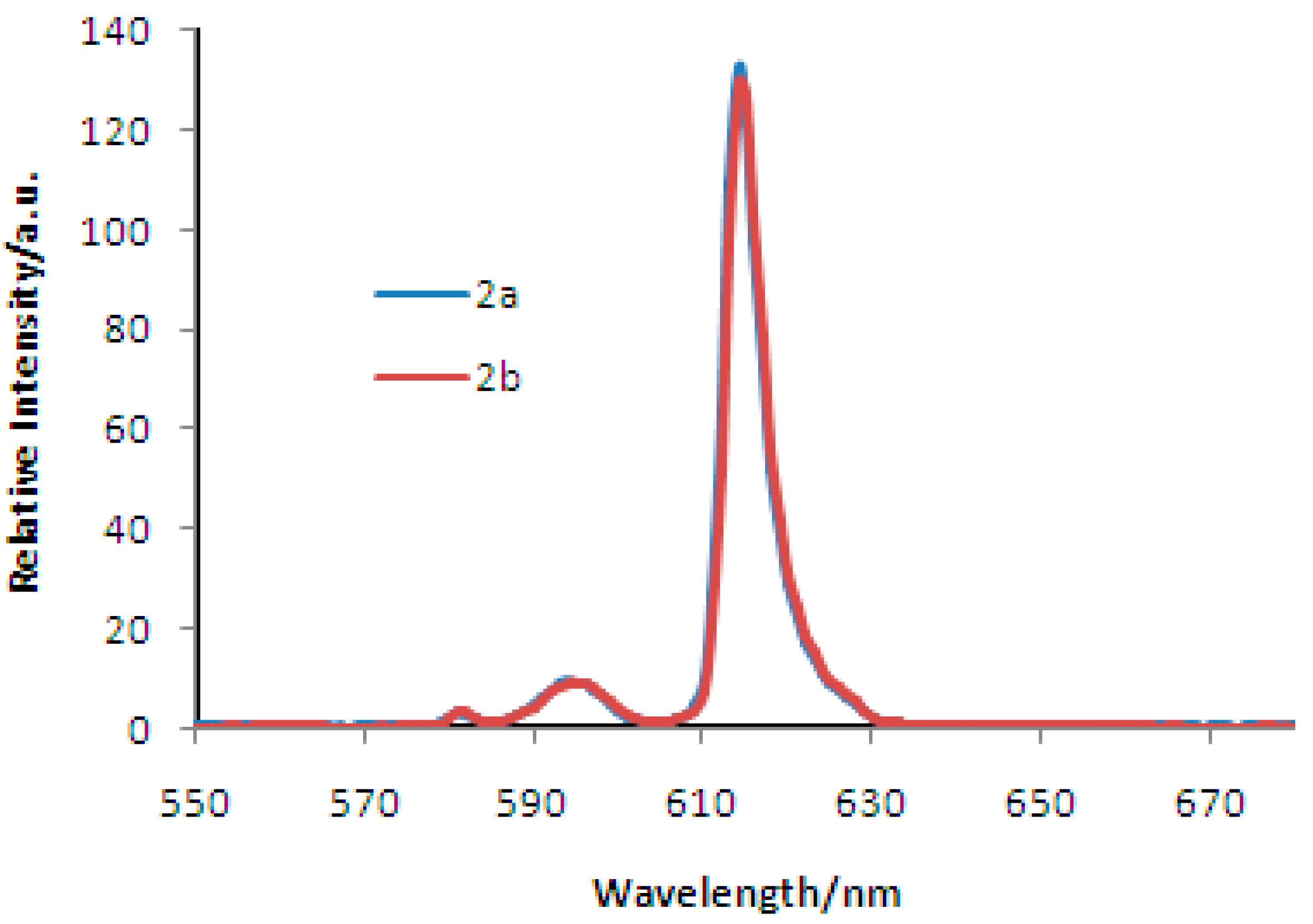
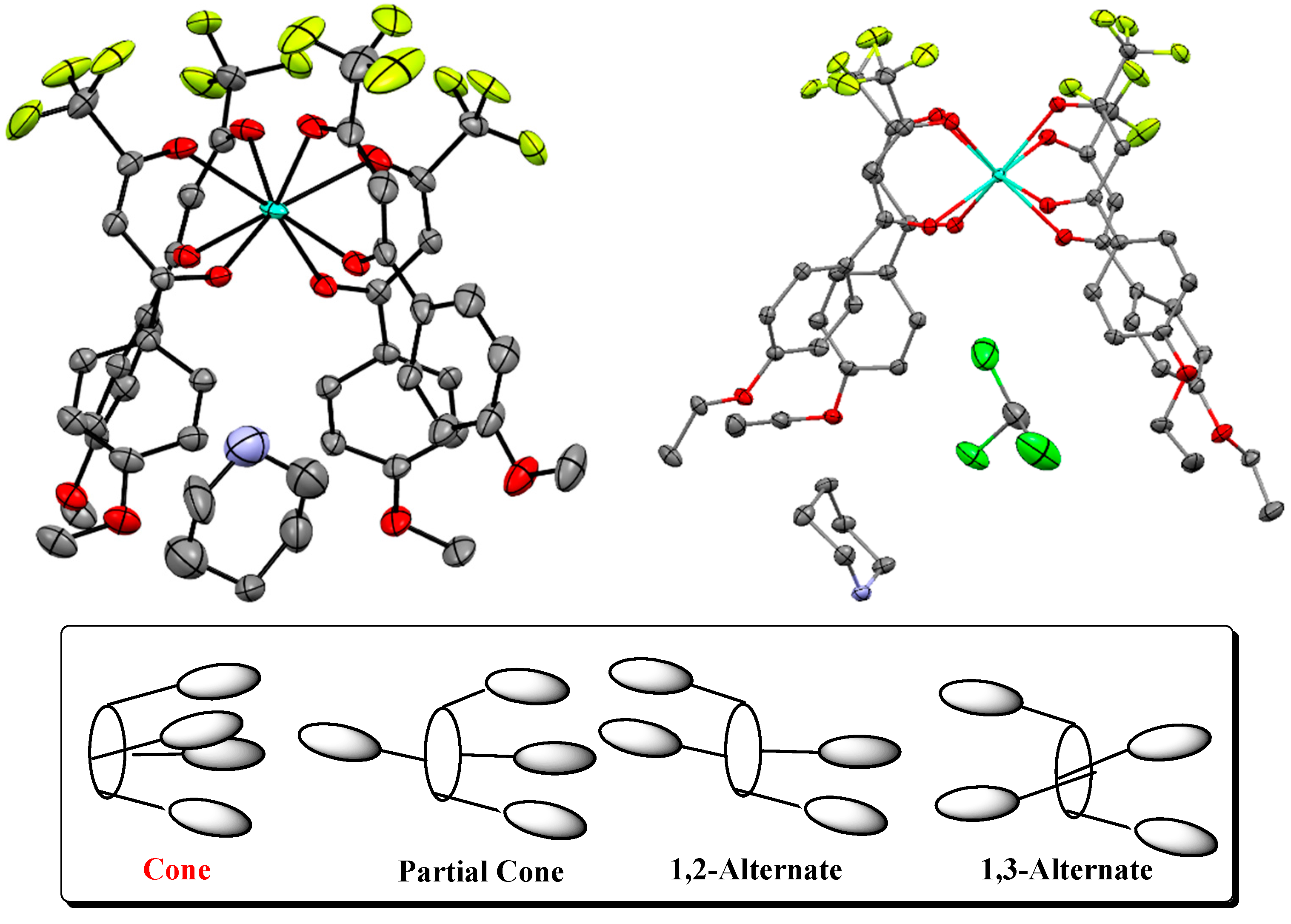
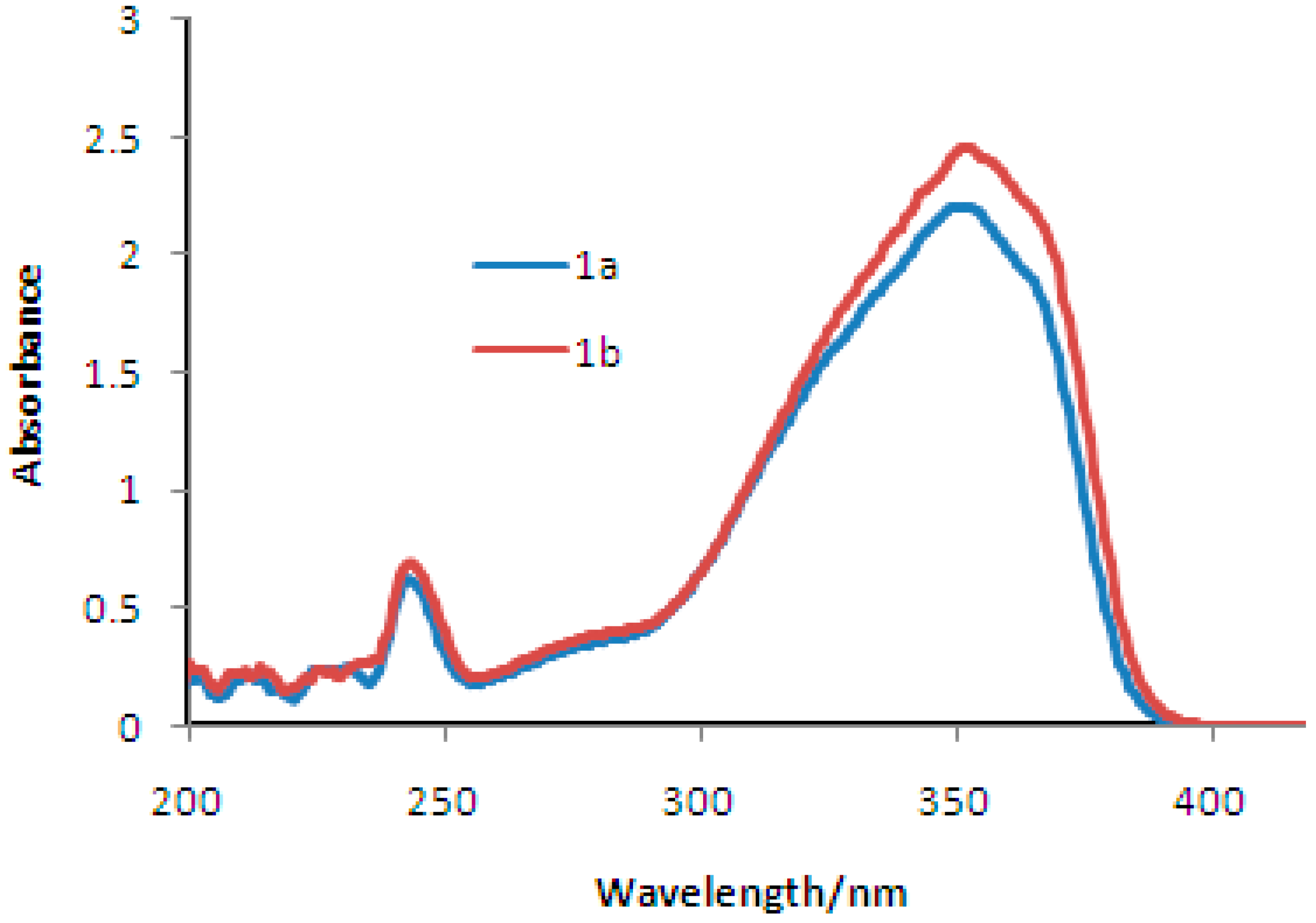
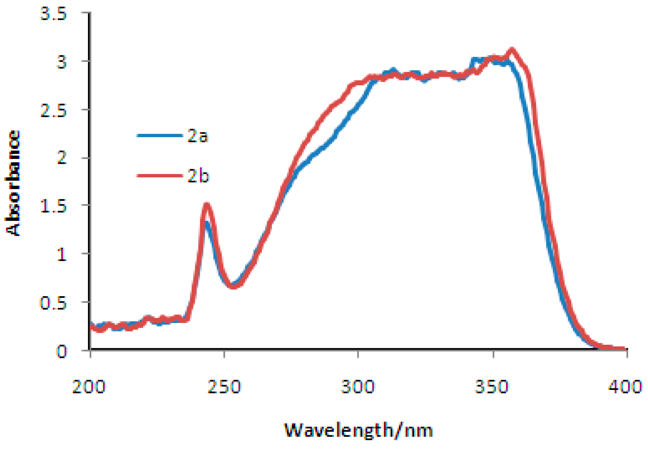
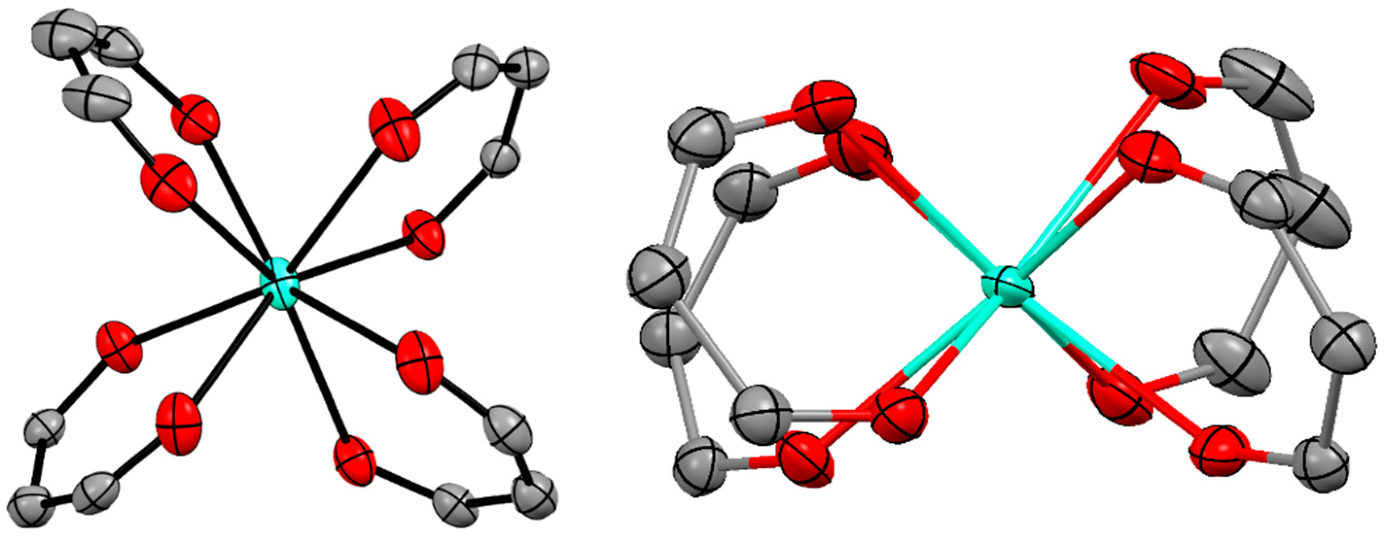
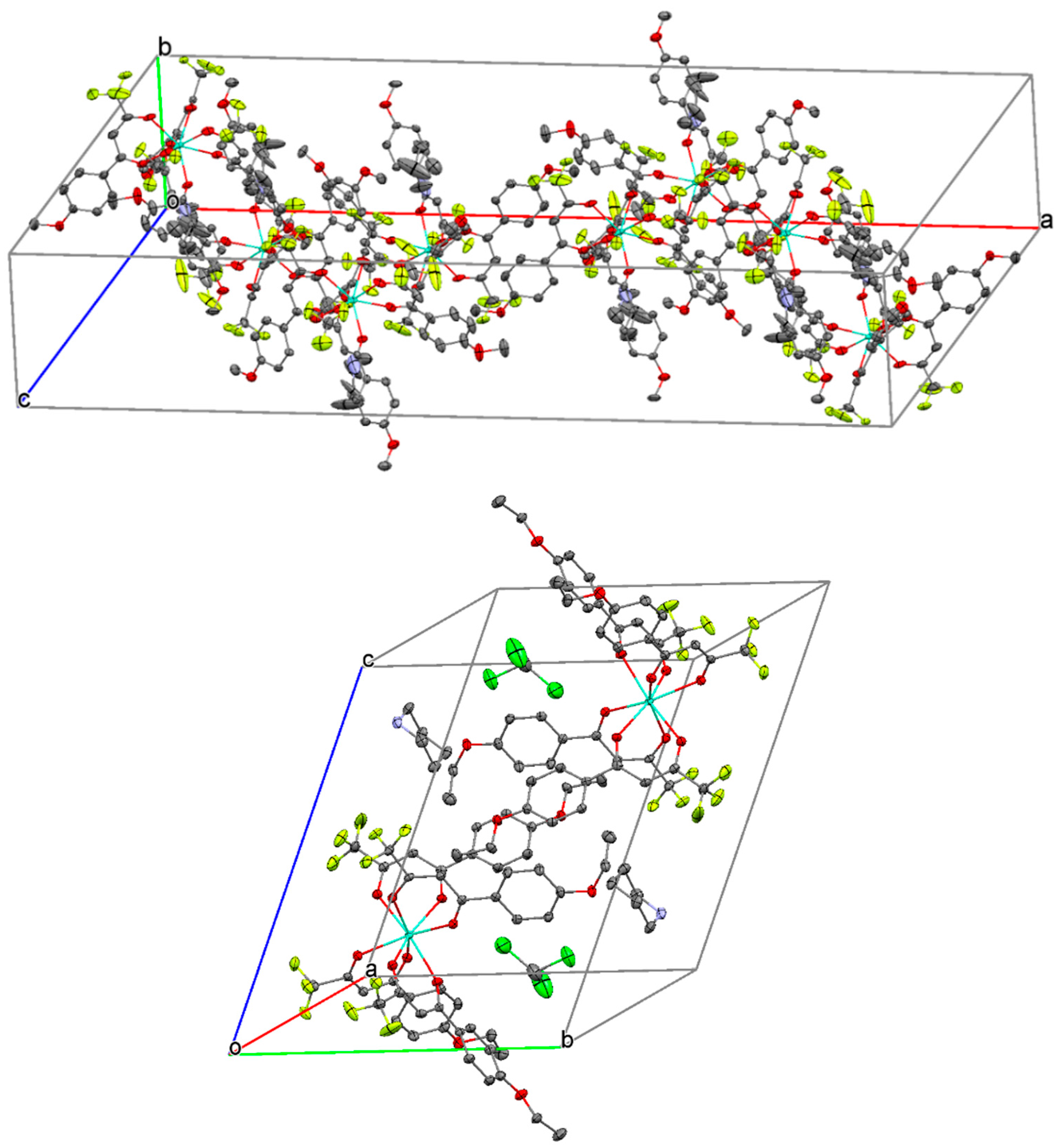
2. Results and Discussion

Synthesis and Spectra Analysis

The two complexes 2a and 2b were synthesized in two steps. In the first step, β-diketone ligands were prepared by Claisen condensation of 4′-methoxy and ethoxy acetophenone with ethyl trifluoroacetate using sodium hydride as a base in THF. Secondly, 1-(4′-methoxy or ethoxy phenyl)-4,4,4-trifluoro-1,3-butanedione 1a and 1b reacted with europium (III) chloride in the presence of piperidine as a base reagent in ethanol solvent according to the method reported in the literature, yielding the corresponding complexes 2a and 2b in very good yields [10]. Complexes 2a and 2b are stable under air and moisture conditions. Structural properties in solution are in line with expectations, as shown by 1H NMR spectroscopy. Although the peaks of the complexes are quite shifted from normal regions due to paramagnetic effect of the europium (III) ion, it can be easily assigned.
In the mass spectrum of 2a, the fragment peak is m/z 888 ([M + H]+ − 1 ligand − piperidinium cation) mainly appears. However, the peak m/z 1133 ([M + H]+ − piperidinium cation) is quite small. In addition, in the mass spectrum of 2b, the main fragment peak is m/z 930 ([M + H]+ − 1 ligand − piperidinium cation) with the small peak m/z 1189 ([M + H]+ − piperidinium cation). This is well explained by the fact that the neutral fragment is more stable than that of the anionic fragment ion.
The UV-vis absorptions for the ligands (1a, 1b) and their corresponding europium complexes (2a, 2b) were measured in dichloromethane solution (1 × 10⁻³ mol/L), and their corresponding spectra is shown in Figure 1 and Figure 2. The ligands showed strong absorption bands at 352 nm and 351 nm, respectively. These strong absorption bands were assigned to the π-π* enol absorptions of the β-diketone ligands. Relatively low intensity absorption bands at 246 nm and 244 nm were assigned to the n-π* enol absorptions of the β-diketone ligands. Complexes 2a and 2b exhibited broad high intensity absorption bands from 342nm to 356 nm, respectively. The absorption coefficients (log ε) of the complexes 2a and 2b were 3.10 and 3.15, respectively. This was attributed to the chelating of four β-diketone ligands with the europium (III) ion.
The emission spectra of the complexes 2a and 2b was recorded in dichloromethane solution (1 × 10⁻³ mol/L), and their emission spectra is shown in Figure 3. Emission spectra of the complexes were measured by exciting the complexes at their absorption maximum wavelengths 352 nm and 351 nm, respectively. The emission spectra of the complexes 2a and 2b showed sharp peaks in the region 590–720 nm associated with 5D07FJ (J = 0–4) transitions of the europium (III) ion. The very high intensity peak was observed at 613 nm due to the 5D07F2 transition, suggesting a highly polarizable chemical environment around the europium (III) ion [11]. This transition was responsible for the red emission of the complexes 2a and 2b. The emission and absorption spectra of complexes 2a and 2b are almost identical, suggesting that the changing substitution on β-diketone ligands from methoxy to ethoxy did not affect the luminescent properties of the complexes 2a and 2b.
To further confirm the absolute structures of complexes 2a and 2b, suitable single crystals for X-ray structure analysis were easily obtained in ethanol solvent using a slow evaporation method. The complex 2a crystallizes in the monoclinic space group C2/c (centrosymmetric, No. 15), the cell unit includes four molecules with four piperidinium ions and with no solvate molecule (Figure 4).
The complex 2b crystallizes in the monoclinic space group P − 1 (non-centrosymmetric, No. 2), the cell unit includes two molecules with two piperidinium ions and with two chloroform molecules (Figure 4).
The europium (III) ions of 2a and 2b are coordinated by a distorted octahedral arrangement of eight oxygen atoms from four chelating β-diketonato ligands (Figure 5). The average Eu1-O (1–8) bond lengths are moderately normal, and these values are ca. 2.357 Å for 2a and 2.392 Å for 2b, respectively. The bond angles in the five membered rings consisting of Eu and 1,3-butanedionato ligands (O-Eu-O) vary from 71.83(15)° to 73.60(16)° for 2a, 70.41(8)° to 71.81(8)° for 2b, respectively (Table 1 and Table 2). These values of bond distances and bond angles are in good agreement with those reported for other analogous europium β-diketonato complexes [12]. The piperidinium cation involving the N1 atom is the most stable chair form in the crystal, and located near the crystallized benzene molecules (Figure 4).
Molecular shapes of these complexes are cone-like structures and the complex molecules have cavities (Figure 4). The difference between 2a and 2b about the inclusion of solvate molecule is well explained by the difference of the largeness of the cavity consisting of the four ligands. The cavity size of the 2b complexes seems to be larger than those of 2a. Differences between these complexes and calix[4]arene are the environment’s internal cavity spaces; in another words, aromatic parts on the calixarenes are oriented-electron surfaces to the cavity center. On the contrary, the aromatic parts on the complexes are aromatic C–H protons oriented to the cavity center (Figure 6). Therefore, the complexes are expected as new compounds for undeveloped fields of host-guest chemistry.

3. Experiments

3.1. Materials and Instrumentation
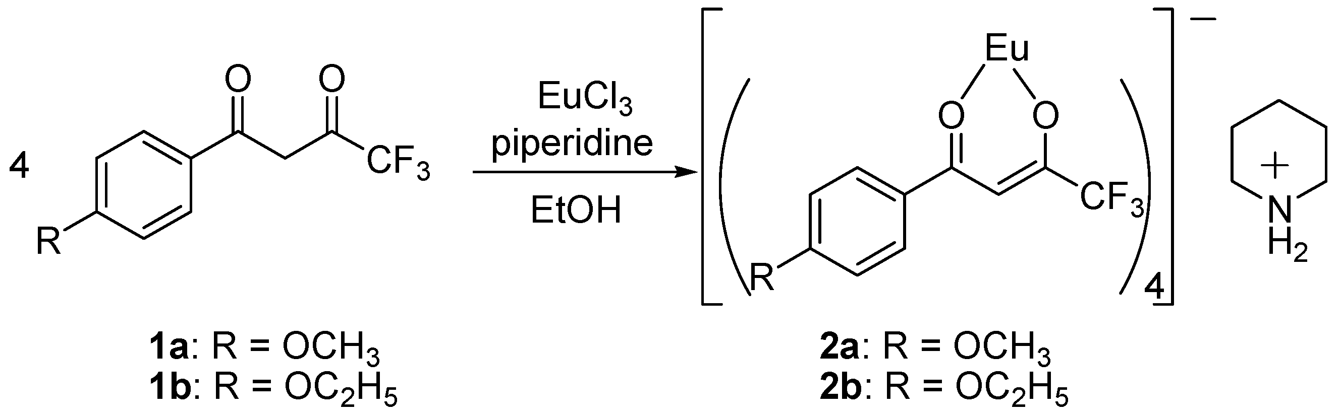
All reagents and solvents were obtained from commercial sources and are used without further purification. The 1H-NMR spectra were recorded on a Bruker AVANCE400S spectrometer (Bruker, Yokohama, Japan) in CDCl3 with tetramethylsilane (Me4Si) as an internal reference. The positive fast atom bombardment (FAB) mass spectrum (MS) of the complex were obtained on a Nippon Densi JEOL JMS-SX102A spectrometer (JEOL, Tokyo, Japan) using NBA (nitrobenzylalcohol) as the matrix and DCM (dichloromethane) as the solvent. The instrument was operated in positive ion mode over an m/z range of 100–2000. Elemental analysis data were recorded on a Yanako MT–4 analyzer (Yanako Group, Kyoto, Japan). A JASCO V-550 spectrophotometer (JASCO Corporation, Tokyo, Japan) was used for obtaining UV-Vis spectra in dichloromethane with 250–900 nm range. HITACHI F-2500 spectrophotometer (Hitachi High-Technologies Corporation, Tokyo, Japan) was used for fluorescence spectra measurements in dichloromethane with 250–900 nm range. CCDC No. 1529390 and 1529592 contain the supplementary crystallographic data for the complexes 2a and 2b, respectively.

3.2. Synthesis

3.2.1. Preparation of Piperidinium Tetrakis{1-(4′-substituted phenyl)-4,4,4-trifluoro-1,3-butanedionato}europate (III) Complexes 2a and 2b (Scheme 1)

Typical Procedure

In a first Schlenk vessel, a solution of europium (III) chloride (0.650 g, 0.41 mmol) and 1-(4′-methoxy or ethoxy phenyl)-4,4,4-trifluoro-1,3-butanedione 1 (1.65 mmol) in absolute ethanol (30 mL) was prepared at room temperature. Under protection from air, a slight excess of piperidine (0.30 mL, 3.0 mmol) was added to the solution, and the two solutions were then combined and stirred at room temperature for 12 h. After filtration, piperidine and most volatile materials were removed from the filtrate on a vacuum line. Under protection from air, the residue was repeatedly washed with small portions (5 mL) of warm, dry ethanol. The residual powders were dissolved with ethanol for crystallization. Without protection from air, the crystallized product was filtered off, washed with two portions of cold ethanol, and dried under reduced pressure, affording 0.371 g of pale-yellow piperidinium tetrakis(1-(4′-methoxy or ethoxy phenyl)-4,4,4-trifluoro-1,3-butanedionato)europate (III) as a powder in 80% yield.

3.2.2. Piperidinium Tetrakis{1-(4′-methoxy phenyl)-4,4,4-trifluoro-1,3-butanedionato}europate (III) 2a

M.p: 186–189 °C. 1H NMR (400 MHz, CDCl3) δH 1.78 (br s, 2H, piperidinium), 2.05 (br s, 4 H, piperidinium), 3.47 (br s, 4 H, piperidinium), 3.95 (s, 12 H, OCH3), 5.04 (s, 4 H, β-diketonato), 6.95 (br s, 8 H, phenyl), 7.99 (br s, 8 H, phenyl). Pos. FAB-MS: m/z 888 ([M + H]+ − 1 ligand − piperidinium cation), 1133 ([M + H]+ − piperidinium cation). Elemental analysis calcd. For C49H44EuF12NO12: Eu, 12.47%; C, 48.29%; H, 3.64%; N, 1.15%. Found: Eu, 12.40%; C, 48.03%; H, 3.87%; N, 1.29%.

3.2.3. Piperidinium Tetrakis{1-(4′-ethoxy phenyl)-4,4,4-trifluoro-1,3-butanedionato}europate (III) 2b

M.p: 148–150 °C. 1H NMR (400 MHz, CDCl3) δΗ 1.49 (d, 12 H, OC2H5, J = 6 Hz), 1.78 (br s, 2 H, piperidinium), 2.05 (br s, 4 H, piperidinium), 3.47 (br s, 4 H, piperidinium), 4.18 (t, 8 H, OC2H5, J = 6 Hz), 5.03 (s, 4 H, β-diketonato), 6.90 (br s, 8 H, phenyl), 8.01(br s, 8 H, phenyl). Pos. FAB-MS: m/z 930 ([M + H]+ − 1 ligand − piperidinium cation), 1189 ([M + H]+ − piperidinium cation). Elemental analysis calcd. For C53H52EuF12NO12: Eu, 11.92%; C, 49.93%; H, 4.11%; N, 1.29%. Found: Eu, 11.50%; C, 49.77%; H, 4.14%; N, 1.29%.

3.3. Data Collection, Refinement and Structural Determination

Single crystals of piperidinium tetrakis{1-(4′-methoxy or ethoxy phenyl)-4,4,4-trifluoro-1,3-butanedionato}europate (III) complexes 2a and 2b were obtained from a solution of chloroform/ethanol at room temperature using slow diffusion. The crystallographic data of these complexes were summarized in Table 3. APEX2 software was used for preliminary determination of the unit cell [13]. The determination of integrated intensities and unit cell refinement were performed using the SAINT program [14]. The structures were solved with SHELIXS-2014/7 [15] and subsequent structure refinements were performed with SHELIX-L2014/7 [15].

4. Conclusions

In conclusion, two new europium (III) complexes 2a and 2b were successfully synthesized, characterized by 1H-NMR spectroscopies, positive FAB-Mass, elemental analysis, and their photophysical properties were evaluated. Further, their absolute structures were determined by X-ray analysis. These complexes 2a and 2b exhibited strong emission at 614 nm, which could find prominent applications in light emitting devices. The luminescent intensities of the complexes 2a and 2b are quite the same. This result implies that the complexes 2a and 2b have similar structures. These strong emissions were attributed to the 5D07F2 transition of europium (III) ions under UV excitation.

Acknowledgments

We are grateful to the Center for Instrumental Analysis, Kyushu Institute of Technology (KITCIA) for the Pos. FAB mass, 1H NMR spectral measurements, elemental analyses, and single crystal X-ray measurements. This research was financially supported by JSPS KAKENH Grant Number 15K05611.

Author Contributions

All authors contributed equally to this contribution.

Conflicts of Interest

The authors declare no conflict of interest.

References

  1. Binnemans, B. Lanthanide-Based Luminescent Hybrid Materials. Chem. Rev. 2009, 109, 4283–4374. [Google Scholar] [CrossRef] [PubMed]
  2. Hasegawa, Y.; Wada, Y.; Yanagida, S. Strategies for the design of luminescent lanthanide(III) complexes and their photonic applications. J. Photochem. Photobiol. C 2004, 5, 183–202. [Google Scholar] [CrossRef]
  3. Parker, D.; Senanayake, P.K.; Gareth Williams, J.A. Luminescent sensors for pH, pO2, halide and hydroxide ions using phenanthridine as a photosensitiser in macrocyclic europium and terbium complexes. J. Chem. Soc. Perkin Trans. 2 1998, 2129–2139. [Google Scholar] [CrossRef]
  4. Robinson, M.R.; O’Reganc, M.B.; Bazan, G.C. Synthesis, morphology and optoelectronic properties of tris [(N-ethylcarbazolyl)(3′,5′-hexyloxybenzoyl) methane](phenanthroline)-europium. Chem. Commun. 2000, 1645–1646. [Google Scholar] [CrossRef]
  5. Manju, B.; Satish, K.; Taxak, V.B.; Priti, B.; Khatkar, S.P. Synthesis, photoluminescent features and intramolecular energy transfer mechanism of europium (III) complexes with fluorinate β-diketone ligand and auxiliary ligands. J. Fluorine Chem. 2015, 178, 6–13. [Google Scholar]
  6. Wang, D.; Liu, H.; Fan, L.; Yin, G.; Hu, Y.; Zheng, J. Synthesis and photoluminescent behavior of Eu(III) complexes with 4,4,4-trifluoro-1-(6-methoxy-naphthalen-2-yl)-butane-1,3-dione. Synth. Met. 2015, 209, 267–272. [Google Scholar] [CrossRef]
  7. Kalinovskaya, I.V.; Mirochnik, A.G. Luminescent Properties of Compounds of Europium(III) with Quinaldic Acid and β-Diketones. Opt. Spectrosc. 2015, 119, 992–995. [Google Scholar] [CrossRef]
  8. Malba, C.M.; Enrichi, F.; Facchin, M.; Demitri, N.; Plaisier, J.R.; Natile, M.M.; Selva, M.; Riello, P.; Perosa, A.; Benedetti, A. Phosphonium-based tetrakis dibenzoylmethane Eu(III) and Sm(III) complexes: Synthesis, crystal structure and photoluminescence properties in a weakly coordinating phosphonium ionic liquid. RSC Adv. 2015, 5, 60898–60907. [Google Scholar] [CrossRef]
  9. Martins, J.P.; Martín-Ramos, P.; Coya, C.; Silva, M.R.; Eusebio, M.E.S.; deAndrés, A.; Álvarez, A.D.; Martín-Gil, J. Highly luminescent pure-red-emitting fluorinated β-diketonate europium(III) complex for full solution-processed OLEDs. J. Lumin. 2015, 159, 17–25. [Google Scholar] [CrossRef]
  10. Melby, L.R.; Rose, N.J.; Abramson, E.; Caris, J.C. Synthesis and Fluorescence of Some Trivalent Lanthanide Complexes. J. Am. Chem. Soc. 1964, 86, 5117–5125. [Google Scholar] [CrossRef]
  11. Richardson, F.S. Terbium(III) and europium(III) ions as luminescent probes and stains for biomolecular systems. Chem. Rev. 1982, 82, 541–552. [Google Scholar] [CrossRef]
  12. Zhang, L.; Li, B.; Zhang, L.; Chen, P.; Liu, S. Synthesis, Characterization, and Luminescent Properties of Europium Complexes with Fluorine Functionalized Phenanthroline. J. Electron. Soc. 2009, 156, H202–H207. [Google Scholar] [CrossRef]
  13. APEX2 Version 2009.9, Bruker AXS Inc.: Tokyo, Japan, 2009.
  14. SAINT Version 7.68A, Bruker AXS Inc.: Tokyo, Japan, 2009.
  15. Sheldrick, G.M. A short history of SHELX. Acta Cryst. A 2008, 64, 112–122. [Google Scholar] [CrossRef] [PubMed]
Figure 1. UV-Visible spectra of the ligands 1a and 1b.
Crystals 07 00085 g001
Figure 2. UV-Visible spectra of the complexes 2a and 2b.
Crystals 07 00085 g002
Figure 3. Emission spectra of the complexes 2a and 2b.
Crystals 07 00085 g003
Figure 4. ORTEP view of the complexes 2a (left) and 2b (right). Ellipsoids are drawn at 50% probability level. Aqua blue, palegreen, blue, red and green ellipsoids show Eu, F, N, O, and Cl atom(s), respectively.
Crystals 07 00085 g004
Figure 5. Top views of eight coordinate structures of europium (III) ion for 2a (left) and 2b (right). Aqua blue and red ellipsoids show Eu and O atoms, respectively.
Crystals 07 00085 g005
Figure 6. Crystal packing diagram of the complexes 2a (above) and 2b (below). Aqua blue, pale green, darkblue, red, and green ellipsoids show Eu, F, N, O, and Cl atoms, respectively.
Crystals 07 00085 g006
Scheme 1. Synthesis of the complexes 2a and 2b.
Crystals 07 00085 sch001
Table 1. Selected bond lengths (Å) and angles (°) for the complex 2a.
Eu1-O12.383(5)Eu1-O22.351(5)
Eu1-O42.373(4)Eu1-O52.331(4)
Eu1-O72.381(5)Eu1-O82.373(5)
Eu1-O102.350(6)Eu1-O112.316(4)
O1-Eu1-O273.60(16)O4-Eu1-O572.67(14)
O7-Eu1-O871.83(15)O10-Eu1-O1172.26(14)
Table 2. Selected bond lengths (Å) and angles (°) for the complex 2b.
Eu1-O12.404(3)Eu1-O22.371(3)
Eu1-O42.439(2)Eu1-O52.339(3)
Eu1-O72.411(2)Eu1-O82.424(3)
Eu1-O102.420(2)Eu1-O112.331(2)
O1-Eu1-O270.41(8)O4-Eu1-O570.63(8)
O7-Eu1-O871.19(8)O10-Eu1-O1171.81(8)
Table 3. Crystallographic data for the complexes 2a and 2b.
Crystal Information2a2b
Empirical formulaC49H44EuF12NO12C53H52EuF12NO12, CHCl3
Formula weight1218.821394.30
Temperature90 K90 K
Wavelength0.71073 Å0.71073 Å
Crystal systemMonoclinicTriclinic
Space groupC2/c (no. 15)P − 1 (no. 2)
Unit cell dimensionsa = 49.553(5) Å
b = 11.2988(12) Å
c = 18.8552(19) Å
β = 111.599(2)°
a = 12.9414(12) Å
b = 15.5698(13) Å
c = 17.5075(16) Å
α = 69.6130(10)°
β = 70.0410(10)°
γ = 76.1220(10)°
Cell volume9815.6(18) Å32880.9(5) Å3
Z 82
Density (calculated)1.650 g/cm31.607 g/cm3
Absorption coefficient1.387 mm−11.327 mm−1
F(000)48961404
Crystal size(mm)0.45 × 0.30 × 0.300.30 × 0.20 × 0.10
θ range for data collection1.77° to 25.03°1.29° to 25.03°
Index ranges−58 ≤ h ≤ 46, −13 ≤ k ≤13 −15 ≤ h ≤ 10, −17 ≤ k ≤ 12
Reflections collected−22 ≤ l ≤ 21−20 ≤ l ≤ 20
Independent reflections8642 [R(int) = 0.0308]9950 [R(int) = 0.0223]
Reflections 7138(25.03°)9356(25.03°)
[I > 2sigma(I)]99.4%97.6%
Completeness to theta°EmpiricalEmpirical
Refinement methodFull-matrix least-squares on F2Full-matrix least-squares on F2
Absorption correctionEmpiricalEmpirical
Data/restraints/ parameters8642/0/6819950/0/752
Goodness-of-fit on F21.1301.168
Final R1 indices [I >2sigma(I)] R1 = 0.0570R1 = 0.0361
wR2 indices (all data)wR2 = 0.1716wR2 = 0.1090

