Article

Control of Cellular Arrangement by Surface Topography Induced by Plastic Deformation

Division of Materials and Manufacturing Science, Graduate School of Engineering, Osaka University, 2-1 Yamada-oka, Suita, Osaka 565-0871, Japan
*
Author to whom correspondence should be addressed.
Crystals 2016, 6(6), 73; https://doi.org/10.3390/cryst6060073
Submission received: 30 April 2016 / Revised: 13 June 2016 / Accepted: 20 June 2016 / Published: 22 June 2016
(This article belongs to the Special Issue Crystal Dislocations)

Abstract

:
The anisotropic microstructure of bone tissue is crucial for appropriate mechanical and biological functions of bone. We recently revealed that the construction of oriented bone matrix is established by osteoblast alignment; there is a quite unique correlation between cell alignment and cell-produced bone matrix orientation governed by the molecular interactions between material surface and cells. Titanium and its alloys are one of the most attractive materials for biomedical applications. We previously succeeded in controlling cellular arrangement using the dislocations of a crystallographic slip system in titanium single crystals with hexagonal close-packing (hcp) crystal lattice. Here, we induced a specific surface topography by deformation twinning and dislocation motion to control cell orientation. Dislocation and deformation twinning were introduced into α-titanium polycrystals in compression, inducing a characteristic surface structure involving nanometer-scale highly concentrated twinning traces. The plastic deformation-induced surface topography strongly influenced osteoblast orientation, causing them to align preferentially along the slip and twinning traces. This surface morphology, exhibiting a characteristic grating structure, controlled the localization of focal adhesions and subsequent elongation of stress fibers in osteoblasts. These results indicate that cellular responses against dislocation and deformation twinning are useful for controlling osteoblast alignment and the resulting bone matrix anisotropy.

Graphical Abstract

1. Introduction

Anisotropic structures are widespread in biological systems; for example, a specific tissue or organ morphology is required to facilitate functionality. In particular, bone tissue exhibits a characteristic anisotropic microstructure derived from the crystallographic orientation of apatite and the related collagen fiber orientation [1]. Collagen fibers are known to align spontaneously; several studies have reported the liquid crystalline character of collagen solutions depending on the concentration [2,3]. The lateral spacing and orientation of collagen fibrils during the anisotropic growth of mineral nanocrystals has also been visualized [4]. Importantly, bone tissue orientation, rather than bone mass, is the dominant factor controlling the mechanical function of bone [5]. Nevertheless, intrinsic anisotropy in bone is difficult to recover and establish even by using an advanced tissue engineering technique [6]. Scaffold-driven control of bone tissue orientation is therefore essential for ideal bone tissue regeneration, which will enable the reconstruction of the original oriented microstructure at the crystallographic level. We previously revealed that bone tissue anisotropy can be controlled by regulating the osteoblast arrangement using topographical [7], mechanical [8], and chemical methods [9]. These findings indicate that cellular orientation controls the bone matrix architecture via molecular-level interactions between cell and material adhesion systems by regulating the following signaling cascade. Controlling cellular arrangement in a favored direction and to a favored degree is important for bone tissue regeneration because its orientation exhibits site-specific anisotropic properties; for example, the apatite orientation in the mandible depends on the locally variable loading direction caused by biting stresses [10].
Plastic deformation in metals involves two prominent mechanisms: slip and twinning. In our previous study, a stepped surface structure introduced by the dislocations in the prismatic slip system in α-titanium single crystals was proposed as a powerful tool for controlling cell arrangement [11]. We reported that osteoblasts are aligned along the slip traces depending on the compressive plastic strain applied to the α-titanium single crystals. Notably, the obtained characteristic surface steps with acute angles between slip planes and the loading axis significantly affected cellular behaviors related to focal adhesion molecule accumulation between the slip traces. In addition to the above asymmetrical features of the obtained surface in slip systems, the twinning plane is symmetrical and the twinned portion of the crystal is a mirror image of the parent crystal. In this study, we examined the possibility of controlling cellular arrangement by introducing a specific surface topography using the twinning and dislocation motion of α-titanium polycrystals. The characteristic surface relief introduced by twinning deformation could not be achieved by other processing technologies involving nanotechnology-based methods such as photolithography. The aims of this study are to control cellular arrangement by modifying surface topography using dislocation motion and deformation twinning and to clarify the molecular interactions between cells and the crystallography-driven surface topography.

2. Results

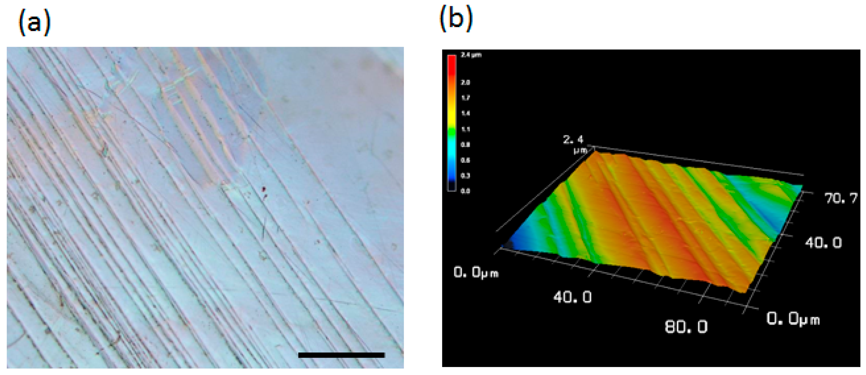
2.1. Surface Topography Induced by Plastic Deformation

α-titanium polycrystals were compressed at a compressive plastic strain of 5%. The obtained specimens showed a characteristic surface relief derived from the dislocation glide and deformation twinning as observed in optical microscopy (Figure 1a) and laser microscopy (Figure 1b). The average height of the resulting step structures analyzed with the laser microscope images was approximately 338.6 ± 34.4 nm.

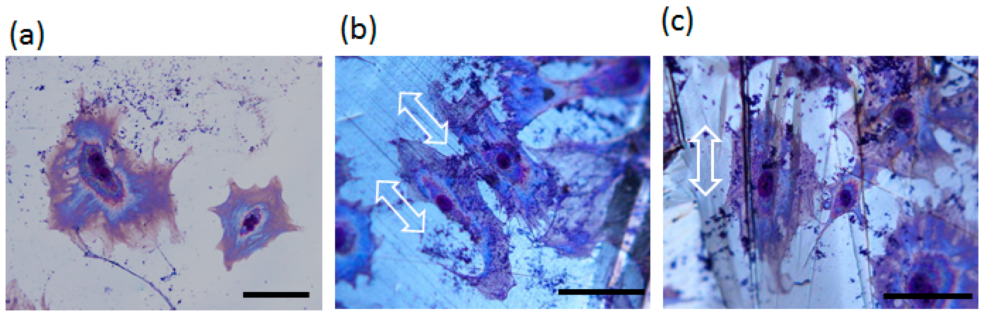
2.2. Cell Orientation

The surface topography introduced by plastic deformation induced osteoblast alignment along the twinning traces and/or slip traces, whereas the cells on the control substrates without deformation showed no preferential orientation. Giemsa staining revealed that cell elongation was effectively controlled to align in the direction of the twinning and/or slip traces (Figure 2).

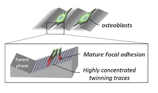
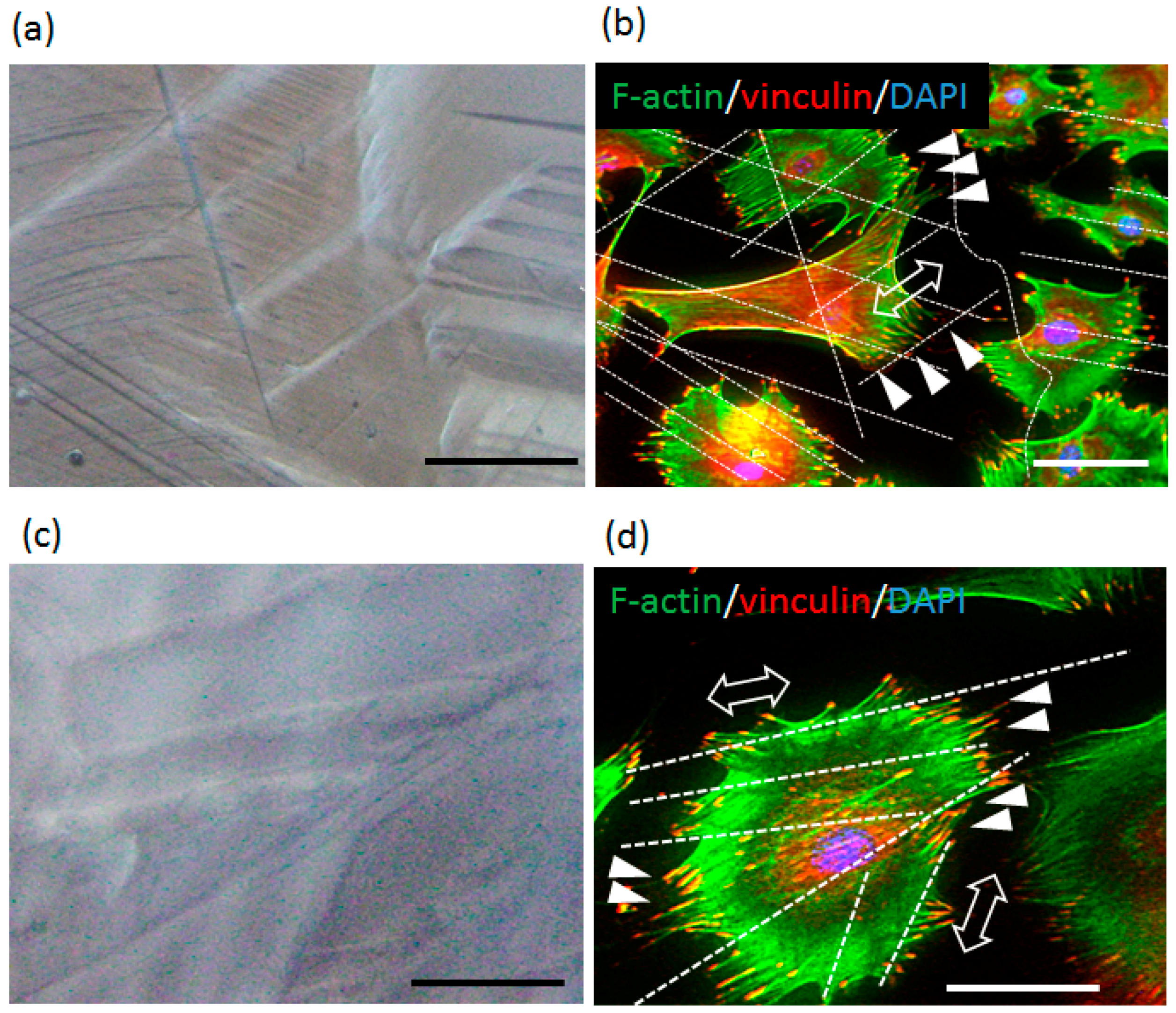
2.3. Plastic Deformation-Induced Cytoskeletal Rearrangement

Cytoskeletal proteins that modulate cell morphology and orientation were successfully visualized by immunocytochemistry. Actin stress fibers were preferentially elongated along the surface twinning traces and/or slip traces (Figure 3a,b). Focal adhesions, which mediate the interactions between cells and material surfaces, accumulated between the twinning traces and/or slip traces with elongated morphology (Figure 3c,d).

3. Discussion

Control of cellular morphology and arrangement is crucial for tissue and organ morphogenesis to achieve proper individual functions [12,13]. In particular, the characteristic anisotropic microstructure of bone tissue is essential for its mechanical function [2]. We previously revealed that the level of osteoblast arrangement determines the degree of apatite orientation in the bone matrix [9]; bone tissue anisotropy is controllable by regulating the osteoblast arrangement in a favored direction and to a favored degree. In this study, we developed a novel approach to control osteoblast arrangement; the surface relief introduced by dislocation glide and twinning deformation was shown to be effective for controlling cellular direction. To the best of our knowledge, this is the first report to reveal the relationship between biological cells and deformation twinning in addition to dislocation motion.
Titanium and its alloys have been the most widely used materials for biomedical applications, particularly for hard tissue replacement because of their excellent mechanical, biological, and corrosion-resistant performance [14]. α-titanium crystallizes in a hexagonal close-packed (hcp) crystal structure. Deformation of hcp materials typically occurs by simultaneous crystallographic slip and deformation twinning, and the resulting mechanical properties are strongly affected by the interactions between these two major mechanisms of plastic deformation [15]. Twinning is commonly observed in hcp materials including titanium and makes a significant contribution to deformation. The twinning modes operating in α-titanium include the following three types: { 1 1 ¯ 02 } < 1 ¯ 101 > , { 1 ¯ 2 1 ¯ 2 } < 1 ¯ 2 1 ¯ 3 > , and { 1 ¯ 2 1 ¯ 1 } < 1 ¯ 2 1 ¯ 6 > . The accompanying dislocation glide can also be observed in plastic deformation of polycrystals. In this study, deformation twinning, including the above twinning systems, and dislocation motion were introduced into the α-titanium polycrystals by compressive plastic deformation.
The surface relief introduced by the dislocation motion and/or deformation twinning show characteristic step-like structures, as depicted in Figure 1. The observed structure is considered to be derived from the surface extrusions and intrusions of atoms dominated by nucleation of different variants and their growth in plastic deformation. The obtained laser microscope images revealed that the surface topography introduced by dislocation and deformation twinning exhibited nanometer-scale step structures derived from the highly concentrated twinning traces.
The osteoblasts showed morphological responses to the surface relief derived from deformation twinning; when the cells encountered the twinned surface, they elongated along the surface steps. In contrast, they showed no preferential alignment on smooth substrate without plastic deformation (Figure 2). The cellular responses are not dependent on the twinning mode, suggesting possible control of cell arrangement by twinning, regardless of crystallographic orientation. Cell morphology is strictly regulated by the mutual activities of the constituent organelles, including cytoskeletal components. Actin stress fibers play a key role in determining cell shape and migration. On the deformed surface, the actin stress fibers are preferentially elongated along the surface relief. In particular, the focal adhesions, which mediate the cell-materials interface and regulate the extracellular matrix architecture depending on their sizes [16], could not overcome the surface relief, dictating the localization between the twinning and/or slip traces and elongation along the steps introduced by compressive deformation (Figure 3d). These results indicated that the focal adhesion assembly was regulated to elongate along the surface concave-convex structures, facilitating cell body alignment. Interestingly, we recently revealed that the osteoblasts aligned along the nanometer-scale grooves produce collagen/apatite bone matrix orthogonal to the cellular direction [4]. This process is considered to be mediated by the mature focal adhesion structures developed in response to the nanotopography. In this study, nanometer-scale surface topography derived from the highly concentrated twinning traces was observed on the substrate surface after plastic deformation. Interestingly, mature focal adhesion structures were found along the twinning face. The cell arrangement along the surface relief, therefore, is considered to be mediated by the mature focal adhesion constructs (Figure 4), and the following bone matrix formation is necessarily revealed including its preferential orientation.

4. Materials and Methods

4.1. Deformation of α-Titanium Polycrystals

Parallelepipeds oriented for compression (dimensions: 2 × 2 × 5 mm3) were cut from the α-titanium (purity: 99.9%) polycrystals by electric discharge machining. These specimens were ground using emery paper (#1000, #2000), following which they were electrochemically polished to obtain reasonably smooth surfaces. Compression tests were carried out using an Instron-type testing machine (AG-5000C; Shimadzu, Kyoto, Japan) at a nominal strain rate of 1.67 × 10⁻⁴ s−1 until the compressive plastic strain reached 5%. The amount of plastic deformation was determined according to our previous report [11]. At 5% plastic deformation, surface topology covers the entire surface independent of crystal grain in polycrystalline.

4.2. Surface Characterization

The deformed surfaces were observed using an optical microscope equipped with Normarski interference contrast (BX60M; Olympus, Tokyo, Japan). Three-dimensional imaging and quantification of the surface topographic features were performed by using a three-dimensional (3D) laser microscope (VK-9700; Keyence, Osaka, Japan). Topographical images of the substrate surfaces were obtained with a scanning pitch of 0.08 μm.

4.3. Cell Culture

Primary osteoblasts were isolated from the calvariae of neonatal mice. Briefly, the calvariae of neonatal C57BL/6 mice were excised under aseptic conditions and placed in ice-cold α-MEM (Invitrogen, Carlsbad, CA, USA). The fibrous tissues around the bone were then removed gently. The calvariae were subjected to a series of collagenase/trypsin digests (collagenase: Wako, Osaka, Japan; trypsin: Nacalai Tesque, Kyoto, Japan) at 37 °C for 15 min each. This process allows the collagen matrix to degrade and makes the isolation of osteoblasts easier. The first two digests were discarded, and supernatants from the 3rd, 4th, and 5th digests were neutralized with α-MEM, pooled, and filtered through a 100-μm mesh (BD Biosciences, San Jose, CA, USA). The filtrates were centrifuged and the resulting pellets were re-suspended in α-MEM containing 10% fetal bovine serum (FBS) for culturing the cells. Cells were seeded onto the fabricated specimens at a density of 6250 cells/cm2.

4.4. Optical Images of Cultured Cells

After culturing the cells for 24 h, they were washed in phosphate-buffered saline (PBS) and fixed with methanol for 10 min. After air-drying, they were stained with 5% Giemsa staining solution (Wako, Osaka, Japan) for 10 min. The cells were then washed in distilled water and air-dried. Optical images were obtained using an optical microscope equipped with Normarski interference contrast (BX60M; Olympus).

4.5. Fluorescent Images of Cultured Cells

After culturing the cells for 24 h, they were incubated in PBS-0.05% Triton X-100 (PBST) containing 1% normal goat serum (NGS) (Invitrogen) for 30 min to block non-specific antibody binding sites. The cells were then incubated with mouse mono-clonal antibodies against vinculin (Sigma Aldrich, St Louis, MO, USA) at 4 °C for 12 h. This step was followed by incubation with Alexa Fluor 546-conjugated anti-mouse IgG (Molecular Probes, Invitrogen) and Alexa Fluor 488-conjugated phalloidin (Molecular Probes, Invitrogen). Finally, the cells were washed with PBST and mounted in Prolong Gold anti-fade reagent containing DAPI (Molecular Probes, Invitrogen). Fluorescent images were obtained using a fluorescence microscope (Biozero; Keyence) and processed using Adobe Photoshop 10.0 software.

5. Conclusions

Control of osteoblast arrangement is essential for anisotropic bone tissue construction. In this study, a crystallography-driven methodology was proposed as a novel tool for controlling cellular arrangement. A specific surface topography derived from dislocation and deformation twinning was introduced into α-titanium polycrystals. Primary osteoblasts were aligned along the twinning traces and/or slip traces, and accumulation of the mature focal adhesions was observed in the same direction. These results indicated that the highly concentrated twinning traces with nanometer-scale relief structures induced the elongation and maturation of focal adhesions and the resulting cellular elongation and alignment. These findings provide important insights into the mechanisms underlying the interactions between cells and materials and the development of biomedical devices that can control bone tissue anisotropy.

Acknowledgment

This work was supported by Grants-in-Aid for Scientific Research (S) from the Japan Society for Promotion of Science (Grant No. 25220912).

Author Contributions

Takayoshi Nakano designed the study; Aira Matsugaki collected and analyzed the data; Aira Matsugaki and Takayoshi Nakano interpreted the data; Aira Matsugaki drafted the manuscript; Takayoshi Nakano revised the manuscript content.

Conflicts of Interest

The authors declare no conflict of interest.

References

  1. Landis, W.J. The strength of a calcified tissue depends in part on the molecular structure and organization of its constituent mineral crystals in their organic matrix. Bone 1995, 16, 533–544. [Google Scholar] [CrossRef]
  2. Onsager, L. The effects of shape on the interactions of colloidal particles. Ann. N. Y. Acad. Sci. 1949, 51, 627–659. [Google Scholar] [CrossRef]
  3. Giraud-Guille, M.M. Liquid crystallinity in condensed type I collagen solutions.a clue to the packing of collagen in extracellular matrices. J. Mol. Biol. 1992, 224, 861–873. [Google Scholar] [CrossRef]
  4. Campi, G.; Fratini, M.; Bukreeva, I.; Ciasca, G.; Burghammer, M.; Brun, F.; Tromba, G.; Mastrogiacomo, M.; Cedola, A. Imaging collagen packing dynamics during mineralization of engineered bone tissue. Acta Biomater. 2015, 23, 309–316. [Google Scholar] [CrossRef] [PubMed]
  5. Ishimoto, T.; Nakano, T.; Umakoshi, Y.; Yamamoto, M.; Tabata, Y. Degree of biological apatite c-axis orientation rather than bone mineral density controls mechanical function in bone regenerated using rBMP-2. J. Bone Miner. Res. 2013, 28, 1170–1179. [Google Scholar] [CrossRef] [PubMed]
  6. Nakano, T.; Kaibara, K.; Ishimoto, T.; Tabata, Y.; Umakoshi, Y. Biological apatite (BAp) crystallographic orientation and texture as a new index for assessing the microstructure and function of bone regenerated by tissue engineering. Bone 2012, 51, 741–747. [Google Scholar] [CrossRef] [PubMed]
  7. Matsugaki, A.; Aramoto, G.; Ninomiya, T.; Sawada, H.; Hata, S.; Nakano, T. Abnormal arrangement of a collagen/apatite extracellular matrix orthogonal to osteoblast alignment is constructed by a nanoscale periodic surface structure. Biomaterials 2015, 37, 134–143. [Google Scholar] [CrossRef] [PubMed]
  8. Matsugaki, A.; Fujiwara, N.; Nakano, T. Continuous cyclic stretch induces osteoblast alignment and formation of anisotropic collagen fiber matrix. Acta Biomater. 2013, 9, 7227–7235. [Google Scholar] [CrossRef] [PubMed]
  9. Matsugaki, A.; Isobe, Y.; Saku, T.; Nakano, T. Quantitative regulation of bone-mimetic, oriented collagen/apatite matrix structure depends on the degree of osteoblast alignment on oriented collagen substrates. J. Biomed. Mater. Res. 2015, 103, 489–499. [Google Scholar] [CrossRef] [PubMed]
  10. Nakano, T.; Kaibara, K.; Tabata, Y.; Nagata, N.; Enomoto, S.; Marukawa, E.; Umakoshi, Y. Unique alignment and texture of biological apatite crystallites in typical calcified tissues analyzed by microbeam X-ray diffractometer system. Bone 2002, 31, 479–487. [Google Scholar] [CrossRef]
  11. Matsugaki, A.; Aramoto, G.; Nakano, T. The alignment of MC3T3-E1 osteoblasts on steps of slip traces introduced by dislocation motion. Biomaterials 2012, 33, 7327–7335. [Google Scholar] [CrossRef] [PubMed]
  12. Goto, T.; Davidson, L.; Asashima, M.; Keller, R. Planar cell polarity genes regulate polarized extracellular matrix deposition during frog gastrulation. Curr. Biol. 2005, 2615, 787–793. [Google Scholar] [CrossRef] [PubMed]
  13. Shindo, T.; Kurihara, H.; Kuno, K.; Yokoyama, H.; Wada, T.; Kurihara, Y.; Imai, T.; Wang, Y.; Ogata, M.; Nishimatsu, H.; et al. ADAMTS-1: A metalloproteinase-disintegrin essential for normal growth, fertility, and organ morphology and function. J. Clin. Investig. 2000, 105, 1345–1352. [Google Scholar] [CrossRef] [PubMed]
  14. Niinomi, M. Mechanical biocompatibilities of titanium alloys for biomedical applications. J. Mech. Behav. Biomed. Mater. 2008, 1, 30–42. [Google Scholar] [CrossRef] [PubMed]
  15. Yoo, M.H.; Lee, J.K. Deformation twinning in h.c.p. metals and alloys. Philos. Mag. A 1991, 63, 987–1000. [Google Scholar] [CrossRef]
  16. Wozniak, M.A.; Modzelewska, K.; Kwong, L.; Keel, P.J. Focal adhesion regulation of cell behavior. Biochim. Biophys. Acta 2004, 1692, 103–119. [Google Scholar] [CrossRef] [PubMed]
Figure 1. (a) Optical microscope and (b) laser microscope image of the surface of the obtained specimens. Scale bar: 100 μm.
Crystals 06 00073 g001
Figure 2. Giemsa staining of the osteoblasts on (a) a smooth surface and (b,c) deformed surfaces. Arrows indicate the direction of twinning and/or slip traces. Scale bar: 100 μm.
Crystals 06 00073 g002
Figure 3. Immunocytochemical analysis of the osteoblasts cultured on surface topography introduced by plastic deformation. Optical microscopic images of (a,c) surface relief and (b,d) cellular cytoskeleton proteins. Arrows indicate the direction of slip and/or twinning traces. Arrowheads indicate the mature focal adhesions accumulated between slip and/or twinning traces. Green, F-actin; red, vinculin; blue, nuclei. Scale bar: 100 μm.
Crystals 06 00073 g003
Figure 4. Schematic illustration of cell alignment along slip and/or twinning traces. The mature focal adhesions elongated along the highly concentrated twinning traces, resulting in cellular alignment in the same direction.
Crystals 06 00073 g004

