Article

Production of New Isoflavone Glucosides from Glycosylation of 8-Hydroxydaidzein by Glycosyltransferase from Bacillus subtilis ATCC 6633

1
Department of Biotechnology, Chia Nan University of Pharmacy and Science, No. 60, Sec. 1, Erh-Jen Rd., Jen-Te District, Tainan 71710, Taiwan
2
Biodiversity Research Center, Academia Sinica, Taipei 115, Taiwan
3
Department of Biological Sciences and Technology, National University of Tainan, Tainan 70005, Taiwan
4
Department of Food Science, National Quemoy University, Kinmen County 892, Taiwan
*
Authors to whom correspondence should be addressed.
Catalysts 2018, 8(9), 387; https://doi.org/10.3390/catal8090387
Submission received: 24 August 2018 / Revised: 4 September 2018 / Accepted: 7 September 2018 / Published: 10 September 2018
(This article belongs to the Special Issue Biocatalysis: Chemical Biosynthesis)

Abstract

:
8-Hydroxydaidzein (8-OHDe) has been proven to possess some important bioactivities; however, the low aqueous solubility and stability of 8-OHDe limit its pharmaceutical and cosmeceutical applications. The present study focuses on glycosylation of 8-OHDe to improve its drawbacks in solubility and stability. According to the results of phylogenetic analysis with several identified flavonoid-catalyzing glycosyltransferases (GTs), three glycosyltransferase genes (BsGT110, BsGT292 and BsGT296) from the genome of the Bacillus subtilis ATCC 6633 strain were cloned and expressed in Escherichia coli. The three BsGTs were then purified and the glycosylation activity determined toward 8-OHDe. The results showed that only BsGT110 possesses glycosylation activity. The glycosylated metabolites were then isolated with preparative high-performance liquid chromatography and identified as two new isoflavone glucosides, 8-OHDe-7-O-β-glucoside and8-OHDe-8-O-β-glucoside, whose identity was confirmed by mass spectrometry and nuclear magnetic resonance spectroscopy. The aqueous solubility of 8-OHDe-7-O-β-glucoside and 8-OHDe-8-O-β-glucoside is 9.0- and 4.9-fold, respectively, higher than that of 8-OHDe. Moreover, more than 90% of the initial concentration of the two 8-OHDe glucoside derivatives remained after 96 h of incubation in 50 mM of Tris buffer at pH 8.0. In contrast, the concentration of 8-OHDe decreased to 0.8% of the initial concentration after 96 h of incubation. The two new isoflavone glucosides might have potential in pharmaceutical and cosmeceutical applications.

1. Introduction

Daidzein and genistein, the major isoflavones found in soybean, have been under intensive investigation in the past few decades due to their potential roles in preventing certain hormone-dependent diseases [1]. In recent years, biotransformation of isoflavones using either wild-type or genetically-engineered microorganisms has also been of interest because the bioactivity of isoflavones dramatically alters after biotransformation. Among the various biotransformations of soy isoflavones, ortho-hydroxylation of soy isoflavones has become a subject of great interest because of the ortho-hydroxydaidzein and ortho-hydroxygenistein derivatives produced, which usually possess higher bioactivities compared with those of the precursors, daidzein and genistein [2].
8-Hydroxydaidzein (8-OHDe) is one of the ortho-hydroxydaidzein derivatives. The compound can be produced from biotransformation of daidzein by either wild Aspergillus oryzae [3,4,5], genetically-engineered Pichia pastoris [6] or Escherichia coli [7,8]. Multiple bioactivities related to 8-OHDe have been reported, including anti-cancer [9], suppression of multidrug resistance [10], anti-tyrosinase [11,12], skin whitening [13,14], anti-aldose reductase [15] and the newly-found anti-inflammatory activity [16,17].
Although 8-OHDe has been identified with many bioactivities, some drawbacks (including low aqueous solubility and stability) limit isoflavone’s application in pharmaceuticals and cosmeceuticals [18]. To improve the drawbacks of 8-OHDe, biotransformation through whole cells or enzyme biocatalysts into 8-OHDe derivatives is a promising strategy. Among various biotransformations, glycosylation, the attachment of a bulky sugar group to the precursor molecules, could improve the chemical stability and aqueous solubility of natural compounds. The aqueous solubility of the corresponding 7-O-glucoside of soy isoflavones is improved about 30-fold [19]. Increasing the aqueous solubility and stability could expand applications of the compounds in advance. Therefore, in the present study, we are interested in investigating the glycosyl-biotransformation of 8-OHDe.
Glycosylation is a common modification reaction in the biosynthesis of natural compounds. Generally, glycosylation is catalyzed by glycosyltransferases (GTs, EC 2.4.x.y), which transfer sugar moieties from the activated donor molecules to specific acceptor molecules [20,21,22]. Our previous studies found that Bacillus subtilis ATCC 6633 could biotransform antcin K, which is a major ergostane triterpenoid from the fruiting bodies of Antrodia cinnamomea, to its glucoside derivatives [23]. In the present study, the B. subtilis strain was found to biotransform 8-OHDe. To identify the biotransformation in advance, three GT genes were cloned from the B. subtilis strain and overexpressed in Escherichia coli. Then, the biotransformation activity of the three purified GT enzymes toward 8-OHDe was determined. The biotransformed metabolites by one positive-active enzyme were isolated and identified. Finally, the aqueous solubility and stability of the 8-OHDe glucoside derivatives were determined.

2. Results and Discussion

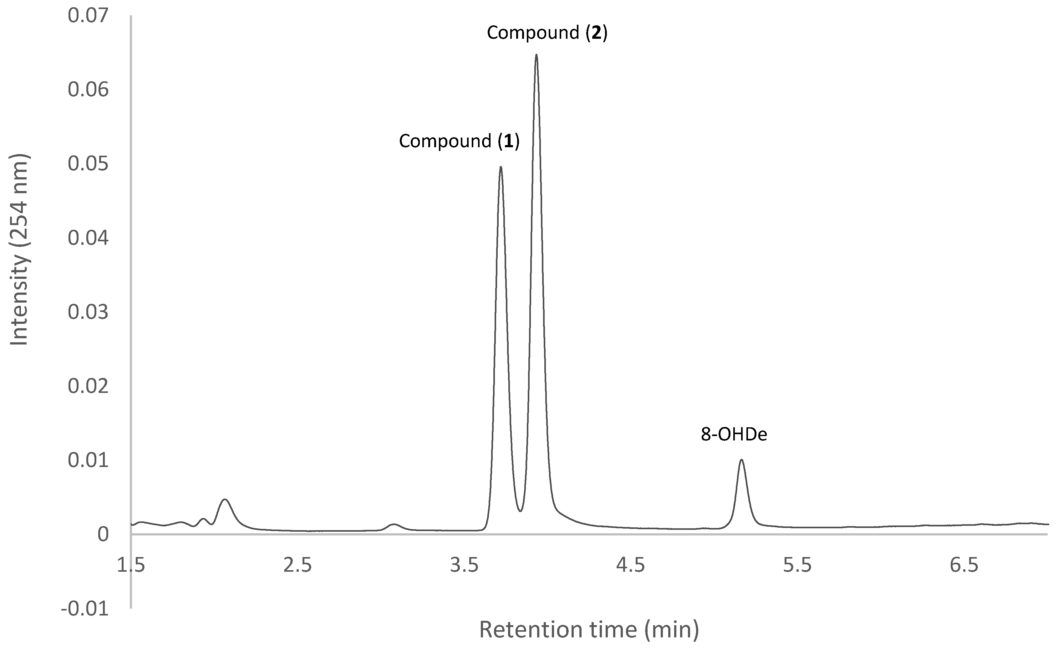
2.1. Confirming Biotransformation of 8-OHDe by Bacillus subtilis ATCC 6633

Our previous studies showed that B. subtilis ATCC 6633 could biotransform the ergostane triterpenoid antcin K to its glucoside derivatives [23]. To confirm whether B. subtilis ATCC 6633 could also biotransform 8-OHDe, the bacterium was cultivated in broth with 8-OHDe, and the fermentation broth was analyzed using ultra-performance liquid chromatography (UPLC).
Figure 1 shows the UPLC analysis of the initial (dashed line) and 24-h (solid line) fermentation broths of the strain B. subtilis ATCC 6633 fed with 8-OHDe. In the figure, 8-OHDe appears at the retention time (RT) of 5.1 min. After 24 h of fermentation, the peak of the precursor decreases, while several new peaks with RTs of between 3 and 4 min appear. To confirm whether the new peaks are from biotransformation of 8-OHDe, biotransformation was conducted in the absence of 8-OHDe. The results showed that the new peaks did not appear at 24 h of the fermentation broth of the strain in the absence of 8-OHDe (Figure S1). Thus, it was concluded that 8-OHDe was biotransformed by the strain B. subtilis ATCC 6633.

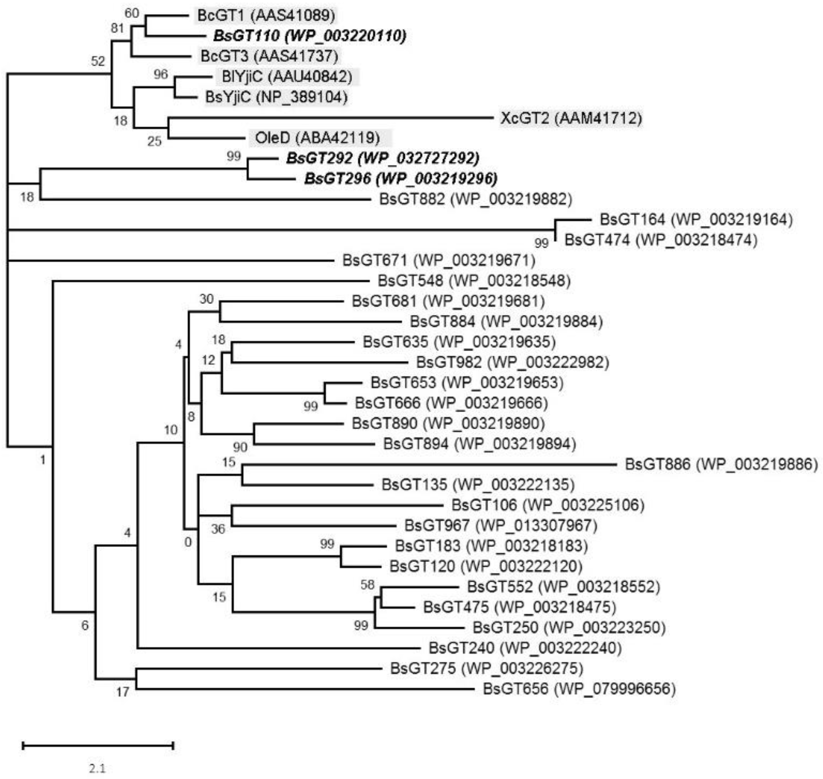
2.2. Phylogenetic Analysis of GTs from B. subtilis ATCC 6633

Although B. subtilis ATCC 6633 has been proven to biotransform the ergostane triterpenoid antcin K [23] and 8-OHDe (Figure 1), biotransformation by using whole cells as biocatalysts usually has lower efficiency than that by using purified enzyme, which can be produced through genetic engineering. Thus, cloning of the putative genes encoding the catalytic enzymes in the biotransformation is a worthy strategy. According to the genome data of B. subtilis ATCC 6633 (GenBank BioProject Accession No. PRJNA43011), there are 28 GTs annotated in the genome. To clone the GT genes from B. subtilis ATCC 6633 responsible for the biotransformation of 8-OHDe, phylogenetic analysis of the annotated GTs from B. subtilis ATCC 6633 was compared with known bacterial GTs, which have been proven to possess glycosylation activity toward flavonoids. The characterized bacterial glycosyltransferases included BlYjiC (AAU40842) from B. licheniformis ATCC 14580 [24], BsYjiC (NP_389104) from B. subtilis 168 [25,26], BcGT-1 (AAS41089) and BcGT-3 (AAS41737) from B. cereus ATCC 10987 [27,28], OleD (ABA42119) from Streptomyces antibioticus [29] and XcGT-2 (AAM41712) from Xanthomonas campestris pv. campestris ATCC 33913 [30]. The results are shown in Figure 2. According to the results of the phylogenetic analysis, all tested six identified flavonoid-catalyzing GTs (gray background in Figure 2) were clustered into one group (Figure 2). Among the 28 BsGTs, only BsGT110 was included in the group. At the same time, BsGT292 and BsGT296 were close to the group. Thus, the three GTs, BsGT110 (GenBank Protein Accession No. WP_003220110), BsGT292 (GenBank Protein Accession No. WP_032727292) and BsGT296 (GenBank Protein Accession No. WP_003219296; bold text in Figure 2), were selected for further functional assay.

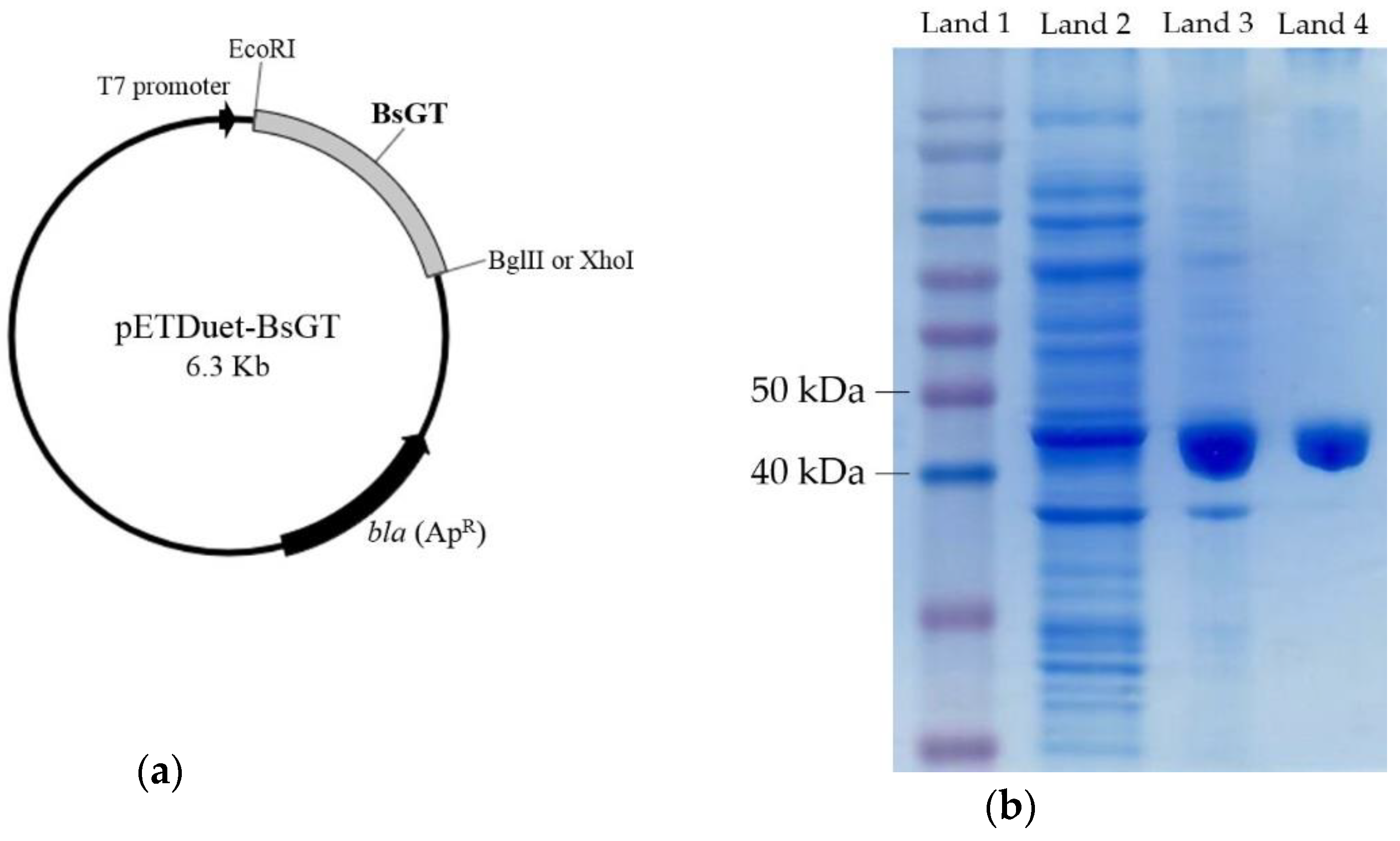
2.3. Cloning, Overexpression, Purification and Activity Assay of BsGTs from B. subtilis ATCC 6633 in E. coli

To identify which enzyme catalyzed the biotransformation, the three BsGT genes were cloned into a pETDuet-1 expression vector (Figure 3a). The recombinant BsGT gene in recombinant E. coli was overexpressed by induction with 0.2 mM of isopropyl β-D-1-thiogalactopyranoside (IPTG), and the protein produced was purified with Ni2+ chelate affinity chromatography. Figure 3b shows the sodium dodecyl sulfate polyacrylamide gel electrophoresis (SDS-PAGE) analysis of the overexpressed and purified BsGT110, while the SDS-PAGE analysis of purified BsGT292 and BsGT296 is in Figure S2.
The purified enzymes were incubated with uridine diphosphate (UDP)-glucose and the precursor 8-OHDe to confirm their biotransformation activity toward 8-OHDe. The results showed that only BsGT110 has biotransformation activity toward 8-OHDe (Figure 4), while neither purified BsGT292, nor BsGT296 showed the activity (Figure S3). The result was consistent with the phylogenetic analysis results (Figure 2), where only BsGT110 was clustered with the group of flavonoid-catalyzing GTs. In addition, the RTs of the two biotransformation metabolites (Figure 4), 3.7 min and 3.9 min for Compound (1) and Compound (2), respectively, were similar to some of the new peaks in the 8-OHDe biotransformation by the strain B. subtilis ATCC 6633 (Figure 1). The results imply that BsGT110 is one of the corresponding enzymes catalyzing 8-OHDe during the biotransformation of 8-OHDe by the strain B. subtilis ATCC 6633.
By comparing the results of the biotransformations using whole cells (Figure 1) and the purified BsGT110 (Figure 4), it is obvious that biotransformation products using the purified BsGT110 (Compound (1) and Compound (2) in Figure 4) were more specific than those using the whole cells (metabolites between RT 3 and 4 min in Figure 1). Moreover, the catalyzing efficiency using the purified BsGT110 (reaction time of 30 min in Figure 4) was higher than that using whole cells (reaction time of 24 h in Figure 1). Therefore, biotransformation by using the purified BsGT110 as biocatalysts has higher efficiency and specificity than those by using whole cells.

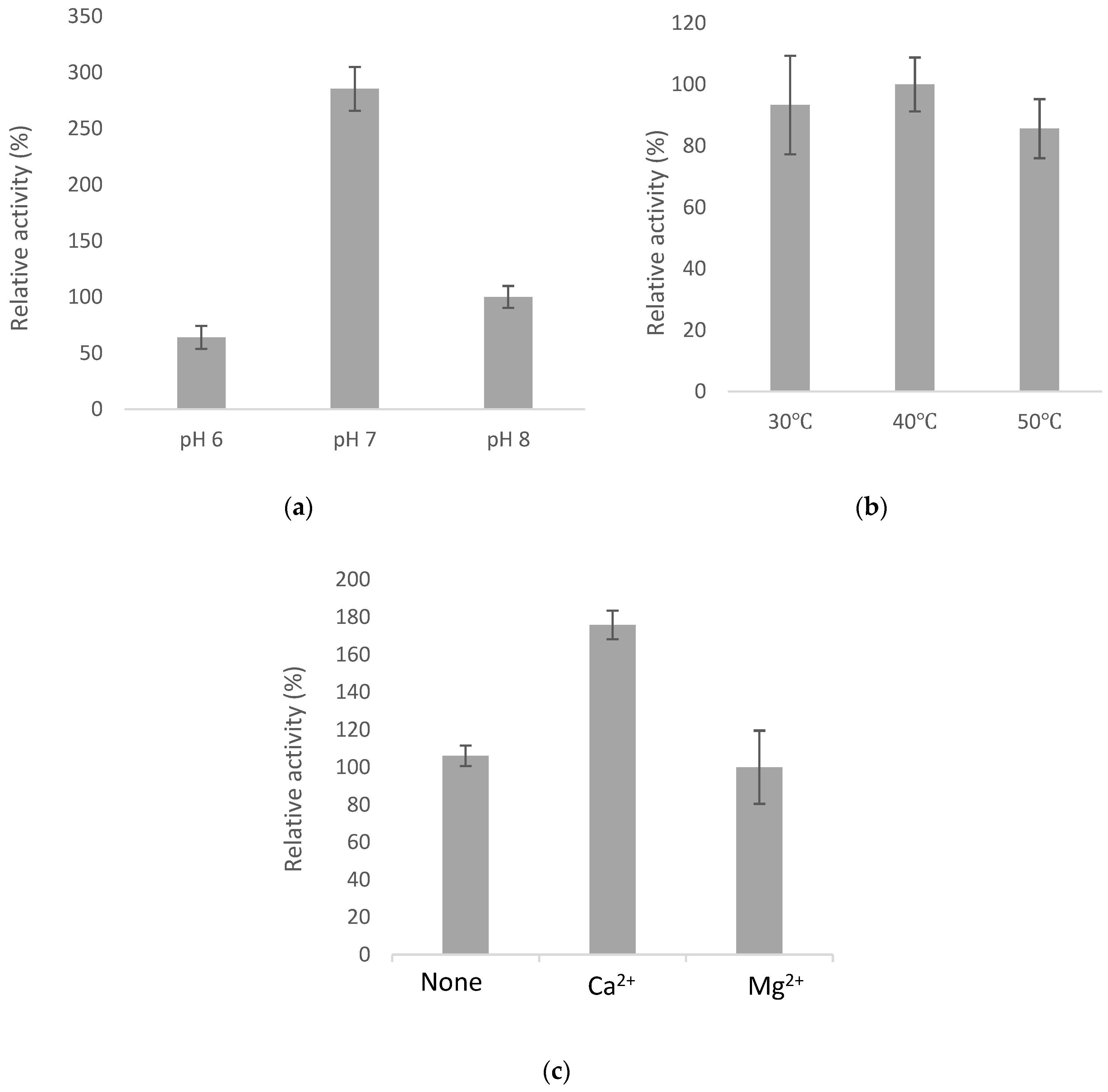
2.4. Optimal Catalyzing Conditions for BsGT110

To determine the optimal catalyzing condition, the activity of BsGT110 at different pH values, temperatures and metal ions was examined. The results are shown in Figure 5. The enzyme activity was very sensitive to pH, and the enzyme activity at pH 7.0 was nearly 3–4-fold higher than that at pH 8.0 and pH 6.0. However, the enzyme activity was insensitive to temperature, and the enzyme activity was not significantly different between 30 °C and 50 °C. In addition, the enzyme favored Ca2+ as its cofactor. In the presence of Ca2+, the enzyme activity was 1.8-fold higher than that at Mg2+ or none. It is known that GTs utilize divalent metal ion cofactors such as Mn2+ and Mg2+. However, Li et al. found that the activity of the GT from Bacillus circulans was enhanced by Ca2+ due to an additional calcium-binding site in the structure [33]. Whether BsGT110 favored Ca2+ as its cofactor due to an additional calcium-binding site needs to be studied in the future. From the results, the optimal catalyzing conditions for BsGT110 are at 40 °C, pH 7.0 with 10 mM of Ca2+.

2.5. Substrate Specificity of BsGT110

To study the substrate specificity of BsGT110, another three ortho-hydroxyisoflavones (3’-hydroxydaidzein, 3’-OHDe; 3’-hydroxygenistein, 3’-OHGe; 6’-hydroxydaidzein, 6-OHDe) were used in the biotransformation assays by BsGT110. The result is shown in Figure 6 and reveals that BsGT110 could also catalyze all the tested ortho-hydroxyisoflavones. However, unlike two major metabolites, Compound (1) and Compound (2), produced in the biotransformation of 8-OHDe by BsGT110 (Figure 4), only one major metabolite appeared in the biotransformations of either 3’-OHDe, 3’-OHGe or 6-OHDe by BsGT110 (Figure 6). Due to very low amounts of the three ortho-hydroxyisoflavones, the biotransformation metabolites from these biotransformations were not identified in advance. In addition, among the ortho-hydroxyisoflavones in the present study, including 3’-OHDe, 3’-OHGe, 6-OHDe and 8-OHDe, only 8-OHDe has been approved to possess bioactivity in human volunteers [13,14]. Therefore, 8-OHDe was used as a substrate for the following study.

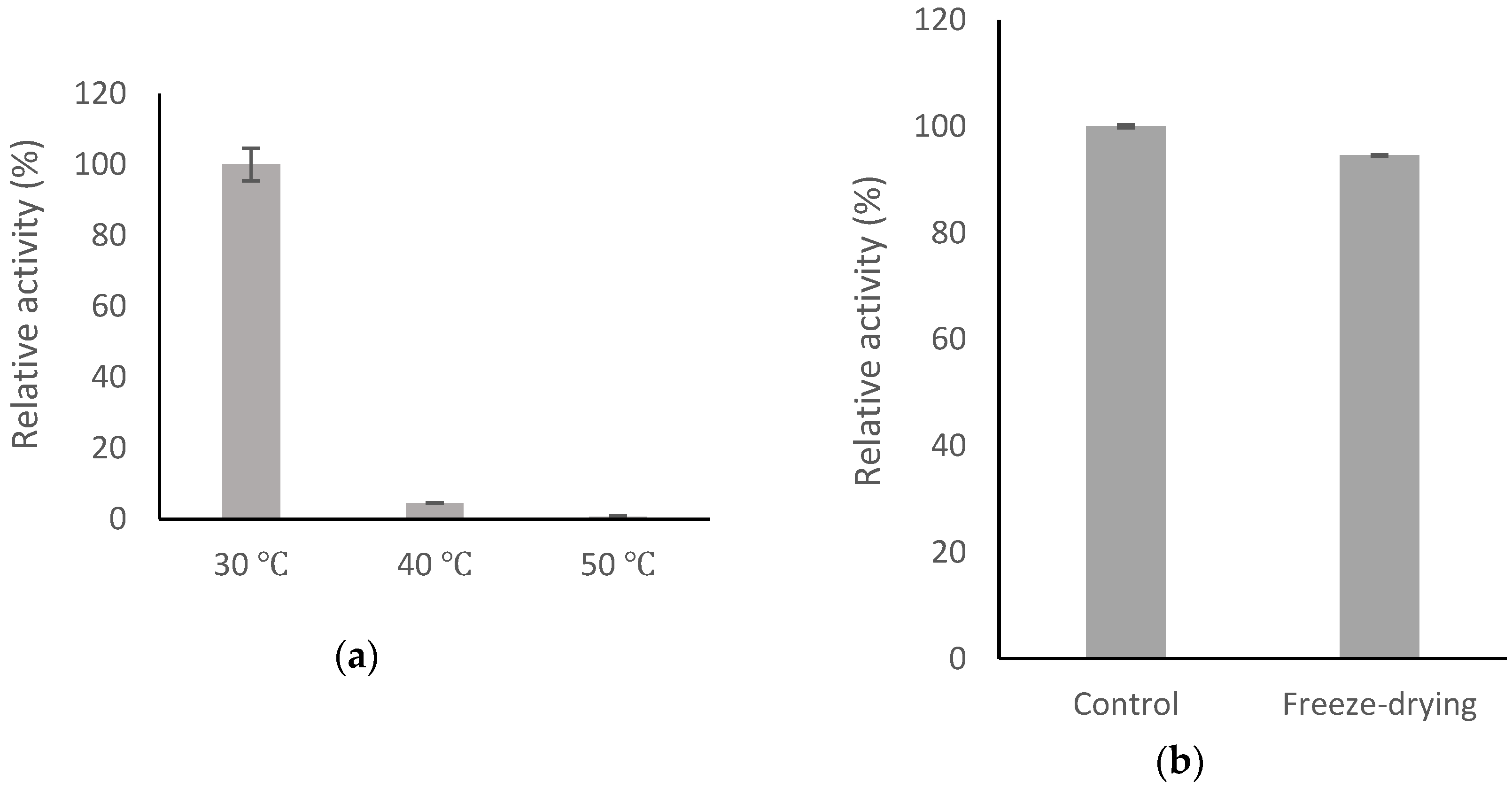
2.6. Stability of BsGT110

To study the stability of BsGT110, BsGT110 was incubated at different temperatures for 1 h or was freeze-dried once, and then, the activity of the treated BsGT110 was determined. The result is shown in Figure 7. The enzyme was not stable above 40 °C for 1 h, while the enzyme was stable after freeze-drying.

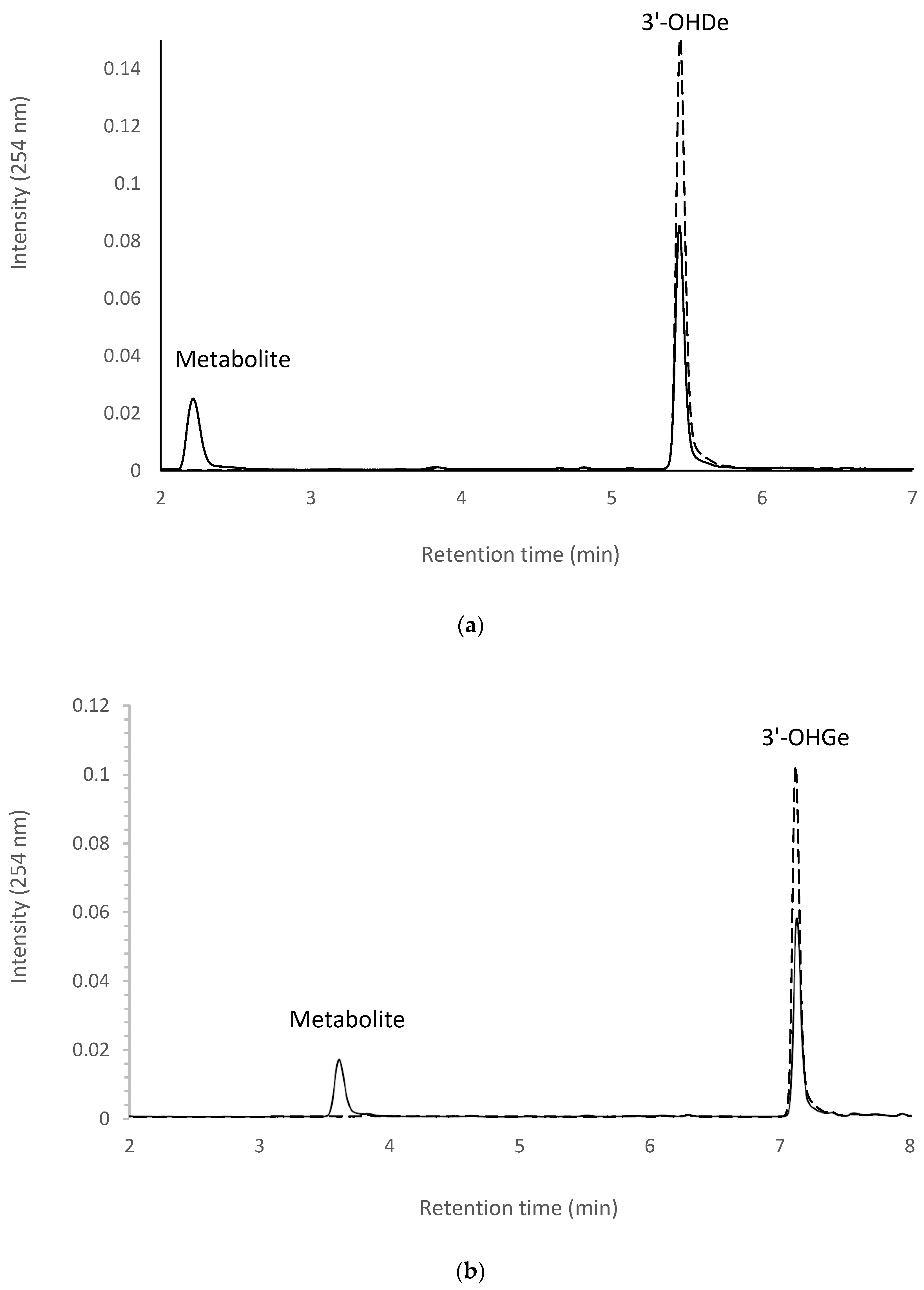
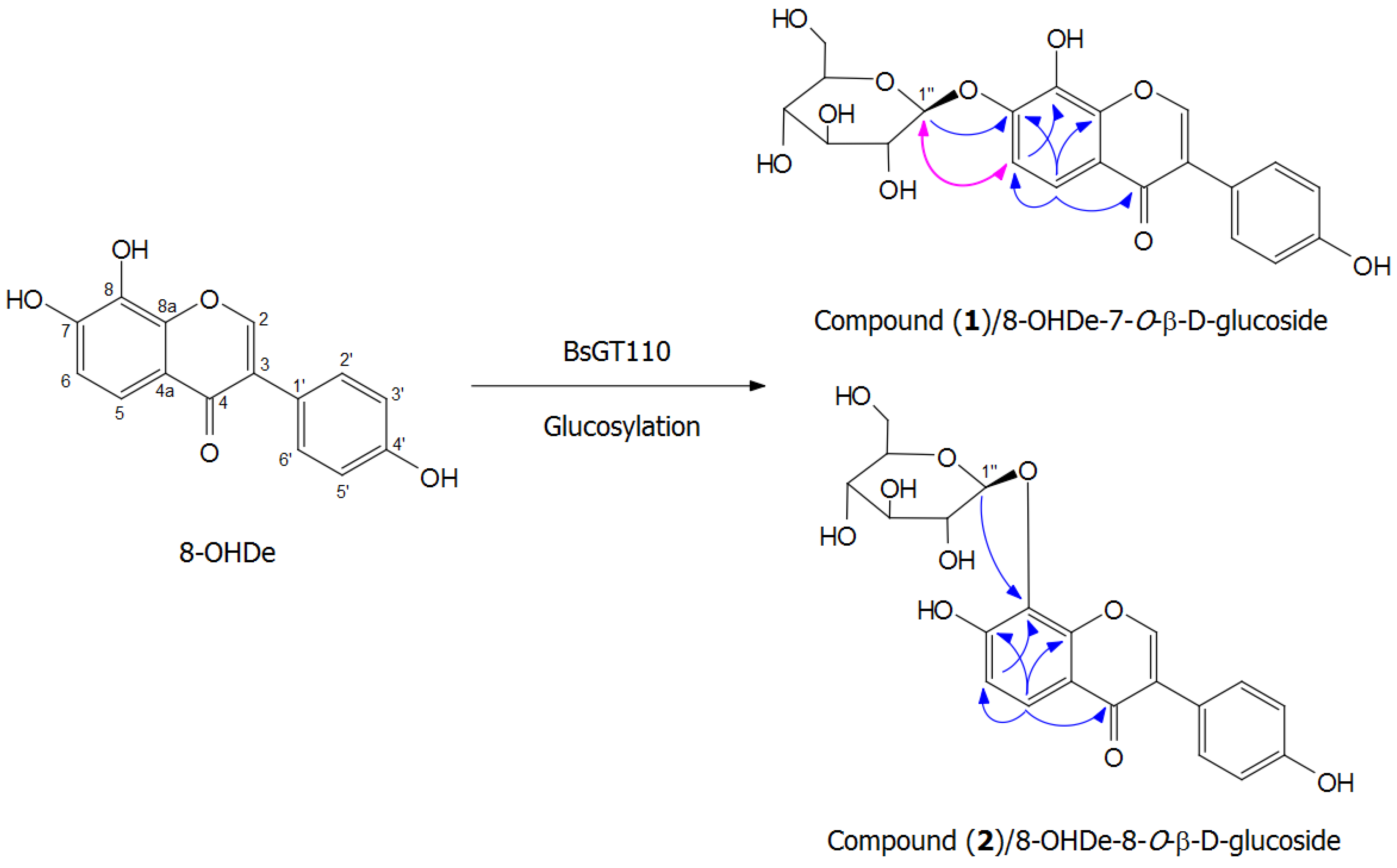
2.7. Isolation and Identification of Biotransformation Metabolites

To resolve the chemical structures of the metabolites, the biotransformation was scaled up at the optimal condition, and the two metabolites were purified with preparative high-performance liquid chromatography (HPLC). From a 40-mL reaction mixture containing 1 mg/mL of 8-OHDe, 10 mM of UDP-glucose, 10 mM of CaCl2 and 50 mM of phosphate buffer at pH 7.0, 17.4 mg and 24.5 mg of Compound (1) and Compound (2) were isolated. Both compounds showed an [M-H]- ion peak at m/z: 431.23 in the electrospray ionization mass (ESI-MS) spectrum corresponding to the molecular formula C21H20O10. Then, 1H and 13C nuclear magnetic resonance (NMR), including distortionless enhancement by polarization transfer (DEPT), heteronuclear single quantum coherence (HSQC), heteronuclear multiple bond connectivity (HMBC), correlation spectroscopy (COSY) and nuclear Overhauser effect spectroscopy (NOESY) spectra, were obtained, and the 1H- and 13C-NMR signal assignments were conducted accordingly (shown in Figures S4–S17). In both compounds, in addition to the signals of the 8-OHDe moiety, which were confirmed by HMBC spectra (full assignments of both compounds are listed in Table S1), seven proton (from 3.18 to 4.93 ppm) and six carbon (from 60 to 105 ppm) signals corresponding to the glucose moiety structure were observed. The anomeric proton signal at δ 4.91 (J = 7.6 Hz) and 4.93 (J = 7.7 Hz) in the 1H-NMR spectra of Compounds (1) and (2), respectively, indicated the β-configuration for the glucopyranosyl moiety. The cross peak of H-1’’ with C-7 (4.91/149.0 ppm) in the HMBC spectrum, as well as the cross peak of H-1’’ with H-6 (4.91/ 7.30 ppm) in the NOESY spectrum demonstrated that the structure of Compound (1) was 8-OHDe-7-O-β-glucoside. The cross peaks of H-1’’ with C-8 (4.93/131.9 ppm) and H-6 with C-8 (7.02/131.9 ppm) in the HMBC spectrum demonstrated that the structure of Compound (2) was 8-OHDe-8-O-β-glucoside. The key HMBC and NOESY correlations of Compounds (1) and (2) and the illustration of the biotransformation process of 8-OHDe by BsGT110 are shown in Figure 8.

2.8. Determination of Aqueous Solubility of 8-OHDe and Its Glucosides

The aqueous solubility of 8-OHDe and the two 8-OHDe glucoside derivatives was examined and is summarized in Table 1. The results revealed that the aqueous solubility of 8-OHDe-7-O-β-glucoside and 8-OHDe-8-O-β-glucoside was 9.0- and 4.9-fold, respectively, higher than that of 8-OHDe.

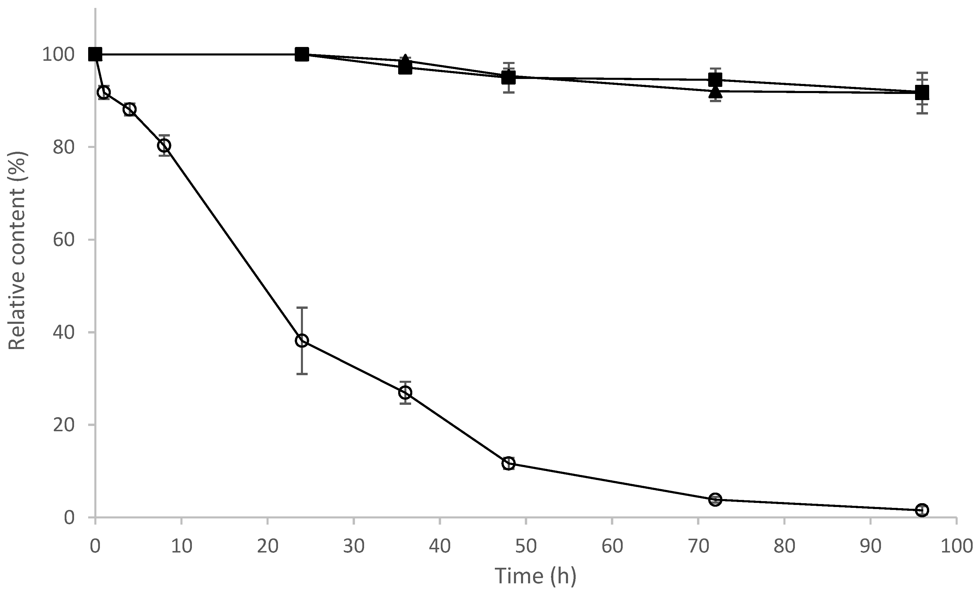
2.9. Determination of Stability of 8-OHDe and Its Glucosides

8-OHDe has been proven to be unstable in alkaline solution [18]. To determine the stability of 8-OHDe and its glucosides, the tested compounds were added in 50 mM of Tris at pH 8.0 and 20 °C. The residues of the compounds were monitored with UPLC at different time intervals. The results are shown in Figure 9. The amount of 8-OHDe decreased to 38% of the initial concentration after 24 h and decreased to 0.8% of the initial concentration after 96 h. In contrast, more than 90% of the two 8-OHDe glucoside derivatives remained after 96 h of incubation. The results revealed that the two 8-OHDe glucoside derivatives were much more stable than 8-OHDe in the aqueous solution.
8-OHDe has been proven to possess multiple bioactivities; especially, 8-OHDe has been proven for its skin-whitening activity in vivo in the skin of mice and human volunteers [13,14]. Some skin-whitening agents maintain skin-whitening activity after glycosylation. For examples, ascorbic acid and hydroquinone are famous skin-whitening agents. Their glucoside derivatives, ascorbic acid 2-glucosdie (AA2G) and arbutin (hydroquinone-glucoside), possess potent skin-whitening activity and are the most well-known skin-whitening agents used in cosmetic markets nowadays. In our previous study, we found that ortho-hydroxyisoflavone glucosides possessed skin-whitening activity in vivo in the skin of mice [34]. Therefore, the 8-OHDe glucosides in the present study might have skin-whitening activity. The experiments to evaluate the in vivo skin-whitening activity in the mice skin of the 8-OHDe glucosides in the present study were conducting in our laboratory.

3. Materials and Methods

3.1. Microorganisms, Animal Cells and Chemicals

Bacillus subtilis ATCC 6633 (BCRC 10447) was purchased from the Bioresources Collection and Research Center (BCRC, Food Industry Research and Development Institute, Hsinchu, Taiwan). 8-OHDe was prepared according to Wu et al.’s [4] method. 3’-OHDe and 3’-OHGe were prepared according to our previous study [35]. 6-OHDe was purchased from Sigma (St. Louis, MO, USA). UDP-glucose was obtained from Cayman Chemical (Ann Arbor, MI, USA). All the materials needed for polymerase chain reaction (PCR), including primers, deoxyribonucleotide triphosphate and Taq DNA polymerase, were purchased from MDBio (Taipei, Taiwan). pETDuet-1 plasmid was purchased from Novagen (Madison, WI, USA). Restriction enzymes and DNA ligase were obtained from New England Biolabs (Ipswich, MA, USA). The other reagents and solvents used were of high quality and were purchased from commercially available sources.

3.2. Identification of Bacteria B. subtilis ATCC 6633 with Biotransformation Activity

Bacillus subtilis ATCC 6633 was cultivated in a 250-mL baffled Erlenmeyer flask containing 20 mL of a modified glucose-nutrient (MGN) medium (5 g/L of peptone, yeast extract, K2HPO4 and NaCl; 20 g/L of glucose) and 20 mg/L of 8-OHDe. After cultivation at 180 rpm, 28 °C for 24 h, 1 mL of the culture was then mixed with an equal volume of methanol. The cell debris was removed by centrifugation at 10,000× g for 10 min. The supernatant from the extracted broth was assayed with UPLC to measure the biotransformation activity.

3.3. UPLC Analysis

The UPLC system (Acquity UPLC H-Class, Waters, Milford, MA, USA) was equipped with an analytic C18 reversed-phase column (Acquity UPLC BEH C18, 1.7 μm, 2.1 i.d. × 100 mm, Waters, Milford, MA, USA). The operation conditions contained a gradient elution using water (A) containing 1% (v/v) acetic acid and methanol (B) with a linear gradient for 7 min with 35% to 80% B at a flow rate of 0.2 mL/min, an injection volume of 0.2 μL and absorbance detection at 254 nm.

3.4. Phylogenetic Analysis of BsGTs

The unrooted phylogenetic tree of the candidate genes was constructed with the maximum likelihood method, using Molecular Evolutionary Genetics Analysis (MEGA X) software (Version 10.0.4, Center for Evolutionary Functional Genomics, The Biodesign Institute, Arizona State University, Tempe, AZ, USA, 1993–2018) [32] with 500 bootstrap replications, the mtREV24+I model [31] and partial deletion.

3.5. Expression and Purification of UGT398 and UGT489

The genomic DNA of B. subtilis ATCC 6633 was isolated using the commercial kit Geno PlusTM (Viogene, Taipei, Taiwan). The three GT target genes, BsGT110 (GenBank Protein Accession No. WP_003220110), BsGT292 (GenBank Protein Accession No. WP_032727292) and BsGT296 (GenBank Protein Accession No. WP_003219296), were amplified from the genomic DNA with PCR with the following primer sets: forward: 5′-CGC GAA TTC ggc taa tgt att aat gat cgg tt-3′; reverse: 5′-CGC AGA TCT tta tgc gtt ggc tga ttg agt tt-3′ for BsGT110; forward: 5′-CGC GAA TTC gat gaa gct tgc ctt tat ctg tac ag-3′; reverse: 5′-CGC CTC GAG tta tga ttt ggc ttt cac aaa aag c-3′ for BsGT292; forward: 5’-CGC GAA TTC Gat gaa aat agc act gat cgc cac ag-3’; reverse: 5’-CGC CTC GAG cta tct gtt ctt ctc ata cac gct g-3’ for BsGT296. Restriction enzyme-recognizing sites were designed at the forward primer (EcoRI, GAATTC) for the three BsGTs and the reverse primer (BglII, AGATCT) for BsGT110 and (XhoI, CTCGAG) for BsGT 292 and BsGT296. The amplified BsGT genes were subcloned into the corresponding sites of the pETDuet-1™ vector to obtain the expression vector pETDuet-BsGT (Figure 3a). In the cloning strategy, the N-terminal fusion with His-tag would allow the expressed proteins to be purified with Ni2+ chelate affinity chromatography. The expression vectors were transformed into E. coli BL21 (DE3) via electroporation to obtain the recombinant E. coli.
The recombinant E. coli was cultivated in 150 mL of Luria–Bertani (LB) medium containing 50 μg/mL of ampicillin with 200 rpm shaking at 37 °C. When the optical density at 600 nm reached 0.6, 0.2 mM IPTG was added to induce expression of the BsGT genes. The cells were continuously cultured in an incubator at 18 °C for another 20 h. At the end of the cultivation, the cells were harvested by centrifugation at 5000 rpm and 4 °C and washed once with 100 mL of phosphate saline buffer (PBS, 50 mM phosphate pH 6.8 and 100 mM NaCl). The cells were resuspended in 5 mL of PBS containing 25 mM of imidazole and broken by sonication at 4 °C. Cell debris was removed by centrifugation at 17,000× g for 30 min 4 °C. The supernatant containing the recombinant UGTs was applied on a Ni2+ chelate affinity column (10 i.d. × 50 mm, Ni Sepharose 6 Fast Flow, GE Healthcare, Chicago, IL, USA). After washing with 20 mL of PBS containing 25 mM of imidazole, the bound proteins were eluted with 15 mL of PBS containing 250 mM of imidazole. The elute protein was dialyzed twice against 50 mM Tris pH 8.0 and 100 mM of NaCl and then concentrated using Macrosep 10K centrifugal filters (Pall, Ann Arbor, MI, USA). The purity and molecular weights of the purified UGTs were analyzed with SDS-PAGE. The protein concentration was measured with the Bradford method with bovine serum albumin as the standard. The final purified proteins were stored at −80 °C in the presence of 50% of glycerol for use.

3.6. In Vitro Biotransformation Assay

The in vitro biotransformation was conducted with the purified BsGTs. The reaction (1 mL) containing 2 μg of the tested enzyme, 0.02 mg/mL of 8-OHDe, 0.4 mM of UDP-glucose, 10 mM of MgCl2 and 50 mM of Tris at pH 8.0 was carried out at 40 °C for 30 min. After the reaction, the mixture was stopped by adding an equal volume of methanol and analyzed with UPLC. To determine the optimal condition, a standard condition was set as 2 µg of the purified BsGT110, 1 mg/mL of 8-OHDe, 10 mM of MgCl2 and 10 mM of UDP-glucose at 50 mM of Tris at pH 8.0 and 40 °C, and the pH, temperature or metal ion in the standard condition was replaced by the tested condition. For pH testing, phosphate buffer (pH 6.0 and 7.0) and Tris buffer (pH 8.0) were used. For metal ion testing, 10 mM of either MgCl2 or CaCl2 were used. Relative activity was obtained by dividing the area of the summation of the two product peaks, Compound (1) and Compound (2), of the reaction in the UPLC profile by that of the reaction at the standard condition.

3.7. Scale-Up, Isolation and Identification of the Biotransformation Product

To purify the biotransformation metabolites, the reaction was scaled up to a 40-mL reaction mixture containing 20 µg of the purified BsGT110, 1 mg/mL of 8-OHDe, 10 mM of UDP-glucose, 10 mM of CaCl2 and 50 mM of phosphate buffer at pH 7.0. After reaction at 40 °C for 30 min, 40 mL of methanol were added to stop the reaction. After being filtrated through a 0.2-μm nylon membrane, the mixture was injected into a preparative YoungLin HPLC system (YL9100, YL Instrument, Gyeonggi-do, Korea). The system was equipped with a preparative C18 reversed-phase column (Inertsil, 10 μm, 20.0 i.d. × 250 mm, ODS 3, GL Sciences, Eindhoven, The Netherlands). The operational conditions for the preparative HPLC analysis were the same as those in the UPLC analysis. The elution corresponding to the peak of the metabolite in the UPLC analysis was collected, concentrated under vacuum and then lyophilized. Finally, 17.4 mg and 24.5 mg of Compound (1) and Compound (2) were isolated, and the structures of the compounds were confirmed with NMR and mass spectral analysis. The mass analysis was performed on a Finnigan LCQ Duo mass spectrometer (ThermoQuest Corp., San Jose, CA, USA) with electrospray ionization (ESI). 1H- and 13C-NMR, DEPT, HSQC, HMBC, COSY and NOESY spectra were recorded on a Bruker AV-700 NMR spectrometer (Bruker Corp., Billerica, MA, USA) at ambient temperature. Standard pulse sequences and parameters were used for the NMR experiments, and all chemical shifts were reported in parts per million (ppm, δ).

3.8. Determination of Solubility

Aqueous solubility of 8-OHDe and its glucoside derivatives was examined as follows. Each compound was vortexed in d.d. H2O for 1 h at 25 °C. The mixture was centrifuged at 10,000× g for 30 min at 25 °C and analyzed with UPLC. The concentrations of the tested compounds were determined based on their peak areas using calibration curves prepared with UPLC analyses of authentic samples.

3.9. Determination of Stability

A stock of 8-OHDe or its glucosides (100 mg/mL in dimethyl sulfoxide) was diluted 100-fold to a concentration of 1 mg/mL in 50 mM of Tris buffer at pH 8.0. Then, the diluted solutions in 1.5-mL tubes covered with alumni fossil to avoid light were placed at 20 °C for 96 h. During the storage time, samples were taken out for the UPLC analysis at the determined interval times.

4. Conclusions

8-OHDe, which has been demonstrated to possess multiple bioactivities, is a valuable isoflavone. However, the compound has very low aqueous solubility and stability, which limit its applications. In the present study, two new 8-OHDe glucoside derivatives, 8-OHDe-7-O-β-glucoside and 8-OHDe-8-O-β-glucoside, were produced through glycosylation of 8-OHDe by the glycosyltransferase BsGT110 from B. subtilis ATCC 6633. The two produced 8-OHDe glucoside derivatives have higher aqueous solubility and stability than those of 8-OHDe, which could expand the use of the two new isoflavone glucosides in pharmaceutical and cosmeceutical applications in the future.

Supplementary Materials

The following are available online at https://www.mdpi.com/2073-4344/8/9/387/s1. Table S1. NMR spectroscopic data for Compound (1)/(2) (in DMSO-d6; 700MHz); Figure S1. The UPLC analysis of 24-h fermentation broth of B. subtilis ATCC 6633 in the absence of 8-OHDe. Figure S2. SDS-PAGE analysis of expressed and purified proteins from recombinant E. coli harboring pETDuet-BsGT292 (a) and pETDuet-BsGT292 (b). Figure S3. Biotransformation of 8-OHDe by the purified BsGT110 (a) and BsGT296 (b). Figure S4. The 1H-NMR (700 MHz, DMSO-d6) spectrum of Compound (1). Figure S5. The 13C-NMR (176 MHz, DMSO-d6) spectrum of Compound (1). Figure S6. The DEPT-90 and DEPT-135 (176 MHz, DMSO-d6) spectra of Compound (1). Figure S7. The HSQC (700 MHz, DMSO-d6) spectrum of Compound (1). Figure S8. The HMBC (700 MHz, DMSO-d6) spectrum of Compound (1). Figure S9. The H-H COSY (700 MHz, DMSO-d6) spectrum of Compound (1). Figure S10. The H-H NOESY (700 MHz, DMSO-d6) spectrum of Compound (1). Figure S11. The 1H-NMR (700 MHz, DMSO-d6) spectrum of Compound (2). Figure S12. The 13C-NMR (176 MHz, DMSO-d6) spectrum of Compound (2). Figure S13. The DEPT-90 and DEPT-135 (176 MHz, DMSO-d6) spectra of Compound (2). Figure S14. The HSQC (700 MHz, DMSO-d6) spectrum of Compound (2). Figure S15. The HMBC (700 MHz, DMSO-d6) spectrum of Compound (2). Figure S16. The H-H COSY (700 MHz, DMSO-d6) spectrum of Compound (2). Figure S17. The H-H NOESY (700 MHz, DMSO-d6) spectrum of Compound (2).

Author Contributions

Conceptualization, T.-S.C. Data curation, C.-M.C., T.-Y.W., S.-Y.Y., J.-Y.W. and T.-S.C. Methodology, C.-M.C., T.-Y.W., S.-Y.Y., J.-Y.W. and T.-S.C. Project administration, T.-S.C. Writing, original draft, C.-M.C., T.-Y.W., J.-Y.W. and T.-S.C. Writing, review and editing, C.-M.C., T.-Y.W., J.-Y.W. and T.-S.C.

Funding

This research was financially supported by grants from the National Scientific Council of Taiwan (Project No. MOST 107-2622-E-024-002-CC3).

Conflicts of Interest

The authors declare no conflicts of interest.

References

  1. Franke, A.A.; Custer, L.J.; Cerna, C.M.; Narala, K.K. Quantitation of phytoestrogens in legumes by HPLC. J. Agric. Food Chem. 1994, 42, 1905–1913. [Google Scholar] [CrossRef]
  2. Chang, T.S. Isolation, bioactivity, and production of ortho-hydroxydaidzein and ortho-hydroxygenistein. Int. J. Mol. Sci. 2014, 15, 5699–5716. [Google Scholar] [CrossRef] [PubMed]
  3. Chang, T.S.; Ding, H.Y.; Tai, S.S.K.; Wu, C.Y. Metabolism of the soy isoflavone daidzein and genistein by the fungi used for the preparation of various fermented soybean foods. Biosci. Biotechnol. Biochem. 2007, 71, 1330–1333. [Google Scholar] [CrossRef] [PubMed]
  4. Wu, S.C.; Chang, C.W.; Lin, C.W.; Hsu, Y.C. Production of 8-hydroxydaidzein polyphenol using biotransformation by Aspergillus oryzae. Food Sci. Technol. Res. 2015, 21, 557–562. [Google Scholar] [CrossRef]
  5. Seo, M.H.; Kim, B.N.; Kim, K.R.; Lee, K.W.; Lee, C.H.; Oh, D.K. Production of 8-hydroxydaidzein from soybean extract by Aspergillus oryzae KACC 40247. Biosci. Biotechnol. Biochem. 2013, 77, 1245–1250. [Google Scholar] [CrossRef] [PubMed]
  6. Chang, T.S.; Chao, S.Y.; Chen, Y.C. Production of ortho-hydroxydaidzein derivatives by a recombinant strain of Pichia pastoris harboring a cytochrome P450 fusion gene. Process. Biochem. 2013, 48, 426–429. [Google Scholar] [CrossRef]
  7. Roh, C.; Choi, K.Y.; Pandey, B.P.; Kim, B.G. Hydroxylation of daidzein by CYP107H1 from Bacillus subtilis 168. J. Mol. Catal. B Enzym. 2009, 59, 248–253. [Google Scholar] [CrossRef]
  8. Choi, K.Y.; Jung, E.O.; Jung, D.H.; Pandey, B.P.; Yun, H.; Park, H.Y.; Kazlauskas, R.J.; Kim, B.G. Cloning, expression and characterization of CYP102D1, a self-sufficient P450 monooxygenase from Streptomyces avermitilis. FEBS J. 2012, 279, 1650–1662. [Google Scholar] [CrossRef] [PubMed]
  9. Funayama, S.; Anraku, Y.; Mita, A.; Komiyama, K.; Omura, S. Structure study of isoflavonoids possessing antioxidant activity from the fermentation broth of Streptomyces sp. J. Antibiot. 1989, 42, 1350–1355. [Google Scholar] [CrossRef] [PubMed]
  10. Lo, Y.L. A potential daidzein derivative enhances cytotoxicity of epirubicin on human colon adenocarcinoma Caco-2 cells. Int. J. Mol. Sci. 2012, 14, 158–176. [Google Scholar] [CrossRef] [PubMed]
  11. Chang, T.S.; Ding, H.Y.; Tai, S.S.K.; Wu, C.Y. Tyrosinase inhibitors isolated from soygerm koji fermented with Aspergillus oryzae BCRC 32288. Food Chem. 2007, 105, 1430–1438. [Google Scholar] [CrossRef]
  12. Chang, T.S. Two potent suicide substrates of mushroom tyrosinase: 7,8,4′-trihydroxyisoflavone and 5,7,8,4′-tetrahydroxyisoflavone. J. Agric. Food Chem. 2007, 55, 2010–2015. [Google Scholar] [CrossRef] [PubMed]
  13. Goh, M.J.; Park, J.S.; Bae, J.H.; Kim, D.H.; Kim, H.K.; Na, Y.J. Effects of ortho-dihydroxyisoflavone derivatives from Korean fermented soybean paste on melanogenesis in B16 melanoma cells and human skin equivalents. Phytother. Res. 2012, 26, 1107–1112. [Google Scholar] [CrossRef] [PubMed]
  14. Tai, S.S.; Lin, C.G.; Wu, M.H.; Chang, T.S. Evaluation of depigmenting activity by 8-hydroxydaidzein in mouse B16 melanoma cells and human volunteers. Int. J. Mol. Sci. 2009, 10, 4257–4266. [Google Scholar] [CrossRef] [PubMed]
  15. Fujita, T.; Funako, T.; Hayashi, H. 8-Hydroxydaidzein, an aldose reductase inhibitor from okara fermented with Aspergillus sp. HK-388. Biosci. Biotechnol. Biochem. 2004, 68, 1588–1590. [Google Scholar] [CrossRef] [PubMed]
  16. Wu, P.S.; Ding, H.Y.; Yen, J.H.; Chen, S.F.; Lee, K.H.; Wu, M.J. Anti-inflammatory activity of 8-hydroxydaidzein in LPS-stimulated BV2 microglial cells via activation of Nrf2-antioxidant and attenuation of Akt/NF-κB-inflammatory signaling pathways, as well as inhibition of COX-2 activity. J. Agric. Food Chem. 2018, 66, 5790–5801. [Google Scholar] [CrossRef] [PubMed]
  17. Kim, E.; Kang, Y.G.; Kim, J.H.; Kim, Y.J.; Lee, T.R.; Lee, J.; Kim, D.; Cho, J.Y. The antioxidant and anti-Inflammatory activities of 8-hydroxydaidzein (8-HD) in activated macrophage-Like RAW264.7 Cells. Int. J. Mol. Sci. 2018, 19, 1828. [Google Scholar] [CrossRef] [PubMed]
  18. Chang, T.S. 8-Hydroxydaidzein is unstable in alkaline solutions. J. Cosmet. Sci. 2009, 60, 353–357. [Google Scholar] [CrossRef] [PubMed]
  19. Shimoda, K.; Hamada, H.; Hamada, H. Synthesis of xylooligosaccharides of daidzein and their anti-oxidant and anti-allergic activities. Int. J. Mol. Sci. 2011, 12, 5616–5625. [Google Scholar] [CrossRef] [PubMed]
  20. Tiwari, P.; Sangwan, R.S.; Sangwan, N.S. Plant secondary metabolism linked glycosyltransferases: An update on expanding knowledge and scopes. Biotechnol. Adv. 2016, 34, 716–739. [Google Scholar] [CrossRef] [PubMed]
  21. Kim, B.G.; Yang, S.M.; Kim, S.Y.; Cha, M.N.; Ahn, J.H. Biosynthesis and production of glycosylated flavonoids in Escherichia coli: Current state and perspectives. Appl. Microbiol. Biotechnol. 2015, 99, 2979–2988. [Google Scholar] [CrossRef] [PubMed]
  22. Hofer, B. Recent developments in the enzymatic O-glycosylation of flavonoids. Appl. Microbiol. Biotechnol. 2016, 100, 4269–4281. [Google Scholar] [CrossRef] [PubMed]
  23. Chang, T.S.; Chiang, C.M.; Siao, Y.Y.; Wu, J.Y. Sequential Biotransformation of Antcin K by Bacillus subtilis ATCC 6633. Catalysts 2018, 8, 349. [Google Scholar] [CrossRef]
  24. Pandey, R.P.; Gurung, R.B.; Parajuli, P.; Koirala, N.; Tuoi, L.T.; Sohng, J.K. Assessing acceptor substrates promiscuity of YjiC-mediated glycosylation towards flavonoids. Car. Res. 2014, 393, 26–31. [Google Scholar] [CrossRef] [PubMed]
  25. Dai, L.; Li, J.; Yang, J.; Zhu, Y.; Men, Y.; Zeng, Y.; Cai, Y.; Dong, C.; Dai, Z.; Zhang, X.; Sun, Y. Use of a promiscuous glycosyltransferase from Bacillus subtilis 168 for the enzymatic synthesis of novel protopanaxtriol-type ginsenosides. J. Agric. Food Chem. 2017, 66, 943–949. [Google Scholar] [CrossRef] [PubMed]
  26. Dai, L.; Li, J.; Yao, P.; Zhu, Y.; Men, Y.; Zeng, Y.; Yang, J.; Sun, Y. Exploiting the aglycon promiscuity of glycosyltransferase Bs-YjiC from Bacillus subtilis and its application in synthesis of glycosides. J. Biotechnol. 2017, 248, 69–76. [Google Scholar] [CrossRef] [PubMed]
  27. Ko, J.H.; Kim, B.G.; Anh, J.H. Glycosylation of flavonoids with a glycosyltransferase from Bacillus cereus. FEMS Mcrobiol. Lett. 2006, 258, 263–268. [Google Scholar]
  28. Ahn, B.C.; Kim, B.G.; Jeon, Y.M.; Lee, E.J.; Lim, Y.; Ahn, J.H. Formation of flavone di-O-glucosides using a glycosyltransferase from Bacillus cereus. J. Microbiol. Biotechnol. 2009, 19, 387–390. [Google Scholar] [CrossRef] [PubMed]
  29. Zhou, M.; Hamza, A.; Zhan, C.G.; Thorson, J.S. Assessing the regioselectivity of OleD-catalyzed glcosylation with a diverse set of acceptors. J. Nat. Prod. 2013, 76, 279–286. [Google Scholar] [CrossRef] [PubMed]
  30. Kim, J.H.; Kim, B.G.; Kim, J.A.; Park, Y.; Lee, Y.J.; Lim, Y.; Ahn, J.H. Glycosylation of flavonoids with E. coli expression glycosyltransferase from Xanthomonas campestris. J. Microbiol. Biotechnol. 2007, 17, 539–542. [Google Scholar] [PubMed]
  31. Adachi, J.; Hasegawa, M. Model of amino acid substitution in proteins encoded by mitochondrial DNA. J. Mol. Evol. 1996, 42, 459–468. [Google Scholar] [CrossRef] [PubMed]
  32. Kumar, S.; Stecher, G.; Li, M.; Knyaz, C.; Tamura, K. MEGA X: Molecular evolutionary genetics analysis across computing platforms. Mol. Biol. Evol. 2018, 35, 1547–1549. [Google Scholar] [CrossRef] [PubMed]
  33. Li, C.; Ban, X.; Gu, Z.; Li, Z. Calcium ion contribution to thermostability of cyltrodetrin glycosyltransferase is closely related to calcium-binding site CaIII. J. Agric. Food Chem. 2013, 61, 8836–8841. [Google Scholar] [CrossRef] [PubMed]
  34. Chang, T.S.; Wang, T.S. 3’-Isoflavone Glycosides Having Whitening and Anti-Aging Effects, Preparing Method and Use Thereof. Taiwan Patent I602580, 21 October 2017. [Google Scholar]
  35. Chiang, C.M.; Wang, T.S.; Chang, T.S. Improving free radical scavenging activity of soy isoflavone glycosides daidzin and genistin by 3’-hydroxylation using recombinant Escherichia coli. Molecules 2016, 21, 1723. [Google Scholar] [CrossRef] [PubMed]
Figure 1. Biotransformation of 8-hydroxydaidzein (8-OHDe) by B. subtilis ATCC 6633. The strain was cultivated in modified glucose nutrient (MGN) media containing 0.02 mg/mL of 8-OHDe. The initial (dashed line) and 24-h (solid line) cultivations of the fermentation broth were analyzed with UPLC. The UPLC operation conditions are described in the Materials and Methods.
Catalysts 08 00387 g001
Figure 2. Unrooted phylogenetic analysis of glycosyltransferase genes by using the maximum likelihood method. The tree with the highest log likelihood (−19,549.15) is shown based on the general reversible mitochondrial model [31]. The percentage of trees in which the associated taxa clustered together is shown next to the branches. Initial trees for the heuristic search were obtained automatically by applying the neighbor-join and BioNJ algorithms to a matrix of pairwise distances estimated using a JTT model and then selecting the topology with the superior log likelihood value. The rate variation model allowed for some sites to be evolutionarily invariable ([+I], 0.00% sites). The tree is drawn to scale, with branch lengths measured in the number of subsstitutions per site. The analysis involved 34 protein sequences. All positions with less than 95% site coverage were eliminated. That is, fewer than 5% alignment gaps, missing data and ambiguous bases were allowed at any position. There were a total of 210 positions in the final dataset. Evolutionary analyses were conducted in Molecular Evolutionary Genetics Analysis (MEGA X) across computing platforms (Version 10.0.4, Center for Evolutionary Functional Genomics, The Biodesign Institute, Arizona State University, Tempe, AZ, USA, 1993–2018) [32].
Catalysts 08 00387 g002
Figure 3. Expression and purification of the BsGTs from B. subtilis ATCC 6633 in E. coli. (a) Diagram of the recombinant expression plasmid; (b) SDS-PAGE analysis of expressed and purified proteins from recombinant E. coli harboring pETDuet-BsGT110. Lane 1: molecular marker; Lane 2: total protein before induction; Lane 3: total protein after 20 h induction; Lane 4: purified protein.
Catalysts 08 00387 g003
Figure 4. Biotransformation of 8-OHDe by the purified BsGT110. Two micrograms of the purified enzyme were incubated with 0.4 mM uridine diphosphate (UDP)-glucose and 0.02 mg/mL of 8-OHDe in the presence of 50 mM Tris at pH 8.0 and 10 mM of MgCl2 at 40 °C for 30 min. After the reaction, the mixtures were analyzed with ultra-performance liquid chromatography (UPLC). The UPLC operation conditions are described in the Materials and Methods.
Catalysts 08 00387 g004
Figure 5. Effects of pH (a), temperature (b) and metal ion (c) on BsGT110 activity. A standard condition was set as 2 μg of the purified BsGT110, 1 mg/mL of 8-OHDe, 10 mM of MgCl2 and 10 mM of UDP-glucose in 50 mM of Tris at pH 8.0 and 40 °C. To determine the optimal reaction condition, the pH, temperature or metal ion in the standard condition was replaced by the tested condition. Relative activity was obtained by dividing the area of the summation of the two product peaks, Compound (1) and Compound (2), of the reaction in the UPLC profile by that of the reaction at the standard condition. The mean (n = 3) is shown, and the standard deviations are represented by error bars.
Catalysts 08 00387 g005
Figure 6. Biotransformation of 3’-hydroxydaidzein (3’-OHDe) (a), 3’-hydroxygenistein (3’-OHGe) (b) and 6’-hydroxydaidzein (6-OHDe) (c) by the purified BsGT110. Two micrograms of the purified enzyme were incubated with 0.4 mM uridine diphosphate (UDP)-glucose and 0.02 mg/mL of 3’-OHDe (a), 3’-OHGe (b) or 0.005 mg/mL of 6-OHDe (c) in the presence of 50 mM phosphate buffer at pH 7.0 and 10 mM of CaCl2 at 40 °C for 30 min. Before (dashed line) and after (solid line) the reaction, the mixtures were analyzed with ultra-performance liquid chromatography (UPLC). The UPLC operation conditions are described in the Materials and Methods.
Catalysts 08 00387 g006aCatalysts 08 00387 g006b
Figure 7. Stability of BsGT110 at different temperatures (a) and with freeze-drying (b). Two micrograms of the purified BsGT110 were pre-incubated at the tested temperature for 1 h or freeze-dried before conduction of the activity assay. A standard condition was set as 2 μg of the purified BsGT110, 1 mg/mL of 8-OHDe, 10 mM of CaCl2 and 10 mM of UDP-glucose in 50 mM of phosphate at pH 7.0 and 40 °C. Relative activity was obtained by dividing the area of the summation of the two product peaks, Compound (1) and Compound (2), of the reaction in the UPLC profile by that of the reaction at the standard condition. The mean (n = 3) is shown, and the standard deviations are represented by error bars.
Catalysts 08 00387 g007
Figure 8. Biotransformation process of 8-OHDe by BsGT110. The key HMBC (H–C, blue arrows) and NOESY (pink arrows) correlations of Compound (1) and Compound (2).
Catalysts 08 00387 g008
Figure 9. Stability of 8-OHDe and its glucosides. One milligram per milliliter of the tested compound was dissolved in 50 mM of Tris buffer at pH 8.0 and stored at 20 °C for 96 h. During the storage time, samples were taken out for the UPLC analysis at the determined interval times. The mean (n = 3) is shown, and the standard deviations are represented by error bars.
Catalysts 08 00387 g009
Table 1. Aqueous solubility of 8-OHDe and its glucoside derivatives.
CompoundAqueous Solubility (mg/L)Fold 1
8-OHDe51.31
8-OHDe-7-O-β-glucoside462.09.0
8-OHDe-8-O-β-glucoside251.14.9
1 The fold of aqueous solubility of 8-OHDe glucoside derivatives is expressed relative to that of 8-OHDe, normalized to 1.

