Article

Optimal Elbow Angle for Extracting sEMG Signals During Fatiguing Dynamic Contraction

by
Mohamed R. Al-Mulla
1,*,
Francisco Sepulveda
2 and
Bader Al-Bader
1
1
Department of Computing Science and Engineering, Kuwait University, P.O. Box 5969, Safat 13060, Kuwait
2
School of Computer Science and Electronic Engineering, University of Essex, Colchester, CO4 3SQ, UK
*
Author to whom correspondence should be addressed.
Computers 2015, 4(3), 251-264; https://doi.org/10.3390/computers4030251
Submission received: 15 May 2015 / Revised: 6 August 2015 / Accepted: 1 September 2015 / Published: 10 September 2015

Abstract

:
Surface electromyographic (sEMG) activity of the biceps muscle was recorded from 13 subjects. Data was recorded while subjects performed dynamic contraction until fatigue and the signals were segmented into two parts (Non-Fatigue and Fatigue). An evolutionary algorithm was used to determine the elbow angles that best separate (using Davies-Bouldin Index, DBI) both Non-Fatigue and Fatigue segments of the sEMG signal. Establishing the optimal elbow angle for feature extraction used in the evolutionary process was based on 70% of the conducted sEMG trials. After completing 26 independent evolution runs, the best run containing the optimal elbow angles for separation (Non-Fatigue and Fatigue) was selected and then tested on the remaining 30% of the data to measure the classification performance. Testing the performance of the optimal angle was undertaken on nine features extracted from each of the two classes (Non-Fatigue and Fatigue) to quantify the performance. Results showed that the optimal elbow angles can be used for fatigue classification, showing 87.90% highest correct classification for one of the features and on average of all eight features (including worst performing features) giving 78.45%.

1. Introduction

When we exercise, the muscles are active and after a long, sustained muscle activity, localised muscle fatigue will occur. Each individual has different muscle characteristics, which makes it impossible to determine a universal fatigue threshold based on a set muscle load and time. If muscle fatigue is not detected, it may result in muscle damage [1]. Localised muscle fatigue has mainly been researched using for two techniques, mechanomyography (MMG) and surface electromyography (sEMG). However, surface electromyography (sEMG) has been used for localised muscle fatigue research for the last 40 years [2], indicating it is a preferred method for muscle fatigue detection by non-invasive means [3].
In an endurance task, fatigue is demonstrated by a decrease in the joint angle to a certain threshold. Masuda et al. combined goniometer and temperature data with features from the sEMG signal analysis to detect both static and dynamic muscle fatigue occurrence at a joint angle of 90 in the knee [4]. With a goniometer, Ravier et al. studied static contractions with an elbow angle of 100 in the biceps brachii to analyse fluctuations, which indicate fatigue in the sEMG signal, based on a shift in the median frequencies [5]. In muscle fatigue research a goniometer is often used together with other signal detection methods, yielding additional useful information about the fatiguing muscle.
Linking the effect of the joint angle on localised muscle fatigue and force production has been studied using sEMG signals. Onishi et al. [6] studied the relationship between EMG and the joint angle during voluntary contraction with maximum effort and the differences in activity among three hamstring muscles during knee flexion. Results showed the EMG activity in both maximum isokinetic and isometric knee flexion varied with changes in the joint angle. Another study found that muscle length in the bices brachii (BB) would influence the sEMG wave form and controlling the elbow angle may be necessary when localised muscle fatigue is analysed using EMG spectral indices [7]. It has been argued that in fatigue occurrence the muscle length has an effect on the EMG center frequency [8]. When the muscle length increases, the joint angle decreases, and these factors are important aspects when studying the EMG center frequency for fatigue detection. Previous research discovered that long muscle length decreases the muscle force and hence the chance of muscle damage decreases [9]. When studying the elbow joint angle, it was found that damage increased in bigger elbow joint angles 100 180 than in smaller elbow angles 50 130 for eccentric exercise, however, the muscles in the elbow flexors may be influenced by the exercise. Nevertheless, the muscle force and muscle damage in the elbow flexors are still dependent upon the elbow angle. Mamghani et al. used both MMG and sEMG signals to investigate time and frequency domain in fatiguing isometric contractions in four different muscles, where the contractions were performed at different MVC (Maximum Voluntary Contraction) at different angles. Results demonstrated that the shortest endurance time occurred at the longest muscle length (smallest angle, 90 at 60% MVC) in the BB (Biceps Brachii). They also found that the MPF (mean power frequency) and the RMS (Rooot mean square) value of the EMG and MMG signal changed according to the joint angle. A study by Oliveira et al. discovered that maximal elbow angles would result in high RMS values in isometric contractions. At a 90 elbow angle the muscle fibres in the BB may be at the optimal length for force production [10].
Due to the stochastic nature of the sEMG signal, the short-time Fourier transform (STFT) is not suited for the signal analysis as it does not provide the optimal frequency or time resolutions even with the short time windows. Instead a wavelet transform (WT), which consists of several wavelet functions (WF), is better suited for sEMG signal decomposition. Wavelet transforms are an effective measure for muscle fatigue detection based on sEMG signals, making it possible to get an accurate fatigue discriminant by using the most appropriate wavelet functions and scaling [11]. This technique may be utilised in an automated fatigue detection system. Kumar et al. discovered that muscle fatigue can be determined by using the wavelets Sym4 (Symlet 4) or Sym5 (Symlet 5), with a signal decomposition at 8 and 9 (out of 10) levels [11]. In order to determine the power spectrum of sEMG signals [12,13] and to analyse muscle fatigue [14], a joint-time frequency method called discrete wavelet transform (DWT) can be utilised.
As the sEMG signal from dynamic contraction is non-stationary, it may be more complicated to use for classification purposes [15]. Previous research utilised a classification method for sEMG signals based on discrete harmonic wavelet packet transform (DHWPT) [16]. Firstly, DHWPT (Discrete Harmonic Wavelet Packet Transform) was used to extract the relative energy of sEMG signals in each frequency band, and secondly, a GA choose the optimal features for reducing feature dimensionality. In a study aiming at differentiating Fatigue and Non-Fatigue segments of the sEMG signal using multiple time window (MTW) features, a GA and information based ranking selects the prominent features [17]. The feature reduction was 45% for the GA compared to 36% for the information based ranking. However, the most accurate classification performance was obtained using the k-nearest neighbour algorithm, based on features selected by information gain ranking. Li et al. developed a NARX recurrent neural network (NARX-RNN) technique to identify and predict the muscular dynamics in eEMG (evoked EMG) using FES (Functional electrical stimulation) [18]. This technique overcame the stochastic and time-dependent nature of the EMG signal, and results showed promising prediction performance of muscle torque estimation. Moshou et al. utilised a self-organising map (SOM) to visualise the onset of fatigue over time. A SOM is an automated detection technique using wavelet coefficients and neural networks [19]. The system adapted to various conditions and was not subject specific.
Numerous researchers have used various muscle fatigue classification methods from sEMG signals, e.g., genetic programming and genetic algorithms [20,21,22,23], statistical analysis [24,25,26], in addition to classification techniques for fatigue detection by using neural networks [27] and linear discriminant analysis (LDA) [28]. A variation of these techniques have been tailored in this study where our GA uses a pseudo-wavelet as the feature extraction technique to determine the optimal elbow angles which best separate between fatigue and non-fatigue segments of the sEMG signal emanating from fatiguing dynamic contractions.

2. Methods

In this research sEMG signals were recorded emanating from the biceps brachii during fatiguing dynamic contractions. The GA selected the optimal elbow angle for fatigue classifications using a previously developed pseudo-wavelet as a feature extraction method [23,29,30,31]. The GA used 26 evolutionary runs to determine the optimal (best separate Non-Fatigue and Fatigue) joint angle window for the best sEMG signal classification. The DBI decided the separation between the two classes (Non-fatigue and Fatigue). For comparison purposes, the classification performance of eight other traditional parameters was also included together with the classification performance of the pseudo-wavelet.

2.1. Data Recording and Pre-Processing

Thirteen athletic, healthy male subjects (mean age 27 . 5 ± 3 . 6 year) volunteered for this research. The study was approved by the University of Essex’s Ethical Committee and all subjects signed an informed consent form prior to taking part in the study.
The participants, all non-smokers, were seated on a “preacher” biceps curl machine to ensure stability and bicep isolation performing the biceps curl tasks. The participants reached physiological fatigue and were encouraged during the trial to reach the complete fatigue stage (unable to continue the exercise).
To evaluate the Maximum Dynamic Strength (MDS) percentage for each participant we used the average of three 100% MDS measurements on three different days to ensure correct estimation. The 100% MDS measurements for each subject were determined by the one-repetition maximum (1 RM), where the subjects managed to keep the correct technique while executing the repetition with the heaviest possible load on a preacher biceps curl machine. In other words 100% MDS is equal to 1 RM. Determining each subject’s 100% MDS allowed estimation for the correct loadings for MDS (40% MDS and 70% MDS) across subjects when conducting the trials.
After establishing the MDS for each subject the trials were carried out. The subjects carried out several submaximal warm-up contractions of the biceps brachii, until the subjects felt comfortable to continue with the trials. After a 2 min resting period, all the 13 participants carried out 3 trials of dynamic exercises with 40% Maximum Dynamic Strength (MDS) and 3 trials of 70% MDS with a one week resting period between trials to ensure full recovery from the bicep fatigue, giving a total of 78 trials. Only one trial was performed per day for each subject in order to avoid injury.
sEMG electrodes (Biometrics Ltd., Newport, UK, Model SX230W). were placed on the participant’s biceps brachii’s lower belly, avoiding the estimated innervation zone and toward the distal tendon to acquire sEMG reading. These electrodes were chosen due to their high quality, designed with an input impedance of more than 10 15 ohms. The goniometer (Biometrics Ltd., Newport, UK) was placed on the lateral side of the arm to measure the elbow angle and arm oscillation.
The myoelectric signal was recorded using one two-channel Single Differential (SD) electrodes (Biometrics Ltd., Newport, UK), (both placed on the biceps brachii with a distance of 2 cm [32]) with A/D conversion at 2000 samples/s. The sEMG signals underwent a rectification and filtering process. The signals were filtered with a dual pass Butterworth filter of order 5, with the pass band being between 10 and 500 Hz. All movement aspects were recorded simultaneously. The goniometer signal has been filtered to remove noise and small oscillations. This has been rectified and calibrated to zero at full elbow extension.
The full range of motion was recorded during the elbow flexor exercises, with the range from 0 (fully extended elbow) to an estimated 150 degrees (fully flexed elbow). The angles vary between subjects due to the differences in physiology. All angles were tested in this research; however, it was the GA that selected the optimal elbow angles, which are shown in the results.

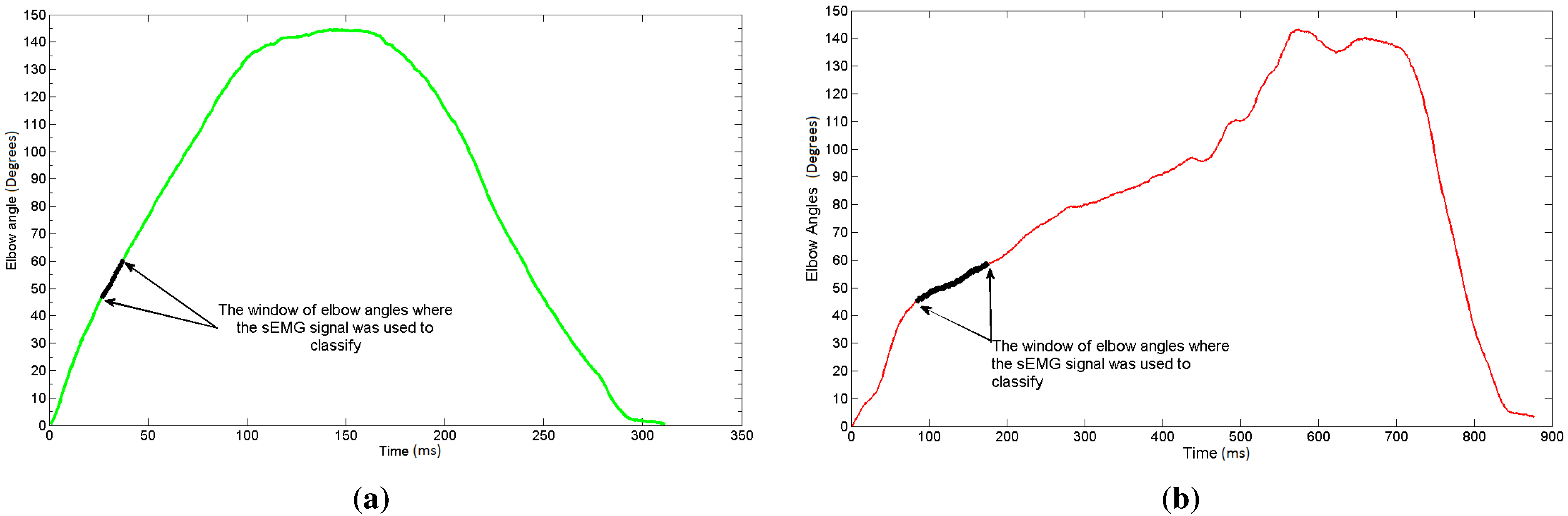
2.2. Labelling the Signals

The sEMG signals were labelled by dividing them into Fatigue and Non-Fatigue epochs. In the beginning of the tasks, when the subjects felt “fresh”, it was understood that the recorded signal indicated Non-Fatigue. Therefore the first repetition was labelled as Non-Fatigue, while the last full repetition was labelled as Fatigue. After the last full repetition, the subjects were unable to continue the set task, hence fatigue had occurred. This labelling method is adapted according to Kumar et al. [11]. The labelling of the signals was later used to train and test the classifier.

2.3. Genetic Algorithms

A Genetic Algorithms (GA) solves linear and nonlinear problems [33]. The GA utilised a pseudo-wavelet (previously evolved by our research group [23,29,30,31]) as the feature extraction technique to determine the optimal elbow angle from sEMG signals, which successfully distinguished between Non-Fatigue and Fatigue classes of the recorded signal. Table 1 displays the parameter settings for the GA runs.
Table 1. Parameter settings for the Genetic Algorithm (GA) runs.
ParameterValue
Independent runs26
Population size5000
Maximum number of generations20
Mutation probability10%
Crossover probability90%
Selection typeTournament, size 5
Termination criterionMaximum number of generations
The GA uses the fitness function to detect the best suited elbow angle in the search space. The modified Davies Bouldin Index (DBI) was used in the fitness function due to it being a simple and effective index. By using modified DBI [34] a calculation of data cluster linear overlap was done by determining the ratio of intracluster spread to intercluster centroid distance. For the DBI, the joint-time frequency decomposition of the pseudo-wavelet utilised every scale and was extracted in one second intervals. Smaller DBI values was an indication of excellent class separation. In this research, the hill climbing technique was reversed by changing the DBI to negative numbers, where the fitness function utilised the hill climbing technique by aiming to obtain the negative DBI closer to zero.

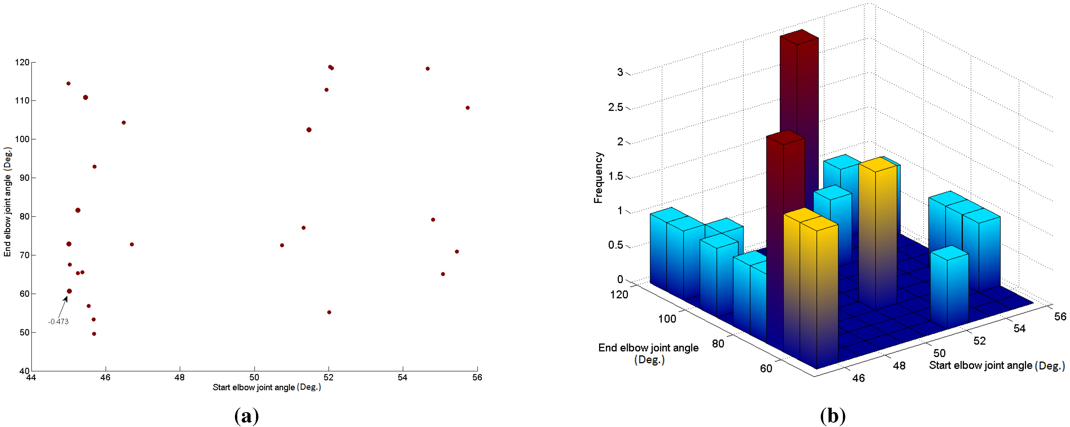
2.4. Evolved Elbow Angle Selection

To find the optimal elbow angle for fatigue detection in the biceps brachii, the GA used the pseudo-wavelet as the feature extraction technique. The pseudo-wavelet used in this study has been developed in previous studies [23,30]. A window of the sEMG signal was chosen based on the starting elbow angle and the ending elbow angle. The starting and ending elbow angle (a window) was decided by the evolutionary process of the GA, by testing the difference (DBI) of the two fatigue stages (Fatigue and Non-Fatigue). The testing facilitated the evolutionary processes by aiming to decrease the DBI, which then enabled the fitness function to maximise the separation between Fatigue and Non-Fatigue. As mentioned, the DBI was changed into negative numbers, enabling the fitness function to use the hill climbing method by trying to obtain (now) negative DBI numbers that are close to zero. The starting and ending angles that generated the best separation of the sEMG signal were later utilised in the classification stage.
Figure 1a displays a single rep when the muscle was fresh (Non-Fatigue) indicating the starting and ending joint angles for one of the subject trials using the best GA run. Figure 1b shows the same joint angles in a fatiguing rep.
Figure 1. (a) Elbow angles during Non-Fatigue rep; (b) Elbow angles during Fatiguing rep. Optimal window of joint angles selected by the GA showing where the Surface electromyographic (sEMG) signal was used for classification .
Computers 04 00251 g001

2.5. Classification

70% of the completed sEMG trials were used for training purposes to determine the optimal elbow angle for feature extraction in the evolutionary computation. The run with the best separation index was chosen, then used in the testing phase with the rest of the 30% of the sEMG signals to calculate the classification performance.
For a comparison between the evolved pseudo-wavelet and eight other feature extraction methods, LDA (linear discriminant analysis) was chosen as the classification method due to its simplicity, being well established and light on computational resources. The input for the training and testing of the LDA classifier was based on the eight parameters derived from the sEMG signal.

2.6. Feature Extraction Techniques

Different feature extraction methods were utilised for testing the optimal angle for the fatigue content in the sEMG signal. In the classification performance these eight features are used for comparison purposes. The eight feature extraction techniques selected for the comparison are:
  • Higher-order statistics (HOS) (HO2 and HO3 were used as they gave the best results.)
  • Mean Frequency (MF)
  • Median Frequency (MDF)
  • Power Spectrum Density (PSD)
  • Root Mean Square (RMS)
  • Daubechies 4 (Db4)
  • Mexican Hat (Mex H)
  • Pseudo-wavelet (p-w)
These feature extraction techniques have been used in several studies on localised muscle fatigue recorded using sEMG signals [11,35,36,37,38,39,40]. The wavelet based methods were used by decomposing the sEMG signal, and then mapping the change in the muscle power output, as conducted in the references [23,30].

3. Results

Table 2 displays the 26 evolutionary runs that generated the optimal (best separate Non-Fatigue and Fatigue) joint angle window with the optimal sEMG signal classification. In the table, the best run is highlighted, which was GA run 19. The window of the joint angles ranged from 45.02 to 60.63, giving the best DBI between Fatigue and Non-Fatigue with a separation index of −0.473.
Table 2. GA runs with the optimal Davies Bouldin Index (DBI).
GA RunElbow Joint 1Elbow Joint 2DBI
154.66118.27–0.491
254.8179.13–0.483
355.74108.17–0.486
455.0865.12–0.487
555.4670.93–0.491
650.7572.56–0.494
746.7172.78–0.483
851.3377.10–0.482
945.5556.87–0.492
1045.0172.87–0.475
1152.08118.39–0.503
1245.2581.60–0.474
1345.45110.82–0.478
1452.04118.76–0.503
1551.94112.88–0.491
1645.6949.57–0.498
1745.6853.35–0.481
1851.47102.53–0.478
1945.0260.63–0.473
2046.49104.29–0.505
2145.7092.98–0.490
2245.3765.55–0.498
2345.2665.36–0.494
2452.0155.24–0.491
2545.00114.42–0.500
2645.0367.56–0.506
Average49.0283.37–0.49
St. Dev4.0723.350.01
Figure 2 illustrates a scatter plot Figure 2a of the results shown in Table 2. Additionally, Figure 2b displays a histogram of the joint elbow angles, where the frequency of joint elbow angles chosen by all the GA runs are shown.
Figure 2. (a) A scatter plot of elbow angles selected by the GA. (larger dots indicate better separation); (b) 3D histogram of elbow joint angles selected by all the GA runs. Elbow joint angles selected by 26 evolutionary runs displayed in a scatter plot and a 3D histogram.
Computers 04 00251 g002
The classification performance for the 13 subjects based on the nine various parameters is displayed in Table 3. The findings shown in this table demonstrate that the elbow joint angles selected by the GA facilitated the classification of the two classes (Non-Fatigue and Fatigue) with remarkable performance based on the different features, giving a range from 72.95% and to 87.90% correct classification performance. In addition, the pseudo-wavelet was the parameter obtaining the best classification average, with the lowest standard deviation. The average classification accuracy for the pseudo-wavelet was substantially higher than the average classification accuracy for the other feature extraction techniques. The parameter giving the second highest average classification was also a wavelet transform (Db4).
Table 3. Classification performance.
SubjectsHO2HO3Mean FreqMedian FreqPSdRMSDb4Mexican HatP-W
183.5883.5888.0682.8480.6088.2986.5717.1690.99
294.8991.9794.8989.0590.5196.3593.4390.5194.16
379.6976.5678.9166.4174.2278.9185.1669.5385.94
468.5267.2882.7274.0764.2082.7283.9580.2588.27
574.1271.9380.7073.2571.4979.3983.7779.3981.58
686.7988.6891.5183.9686.7989.6290.5784.9193.40
775.1773.1583.8979.1971.1483.2277.1883.8982.55
873.3374.8171.1165.1975.5673.3379.2671.1187.41
991.1889.7188.2485.2991.1892.6594.1289.7192.65
1071.1968.6483.0574.5869.4981.3685.5977.9786.44
1159.4663.0663.9666.6762.1667.5781.9867.5793.69
1253.4453.0562.6053.8256.4964.5064.1253.0583.59
1369.1469.1482.7279.0166.6783.9582.1083.3382.10
Average75.4274.7480.9574.8773.8881.6883.6872.9587.90
St. Dev11.8711.329.819.8810.839.257.7219.654.67
Figure 3 illustrates a graphical representation of the classification performance displayed in Table 3.
Figure 3. Graphical representation of the classification performance.
Computers 04 00251 g003

4. Discussion

In this study the optimal elbow angle was determined by the GA. The GA utilised a window that gave highest separation between Non-fatigue and Fatigue content of the sEMG signals at different elbow angles. As the signal emanated from fatiguing dynamic contractions involving continuous movement of the elbow angle, a window of optimal elbow angle was chosen instead of only one specific elbow angle degree. The GA run with the best separation, run 19, used a window of elbow angles from 45.02 to 60.63 , while the average window had a range from 49.022 to 83.372 . This finding means that the fatigue content of the sEMG signal from dynamic contractions are found in the smaller elbow angles, which means at longer muscle length. This falls in line with previous research that discovered that with longer muscle length fatigue occurs more quickly [8,9,41].
This research produced excellent classification results for sEMG signal classification emanating from fatiguing dynamic contractions in the biceps brachii. The correct classification performance for the different features ranged from 72.95% and to 87.90%. These results fall in line with previous research on fining the optimal angle for MMG signals from dynamic contractions, where the classification performance depending on the feature ranged from 52.74% to 80.63% classification performance [31]. The comparison of these results show that the sEMG signal is performing better than the MMG signal, which is further discussed in another study [42].
The classification performance of the various feature extraction methods performed less well than the classification based on the GA using the pseudo-wavelet as a feature, although the classification average for the other common parameters were fairly good (more than 70%). The second best average classification performance was by Db4, which is also a wavelet function, indicating that wavelets functions enables good performance of sEMG fatigue classification from dynamic contraction. This observation may be due to the stochastic nature of the signals, as argued by some researchers [11,15].
Using the eight different feature extraction methods by the GA for classifying the sEMG signals suggests this method produced fairly high classification performance. Some of the time and frequency domain parameters, such as the RMS and mean frequency performed well (with an average of more than 80%).
Previous studies found that both the mean power frequency and RMS for sEMG signal changed in accordance to the elbow angle [41]. Other research found that maximal elbow angles would result in high RMS values in isometric contractions [10]. In addition, changes in the elbow angle (for isometric contractions) gives a shift in the median frequencies of the EMG signal at fatigue occurrence [5]. The findings in these previous studies, as well as the performance of the different feature extraction methods in this study, showed that these parameters are useful in finding the optimal angle. The PSD is giving one of the lowest classification averages, which may be due to the muscle length influencing the sEMG wave form, and therefore the elbow angle is a control mechanism when analysing the sEMG signal’s spectral indices [7].
The methodology used in this study is fairly new although it has been applied in previous research on MMG in finding the optimal elbow angle [31]. A previous study successfully used the GA to select the feature for reducing dimensionality in classifying sEMG signals [16]. This suggests that the approach in this study is useful in future research on sEMG classification.

5. Conclusions

This research showed it is possible to find the optimal elbow angle for fatigue classification recorded using sEMG signals from dynamic contractions. The GA, which utilised a pseudo-wavelet as a feature extraction method, gave good separation between the two classes of fatigue (Non-Fatigue and Fatigue). The pseudo-wavelet as a feature extraction method selected by the GA produced excellent classification results, even when compared to other traditional feature extraction methods used by the GA.

Acknowledgments

This work was supported and funded by Kuwait University Research Grant No. QS01/14.

Author Contributions

Mohamed R. Al-Mulla led the study, performed the experiments and data analysis and led the writing of the paper. Francisco Sepulveda made recommendations on the methodology towards compliance with scientific and ethical standards, provided the equipment for the experiments, and helped with the content and language in the paper manuscript. Bader Al-Bader helped with the contents, language and the graphics within the paper. All authors have read and approved the final manuscript.

Conflicts of Interest

The authors declare no conflict of interest.

References

  1. Garrett, W.E. Muscle strain injuries: Clinical and basic aspects. Med. Sci. Sports Exerc. 1990, 22, 436–443. [Google Scholar] [CrossRef] [PubMed]
  2. Edwards, R. Human muscle function and fatigue. Ciba Found. Symp. 1981, 82, 1–18. [Google Scholar] [PubMed]
  3. Lindstrom, L.; Kadefors, R.; Petersen, I. An electromyographic index for localized muscle fatigue. J. Appl. Physiol. 1977, 43, 750–754. [Google Scholar] [PubMed]
  4. Masuda, K.; Masuda, T.; Sadoyama, T.; Inaki, M.; Katsuta, S. Changes in surface EMG parameters during static and dynamic fatiguing contractions. J. Electromyogr. Kinesiol. 1999, 9, 39–46. [Google Scholar] [CrossRef]
  5. Ravier, P.; Buttelli, O.; Jennane, R.; Couratier, P. An EMG fractal indicator having different sensitivities to changes in force and muscle fatigue during voluntary static muscle contractions. J. Electromyogr. Kinesiol. 2005, 15, 210–221. [Google Scholar] [CrossRef] [PubMed]
  6. Onishi, H.; Yagi, R.; Oyama, M.; Akasaka, K.; Ihashi, K.; Handa, Y. EMG-angle relationship of the hamstring muscles during maximum knee flexion. J. Electromyogr. Kinesiol. 2002, 5, 399–406. [Google Scholar] [CrossRef]
  7. Okada, M. Effect of muscle length on surface EMG wave forms in isometric contractions. Eur. J. Appl. Physiol. Occup. Physiol. 1987, 56, 482–486. [Google Scholar] [CrossRef] [PubMed]
  8. Doud, J.R.; Walsh, J.M. Muscle fatigue and muscle length interaction: Effect on the EMG frequency components. Electromyogr. Clin. Neurophysiol. 1995, 35, 331–339. [Google Scholar] [PubMed]
  9. Nosaka, K.; Sakamoto, K. Effect of elbow joint angle on the magnitude of muscle damage to the elbow flexors. Med. Sci. Sports Exerc. 2001, 33, 22–29. [Google Scholar] [CrossRef] [PubMed]
  10. Oliveira, L.F.; Matta, T.T.; Alves, D.S.; Garcia, M.A.; Vieira, T.M. Effect of the shoulder position on the biceps brachii EMG in different dumbbell curls. J. Sports Sci. Med. 2009, 8, 24–29. [Google Scholar] [PubMed]
  11. Kumar, D.K.; Pah, N.D.; Bradley, A. Wavelet analysis of surface electromyography to determine muscle fatigue. IEEE Trans. Neural. Syst. Rehabil. Eng. 2003, 11, 400–406. [Google Scholar] [CrossRef] [PubMed]
  12. Bonato, P.; Roy, S.H.; Knaflitz, M.; de Luca, C.J. Time-frequency parameters of the surface myoelectric signal for assessing muscle fatigue during cyclic dynamic contractions. IEEE Trans. Neural. Syst. Rehabil. Eng. 2001, 48, 745–753. [Google Scholar] [CrossRef] [PubMed]
  13. Karlsson, S.; Yu, J.; Akay, M. Enhancement of spectral analysis of myoelectric signals during static contractions using wavelet methods. IEEE Trans.Biomed. Eng. 1999, 46, 670–684. [Google Scholar] [CrossRef] [PubMed]
  14. Sparto, P.J.; Parnianpour, M.; Barria, E.A.; Jagadeesh, J.M. Wavelet analysis of electromyography for back muscle fatigue detection during isokinetic constant-torque exertions. Spine 1999, 24, 1791–1798. [Google Scholar] [CrossRef] [PubMed]
  15. Khezri, M.; Jahed, M. Real-time intelligent pattern recognition algorithm for surface EMG signals. Biomed. Eng. Online 2007. [Google Scholar] [CrossRef] [PubMed]
  16. Wang, G.; Yan, Z.; Hu, X.; Xie, H.; Wang, Z. Classification of surface EMG signals using harmonic wavelet packet transform. Physiol. Meas. 2007, 27, 1255–1267. [Google Scholar] [CrossRef] [PubMed]
  17. Venugopal, G.; Navaneethakrishna, M.; Ramakrishnan, S. Extraction and analysis of multiple time window features associated with muscle fatigue conditions using semg signals. Expert Syst. Appl. 2014, 41, 2652–2659. [Google Scholar] [CrossRef]
  18. Li, Z.; Hayashibe, M.; Fattal, C.; Guiraud, D. Muscle fatigue tracking with evoked EMG via recurrent neural network: Toward personalized neuroprosthetics. Comput. Intell. Mag. IEEE 2014, 9, 38–46. [Google Scholar] [CrossRef]
  19. Moshou, D.; Hostens, I.; Papaioannou, G.; Ramon, H. Dynamic muscle fatigue detection using self-organizing maps. Appl. Soft Comput. 2005, 5, 391–398. [Google Scholar] [CrossRef]
  20. Kattan, A.; Al-Mulla, M.R.; Sepulveda, F.; Poli, R. Detecting localised muscle fatigue during isometric contraction using genetic programming. In Proceedings of the IJCCI, Madeira, Portugal, 5–7 October 2009; pp. 292–297.
  21. Al-Mulla, M.R.; Sepulveda, F.; Colley, M.; Kattan, A. Classification of localized muscle fatigue with genetic programming on sEMG during isometric contraction. In Proceedings of the Annual International Conference of the IEEE Engineering in Medicine and Biology Society EMBC, Minneapolis, MN, USA, 3–6 September 2009; pp. 2633–2638.
  22. Al-Mulla, M. Evolutionary computation extracts a super sEMG feature to classify localized muscle fatigue during dynamic contractions. In Proceedings of the Computer Science and Electronic Engineering Conference (CEEC), Essex, UK, 12–13 September 2012; pp. 220–224.
  23. Al-Mulla, M.R.; Sepulveda, F.; Colley, M. Evolved pseudo-wavelet function to optimally decompose sEMG for automated classification of localized muscle fatigue. Med. Eng. Phys. 2011, 33, 411–417. [Google Scholar] [CrossRef] [PubMed]
  24. Al-Mulla, M.R.; Sepulveda, F.; Colley, M.; Al-Mulla, F. Statistical class separation using sEMG features towards automated muscle fatigue detection and prediction. In Proceedings of the International Congress on Image and Signal Processing, Tianjin, China, 17–19 October 2009; pp. 1–5.
  25. Al-Mulla, M.R.; Sepulveda, F. A Novel Feature Assisting in the Prediction of sEMG Muscle Fatigue Towards a Wearable Autonomous System. In Proceedings of the 16th IEEE International Mixed-Signals, Sensors and Systems Test Workshop (IMS3TW’10), La Grande Motte, France, 7–9 June 2010.
  26. Al-Mulla, M.R.; Sepulveda, F. Novel feature modelling the prediction and detection of sEMG muscle fatigue towards an automated wearable system. Sensors 2010, 10, 4838–4854. [Google Scholar] [CrossRef] [PubMed]
  27. Al-Mulla, M.R.; Sepulveda, F. Predicting the time to localized muscle fatigue using ANN and evolved sEMG feature. In Proceedings of the IEEE International Conference on Autonomous and Intelligent Systems, (AIS 2010), Povoa de Varzim, Portugal, 21–23 July 2010; pp. 1–6.
  28. Al-Mulla, M.R.; Sepulveda, F.; Colley, M. An autonomous wearable system for predicting and detecting localised muscle fatigue. Sensors 2011, 11, 1542–1557. [Google Scholar] [CrossRef] [PubMed] [Green Version]
  29. Al-Mulla, M.R.; Sepulveda, F. Novel pseudo-wavelet function for MMG signal extraction during dynamic fatiguing contractions. Sensors 2014, 14, 9489–9504. [Google Scholar] [CrossRef] [PubMed]
  30. Al-Mulla, M.R.; Sepulveda, F. Super wavelet for sEMG signal extraction during dynamic fatiguing contractions. J. Med. Syst. 2015, 39, 1–9. [Google Scholar] [CrossRef] [PubMed]
  31. Al-Mulla, M.R.; Sepulveda, F.; Souod, M. Optimal Elbow Angle for MMG Signal Classification of Biceps Brachii during Dynamic Fatiguing Contraction. In Proceedings of the IWBBIO, International Work-Conference on Bioinformatics and Biomedical Engineering, Granada, Spain, 15–17 April 2015.
  32. Konrad, P. The ABC of EMG: A Practical Introduction to Kinesiological Electromyography; Noraxon, Inc.: Scottsdale, AZ, USA, 2005. [Google Scholar]
  33. Michalewicz, Z. Genetic Algorithms + Data Structures = Evolution Programs; Springer-Verlag: New York, NY, USA, 1996. [Google Scholar]
  34. Sepulveda, F.; Meckes, M.; Conway, B. Cluster separation index suggests usefulness of non-motor EEG channels in detecting wrist movement direction intention. In Proceedings of the IEEE Conference on Cybernetics and Intelligent Systems, Singapore, 1–3 December 2004; pp. 943–947.
  35. Merletti, R.; Farina, D.; Gazzoni, M. The linear electrode array: A useful tool with many applications. J. Electromyogr. Kinesiol. 2003, 13, 37–47. [Google Scholar] [CrossRef]
  36. De Luca, C.J. The use of surface electromyography in biomechanics. J. Appl. Biomech. 1997, 13, 135–163. [Google Scholar]
  37. Ortengren, R.; Andersson, G.; Broman, H.; Magnusson, R.; Petersen, I. Vocational electromyography: Studies of localized muscle fatigue at the assembly line. Ergonomics 1975, 18, 157–174. [Google Scholar] [CrossRef] [PubMed]
  38. Hagberg, M. Work load and fatigue in repetitive arm elevations. Ergonomics 1981, 24, 543–555. [Google Scholar] [CrossRef] [PubMed]
  39. Raez, M.B.; Hussain, M.S.; Mohd-Yasin, F. Techniques of EMG signal analysis: Detection, processing, classification and applications. Biol. Proc. Online 2006, 8, 11–35. [Google Scholar] [CrossRef] [PubMed]
  40. Walker, J.S. A Primer on Wavelets and Their Scientific Applications; Chapman and Hall/CRC: Boca Raton Fla, FL, USA, 2000. [Google Scholar]
  41. Mamghani, N.K.; Shimomura, Y.; Iwanaga, K.; Katsuur, T. Mechanomyogram and Electromyogram Responses of Upper Limb during Sustained Isometric Fatigue with Varying Shoulder and Elbow Postures. J. Physiol. Anthropol. 2002, 21, 29–43. [Google Scholar] [CrossRef]
  42. Al-Mulla, M.R.; Sepulveda, F.; Al-Bader, B. Optimal Elbow Angle for Extraction of sEMG and MMG Signals During Dynamic Fatiguing Contractions, Mobihealth. In Proceedings of the 5th EAI International Conference on Wireless Mobile Communication and Healthcare—Transforming Healthcare Through Innovations in Mobile And Wireless Technologies, London, UK, 14–16 October 2015. in press.

