
South Asian Medicinal Compounds as Modulators of Resistance to Chemotherapy and Radiotherapy

Abstract
:1. Introduction
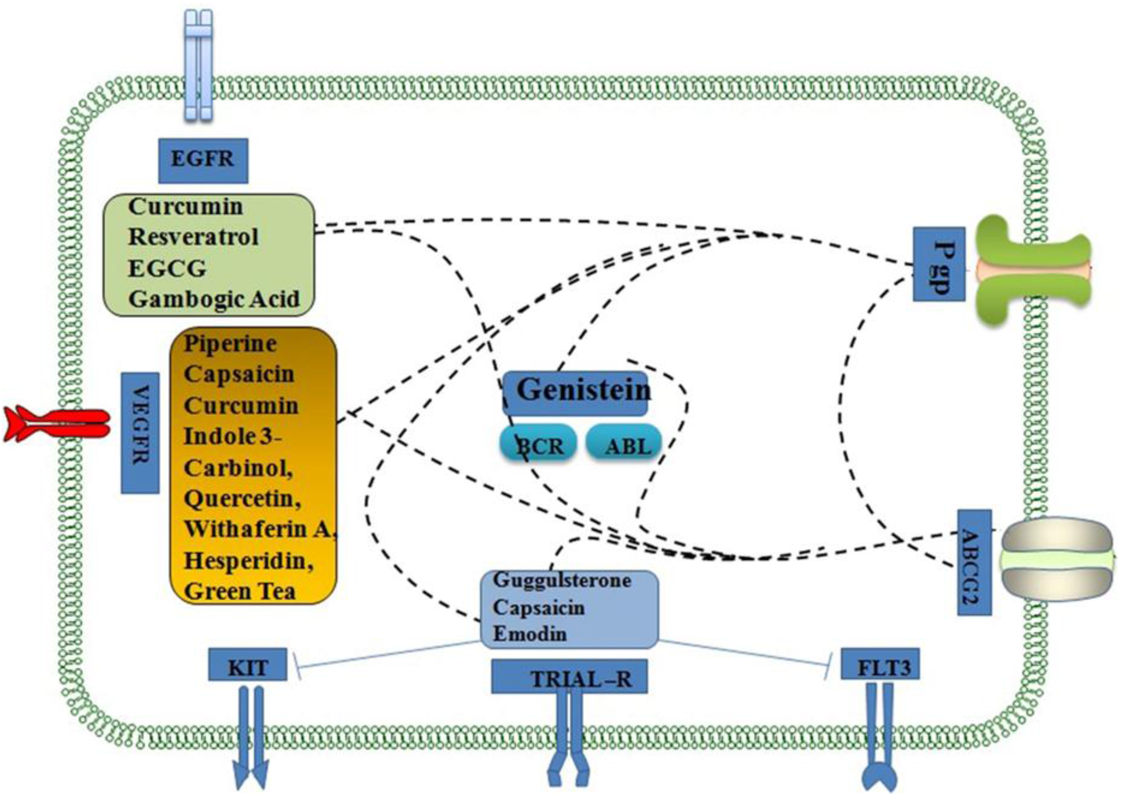
2. South Asian Medicinal Compounds as Chemosensitizers and Radiosensitizers
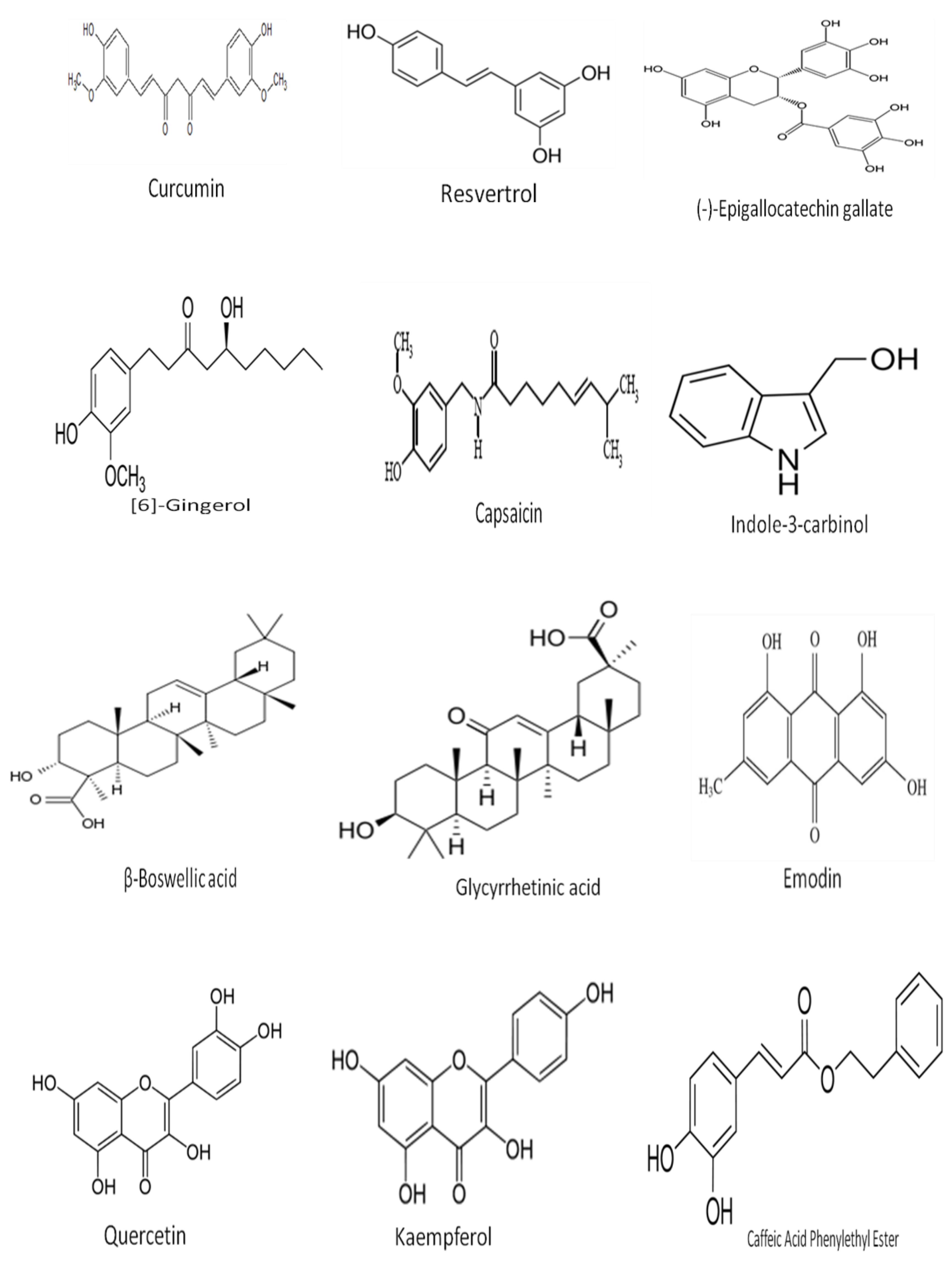
3. Phytochemicals as Chemosensitizers
4. Radioresistance
5. Radiosensitization
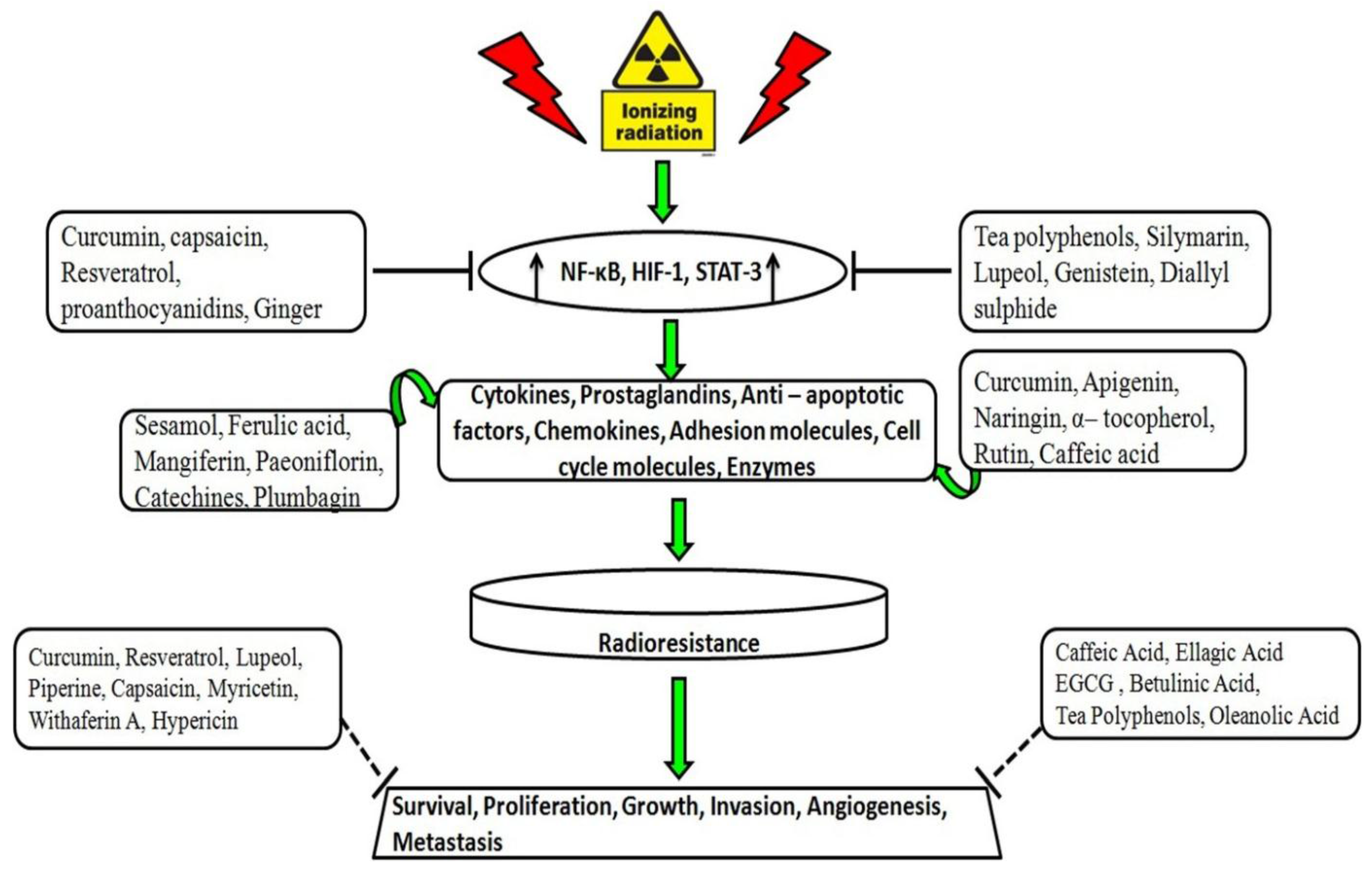
6. Radiosensitizing Properties of South Asian Medicinal Compounds
7. Radioprotective Effects of Natural Compounds
8. Conclusions
Acknowledgements
Author Contributions
Conflicts of Interest
References
- Jain, J.B.; Kumane, S.C.; Bhattacharya, S. Medicinal flora of Madhya Pradesh and Chhatisgarh-A review. Indian J. Tradit. Know. 2006, 5, 237–242. [Google Scholar]
- Garg, A.; Darokar, M.P.; Sundaresan, V.; Faridi, U.; Luqman, S.R.; Khanuja, S.P.S. Anticancer activity of some medicinal plants from high altitude evergreen elements of Indian Western Ghats. J. Res. Educ. Indian Med. 2007, 13, 1–6. [Google Scholar]
- Molnar, J.; Engi, H.; Hohmann, J.; Molnar, P.; Deli, J.; Wesolowska, O.; Michalak, K.; Wang, Q. Reversal of multidrug resitance by natural substances from plants. Curr. Top. Med. Chem. 2010, 10, 1757–1768. [Google Scholar] [PubMed]
- Zhang, S.; Morris, M.E. Effects of the flavonoids biochanin A, morin, phloretin, and silymarin on P-glycoprotein-mediated transport. J. Pharm. Exp. Ther. 2003, 304, 1258–1267. [Google Scholar] [CrossRef] [PubMed]
- Aggarwal, B.B.; Ichikawa, H.; Garodia, P.; Weerasinghe, P.; Bhatt, I.D.; Pandey, M.K.; Shishodia, S.; Nair, M.G. From traditional Ayurvedic medicine to modern medicine: Identification of therapeutic targets for suppression of inflammation and cancer. Expert. Opin. Ther. Targets 2006, 10, 87–118. [Google Scholar] [CrossRef] [PubMed]
- Dhar, M.L.; Dhar, M.M.; Dhawan, B.N.; Mehrotra, B.N.; Ray, C. Screening of Indian plants for biological activity: I. Indian. J. Exp. Biol. 1968, 6, 232–247. [Google Scholar] [PubMed]
- Singh, S.S.; Pandey, S.C.; Srivastava, S.; Gupta, V.S.; Patro, B.; Ghosh, A.C. Chemistry and medicinal properties of Tinospora cordifolia (Guduchi). Indian J. Pharmacol. 2003, 35, 83–91. [Google Scholar]
- Bharti, A.C.; Donato, N.; Aggarwal, B.B. Curcumin (diferuloylmethane) inhibits constitutive and IL-6-inducible STAT3 phosphorylation in human multiple myeloma cells. J. Immunol. 2003, 171, 3863–3871. [Google Scholar] [CrossRef] [PubMed]
- Anto, R.J.; Mukhopadhyay, A.; Denning, K.; Aggarwal, B.B. Curcumin (diferuloylmethane) induces apoptosis through activation of caspase-8, BID cleavage and cytochrome c release: Its suppression by ectopic expression of Bcl-2 and Bcl-xl. Carcinogenesis 2002, 23, 143–150. [Google Scholar] [CrossRef] [PubMed]
- Mukhopadhyay, A.; Bueso-Ramos, C.; Chatterjee, D.; Pantazis, P.; Aggarwal, B.B. Curcumin downregulates cell survival mechanisms in human prostate cancer cell lines. Oncogene 2001, 20, 7597–7609. [Google Scholar] [CrossRef] [PubMed]
- Bible, K.C.; Kaufmann, S.H. Flavopiridol: A cytotoxic flavone that induces cell death in noncycling A549 human lung carcinoma cells. Cancer Res. 1996, 56, 4856–4861. [Google Scholar] [PubMed]
- Manna, S.K.; Mukhopadhyay, A.; Aggarwal, B.B. Resveratrol suppresses TNF-induced activation of nuclear transcription factors NF-kappa B, activator protein-1, and apoptosis: Potential role of reactive oxygen intermediates and lipid peroxidation. J. Immunol. 2000, 164, 6509–6519. [Google Scholar] [CrossRef] [PubMed]
- Kim, D.M.; Koo, S.Y.; Jeon, K.; Kim, M.H.; Lee, J.; Hong, C.Y.; Jeong, S. Rapid induction of apoptosis by combination of flavopiridol and tumor necrosis factor (TNF)-alpha or TNF-related apoptosis-inducing ligand in human cancer cell lines. Cancer Res. 2003, 63, 621–626. [Google Scholar] [PubMed]
- Murakami, A.; Takahashi, D.; Kinoshita, T.; Koshimizu, K.; Kim, H.W.; Yoshihiro, A.; Nakamura, Y.; Jiwajinda, S.; Terao, J.; Ohigashi, H. Zerumbone, a Southeast Asian ginger sesquiterpene, markedly suppresses free radical generation, proinflammatory protein production, and cancer cell proliferation accompanied by apoptosis: The alpha,beta-unsaturated carbonyl group is a prerequisite. Carcinogenesis 2002, 23, 795–802. [Google Scholar] [CrossRef] [PubMed]
- Kirtikar, K.R.; Basu, B.D. Indian Medicinal Plant; Lalit Mohan Publication: Calcutta, India, 1935. [Google Scholar]
- Takada, Y.; Andreeff, M.; Aggarwal, B.B. Indole-3-carbinol suppresses NF-kappaB and Ikappa-β alpha kinase activation, causing inhibition of expression of NF-kappaB-regulated antiapoptotic and metastatic gene products and enhancement of apoptosis in myeloid and leukemia cells. Blood 2005, 106, 641–649. [Google Scholar] [CrossRef] [PubMed]
- Behera, B.; Devi, K.S.; Mishra, D.; Maiti, S.; Maiti, T.K. Biochemical analysis and antitumour effect of Abrus precatorius agglutinin derived peptides in Ehrlich's ascites and B16 melanoma mice tumour model. Environ. Toxicol. Phar. 2014, 38, 288–296. [Google Scholar] [CrossRef] [PubMed]
- Dhiman, A.; Hiremath, S.K.; Pathak, M.; Mishra, D. A review article on anticancerous drugs in ayurveda and screened anticancer activity of medicinal plants. Int. J. Ayu. Alt. Med. 2014, 2, 54–60. [Google Scholar]
- Rai, M.; Jogee, P.S.; Agarkar, G.; Santos, C.A. Anticancer activities of Withania somnifera: Current research, formulations, and future perspectives. Pharm. Biol. 2015, 7, 1–9. [Google Scholar]
- Nasri, H.; Baradaran, A.; Shirzad, H.; Rafieian-Kopaei, M. New concepts in nutraceuticals as alternative for pharmaceuticals. Int. J. Prev. Med. 2014, 5, 1487–1499. [Google Scholar] [PubMed]
- Di Domenico, F.; Foppoli, C.; Coccia, R.; Perluigi, M. Antioxidants in cervical cancer: Chemopreventive and chemotherapeutic effects of polyphenols. Biochim. Biophys. Acta 2012, 1822, 737–747. [Google Scholar] [CrossRef] [PubMed]
- Baguley, B.C. Multidrug resistance in cancer. Methods Mol. Biol. 2010, 596, 1–14. [Google Scholar] [PubMed]
- Cort, A.; Ozben, T. Natural product modulators to overcome multidrug resistance in cancer. Nutr. Cancer 2015, 67, 411–423. [Google Scholar] [CrossRef] [PubMed]
- Chorawala, M.R.; Oza, P.M.; Shah, G.B. Mechanisms of anticancer drugs resistance: An overview. Int. J. Pharm. Sci. Drug Res. 2012, 4, 1–9. [Google Scholar]
- Dean, M.; Hamon, Y.; Chimini, G. The human ATP-binding cassette (ABC) transporter super family. J. Lipid Res. 2001, 42, 1007–1017. [Google Scholar] [PubMed]
- Coley, H.M. Overcoming multidrug resistance in cancer: Clinical studies of p-glycoprotein inhibitors. Methods Mol. Biol. 2010, 596, 341–358. [Google Scholar] [PubMed]
- Gottesman, M.M.; Ambudkar, S.V.; Xia, D. Structure of a multidrug transporter. Nat. Biotechnol. 2009, 27, 546–547. [Google Scholar] [CrossRef] [PubMed]
- Krishna, R.; Mayer, L.D. Multidrug resistance (MDR) in cancer: Mechanisms, reversal using modulators of MDR and the role of MDR modulators in influencing the pharmacokinetics of anticancer drugs. Eur. J. Pharm. Sci. 2000, 11, 265–283. [Google Scholar] [CrossRef]
- Kitagawa, S. Inhibitory Effects of Polyphenols on P-Glycoprotein-Mediated Transport. Biol. Pharm. Bull. 2006, 29, 1–6. [Google Scholar] [CrossRef] [PubMed]
- Chavan, S.S.; Damale, M.G.; Shamkuwar, P.B.; Pawar, D.P. Traditional Medicinal Plants for anticancer activity. Int. J. Curr. Pharm. Res. 2013, 5, 50–54. [Google Scholar]
- Shishodia, S.; Potdar, P.; Gairola, C.G.; Aggarwal, B.B. Curcumin (diferuloylmethane) downregulates cigarette smoke-induced NFkappaB activation through inhibition of IkappaBalpha kinase in human lung epithelial cells: Correlation with suppression of COX-2, MMP-9 and cyclin D1. Carcinogenesis 2003, 24, 1269–1279. [Google Scholar] [CrossRef] [PubMed]
- Bharti, A.; Takada, Y.; Aggarwal, B. Curcumin (diferuloylmethane) inhibits receptor activator of NF-kappa B ligand-induced NF-kappa B activation in osteoclast precursors and suppresses osteoclastogenesis. J. Immunol. 2004, 172, 5940–5947. [Google Scholar] [CrossRef] [PubMed]
- Bharti, A.C.; Shishodia, S.; Reuben, J.M.; Weber, D.; Alexanian, R.; Raj-Vadhan, S.; Estrov, Z.; Talpaz, M.; Aggarwal, B.B. Nuclear factor-kappaB and STAT3 are constitutively active in CD138+ cells derived from multiple myeloma patients, and suppression of these transcription factors leads to apoptosis. Blood 2004, 103, 3175–3184. [Google Scholar] [CrossRef] [PubMed]
- Iersel, M.L.; Ploemen, J.P.; Struik, I.; van Amersfoort, C.; Keyzer, A.E.; Schefferlie, J.G.; van Bladeren, P.J. Inhibition of glutathione S-transferase activity in human melanoma cells by alpha,beta-unsaturated carbonyl derivatives. Effects of acrolein, cinnamaldehyde, citral, crotonaldehyde, curcumin, ethacrynic acid, and trans-2-hexenal. Chem. Biol. Interact. 1996, 102, 117–132. [Google Scholar] [CrossRef]
- Duvoix, A.; Morceau, F.; Delhalle, S.; Schmitz, M.; Schnekenburger, M.; Galteau, M.M.; Dicato, M.; Diederich, M. Induction of apoptosis by curcumin: Mediation by glutathione S-transferase P1-1 inhibition. Biochem. Pharmacol. 2003, 66, 1475–1483. [Google Scholar] [CrossRef]
- Liang, C.; Li, H.; Shen, C.; Lai, J.; Shi, Z.; Liu, B.; Tao, H.M. Genistein potentiates the anti-cancer effects of gemcitabine in human osteosarcoma via the downregulation of Akt and nuclear factor-kappaB pathway. Anticancer Agents Med. Chem. 2012, 12, 554–563. [Google Scholar] [CrossRef] [PubMed]
- Zhang, B.; Shi, Z.L.; Liu, B.; Yan, X.B.; Feng, J.; Tao, H.M. Enhanced anticancer effect of gemcitabine by genistein in osteosarcoma: The role of Akt and nuclear factor-kappaB. Anticancer Drugs 2010, 21, 288–296. [Google Scholar] [CrossRef] [PubMed]
- Park, S.Y.; Seol, D.W. Regulation of Akt by EGF-R inhibitors, a possible mechanism of EGF-R inhibitor-enhanced TRAIL-induced apoptosis. Biochem. Biophys. Res. Commun. 2002, 295, 515–518. [Google Scholar] [CrossRef]
- Jian, L.; Xie, L.P.; Lee, A.H.; Binns, C.W. Protective effect of green tea against prostate cancer: A case-control study in southeast China. Int. J. Cancer 2004, 108, 130–135. [Google Scholar] [CrossRef] [PubMed]
- Du, G.; Lin, H.; Wang, M.; Zhang, S.; Wu, X.; Lu, L.; Ji, L.; Yu, L. Quercetin greatly improved therapeutic index of doxorubicin against 4T1 breast cancer by its opposing effects on HIF-1alpha in tumor and normal cells. Cancer Chemother. Pharmacol. 2010, 65, 277–287. [Google Scholar] [CrossRef] [PubMed]
- Zhang, L.; Hung, M.C. Sensitization of HER-2/neuoverexpressing non-small cell lung cancer cells to chemotherapeutic drugs by tyrosine kinase inhibitor emodin. Oncogene 1996, 12, 571–576. [Google Scholar] [PubMed]
- Li, J.; Liu, P.; Mao, H.; Wanga, A.; Zhang, X. Emodin sensitizes paclitaxel-resistant human ovarian cancer cells to paclitaxel-induced apoptosis in vitro. Oncol. Rep. 2009, 21, 1605–1610. [Google Scholar] [CrossRef] [PubMed]
- Huang, X.Z.; Wang, J.; Huang, C.; Chen, Y.Y.; Shi, G.Y.; Hu, Q.S.; Yi, J. Emodin enhances cytotoxicity of chemotherapeutic drugs in prostate cancer cells: The mechanisms involve ROS-mediated suppression of multidrug resistance and hypoxia inducible factor-1. Cancer Biol. Ther. 2008, 7, 468–475. [Google Scholar] [CrossRef] [PubMed]
- Zhang, L.; Lau, Y.K.; Xia, W.; Hortobagyi, G.N.; Hung, M.C. Tyrosine kinase inhibitor emodin suppresses growth of HER-2/neu-overexpressing breast cancer cells in athymic mice and sensitizes these cells to the inhibitory effect of paclitaxel. Clin. Cancer Res. 1999, 5, 343–353. [Google Scholar] [PubMed]
- Sallman, D.A.; Chen, X.; Zhong, B.; Gilvary, D.L.; Zhou, J.; Wei, S.; Djeu, J.Y. Clusterin mediates TRAIL resistance in prostate tumor cells. Mol. Cancer Ther. 2007, 6, 2938–2947. [Google Scholar] [CrossRef] [PubMed]
- Ivanov, V.N.; Partridge, M.A.; Johnson, G.E.; Huang, S.X.; Zhou, H.; Hei, T.K. Resveratrol sensitizes melanomas to TRAIL through modulation of antiapoptotic gene expression. Exp. Cell Res. 2008, 314, 1163–1176. [Google Scholar] [CrossRef] [PubMed]
- Shankar, S.; Chen, Q.; Siddiqui, I.; Sarva, K.; Srivastava, R.K. Sensitization of TRAIL-resistant LNCaP cells by resveratrol (3, 4’, 5 tri-hydroxystilbene): Molecular mechanisms and therapeutic potential. J. Mol. Signal 2007. [Google Scholar] [CrossRef] [PubMed]
- Jazirehi, A.R.; Bonavida, B. Resveratrol modifies the expression of apoptotic regulatory proteins and sensitizes non-Hodgkin’s lymphoma and multiple myeloma cell lines to paclitaxel-induced apoptosis. Mol. Cancer Ther. 2004, 3, 71–84. [Google Scholar] [PubMed]
- Quan, F.; Pan, C.; Ma, Q.; Zhang, S.; Yan, L. Reversal effect of resveratrol on multidrug resistance in KBv200 cell line. Biomed. Pharmacother. 2008, 62, 622–629. [Google Scholar] [CrossRef] [PubMed]
- Rezk, Y.A.; Balulad, S.S.; Keller, R.S.; Bennett, J.A. Use of resveratrol to improve the effectiveness of cisplatin and doxorubicin: Study in human gynecologic cancer cell lines and in rodent heart. Am. J. Obstet Gynecol. 2006, 194, 23–26. [Google Scholar] [CrossRef] [PubMed]
- Rigolio, R.; Miloso, M.; Nicolini, G.; Villa, D.; Scuteri, A.; Simone, M.; Tredici, G. Resveratrol interference with the cell cycle protects human neuroblastoma SH-SY5Y cell from paclitaxel-induced apoptosis. Neurochem. Int. 2005, 46, 205–211. [Google Scholar] [CrossRef] [PubMed]
- Scarlatti, F.; Sala, G.; Ricci, C.; Maioli, C.; Milani, F.; Minella, M.; Botturi, M.; Ghidoni, R. Resveratrol sensitization of DU145 prostate cancer cells to ionizing radiation is associated to ceramide increase. Cancer Lett. 2007, 253, 124–130. [Google Scholar] [CrossRef] [PubMed]
- Pirzadeh, S.; Fakhari, S.; Jalili, A.; Mirzai, S.; Ghaderi, B.; Haghshenas, V. Glycyrrhetinic acid induces apoptosis in Leukemic HL60 cells through upregulating of CD95/ CD178. Int. J. Mol. Cell Med. 2014, 3, 272–278. [Google Scholar] [PubMed]
- Jung, G.R.; Kim, K.J.; Choi, C.H.; Lee, T.B.; Han, S.I.; Han, H.K.; Lim, S.C. Effect of betulinic acid on anticancer drug-resistant colon cancer cells. Basic Clin. Pharmacol. Toxicol. 2007, 101, 277–285. [Google Scholar] [CrossRef] [PubMed]
- Nabekura, T.; Hiroi, T.; Kawasaki, T.; Uwai, Y. Effects of natural nuclear factor-kappa B inhibitors on anticancer drug efflux transporter human P-glycoprotein. Biomed. Pharmacother. 2015, 70, 140–145. [Google Scholar] [CrossRef] [PubMed]
- Suttana, W.; Mankhetkorn, S.; Poompimon, W.; Palagani, A.; Zhokhov, S.; Gerlo, S.; Haegeman, G.; Berghe, W.V. Differential chemosensitization of P-glycoprotein overexpressing K562/Adr cells by withaferin A and Siamois polyphenols. Mol. Cancer 2010. [Google Scholar] [CrossRef] [PubMed] [Green Version]
- Ikegawa, T.; Ushigome, F.; Koyabu, N.; Morimoto, S.; Shoyama, Y.; Naito, M.; Tsuruo, T.; Ohtani, H.; Sawada, Y. Inhibition of P-glycoprotein by orange juice components, polymethoxyflavones in adriamycin-resistant human myelogenous leukemia (K562/ADM) cells. Cancer Lett. 2000, 160, 21–28. [Google Scholar] [CrossRef]
- Ikegawa, T.; Ohtani, H.; Koyabu, N.; Juichi, M.; Iwase, Y.; Ito, C.; Furukawa, H.; Naito, M.; Tsuruo, T.; Sawada, Y. Inhibition of P-glycoprotein by flavonoid derivatives in adriamycin-resistant human myelogenous leukemia (K562/ADM) cells. Cancer Lett. 2002, 177, 89–93. [Google Scholar] [CrossRef]
- Mitsunaga, Y.; Takanaga, H.; Matsuo, H.; Naito, M.; Tsuruo, T.; Ohtani, H.; Sawada, Y. Effect of bioflavonoids on vincristine transport across blood-brain barrier. Eur. J. Pharmacol. 2000, 395, 193–201. [Google Scholar] [CrossRef]
- Chieli, E.; Romiti, N.; Cervelli, F.; Tongiani, R. Effects of flavonols on Pglycoprotein activity in cultured rat hepatocytes. Life Sci. 1995, 57, 1741–1751. [Google Scholar] [CrossRef]
- Shin, S.C.; Choi, J.S.; Li, X. Enhanced bioavailability of tamoxifen after oral administration of tamoxifen with quercetin in rats. Int. J. Pharm. 2006, 313, 144–149. [Google Scholar] [CrossRef] [PubMed]
- Choi, J.S.; Jo, B.W.; Kim, Y.C. Enhanced paclitaxel bioavailability after oral administration of paclitaxel or prodrug to rats pretreated with quercetin. Eur. J. Pharm. Biopharm. 2004, 57, 313–318. [Google Scholar] [CrossRef] [PubMed]
- Limtrakul, P.; Khantamat, O.; Pintha, K. Inhibition of P-glycoprotein function and expression by kaempferol and quercetin. J. Chemother. 2005, 17, 86–95. [Google Scholar] [CrossRef] [PubMed]
- Sharma, V.; Joseph, C.; Ghosh, S.; Agarwal, A.; Mishra, M.K.; Sen, E. Kaempferol induces apoptosis in glioblastoma cells through oxidative stress. Mol. Cancer Ther. 2007, 6, 2544–2553. [Google Scholar] [CrossRef] [PubMed]
- Wang, S.; Chen, R.; Zhong, Z.; Shi, Z.; Chen, M.; Wang, Y. Epigallocatechin-3-gallate potentiates the effect of curcumin in inducing growth inhibition and apoptosis of resistant breast cancer cells. Am. J. Chin. Med. 2014, 42, 1279–1300. [Google Scholar] [CrossRef] [PubMed]
- Qian, F.; Wei, D.; Zhang, Q.; Yang, S. Modulation of P-glycoprotein function and reversal of multidrug resistance by (−)-epigallocatechin gallate in human cancer cells. Biomed. Pharmacother. 2005, 59, 64–69. [Google Scholar] [CrossRef] [PubMed]
- Jodoin, J.; Demeule, M.; Beliveau, R. Inhibition of the multidrug resistance P-glycoprotein activity by green tea polyphenols. Biochim. Biophys. Acta 2002, 1542, 149–159. [Google Scholar] [CrossRef]
- Castro, A.F.; Altenberg, G.A. Inhibition of drug transport by genistein in multidrugresistant cells expressing P-glycoprotein. Biochem. Pharmacol. 1997, 53, 89–93. [Google Scholar] [CrossRef]
- Sak, K. Cytotoxicity of dietary flavonoids on different human cancer types. Pharmacogn. Rev. 2014, 8, 122–146. [Google Scholar] [CrossRef] [PubMed]
- Luo, H.; Daddysman, M.K.; Rankin, G. O.; Jiang, B.H. Kaempferol enhances cisplatin’s effect on ovarian cancer cells through promoting apoptosis caused by down regulation of cMyc. Cancer Cell Int. 2010. [Google Scholar] [CrossRef] [PubMed]
- Piao, Y.; Shin, S.C.; Choi, J.S. Effects of oral kaempferol on the pharmacokinetics of tamoxifen and one of its metabolites, 4-hydroxytamoxifen, after oral administration of tamoxifen to rats. Biopharm. Drug Dispos. 2008, 29, 245–249. [Google Scholar] [CrossRef] [PubMed]
- Chen, H.; Landen, C.N.; Li, Y.; Alvarez, R.D.; Tollefsbol, T.O. Epigallocatechin gallate and sulforaphane combination treatment induce apoptosis in paclitaxel-resistant ovarian cancer cells through hTERT and Bcl-2 down-regulation. Exp. Cell Res. 2013, 319, 697–706. [Google Scholar] [CrossRef] [PubMed]
- Luo, T.; Wang, J.; Yin, Y.; Hua, H.; Jing, J.; Sun, X.; Li, M.; Zhang, Y.; Jiang, Y. (−)-Epigallocatechin gallate sensitizes breast cancer cells to paclitaxel in a murine model of breast carcinoma. Breast Cancer Res. 2010. [Google Scholar] [CrossRef] [PubMed]
- Park, S.; Kim, J.H.; Hwang, Y.I.; Jung, K.S.; Jang, Y.S.; Jang, S.H. Schedule-dependent effect of Epigallocatechin-3-Gallate (EGCG) with paclitaxel on H460 cells. Tuberc. Respir Dis. (Seoul) 2014, 76, 114–119. [Google Scholar] [CrossRef] [PubMed]
- Rak, S.; Cimbora-Zovko, T.; Gajski, G.; Dubravčić, K.; Domijan, A.M.; Delaš, I.; Garaj-Vrhovac, V.; Batinić, D.; Sorić, J.; Osmak, M. Carboplatin resistant human laryngeal carcinoma cells are cross resistant to curcumin due to reduced curcumin accumulation. Toxicol. In Vitro 2013, 27, 523–532. [Google Scholar] [CrossRef] [PubMed] [Green Version]
- Sreenivasan, S.; Krishnakumar, S. Synergistic effect of curcumin in combination with anticancer agents in human retinoblastoma cancer cell lines. Curr. Eye Res. 2015, 40, 1153–1165. [Google Scholar] [CrossRef] [PubMed]
- Saha, S.; Adhikary, A.; Bhattacharyya, P.; Das, T.; Sa, G. Death by design: Where curcumin sensitizes drug-resistant tumours. Anticancer Res. 2012, 32, 2567–2584. [Google Scholar] [PubMed]
- Cridge, B.J.; Larsen, L.; Rosengren, R.J. Curcumin and its derivatives in breast cancer: Current developments and potential for the treatment of drug-resistant cancers. Oncol. Discov. 2013. [Google Scholar] [CrossRef]
- Ganta, S.; Amiji, M. Coadministration of Paclitaxel and curcumin in nanoemulsion formulations to overcome multidrug resistance in tumor cells. Mol. Pharmacol. 2009, 6, 928–939. [Google Scholar] [CrossRef] [PubMed]
- Limtrakul, P.; Anuchapreeda, S.; Buddhasukh, D. Modulation of human multidrug-resistance MDR-1 gene by natural curcuminoids. BMC Cancer 2004. [Google Scholar] [CrossRef] [PubMed]
- Anuchapreeda, S.; Leechanachai, P.; Smith, M.M.; Ambudkar, S.V.; Limtrakul, P.N. Modulation of P-glycoprotein expression and function by curcumin in multidrug-resistant human KB cells. Biochem. Pharmacol. 2002, 64, 573–578. [Google Scholar] [CrossRef]
- Wang, P.; Yang, H.L.; Yang, Y.J.; Wang, L.; Lee, S.C. Overcome cancer cell drug resistance Using natural products. Evid. Based Complement. Alternat. Med. 2015. [Google Scholar] [CrossRef] [PubMed]
- Yunos, N.M.; Beale, P.; Yu, J.Q.; Huq, F. Synergism from sequenced combinations of curcumin and epigallocatechin-3-gallate with cisplatin in the killing of human ovarian cancer cells. Anticancer Res. 2011, 31, 1131–1140. [Google Scholar] [PubMed]
- Cromie, M.M.; Gao, W. Epigallocatechin-3-gallate enhances the therapeutic effects of leptomycin B on human lung cancer a549 cells. Oxid. Med. Cell Longev. 2015. [Google Scholar] [CrossRef] [PubMed]
- Frampton, G.A.; Lazcano, E.A.; Li, H.; Mohamad, A.; DeMorrow, S. Resveratrol enhances the sensitivity of cholangiocarcinoma to chemotherapeutic agents. Lab. Invest. 2010, 90, 1325–1338. [Google Scholar] [CrossRef] [PubMed]
- Lee, S.H.; Koo, B.S.; Park, S.Y.; Kim, Y.M. Anti-angiogenic effects of resveratrol in combination with 5-fluorouracil on B16 murine melanoma cells. Mol. Med. Rep. 2015, 12, 2777–2783. [Google Scholar] [CrossRef] [PubMed]
- Kweon, S.H.; Song, J.H.; Kim, T.S. Resveratrol-mediated reversal of doxorubicin resistance in acute myeloid leukemia cells via downregulation of MRP1 expression. Biochem. Biophys. Res. Commun. 2010, 395, 104–110. [Google Scholar] [CrossRef] [PubMed]
- Hsieh, T.C.; Wu, J.M. Resveratrol: Biological and pharmaceutical properties as anticancer molecule. Biofactors 2010, 36, 360–369. [Google Scholar] [CrossRef] [PubMed]
- Cavaliere, V.; Papademetrio, D.L.; Lombardo, T.; Costantino, S.N.; Blanco, G.A.; Alvarez, E.M. Caffeic acid phenylethyl ester and MG132, two novel nonconventional chemotherapeutic agents, induce apoptosis of human leukemic cells by disrupting mitochondrial function. Target Oncol. 2014, 9, 25–42. [Google Scholar] [CrossRef] [PubMed]
- Sugiyama, T.; Sadzuka, Y. Combination of theanine with doxorubicin inhibits hepatic metastasis of M5076 ovarian sarcoma. Clin. Cancer Res. 1999, 5, 413–416. [Google Scholar] [PubMed]
- Liang, Y.R.; Liu, C.; Xiang, L.P.; Zheng, X.Q. Health Benefits of Theanine in Green Tea: A Review. Trop. J. Pharm. Res. 2015, 14, 1943–1949. [Google Scholar] [CrossRef]
- Bhardwaj, R.K.; Glaeser, H.; Becquemont, L.; Klotz, U.; Gupta, S.K.; Fromm, M.F. Piperine, a major constituent of black pepper, inhibits human P-glycoprotein and CYP3A4. J. Pharmacol. Exp. Ther. 2002, 302, 645–650. [Google Scholar] [CrossRef] [PubMed]
- Meral, O.; Alpay, M.; Kismali, G.; Kosova, F.; Cakir, D.U.; Pekcan, M.; Yigit, S.; Sel, T. Capsaicin inhibits cell proliferation by cytochrome c release in gastric cancer cells. Tumour Biol. 2014, 35, 6485–6492. [Google Scholar] [CrossRef] [PubMed]
- Xia, Q.; Wang, Z.Y.; Li, H.Q.; Diao, Y.T.; Li, X.L.; Cui, J.; Chen, X.L.; Li, H. Reversion of p-glycoprotein-mediated multidrug resistance in human leukemic cell line by diallyl trisulfide. Evid. Based Complement. Alternat. Med. 2012. [Google Scholar] [CrossRef] [PubMed]
- Arora, A.; Seth, K.; Shukla, Y. Reversal of P-glycoprotein-mediated multidrug resistance by diallyl sulfide in K562 leukemic cells and in mouse liver. Carcinogenesis 2004, 25, 941–949. [Google Scholar] [CrossRef] [PubMed]
- Rao, P.S.S.; Midde, N.M.; Miller, D.D.; Chauhan, S.; Kumar, A.; Kumar, S. Diallyl Sulfide: Potential Use in Novel Therapeutic Interventions in Alcohol, Drugs, and Disease Mediated Cellular Toxicity by Targeting Cytochrome P450 2E1. Curr. Drug Metab. 2015, 16, 486–503. [Google Scholar] [CrossRef] [PubMed]
- Nabekura, T.; Yamaki, T.; Ueno, K.; Kitagawa, S. Inhibition of P-glycoprotein and multidrug resistance protein 1 by dietary phytochemicals. Cancer Chemother. Pharmacol. 2008, 62, 867–873. [Google Scholar] [CrossRef] [PubMed]
- Nabekura, T. Overcoming multidrug resistance in human cancer cells by natural compounds. Toxins (Basel). 2010, 2, 1207–1224. [Google Scholar] [CrossRef] [PubMed]
- Bueno Pérez, L.; Pan, L.; Sass, E.; Gupta, S.V.; Lehman, A.; Kinghorn, A.D.; Lucas, D.M. Potentiating effect of the flavonolignan (-)-hydnocarpin in combination with vincristine in a sensitive and P-gp-expressing acute lymphoblastic leukemia cell line. Phytother. Res. 2013, 27, 1735–1738. [Google Scholar] [CrossRef] [PubMed]
- Febriansah, R.; Putri, D.D.; Sarmoko; Nurulita, N.A.; Meiyanto, E.; Nugroho, A.E. Hesperidin as a preventive resistance agent in MCF-7 breast cancer cells line resistance to doxorubicin. Asian Pac. J. Trop. Biomed. 2014, 4, 228–233. [Google Scholar] [CrossRef]
- Nygren, P.; Glimelius, B. The Swedish Council on Technology assessment in Health Care (SBU) report on Cancer Chemotherapy––Project objectives, the working process, key definitions and general aspects on cancer trial methodology and interpretation. Acta Oncol. 2001, 40, 155–165. [Google Scholar] [PubMed]
- Impicciatore, G.; Sancilio, S.; Miscia, S.; Di Pietro, R. Nutlins and ionizing radiation in cancer therapy. Curr. Pharm. Des. 2010, 16, 1427–1442. [Google Scholar] [CrossRef] [PubMed]
- Sun, L.; Cabarcas, S.M.; Farrar, W.L. Radioresistance and Cancer Stem Cells: Survival of the Fittest. J. Carcinogene Mutagene 2011, 4172, 2157–2518. [Google Scholar]
- Biard, D.S.; Martin, M.; Rhun, Y.L.; Duthu, A.; Lefaix, J.L.; May, P. Concomitant p53 gene mutation and increased radiosensitivity in rat lung embryo epithelial cells during neoplastic development. Cancer Res. 1994, 54, 3361–3364. [Google Scholar] [PubMed]
- Sklar, M.D. The ras oncogene increases the intrinsic resistance of NIH 3T3 cells to ionizing radiation. Science 1988, 239, 645–647. [Google Scholar] [CrossRef] [PubMed]
- Kasid, U.; Pfeifer, A.; Weichselbaum, R.R.; Dritschilo, A.; Mark, G.E. The raf oncogene is associated with a radiation resistant human laryngeal cancer. Science 1987, 237, 1039–1041. [Google Scholar] [CrossRef] [PubMed]
- Lee, J.U.; Hosotani, R.; Wada, M.; Doi, R.; Kosiba, T.; Fujimoto, K.; Miyamoto, Y.; Tsuji, S.; Nakajima, S.; Nishimura, Y.; et al. Role of bcl-2 family proteins (Bax, Bcl-2, Bcl-X) on cellular susceptibility to radiation in pancreatic cancer cells. Eur. J. Cancer 1999, 35, 1374–1380. [Google Scholar] [CrossRef]
- Asanuma, K.; Moriai, R.; Yajima, T.; Yagihashi, A.; Yamada, M.; Kobayashi, D.; Watanabe, N. Survivin as a radioresistant factor in pancreatic cancer. Jpn. J. Cancer Res. 2000, 91, 1204–1209. [Google Scholar] [CrossRef] [PubMed]
- Bao, S.; Wu, Q.; McLendon, R.E.; Hao, Y.; Shi, Q.; Hjelmeland, A.B.; Dewhirst, M.W.; Bigner, D.D.; Rich, J.N. Glioma stem cells promote radioresistance by preferential activation of the DNA damage response. Nature 2006, 444, 756–760. [Google Scholar] [CrossRef] [PubMed]
- Zhang, S.; Wang, L.; Liu, H.; Zhao, G.; Ming, L. Enhancement of recombinant myricetin on the radiosensitivity of lung cancer A549 and H1299 cells. Diagn. Pathol. 2014. [Google Scholar] [CrossRef] [PubMed]
- Sheehan, J.P.; Shaffrey, M.E.; Gupta, B.; Larner, J.; Rich, J.N.; Park, D.M. Improving the radiosensitivity of radioresistant and hypoxic glioblastoma. Future Oncol. 2010, 6, 1591–1601. [Google Scholar] [CrossRef] [PubMed]
- Zhang, M.; Chakravarti, A. Novel radiationenhancing agents in malignant gliomas. Semin. Radiat. Oncol. 2006, 16, 29–37. [Google Scholar] [CrossRef] [PubMed]
- Henriksson, R.; Malmstrom, A.; Bergstrom, P.; Bergh, G.; Trojanowski, T.; Andreasson, L.; Blomquist, E.; Jonsborg, S.; Edekling, T.; Salander, P.; et al. High-grade astrocytoma treated concomitantly with estramustine and radiotherapy. J. Neurooncol. 2006, 78, 321–326. [Google Scholar] [CrossRef] [PubMed]
- Sartor, C.I. Mechanisms of disease: Radiosensitization by epidermal growth factor receptor inhibitors. Nat. Clin. Pract. Oncol. 2004, 1, 80–87. [Google Scholar] [CrossRef] [PubMed]
- Mehta, M.P.; Shapiro, W.R.; Phan, S.C.; Gervais, R.; Carrie, C.; Chabot, P.; Patchell, R.A.; Glantz, M.J.; Recht, L.; Langer, C.; et al. Motexafin gadolinium combined with prompt whole brain radiotherapy prolongs time to neurologic progression in non-small-cell lung cancer patients with brain metastases: Results of a Phase III trial. Int. J. Radiat. Oncol. Biol. Phys. 2009, 73, 1069–1076. [Google Scholar] [CrossRef] [PubMed]
- Criswell, T.; Leskov, K.; Miyamoto, S.; Luo, G.; Boothman, D.A. Transcription factors activated in mammalian cells after clinically relevant doses of ionizing radiation. Oncogene 2003, 22, 5813–5827. [Google Scholar] [CrossRef] [PubMed]
- Deorukhkar, A.; Krishnan, S.; Sethi, G.; Aggarwal, B.B. Back to basics: How natural products can provide the basis for new therapeutics. Expert Opin. Investig. Drugs 2007, 16, 1753–1773. [Google Scholar] [CrossRef] [PubMed]
- Fabricant, D.S.; Farnsworth, N.R. The value of plants used in traditional medicine for drug discovery. Environ. Health Perspect. 2001, 109, 69–75. [Google Scholar] [CrossRef] [PubMed]
- Donehower, R.C. The clinical development of paclitaxel: A successful collaboration of academia, industry and the National Cancer Institute. Stem Cells 1996, 14, 25–28. [Google Scholar] [CrossRef] [PubMed]
- Shao, R.G.; Cao, C.X.; Zhang, H.; Kohn, K.W.; Wold, M.S.; Pommier, Y. Replication-mediated DNA damage by camptothecin induces phosphorylation of RPA by DNA-dependent protein kinase and dissociates RPA:DNA-PK complexes. EMBO J. 1999, 18, 1397–1406. [Google Scholar] [CrossRef] [PubMed]
- Chen, Y.; Pandya, K.J.; Feins, R.; Johnstone, D.W.; Watson, T.; Keng, P.C. Toxicity profile and pharmacokinetic study of a phase I low-dose schedule-dependent radiosensitizing paclitaxel chemoradiation regimen for inoperable non-small-cell lung cancer. Int. J. Radiat. Oncol. Biol. Phys. 2008, 71, 407–413. [Google Scholar] [CrossRef] [PubMed]
- Kurdoglu, B.; Cheong, N.; Guan, J.; Corn, B.W.; Curran, W.J., Jr.; Iliakis, G. Apoptosis as a predictor of paclitaxel-induced radiosensitization in human tumor cell lines. Clin. Cancer Res. 1999, 5, 2580–2587. [Google Scholar] [PubMed]
- Girdhani, S.; Bhosle, S.M.; Thulsidas, S.A.; Kumar, A.; Mishra, K.P. Potential of radiosensitizing agents in cancer chemoradiotherapy. J. Cancer Res. Ther. 2005, 1, 129–131. [Google Scholar] [PubMed]
- Arora, R.; Gupta, D.; Chawla, R.; Sagar, R.; Sharma, A.; Kumar, R.; Prasad, J.; Singh, S.; Samanta, N.; Sharma, R.K. Radioprotection by plant products: Present status and future prospects. Phytother. Res. 2005, 19, 1–22. [Google Scholar] [CrossRef] [PubMed]
- Gault, N.; Lefaix, J.L. Infrared microspectroscopic characteristics of radiation-induced apoptosis in human lymphocytes. Radiat. Res. 2003, 160, 238–250. [Google Scholar] [CrossRef] [PubMed]
- Hosseinimehr, S.J. Flavonoids and genomic instability induced by ionizing radiation. Drug Discov. Today 2010, 15, 907–918. [Google Scholar] [CrossRef] [PubMed]
- Jebahi, S.; Saoudi, M.; Farhat, L.; Oudadesse, H.; Rebai, T.; Kabir, A.; El Feki, A.; Keskes, H. Effect of novel curcumin-encapsulated chitosan-bioglass drug on bone and skin repair after gamma radiation: Experimental study on a Wistar rat model. Cell Biochem. Funct. 2015, 33, 150–159. [Google Scholar] [CrossRef] [PubMed]
- Rajendra Prasad, N.; Menon, V.P.; Vasudev, V.; Pugalendi, K.V. Radioprotective effect of sesamol on γ-radiation induced DNA damage, lipid peroxidation and antioxidants levels in cultured human lymphocytes. Toxicology 2005, 209, 225–235. [Google Scholar] [CrossRef] [PubMed]
- Khan, S.; Kumar, A.; Adhikari, J.S.; Rizvi, M.A.; Chaudhury, N.K. Protective effect of sesamol against Co γ-ray-induced hematopoietic and gastrointestinal injury in C57BL/6 male mice. Free Radic. Res. 2015, 49, 1344–1361. [Google Scholar] [CrossRef] [PubMed]
- Rajendra Prasad, N.; Srinivasan, M.; Pugalendi, K.V.; Menon, V.P. Protective effect of ferulic acid on γ-radiation-induced micronuclei, dicentric aberration and lipid peroxidation in human lymphocytes. Mutat. Res. 2006, 603, 129–134. [Google Scholar] [CrossRef] [PubMed]
- Maurya, D.K.; Devasagayam, T.P. Ferulic acid inhibits gamma radiation-induced DNA strand breaks and enhances the survival of mice. Cancer Biother. Radiopharm. 2013, 28, 51–57. [Google Scholar] [CrossRef] [PubMed]
- Sharma, N.K. Modulation of radiation-induced and mitomycin C-induced chromosome damage by apigenin in human lymphocytes in vitro. J. Radiat. Res. 2013, 54, 789–797. [Google Scholar] [CrossRef] [PubMed]
- Rodeiro, I.; Delgado, R.; Garrido, G. Effects of a Mangifera indica L. stem bark extract and mangiferin on radiation-induced DNA damage in human lymphocytes and lymphoblastoid cells. Cell Prolif. 2014, 47, 48–55. [Google Scholar] [CrossRef] [PubMed]
- Manna, K.; Das, U.; Das, D.; Kesh, S.B.; Khan, A.; Chakraborty, A.; Dey, S. Naringin inhibits gamma radiation-induced oxidative DNA damage and inflammation, by modulating p53 and NF-κB signaling pathways in murine splenocytes. Free Radic. Res. 2015, 49, 422–439. [Google Scholar] [CrossRef] [PubMed]
- Li, C.R.; Zhou, Z.; Zhu, S.; Sun, Y.N.; Dai, J.N.; Wang, S.Q. Protective effect of paeoniflorin on irradiation-induced cell damage involved in modulation of reactive oxygen species and the mitogen-activated protein kinases. Int. J. Biochem. Cell Biol. 2007, 39, 426–438. [Google Scholar] [CrossRef] [PubMed]
- Yu, J.; Zhu, X.; Qi, X.; Che, J.; Cao, B. Paeoniflorin protects human EA.hy926 endothelial cells against gamma-radiation induced oxidative injury by activating the NF-E2-related factor 2/heme oxygenase-1 pathway. Toxicol Lett. 2013, 218, 224–234. [Google Scholar] [CrossRef] [PubMed]
- Shimoi, K.; Masuda, S.; Shen, B.; Furugori, M.; Kinae, N. Radioprotective effects of antioxidative plant flavonoids in mice. Mutat. Res. 1996, 350, 153–161. [Google Scholar] [CrossRef]
- Rahul, C.; Chatterjee, S.; Sharma, D.; Sumit, G.; Prasad, V.; Arun, S.; Poduval, T.B. Immunomodulatory and radioprotective effects of lignans derived from fresh nutmeg mace (Myristica fragrans) in mammalian splenocytes. Inter. Immunopharm. 2008, 8, 661–669. [Google Scholar]
- Pradeep, K.; Ko, K.C.; Choi, M.H.; Kang, J.A.; Chung, Y.J.; Park, S.H. Protective effect of hesperidin, a citrus flavanoglycone, against γ-radiation-induced tissue damage in Sprague-Dawley rats. J. Med. Food. 2012, 15, 419–427. [Google Scholar] [CrossRef] [PubMed]
- Gomes, C.C.; Ramos-Perez, F.M.; Perez, D.E.; Novaes, P.D.; Bóscolo, F.N.; Almeida, S.M. Radioprotective effect of vitamin E in parotid glands: A morphometric analysis in rats. Braz. Dent. J. 2013, 24, 183–187. [Google Scholar] [CrossRef] [PubMed]
- Richi, B.; Kale, R.K.; Tiku, A.B. Radio-modulatory effects of green tea catechin EGCG on pBR322 plasmid DNA and murine splenocytes against gamma-radiation induced damage. Mutat. Res. 2012, 747, 62–70. [Google Scholar] [CrossRef] [PubMed]
- Nayak, V.; Uma Devi, P. Protection of mouse bone marrow against radiation induced chromosome damage and stem cell death by the Ocimum Flavonoids orientin and vicenin. Radiat. Res. 2005, 163, 165–171. [Google Scholar] [CrossRef] [PubMed]
- Ramadan, L.A.; Roushdy, H.M.; Abu Senna, G.M.; Amin, N.E.; EL-Deshw, O.A. Radioprotective effect of silymarin against radiation induced hepatotoxicity. Pharmacol. Res. 2002, 45, 447–454. [Google Scholar] [CrossRef] [PubMed]
- Rao, B.N.; Archana, P.R.; Aithal, B.K.; Rao, B.S. Protective effect of Zingerone, a dietary compound against radiation induced genetic damage and apoptosis in human lymphocytes. Eur. J. Pharmacol. 2011, 657, 59–66. [Google Scholar] [CrossRef] [PubMed]
- Patil, S.L.; Rao, N.B.; Somashekarappa, H.M.; Rajashekhar, K.P. Antigenotoxic potential of rutin and quercetin in Swiss mice exposed to gamma radiation. Biomed. J. 2014, 37, 305–313. [Google Scholar] [CrossRef] [PubMed]
- Saada, H.N.; Rezk, R.G.; Eltahawy, N.A. Lycopene protects the structure of the small intestine against gamma-radiation-induced oxidative stress. Phytother. Res. 2010. [Google Scholar] [CrossRef] [PubMed]
- Gandhi, N.M.; Maurya, D.K.; Salvi, V.; Kapoor, S.; Mukherjee, T.; Nair, C.K.K. Radioprotection of DNA by glycyrrhizic acid through scavenging free radicals. J. Radiat. Res. 2004, 45, 461–468. [Google Scholar] [CrossRef] [PubMed]
- Jagetia, G.C.; Venkatesha, V.A. Treatment of mice with stem bark extract of Aphanamixis polystachya reduces radiation-induced chromosome damage. Int. J. Radiat. Biol. 2006, 82, 197–209. [Google Scholar] [CrossRef] [PubMed]
- Zbikowska, H.M.; Antosik, A.; Szejk, M.; Bijak, M.; Nowak, P. A moderate protective effect of quercetin against γ-irradiation-and storage-induced oxidative damage in red blood cells for transfusion. Int. J. Radiat. Biol. 2014, 90, 1201–1210. [Google Scholar] [CrossRef] [PubMed]
- Zhang, R.; Kang, K.A.; Kang, S.S.; Park, J.W.; Hyun, J.W. Morin (2',3,4',5,7-pentahydroxyflavone) protected cells against γ-radiation-induced oxidative stress. Basic Clin. Pharmacol. Toxicol. 2011, 108, 63–72. [Google Scholar] [CrossRef] [PubMed]
- Parihar, V.K.; Dhawan, J.; Kumar, S.; Manjula, S.N.; Subramanian, G.; Unnikrishnan, M.K.; Rao, C.M. Free radical scavenging and radioprotective activity of dehydrozingerone against whole body gamma irradiation in Swiss albino mice. Chem. Biol. Interact. 2007, 170, 49–58. [Google Scholar] [CrossRef] [PubMed]
- Mozdarani, H.; Ghoraeian, P. Modulation of gamma-ray-induced apoptosis in human peripheral blood leukocytes by famotidine and vitamin C. Mutat. Res. 2008, 649, 71–78. [Google Scholar] [CrossRef] [PubMed]
- Burlage, F.R.; Roesink, J.M.; Faber, H.; Vissink, A.; Langendijk, J.A.; Kampinga, H.H.; Coppes, R.P. Optimum dose range for the amelioration of long term radiation-induced hyposalivation using prophylactic pilocarpine treatment. Radiother. Oncol. 2008, 86, 347–353. [Google Scholar] [CrossRef] [PubMed]
- Cho, J.H.; Hong, W.G.; Jung, Y.J.; Lee, J.; Lee, E.; Hwang, S.G.; Um, H.D.; Park, J.K. Γ-Ionizing radiation-induced activation of the EGFR-p38/ERK-STAT3/CREB-1-EMT pathway promotes the migration/invasion of non-small cell lung cancer cells and is inhibited by podophyllotoxin acetate. Tumour Biol. 2015. [Google Scholar] [CrossRef] [PubMed]
- Zhang, W.; Anker, L.; Law, R.E.; Hinton, D.R.; Gopalakrishna, R.; Pu, Q.; Gundimeda, U.; Weiss, M.H.; Couldwell, W.T. Enhancement of radiosensitivity in human malignant glioma cells by hypericin in vitro. Clin. Cancer Res. 1996, 2, 843–846. [Google Scholar] [PubMed]
- You, H.; Wei, L.; Sun, W.L.; Wang, L.; Yang, Z.L.; Liu, Y.; Zheng, K.; Wang, Y.; Zhang, W.J. The green tea extract epigallocatechin-3-gallate inhibits irradiation-induced pulmonary fibrosis in adult rats. Int. J. Mol. Med. 2014, 34, 92–102. [Google Scholar] [CrossRef] [PubMed]
- Byun, E.B.; Sung, N.Y.; Park, J.N.; Yang, M.S.; Park, S.H.; Byun, E.H. Gamma-irradiated resveratrol negatively regulates LPS-induced MAPK and NF-κB signaling through TLR4 in macrophages. Int. Immunopharmacol. 2015, 25, 249–259. [Google Scholar] [CrossRef] [PubMed]
- Seong, K.M.; Yu, M.; Lee, K.S.; Park, S.; Jin, Y.W.; Min, K.J. Curcumin mitigates accelerated aging after irradiation in Drosophila by reducing oxidative stress. Biomed Res. Int. 2015. [Google Scholar] [CrossRef] [PubMed]
- Kasten-Pisula, U.; Windhorst, S.; Dahm-Daphi, J.; Mayr, G.; Dikomey, E. Radiosensitization of tumour cell lines by the polyphenol Gossypol results from depressed double-strand break repair and not from enhanced apoptosis. Mol. Radiobiol. 2007, 83, 296–303. [Google Scholar] [CrossRef] [PubMed]
- Bache, M.; Zschornak, M.P.; Passin, S.; Kessler, J.; Wichmann, H.; Kappler, M.; Paschke, R.; Kaluđerović, G.N.; Kommera, H.; Taubert, H.; et al. Increased betulinic acid induced cytotoxicity and radiosensitivity in glioma cells under hypoxic conditions. Radiat. Oncol. 2011. [Google Scholar] [CrossRef] [PubMed]
- Nair, S.; Nair, R.R.; Srinivas, P.; Srinivas, G.; Pillai, M.R. Radiosensitizing Effects of Plumbagin in Cervical Cancer Cells Is Through Modulation of Apoptotic Pathway. Mol. Carcinog. 2008, 47, 22–33. [Google Scholar] [CrossRef] [PubMed]
- Yang, E.S.L.; Choi, M.J.; Kim, J.H.; Choi, K.S.; Kwon, T.K. Withaferin A enhances radiation-induced apoptosis in Caki cells through induction of reactive oxygen species, Bcl-2 downregulation and Akt inhibition. Chem. Biol. Interact. 2011, 190, 9–15. [Google Scholar] [CrossRef] [PubMed]
- Bhoslea, S.M.; Huilgola, N.G.; Mishra, K.P. Enhancement of radiation-induced oxidative stress and cytotoxicity in tumor cells by ellagic acid. Clin. Chim. Acta 2005, 359, 89–100. [Google Scholar] [CrossRef] [PubMed]
- Mansour, H.H.; Tawfik, S.S. Early treatment of radiation-induced heart damage in rats by caffeic acid phenethyl ester. Eur. J. Pharmacol. 2012, 692, 46–51. [Google Scholar] [CrossRef] [PubMed]
- Kim, I.G.; Kim, J.S.; Lee, J.H.; Cho, E.W. Genistein decreases cellular redox potential, partially suppresses cell growth in HL‑60 leukemia cells and sensitizes cells to γ‑radiation‑induced cell death. Mol. Med. Rep. 2014, 10, 2786–2792. [Google Scholar] [CrossRef] [PubMed]
- Puthli, A.; Tiwari, R.; Mishra, K.P. Biochanin A enhances the radiotoxicity in colon tumor cells in vitro. J. Environ. Pathol. Toxicol. Oncol. 2013, 32, 189–203. [Google Scholar] [CrossRef] [PubMed]
- Venier, N.A.; Colquhoun, A.J.; Sasaki, H.; Kiss, A.; Sugar, L.; Adomat, H.; Fleshner, N.E.; Klotz, L.H.; Venkateswaran, V. Capsaicin enhances the effect of radiation in prostate cancer through NF-κB suppression. Cancer Res. 2013. [Google Scholar] [CrossRef]
- Tak, J.K.; Lee, J.H.; Park, J.W. Resveratrol and piperine enhance radiosensitivity of tumor cells. BMB Rep. 2012, 45, 242–246. [Google Scholar] [CrossRef] [PubMed]
- Jin, Y.; Lyu, Y.; Tang, X.; Zhang, Y.; Chen, J.; Zheng, D.; Liang, Y. Lupeol enhances radiosensitivity of human hepatocellular carcinoma cell line SMMC-7721 in vitro and in vivo. Int. J. Radiat. Biol. 2015, 91, 202–208. [Google Scholar] [CrossRef] [PubMed]
- Wang, J.; Yu, M.; Xiao, L.; Xu, S.; Yi, Q.; Jin, W. Radiosensitizing effect of oleanolic acid on tumor cells through the inhibition of GSH synthesis in vitro. Oncol. Rep. 2013, 30, 917–924. [Google Scholar] [PubMed]


| Name of the Plant | Bioactive Compounds |
|---|---|
| Withania somnifera L. | WithaferinA, withanolide |
| Phyllanthus amarus | Nirtetralin, niranthrin, phyllanthin, phyltetralin |
| Aegiceras corniculatum L. | Embilins |
| Annona muricataLinn | Annomuricins, bullatacin |
| Cedrus deodara | (−)-Wikstromal, (−)-matairesinoland dibenzylbutyrolactol |
| Nothapodytes foetida Miers | Camptothecin, irinotecan |
| Boswellia serrata Roxb. | Boswellic acid, acetyl-β-boswellic acid, α-pinene |
| Andrographis paniculata | Andrographolide, betulin, betulinic acid |
| Centella asiatica L. | Asiaticoside, hydrocotyline, vallerine, pectic acid, stigmasterol, thankunosides and ascorbic acid |
| Coscinium fenestratum | Berberine, palmatine, 8-oxoprotoberberine, oxypalmatine, berberrubine |
| Amoora rohituka Roxb. | Amooranin, rohitukine |
| Trichopus zeylanicus | Glycophosphosphingolipids |
| Bruguiera gymnorrhizais L. | Diterpenoids and pimaren |
| Cuscuta reflexa Roxb. | Cuscutin, amarbelin, β-sterol, stigmasterol, kaempferol, dulcitol, myricetin, quercetin, coumarin and oleanolic acid |
| Dendrophthoe falcata (L.F.) | Quercetrin, catechin, gallic acid, chebulinic acid, oleonolic acid, β-amyrin-O-acetate, leucocynidin, β-sitosterol and stigmasterol |
| Dioscorea bulbifera L. | Kaempferol- 3,5-dimethyl ether, caryatin, (L)-catechin, myricetin, quercetin-3-O galactopyranoside, myricetin-3-O-galactopyranoside, diosbulbin B |
| Embelia ribes Burm. | Quercitol, diethylnitrosamine/Phenobarbital |
| Ficus benghalensis L. | Lupeol, psoralen and β-sisterol |
| Ficus religiosa L. | Quercetin and qyricetin, stigmasterol and β-sitosterol |
| Hibiscus tiliaceus L. | Stigmasterol β-sitosterol, quercetin, kaempferol |
| Jatropha gossypiifolia L. | Falodone and jatrophone |
| Vitex agnus-castus | Agnucastoside A, agnucastoside B and agnucastoside C, aucubin, agnuside, mussaenosidic acid |
| Mollugo pentaphylla L. | Apigenin and mollupentin, mmollugogenol A, mollugogenol B, mollugogenol D, oleanolic acid and β-sitosterol |
| Nelumbo nucifera Willd. | Liensinine, neferine, pronuciferine, isoliensinine, negferine, asimilobine, nuciferine, remrefidine, isoliensinine, myricetin, quercetin, leucocyanidin, kaempferol, astragalin |
| Nyctanthes arbor-tristis L. | Phenylpropanoid glycosides, carotenoid glucosides, phenyl-propanoid glycoside cardiac glycosides, polysaccharides, β-sitosterol, β-amyrin, hentriacontane benzoic acid, nyctanthic acid, friedelin, lupeol, oleanolic acid, 6β-hydroxylonganin alkaloids, phlobatanins, terpenoids |
| Eugenia singampattiana | 4-Hydroxybenzoic acid, caffeic acid, rutin, ferulic acid, coumaric acid, epigallocatechin gallate, quercetin, myricetin, and kaempferol |
| Solanum nigrum L. | Gentisic acid, luteolin, apigenin, kaempferol, m-coumaric acid, anthocyanidin, lunasin |
| Strychnos nux-vomica | Brucine, diaboline |
| Zingiber officinale | Gingerenonea, gingeols, shogaols, zingerone |
| Chloroxylon swietenia | Coumarins xanthyletin, xanthoxyletin and 7-demethylsuberosin |
| Podophyllum hexandrum B | Podophyllin, astragalin |
| Linum usitatissimum | Cynogenetic glycosides |
| Glycyrrhiza glabra | Glycyrrhizin |
| Catharanthus roseus | Vinblastine, vincristine, alstonine, ajmalicine and reserpine |
| Camellia sinensis | Epigallocatechin gallate |
| Aloe ferox, Aloe barbadenis | Aloe-emodin, emodin, aloin acemannan |
| Allium sativum | Alliin, allicin alliin, alliinase, S-allylcysteine, diallyldisulphide, diallyltrisulphide and methylallyltrisuphide. |
| Curcuma longa | Curcumin |
| Capsicum annuum | Capsaicin |
| Piper nigrum | Piperidine, piperine |
| Radioresistance | Chemoresistance |
|---|---|
| Tumor hypoxic condition | Increased drug efflux and decreased drug uptake |
| Increased cellular production of cellular antioxidants | Inactivation of apoptosis |
| Activation of certain proto-oncogenes, and stromal interactions | Increased drug metabolism and drug compartmentalization |
| Amplification of DNA repair genes | Increase in the repair of DNA damage |
| Cancer stem cells as contributors to radioresistance | Increased or altered the drug targets |
| Survival signals favoured by transcription factors | Survival signals favoured by transcription factors |
| Natural Products | Anticancer Drugs | Experimental Models | Mechanism of Action | Ref. |
|---|---|---|---|---|
| Morin | Doxorubicin | MCF-7 MDA435/LCC6 cells | Inhibit P-gp-mediated drug efflux and potentiate doxorubicin cytotoxicity in P-gp positive cells. | [4] |
| Vincristine | K562 and K562/ADM | Pentaethylmorin remarkably increased the drug uptake in MDR cells. | [57] | |
| Daunorubicin | Multidrug resistant human breast cancer cell lines | Increased [3H]daunorubicin accumulation in MDR breast cell lines. | [4] | |
| Doxorubicin | MCF-7 and MDA435/L | Potentiates doxorubicin cytotoxicity in MDA435/L cells. | [4] | |
| Biochanin A | Doxorubicin | MCF-7 MDA435/LCC6 cells | Biochanin A can potentiate doxorubicin cytotoxicity in Pgp positive cells | [4] |
| Quercetin | Vincristine | K562 and K562/ADM | Pentamethyl quercetin and pentaallylquercetin remarkably increase drug uptake | [58] |
| Vincristine | MBEC4 cells and ddY mice | Increased drug uptake in cells and enhanced brain-to-plasma concentration ratio in mice | [59] | |
| Doxorubicin | Cultured rat hepatocytes | Reduced drug retention with increase in its efflux | [60] | |
| Tamoxifen | Female SD rats | AUC, Ka, Cmax increased | [61] | |
| Paclitaxel | Male SD rats | AUC, Ka, Cmax increased | [62] | |
| Vinblastine and paclitaxel | MDR KB-V1 cells | Reduced P-gp expression and function. | [63] | |
| Phloretin | Doxorubicin | MCF-7 MDA435/LCC6 cells | Inhibit P-gp-mediated drug efflux; phloretin can potentiate doxorubicin cytotoxicity in P-gp positive cells. | [4] |
| Nobiletin | Vincristine | K562/ADM | Increased drug uptake in K562/ADM cells. | [57] |
| Chrysin | Vincristine | MBEC4 cells and ddY mice | Increased drug uptake in cells and enhanced brain-to-plasma concentration ratio in mice | [59] |
| Kaempferol | Doxorubicin | Cultured rat hepatocytes | Kaempferol potentiated the toxic effect of chemotherapeutic agent and decreasing the efflux of doxorubicin | [64] |
| (–)-Epigallocatechin gallate (EGCG) | Doxorubicin | P-gp over-expressing KB-C2 cells | Increased drug accumulation | [65,66] |
| Green tea polyphenols | Vinblastine | Multidrug-resistant cell line CH(R)C5 | Potentiates the vinblastine cytotoxicity in CH(R)C5 cells. | [67] |
| Genistein | Rhodamine 123 and daunorubicin | P-gp-expressing cells | Elevation in intracellular drug accumulation | [68] |
| Kaempferol | Vinblastine and paclitaxel | MDR KB-V1 cells | Reduced P-gp expression and function | [69,70] |
| Tamoxifen | male rats | AUC, Ka, Cmax increased | [71] | |
| Heptamethoxyflavone | vincristine | K562/ADM | Increased uptake of [3H] vincristine | |
| Phloretin, silymarin | Daunorubicin& doxorubicin | Multidrug resistant human breast cancer cell lines MCF- 7 and MDA435/L | Increased [3 H]Daunomycin accumulation & potentiated doxorubicin Cytotoxicity | [4] |
| EGCG | Paclitaxel | Breast cancer cells (4T1, MCF-7, and MDA-MB- 231) Female Balb/c mice (4T1) cells xenograft) and various carcinoma cells | Induces apoptosis and increased endoplasmic reticulum chaperone GRP78 expression in tumor tissues. Decreases PCNA immunostaining | [72,73,74] |
| Curcumin | Carboplatin | NSCLC cell line, A549 | Suppression of NF-κB via inhibition of the Akt/IKKα pathway and enhanced ERK1/2 activity | [75,76,77] |
| Paclitaxel | Cervical cancer cells | Down-regulation of paclitaxel-induced activation of NF-κB, Akt, and Bcl-2 | [78,79] | |
| Vincristine/ vinblastine | Multidrug resistant KB cells, human multiple myeloma cells | Down-regulated NF-κB or P-gp | [80,81,82] | |
| EGCG + Curcumin | Cisplatin | Ovarian cancer, A2780, A2780cisR and A2780ZD0473R cells | Lower concentrations and shorter time gap between the two treatments produces higher cytotoxic effects | [83,84] |
| Resveratrol | 5-FU | Chemoresistant cholangiocarcinoma tumor model and B16 murine melanoma cells | Down-regulates Cyp1b1 expression and suppresses cell growth and angiogenesis | [85,86] |
| Paclitaxel | nonHodgkin’s lymphoma and multiple myeloma cell lines, KBv200 | Down-regulation of Bcl-2 family members and MDR1/P-gp. Down-regulation of Bcl-2 family members and MDR1/P-gp. | [48] | |
| Doxorubicin or vincristine | human uterine cancer cells , doxorubicin-resistant acute myeloid leukemia cells | Down-regulation of MDR1/P-gp and Bcl-2 | [87,88] | |
| Caffeic Acid Phenylethyl Ester | Vincristine and Doxorubicine | PL104 cells | CAPE an enhancement of cell death | [89] |
| Green tea | Doxorubicin | M5076 sarcoma | Increase in accumulation of the antitumor agent | [90,91] |
| Withaferin A and Siamois | Doxorubicin | K562 and K562/Adr cells | Transcriptional inhibition of NF-κB-, AP1- and Nrf2- and overcome the P-gp-coupled attenuation of caspase-dependent apoptosis in K562/Adr cells | [56] |
| Piperine | Cyclosproine A | Human colon carcinoma cell line (Caco-2) | Piperine might affect disposition of drugs that are substrates for both P-glycoprotein and CYP3A4 | [92] |
| Capsaicin | 5-flourouracil | Gastric cancer cell line HGC-27 | Capsaicin has the potential to treat gastric carcinoma with 5-FU in vitro | [93] |
| Diallyltrisulfide | Doxorubicin | K562/A02 cells | Increased expression of Caspase-3 and down-regulation of NF-κB/p65, increasing intracellular adriamycin concentration and inducing apoptosis | [94] |
| Diallylsulfide | Vinblastine | K562 cells | Enhanced cytotoxic activity of vinblastine as well as other Vinca alkaloids | [95,96] |
| Emodin | Paclitaxel | MDA-MB-361, MDA-MB-453, BT-483, SKBr-3, and BT-474 cells | Sensitizes HER-2/neu-overexpressing breast cancer cells | [44] |
| Glycyrrhetinic acid | Daunorubicin | Human carcinoma KB-C2 cells and human MRP1 gene-transfected KB/MRP cells | Dual inhibitory effects on P-glycoprotein and MRP1 | [97,98] |
| (-)-Hydnocarpin | Vincristine | Acute lymphoblastic leukemia cell line | Hydnocarpin potentiating the effect of vincristine in a multidrug-resistant cell line | [99] |
| Hesperidin | Doxorubicin | MCF-7 cell line, doxorubicin resistant (MCF-7/Dox) cells | Co-chemotherapy application of doxorubicin and hesperidin on MCF-7/Dox cells showed synergism effect through inhibition of Pgp expression. | [100] |
| Name of the Hytochemicals | Concentration Studied | Radiation Dose | Radioprotective/Radiosensitizing Effect | References |
|---|---|---|---|---|
| (i) Radioprotective phytochemicals | ||||
| Curcumin | 50 µg/mL | 1.5 Gy | Curcumin-encapsulated bioglass-chitosan might have promising potential applications for wound healing resulting from gamma radiation. | [127] |
| Sesamol | 10 μg/mL | 4 Gy | (i) Renders protection on γ-radiation induced DNA damage, and antioxidants depletion in cultured human lymphocytes. | [128,129] |
| 100 mg/kg | 7.5 Gy | (ii) Acts as a single prophylactic dose protects hematopoietic and GI systems against γ-radiation-induced injury in mice. | ||
| Ferulic acid | 10 μg/mL | 4 Gy | Prevents γ-radiation-induced micronuclei and dicentric aberration in human lymphocytes. | [130,131] |
| 50 mg/kg | 4 Gy | Enhances the survival of mice possibly by decreasing DNA damage as examined by γH2AX foci, micronuclei formation, and comet assay. | ||
| Apigenin | 10 μg/mL | 3 Gy | Significantly reduced (p < 0.01) the frequency of mitomycin C-induced micronuclei. | [132] |
| Mangiferin | 5–25 μg/mL | 5 Gy | Protects against gamma radiation-induced DNA damage and acts as an antioxidant or pro-oxidant product | [133] |
| Naringin | 50 and 100 µM | 6 Gy | Prevents radiation-induced multiple cellular anomalies. | [134] |
| Paeoniflorin | 200 μg/mL | 4 Gy | Offers protection against radiation-induced cell damage through modulation of reactive oxygen species and the mitogen-activated protein kinases in thymocytes. | [135] |
| 50–200 µg/mL | 10 Gy | Protected EA.hy926 cells against radiation-induced injury through the Nrf2/HO-1 pathway. | [136] | |
| Luteolin | 10 μmol/kg b.wt. | 6 Gy | Radioprotective effects through antioxidative property in mice. | [137] |
| Lignans from Myristica fragrans | 500 µg/mL | 4.26 Gy | Radioprotection through immunomodulation in mammalian splenocytes. | [138] |
| Hesperidin | 50–100 mg/kg b.wt. | 5 Gy | Protects against γ-radiation-induced cellular damage and oxidative stress in rats. | [139] |
| α-tocopherol | 360 mg/kg b.wt. | 15 Gy | Radioprotective effect of Vitamin E in Parotid Glands in rats. | [140] |
| Catechines | 100 μM | 3 Gy | Protects pBR322 DNA under acellular conditions and normal splenocytes under cellular conditions, against γ-radiation-induced damage. | [141] |
| Orientin | 17.5 μM | 4 Gy | Promotes stem cell survival, exogenous spleen colony formation (CFU-S). | [142] |
| Silymarin | 50 mg/kg | 3 Gy | Protects experimental animals from radiation-induced hepatotoxicity. | [143] |
| Zingerone | 10 μg/mL | 2 Gy | Prevents radiation-induced genetic damage and apoptosis in human lymphocytes. | [144] |
| Rutin | 10 mg/kg b.wt. | 3 Gy | Mitigates radiation-induced mortality and cytogenetic damage, which attributes to scavenging of radiation-induced free radicals. | [145] |
| Lycopene | 5 mg/kg b.wt. | 6 Gy | Protects the small intestine against radiation-induced damage. | [146] |
| Glycyrrhizic acid | 4 mM | 1.25 Gy | Offers protection against γ-radiation-induced DNA damage to plasmid pBR322 in vitro, human peripheral blood leukocytes and bone marrow cells in vivo. | [147] |
| Naringin | 7.5 mg/kg b.wt. | 1–5 Gy | Protects mouse bone marrow cells against radiation-induced chromosomal aberrations and lipid peroxidation. | [148] |
| Quercetin | 2–50 μM | 30 Gy | Protects against radiation- and storage-induced oxidative damage to RBCs. | [149] |
| Morin | 25 μM | 10 Gy | Protects against oxidative stress induced by radiation via reduction of ROS and attenuation of the SEK1-JNK-AP-1 pathway. | [150] |
| Dehydrozingerone | 100 mg/kg b.wt. | 10 Gy | Exhibits radioprotective activity in whole body gamma irradiated Swiss albino mice through free radical scavenging. The DMF value was found to be 1.09. | [151] |
| Famotidine | 200 µg/mL | 4–12 Gy | Suppresses radiation-induced apoptosis with various doses of gamma-irradiation via radical scavenging and intracellular antioxidation mechanism. | [152] |
| Pilocarpine | 4 mg/kg b.wt. | 15 Gy | Amelioration of long term radiation-induced hyposalivation using prophylactic pilocarpine treatment | [153] |
| Podophyllotoxin | 10 nM | 10 Gy | Derivatives are used in combination with IR therapy and suggest that the EGFR–p38/ERK–STAT3/CREB-1–EMT pathway might be a useful target for suppressing metastasis. | [154] |
| (ii) Radiosensitizing phytochemicals | ||||
| Hypericin | 5 µM | 8 Gy | Enhancement of radiosensitivity in human malignant glioma cells. | [155] |
| Epigallocatechin-galate | 25 mg/kg b.wt. | 22 Gy | Inhibits irradiation-induced pulmonary fibrosis. | [156] |
| Resveratrol | 1 mg/mL | 70 kGy | Reduces toxicity and plays a potent role in the treatment of inflammatory disease. | [157] |
| Curcumin | 100 μM | 10 Gy | Acts as radiosensitizer through prooxidant mechanisms in cancer cells. | [158] |
| Gossypol | 2 μM | 6 Gy | Radiosensitization of tumour cell lines by depressig double-strand break repair mechanism. | [159] |
| Betulinic acid | 20 μM | 2 Gy | Induces cytotoxicity and radiosensitivity in glioma cells under hypoxic conditions. | [160] |
| Plumbagin | 750 nM | 2 Gy | Radiosensitizing effects in cervical cancer cells through modulation of apoptotic pathway. | [161] |
| Withaferin A | 4 μM | 10 Gy | Enhances radiation-induced apoptosis in Caki cells through induction of reactive oxygen species, Bcl-2 downregulation and Akt inhibition. | [162] |
| Ellagic acid | 100 μmol/L | 6 Gy | Enhances radiation-induced oxidative stress and subsequent cytotoxicity in tumor cells. | [163] |
| Caffeic acid | 10μmol/kgb.wt. | 7 Gy | Exhibits curable effects on gamma irradiation-induced cardiac-oxidative impairment in rats. | [164] |
| Genistein | 20 μM | 5 Gy | Acts as a prooxidant in HL-60 cells, increases ionizing radiation-induced cell cycle arrest and sensitivity to apoptotic cell death in human promyeloid leukemia HL-60 cells. | [165] |
| Myricetin | 25 µM | 2 Gy | Enhances radiosensitivity of lung cancer A549 and H1299 cells. | [114] |
| Biochanin A | 1–100 µM | 2 Gy | Enhances radiotoxicity in colon tumor cells. | [166] |
| Capsaicin | 1–10 µM | 1–8 Gy | Increases radiation effects in prostate cancer. | [167] |
| Piperine | 40 μM | 15 Gy | Enhances radiosensitivity of tumor cells through oxidative mechanism. | [168] |
| Lupeol | 30 μmol/L | 4 Gy | Enhances radiosensitivity of human hepatocellular carcinoma cell line SMMC-7721 in vitro and in vivo. | [169] |
| Oleanolic acid | 35 µg/mL | 250 Gy/min | Radiosensitizes tumor cells through the inhibition of GSH synthesis in vitro. | [170] |
© 2016 by the authors; licensee MDPI, Basel, Switzerland. This article is an open access article distributed under the terms and conditions of the Creative Commons by Attribution (CC-BY) license (http://creativecommons.org/licenses/by/4.0/).
Share and Cite
Prasad, N.R.; Muthusamy, G.; Shanmugam, M.; Ambudkar, S.V. South Asian Medicinal Compounds as Modulators of Resistance to Chemotherapy and Radiotherapy. Cancers 2016, 8, 32. https://doi.org/10.3390/cancers8030032
Prasad NR, Muthusamy G, Shanmugam M, Ambudkar SV. South Asian Medicinal Compounds as Modulators of Resistance to Chemotherapy and Radiotherapy. Cancers. 2016; 8(3):32. https://doi.org/10.3390/cancers8030032
Chicago/Turabian StylePrasad, N. Rajendra, Ganesan Muthusamy, Mohana Shanmugam, and Suresh V. Ambudkar. 2016. "South Asian Medicinal Compounds as Modulators of Resistance to Chemotherapy and Radiotherapy" Cancers 8, no. 3: 32. https://doi.org/10.3390/cancers8030032