Review

Hedgehog Signaling in the Maintenance of Cancer Stem Cells

by
Catherine R. Cochrane
1,2,
Anette Szczepny
1,2,
D. Neil Watkins
3,4,5 and
Jason E. Cain
1,2,*
1
Centre for Cancer Research, Hudson Institute of Medical Research, Clayton, Victoria 3168, Australia
2
Department of Molecular and Translational Science, Monash University, Clayton, Victoria 3168, Australia
3
The Kinghorn Cancer Centre, Garvan Institute of Medical Research, Darlinghurst, New South Wales 2010, Australia
4
UNSW Faculty of Medicine, Randwick, New South Wales 2031, Australia
5
Department of Thoracic Medicine, St Vincent's Hospital, Darlinghurst, New South Wales 2010, Australia
*
Author to whom correspondence should be addressed.
Cancers 2015, 7(3), 1554-1585; https://doi.org/10.3390/cancers7030851
Submission received: 22 June 2015 / Revised: 31 July 2015 / Accepted: 3 August 2015 / Published: 11 August 2015

Abstract

:
Cancer stem cells (CSCs) represent a rare population of cells with the capacity to self-renew and give rise to heterogeneous cell lineages within a tumour. Whilst the mechanisms underlying the regulation of CSCs are poorly defined, key developmental signaling pathways required for normal stem and progenitor functions have been strongly implicated. Hedgehog (Hh) signaling is an evolutionarily-conserved pathway essential for self-renewal and cell fate determination. Aberrant Hh signaling is associated with the development and progression of various types of cancer and is implicated in multiple aspects of tumourigenesis, including the maintenance of CSCs. Here, we discuss the mounting evidence suggestive of Hh-driven CSCs in the context of haematological malignancies and solid tumours and the novel strategies that hold the potential to block many aspects of the transformation attributed to the CSC phenotype, including chemotherapeutic resistance, relapse and metastasis.

Graphical Abstract

1. Introduction

Cancer is a term encompassing a broad spectrum of disease uniformly defined by uncontrolled growth and underpinned by genomic instability, leading to further genetic diversity and intratumoural cellular and functional heterogeneity. As a result, two mutually non-exclusive models have been proposed to account for this tumour heterogeneity: the stochastic or clonal evolution model and the cancer stem cell model [1]. The conventional stochastic model postulates that all cancer cells within a tumour adapt and evolve to produce genetically- and phenotypically-distinct tumourigenic cells [2]. In contrast, the cancer stem cell model suggests the existence of a small population of primitive tumour cells that share many properties with somatic stem cells and the capacity to evolve into all cell types within a tumour, termed cancer stem cells (CSCs) [3,4]. Despite an accumulation of experimental evidence supporting this latter model, strong debate has ensued over the existence of CSCs. This is, in part, attributed to controversy surrounding the cell of origin, lack of a universal CSC marker and the limitations of the experimental techniques used to isolate and functionally-characterize CSCs.
CSCs represent a fractional cell population that exhibits unlimited self-renewal potential, has the ability to give rise to all cell types within a tumour and is resistant to many traditional cancer therapies that affect the more differentiated tumour bulk cells [5]. Although the existence of CSCs remains highly controversial, the CSC hypothesis is of considerable clinical importance, potentially explaining tumour insensitivity to chemotherapies, disease progression and relapse. To date, the cellular mechanisms that regulate CSC maintenance are poorly understood, although mounting evidence has implicated key developmental signaling pathways, including Hedgehog, Wnt and Notch, whose roles in regulating embryonic and adult stem and progenitor cells are better defined [6].
Although critical during embryonic organogenesis and adult homeostasis following repair and injury [7,8], aberrant activation of the Hedgehog (Hh) pathway also controls multiple aspects of tumourigenesis [9]. Together with a major role in maintaining the self-renewing capacity of adult somatic stem cells [10,11], it is not surprising that the Hh signaling has been widely implicated in CSC function and maintenance. If CSCs are the driving force behind tumour maintenance and growth, then understanding the role that Hh plays in regulating CSCs is of vital importance. This review focuses on the evidence that exists in favour of the CSC model, specifically the role of the Hedgehog pathway in CSCs in a variety of haematological malignancies and solid tumours, and highlights the strategies that hold the potential to block many aspects of transformation attributed to the CSC phenotype, through inhibition of the Hh signaling pathway.

2. The Hedgehog Signaling Network

Hedgehog signaling involves a wide variety of cellular and molecular mechanisms, such as protein trafficking, protein-protein interactions, positive and negative feedback loops and post-translational modifications, including phosphorylation, lipidation and proteolytic cleavage. This enables tight regulation of Hh signaling in a temporally- and spatially-specific manner, a key requirement for tissue patterning, cell fate determination and self-renewal.

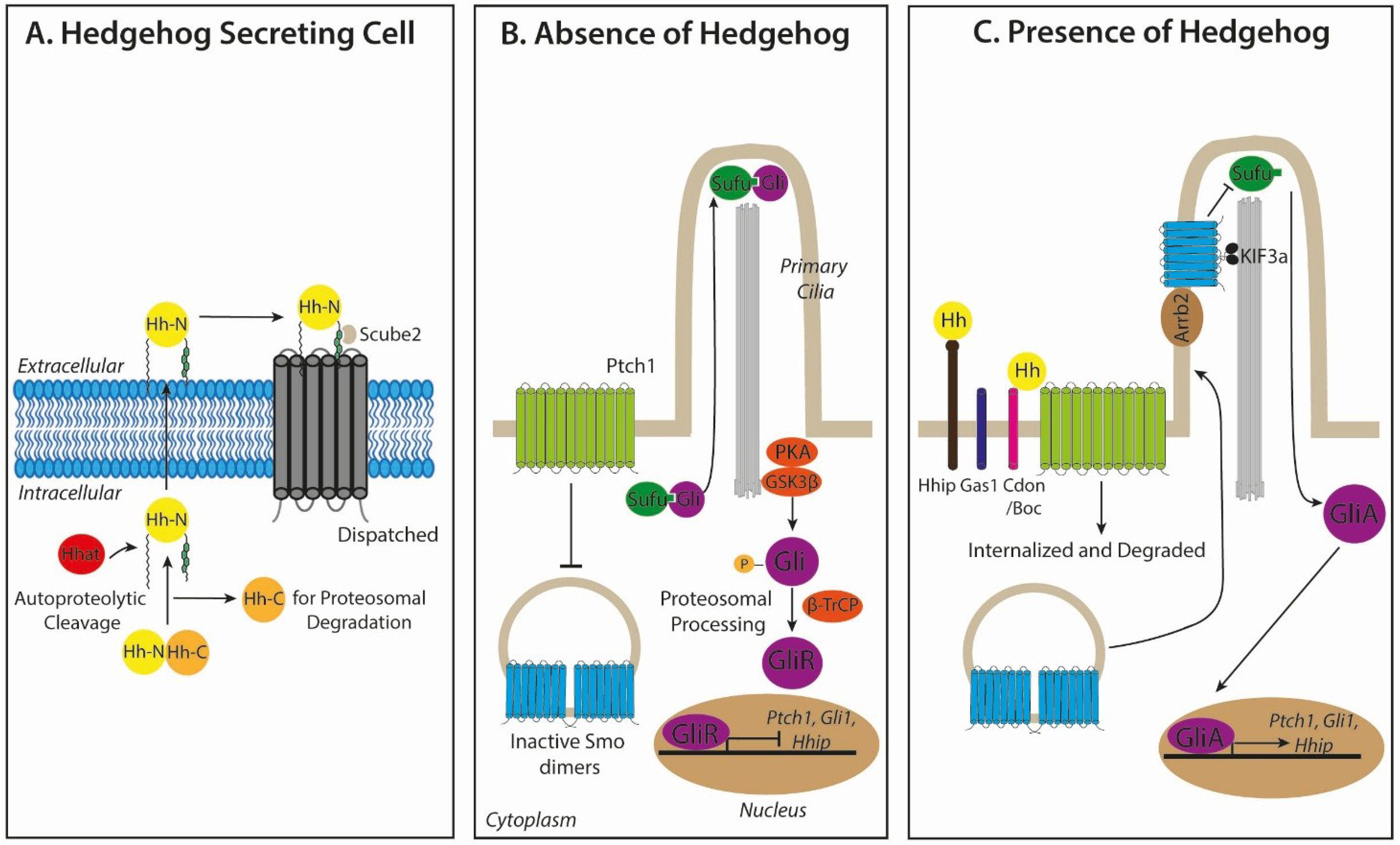
2.1. Hedgehog Biogenesis and Secretion

The three mammalian Hh ligands, Sonic Hedgehog (Shh), Indian Hedgehog (Ihh) and Desert Hedgehog (Dhh), are synthesized as precursor proteins that undergo autoproteolytic cleavage to produce an N-terminal signaling protein with dual lipid modifications [12,13] (Figure 1A). Cleavage of the carboxyl-terminal peptide and subsequent transfer of a cholesterol moiety on the resulting C-terminus leads to Hh ligand retention at the plasma membrane. Hedgehog acyltransferase (Hhat) catalyses the addition of a palmitoyl group on the N-terminus [14,15], promoting the association of the ligand to sterol-rich membrane microdomains to restrict ligand mobility [16,17]. Dispatched (Disp), a large multi-pass transmembrane protein, in synergy with Scube2, a secreted glycoprotein, bind to distinct components of the C-terminal cholesterol group to generate the release of Hh ligand from the plasma membrane and shelter lipidated Hh from the aqueous microenvironment [18,19]. Additionally, Hh contains the ability to form monomers and large multimers through their cholesterol linkages [20,21,22]. Diffusion of Hh ligand is negatively regulated by the membrane protein Hh-interacting protein 1 (Hhip1), which competes with the receptor Patched (see below) for ligand binding through association of the Zn2+ containing pseudo-active site in Hh ligands [23,24]. Similarly, the glycophosphatidylinositol (GPI)-linked heparan sulphate proteoglycan, Glypican-3, (Gpc3), is able to sequester Hh and prevent long-range ligand distribution [25,26,27].

2.2. Hedgehog Signal Transduction

Hh signaling is initiated by the binding of Hh ligand to its corresponding receptor, Patched (Ptch1, and to a lesser extent, Ptch2), a twelve-pass transmembrane protein located on Hh-responsive cells [28,29]. This process is also facilitated by co-receptors, CAM-Related/Downregulated by Oncogenes (Cdon), Brother of Cdon (Boc), and Growth Arrest Specific 1 (Gas1), which form distinct multimolecular complexes with Ptch1 to promote high-affinity Hh ligand binding [30,31]. In the absence of Hh ligand, Ptch1 constitutively represses Smoothened (Smo), a seven-transmembrane domain receptor of the G-protein-coupled receptor (GPCR) superfamily, preventing the translocation of Smo to primary cilia [32,33] (Figure 1B). Smo exists as inactive internalized dimers, where the cytoplasmic tails of each monomer are in a closed configuration maintained by electrostatic forces between arginine and asparagine clusters at the C-terminus [33,34,35]. In the absence of active Smo in the ciliary membrane, the Glioma-associated oncogene (Gli) family of latent zinc-finger transcriptional mediators, Gli1, Gli2 and Gli3, are retained in a complex with the negative regulator, Suppressor of fused (Sufu), at the ciliary tip [36,37]. In this state, Gli2 and Gli3 are phosphorylated by Protein kinase A (PKA) [38] and Glycogen synthase kinase 3β (GSK3β), creating a binding site for the adaptor protein β-transducin repeat containing protein (β-TrCP) [39]. The Gli/β-TrCP complex becomes subject to ubiquitination mediated by the Cul1-based E3 ligase, resulting in partial proteasomal degradation to form transcriptional repressors (Gli2R and Gli3R), which translocate to the nucleus and repress Hh target genes [40]. Gli1 is unable to be processed in this way and only occurs as a full-length transcriptional activator [41].
Figure 1. Mammalian Hedgehog (Hh) signaling. (A) Hh ligand precursor proteins are autoproteolytically cleaved to generate an N-terminal protein that undergoes dual lipid modification, consisting of an N-terminal palmitoyl group and a C-terminal cholesterol moiety, which promotes the binding of ligand to sterol-rich membrane microdomains to restrict mobility. The release of active Hh ligand is then mediated by Dispatched in synergy with Scube2. (B) In the absence of ligand at the responding cell, Patched 1 (Ptch1) constitutively inhibits Smoothened (Smo), preventing its ciliary localization. In this state, Gli proteins are retained in a complex with Suppressor of Fused (Sufu) at the ciliary tip. The recruitment of protein kinase A (PKA), glycogen synthase kinase 3β (GSK3β) and β-transducin repeat-containing protein (β-TrCP) to this complex results in partial proteasomal degradation to form Gli transcriptional repressors (GliR) that translocate to the nucleus and repress Hh target genes. (C) In the presence of the ligand, Hh binding to Ptch1 relieves repression of Smo, triggering its interaction with β-arrestin (Arrb2) and Kif3a and subsequent ciliary localization. This facilitates the release of Gli from Sufu, bypassing proteolytic cleavage into a repressor form, and full-length Gli activators (GliA) translocate to the nucleus to activate Hh target genes. High affinity Hh ligand-Ptch1 binding is facilitated by distinct multimolecular complexes with CAM-Related/Downregulated by Oncogenes (Cdon), Brother of Cdon (Boc) and Growth Arrest Specific 1 (Gas1). Ptch1, Gli1 and Hhip are robust Hh target genes.
Cancers 07 00851 g001
In the presence of Hh ligand, Ptch1 relieves the basal repression of inactive Smo by neutralizing the electrostatic interactions between Smo dimers through G-protein coupled receptor kinase-2 (Grk2)-mediated phosphorylation of the adjacent domain in the C-terminus, promoting an open conformation of active Smo [42,43] (Figure 1C). Simultaneously, Ptch1 becomes internalized and degraded by lysosomes. Smo associates with β-Arrestin (Arrb2) [35,44] and the intraflagellar microtubule motor protein Kif3a, within the ciliary membrane, facilitating the release of full-length transcriptionally-active Gli proteins (GliA) from Sufu, thereby bypassing proteasomal proteolytic cleavage and processing [35,45]. GliA proteins then translocate to the nucleus and transcriptionally activate Hh target genes. Direct targets for GliA are the Hh pathway genes, Gli1, Ptch1 and Hhip, positive and negative regulators of Hh signaling, promoting feedback loops to enhance or reduce the Hh response [46]. Whilst canonical Hh signaling culminates in Gli-mediated transcription, there is growing evidence for “non-canonical” Hh signaling mechanisms. In this case, signaling may occur via Hh signaling components in alternative ways to the canonical paradigm [47,48,49,50,51,52,53,54,55,56,57,58,59,60,61,62]. Since a role for non-canonical Hh signaling in CSC maintenance is yet to be elucidated, this review will focus on the canonical pathway.

3. Roles for Hedgehog Signaling in Cancer

3.1. Modes of Signaling in Hh-Pathway-Dependent Cancers

The Hh pathway plays a crucial role during organogenesis in the developing embryo, by orchestrating reciprocal communicative events between different cells and tissues. The effect of Hh signaling varies according to the receiving cell type, by directing either cell proliferation, cell fate determination, epithelial-to-mesenchymal transitions and the rearrangement of cells by motility and adhesion changes [63]. Therefore, it is not surprising that inappropriate activation of Hh signaling in the adult can contribute to the initiation, growth and maintenance of cancer. Active Hh signaling can also induce treatment failure in cancer patients, by limiting chemotherapeutic responses or by actively inducing more aggressive and therapy-resistant tumours.
The major mechanisms by which the Hh pathway is aberrantly activated in cancer can be attributed to mutations of Hh pathway constituents (Type I: ligand-independent), excessive expression of Hh pathway ligands (Type II–IIIb: ligand-dependent) and the generation of a cancer stem cell (CSC) phenotype (Type IV) (Figure 2 and Figure 3). It is becoming increasingly apparent that it is essential to know which of these modes of signaling are in operation when evaluating experimental models of Hh-dependent cancer and also for considering the design of future tumour therapies involving Hh pathway inhibitors. Indeed, the use of the Hh pathway antagonists in clinical trials has shown promise in tumours driven by ligand-independent mechanisms, but so far has been underwhelming for those driven by ligand-dependent mechanisms. To further complicate matters, these signaling modes are not mutually exclusive and contain the ability to co-exist in parallel.
Figure 2. Modes of signaling in Hh pathway-dependent cancer. (A) Type I: ligand-independent, tumour cell-intrinsic signaling tumours exhibit mutations in the Hh pathway components that promote cell-intrinsic growth and survival. Loss of function mutations in Ptch1 (red cross), activating mutations of Smo and GliA amplifications (yellow stars), are common in these tumours. (B) Type II: ligand-dependent, autocrine stimulation is characterized by the response to the Hh ligand that is self-secreted. (C) Type III: ligand-dependent, paracrine signaling is defined by the secretion of the Hh ligand from the tumour cells that acts on adjacent stroma, in turn creating a favourable microenvironment for tumour growth. (D) In contrast, in Type IIIb ligand-dependent, reverse paracrine signaling, the Hh ligand is secreted by the adjacent stroma and acts on the tumour cells.
Cancers 07 00851 g002
Figure 3. Hh signaling in cancer stem cells (CSCs). CSCs respond to the Hh ligand, secreted by adjacent stromal cells, tumour cells or the CSCs themselves, to maintain a stemness signature by the regulation of pluripotency genes, including Nanog, Sox2 and Bmi1. CSCs are resistant to conventional chemotherapeutics, surviving treatment before expanding and deriving the heterogeneous tumour bulk population, resulting in disease relapse.
Cancers 07 00851 g003

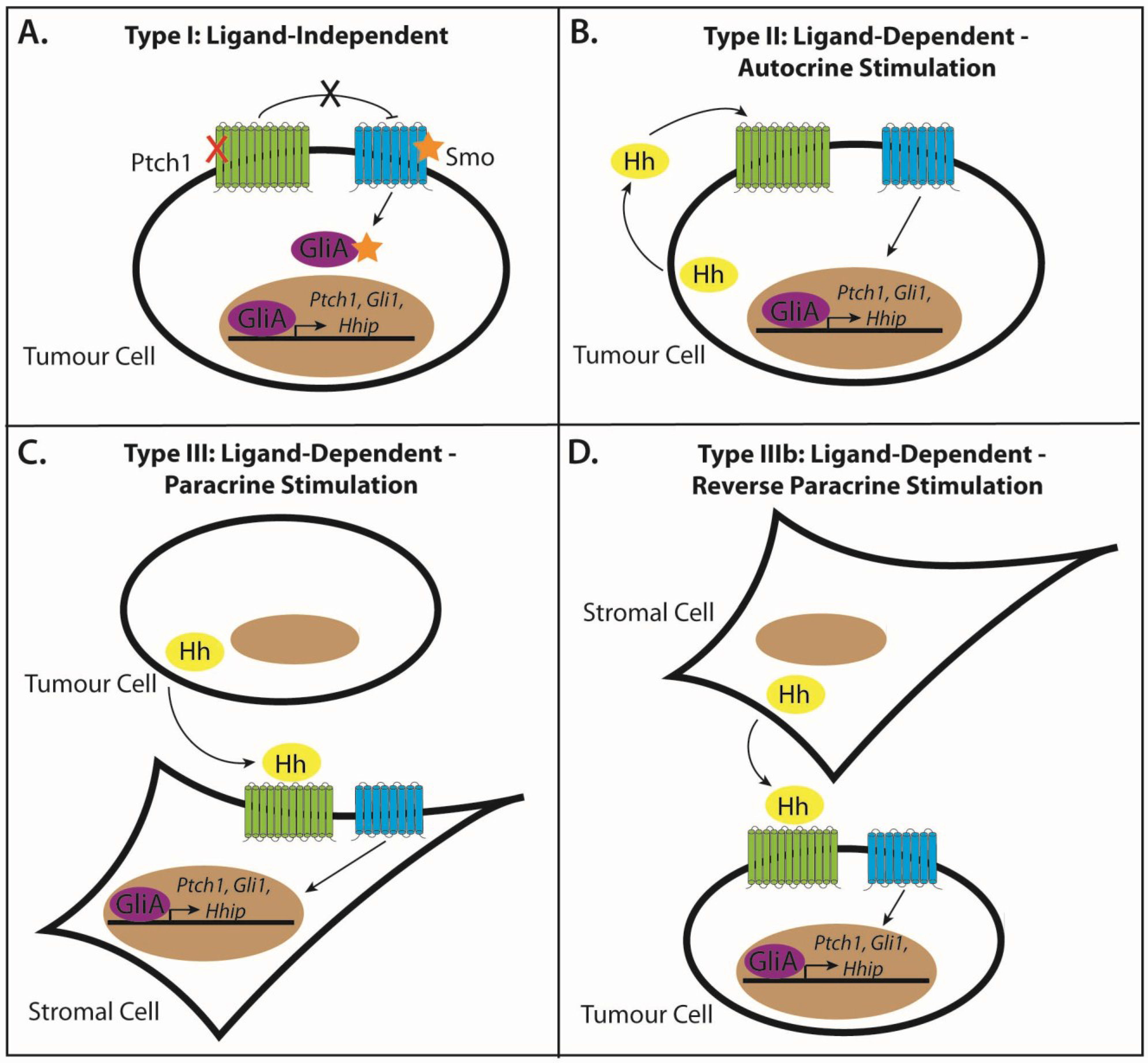
3.2. Type I: Ligand-Independent, Tumour Cell-Intrinsic Signaling

The association between Hh signaling and tumourigenesis was initially established in patients diagnosed with Gorlin syndrome, or nevoid basal cell carcinoma syndrome (NBCCS), where almost all cases are characterized by PTCH1 loss of heterozygosity, leading to ligand-independent constitutive Hh pathway activation [64,65]. Tumours of type 1 origin have genetic aberrations in the Hh pathway components that promote cell-intrinsic growth and survival properties of the tumours (Figure 2A). Analysis of human cancer tissue and mouse models of Hh pathway activation have revealed that inactivating mutations, including deletions, mRNA splice-site and nonsense mutations in PTCH1 [66], SUFU [67,68,69] or activating missense mutations in SMO, SmoM2 (Trp535Leu) [70,71], or gene amplifications and translocations of GLI1 or GLI2 [72], usually in combination with the inactivation of additional tumour suppressor genes [73], are sufficient to form a variety of sporadic tumours [74]. This is especially the case for basal cell carcinomas (BCCs), a skin tumour of keratinocytes, medulloblastoma, a paediatric cancer of the cerebellum and rhabdomyosarcoma [75,76,77].
These findings implicate activating Hh pathway mutations as initiating events in tumourigenesis; therefore, Hh ligand independent tumours make excellent candidates for Hh pathway inhibitor therapy [78]. However, at what level of the signaling pathway a cancer cell has acquired such a pathway-activating genetic aberration will evidently determine whether, or not, a specific inhibitor is efficacious, as tumours with activating mutations downstream of SMO will be insensitive to the majority of Hh pathway inhibitors under development today.

3.3. Type II: Ligand-Dependent, Autocrine Signaling

The vast majority of tumours in which Hh signaling has been implicated lack mutations in the pathway and are dependent on upstream pathway activation driven by the Hh ligand. In this instance, tumour cells have been proposed to self-secrete Hh ligand in order to stimulate signaling, termed autocrine or juxtacrine Hh signaling [74,79] (Figure 2B). This was firstly based on tumour cells expressing both Hh ligand and downstream Hh signaling constituents, where growth was significantly inhibited by the naturally-occurring Smo antagonist, cyclopamine, in the absence of tumour stroma [80,81,82]. Whilst the non-specificity of cyclopamine initially cast some doubt over the interpretation of this finding [83], many more recent in vitro and in vivo studies using the Hh ligand-neutralizing antibody 5E1, RNAi-mediated knockdown of SMO or GLI1, GLI antagonists, such as GANT61, and treatment with various other specific small molecule SMO antagonists have demonstrated similar findings [84,85,86]. Therefore, tumours characterized by type II signaling are susceptible to Hh inhibition at either the level of Hh ligand, Smo or Gli.

3.4. Type III: Ligand-Dependent, Paracrine Signaling

During development, Hh signaling predominantly utilizes the paracrine mode of signaling whereby Hh ligands are produced and secreted by the epithelium to act on adjacent mesenchymal cells [87,88]. Therefore, it is conceivable to think that this mode of signaling would also be utilized to promote tumourigenesis. Indeed, emerging evidence suggests that several tumours, believed to utilize autocrine signaling, might instead, or in addition to this, function through paracrine effects on the adjacent stroma [89,90] (Figure 2C). The responding stroma, in turn, creates a favourable microenvironment that supports tumour growth, by supplying growth and survival factors in order to increase blood vasculature [91]. Human prostate, pancreatic, ovarian and colorectal cancers are thought to activate the Hh pathway via paracrine stimulation, and this response can be blocked by specific Hh inhibitors [92,93]. The precise paracrine feedback mechanisms thought to signal from the stroma to tumour cells remains to be elucidated. However, recent evidence suggests that the IGF and the Wnt signaling pathways are the likely candidates, as the insulin-like growth factor gene (Igf1), IGF pathway binding proteins and Wnt signaling molecules in the stroma were similarly modulated to Gli1 and Ptch1 in Hh inhibitor-treated tumour xenografts [90]. Therefore, it is probable that the stromal microenvironment responds to the Hh ligand secreted from tumour cells, to initiate the production and release of pro-angiogenic stromal feedback factors, promoting growth and survival signals back to the tumour [94,95].

3.5. Type IIIb: Ligand-Dependent, Reverse Paracrine Signaling

In this variant of paracrine signaling, stromal cells produce and secrete Hh ligands to influence tumour cells [89,96] (Figure 2D). Classified as type IIIb signaling, this mechanism of Hh signaling has been observed in haematological malignancies, including B-cell lymphomas, multiple myelomas and leukemias, in which Hh secreted from the bone marrow stroma is essential for the survival of cancerous B cells through the upregulation of the anti-apoptotic factor, Bcl2 [97,98]. Stromal Hh was also found in endothelial cells of high-grade, platelet-derived growth factor (PDGF)-induced gliomas [99]. Again, this tumour growth is impeded in response to the inhibition of the Hh pathway with either cyclopamine, 5E1 or the small molecule SMO antagonist, SANT-1 [97].

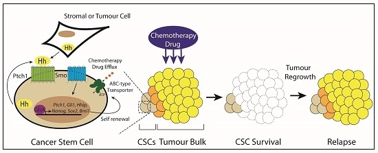
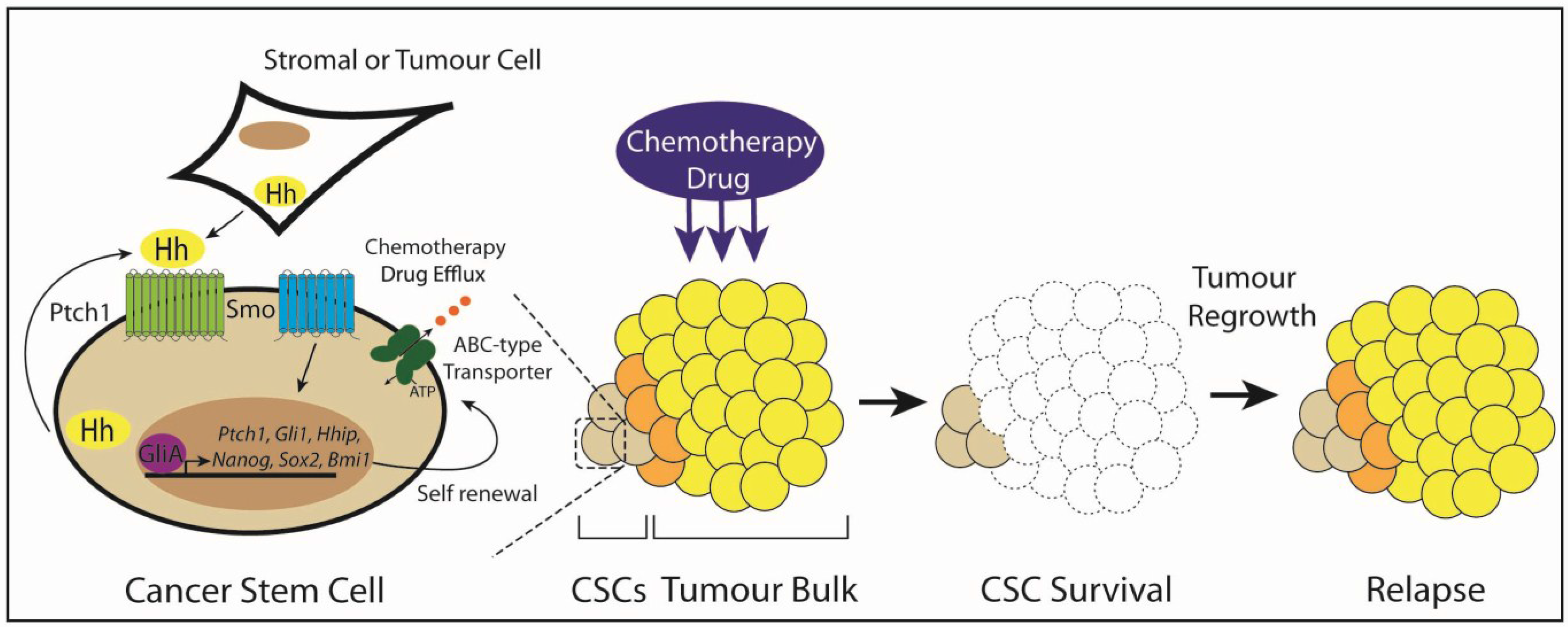
3.6. Type IV: Cancer Stem Cells

An alternative model to the Type I–III Hh pathway stimulation proposes that Hh signaling is important for the existence of a subpopulation of tumour cells that exhibit stem cell-like properties (Figure 3). This rare subset of tumour-initiating cells, termed cancer stem cells (CSCs), are proposed to maintain a self-renewing reservoir and differentiate into transient amplifying cells to produce a state of cellular heterogeneity within a tumour [4,5]. Hh signaling is believed to drive the CSC phenotype through the subverted regulation of stemness-determining genes. Indeed, Nanog, a transcription factor that acts as a master determinant of both embryonic stem cell self-renewal and the re-programming of differentiated somatic cells to pluripotency, is a direct transcriptional target of the Hh signaling pathway [100]. Furthermore, Hh signaling maintains a stemness signature in multiple cancers by driving the expression of stemness regulating genes, such as Oct4, Sox2 and Bmi1 [101,102,103]. While Hh-driven CSCs have been validated for numerous haematological malignancies, their existence in solid tumours remains more controversial. The emerging role for Hh signaling in the maintenance of both haematological malignancies and solid tumour CSCs is discussed in more detail below (also summarized in Table 1).
Table 1. Summary of evidence for the role of Hedgehog signaling in cancer stem cells.
Tumour TypeCSC MarkerStemness Genes ExpressedMode of Hedgehog InhibitionExperimental EvidenceCombination TherapyRefs.
Chronic Myeloid Leukaemia (CML)CD34+, Lin, Sca+, cKit+-Cyclopamine, Bcr-Abl infected Smo−/− embryonic liver cells, Smo KO in CML mouse model, PF-0444991314-fold reduction in CML LSCs, 60% of mice survived after 7 weeksCyclopamine and nilotinib, PF-04449913 and dasatinib[104,105,106,107,108]
Acute Myeloid Leukaemia (AML)--IPI-926, PF-04449913, Cyclopamine, Endogenous Hhip, 5E1Inhibits self-renewal and promotes myelomonocytic differentiationSorafenib and IPI-926, cyclopamine or Hhip or 5E1 and cytarabine[109,110,111,112]
Acute Lymphoblastic Leukaemia(ALL)--Cyclopamine, IPI-926, KAAD-cyclopamine, SANT-1Reduces long-term self-renewal in B-ALL, promotes apoptosis in T-ALL-[113,114]
Multiple MyelomaCD138neg, CD19+-Cyclopamine, 5E1Reduces CD138neg self-renewal by inducing plasma cell differentiation-[98]
GliomaCD133+, ALDH1+, ABCG2+NANOG, OCT4, SOX2, NESTIN, BMI1CyclopamineAbolishes tumour engraftmentCyclopamine, temozolomide and/or 10 Gys of radiation[101,115,116,117]
Breast CancerCD44+, CD24−/low, Lin, ALDH1+p63, OCT4, NESTIN, NANOG, BMI1CyclopamineReduces mammosphere self-renewal and secondary formation-[118,119,120]
Small Cell Lung Cancer-BMP4, NESTIN, ASH-1Cyclopamine, LDE-225, shSMO, 5E1Prevents tumour relapse in LX22 xenograftsLDE-225, carboplatin and etoposide or, GDC-0449 and cisplatin[82,121,122]
Non-Small Cell Lung Cancer-SOX2, OCT4, NANOG, ALDHA1siSHH GDC-0449Decreases colony formation and growth in soft agarGDC-0440, erlotinib and cisplatin[103,122,123]
Gastric CancerCD44+, CD24+SOX2, NANOGCyclopamine, Vismodegib, 5E1, shSMOReduces CD44+ tumourspheres and number and diameter of coloniesVismodegib, 5-flurouracil and/or cisplatin or cyclopamine, oxaliplatin and mitomycin[124,125]
Colon CancerCD133+NANOG, OCT4shSMO CyclopamineReduction of the CD133+ CSC population-[83,102]
Pancreatic CancerCD44+, CD24+, ESA+NANOG, OCT4GDC-0449, Cyclopamine derivative - CyTReduces tumoursphere viability and chemoresistanceCyT and 2 Gys of radiation[86,126,127,128,129,130,131,132]
Prostate Cancer-NANOG, OCT4Cyclopamine, shGLI1,2, GANT61Suppresses tumoursphere and colony formationCyclopamine and paclitaxel[133,134,135,136]
Metastatic MelanomaALDH+SOX2, NANOG, OCT4, KLF4shSMO, shGLI1Reduces ALDH+ melanospheres fraction, clonogenicity and xenograft growth-[137,138,139]

4. Evidence for the Role of Hedgehog Signaling in Cancer Stem Cell Maintenance

4.1. Leukemic Stem Cells

Much of our knowledge of CSC biology is derived from studies on normal and malignant haematopoiesis, which has led to the identification of hematopoietic stem cells (HSCs) and its malignant counterpart, the leukemic stem cell (LSC) [140,141]. The stem cell theory of cancer, which postulates that malignancy arises from the transformation of adult somatic stem cells, is an attractive hypothesis within the hematopoietic system, as evidence indicates that the cell surface phenotype, CD34+ CD38, is shared between LSCs and HSCs [141,142,143]. In addition, cytogenetic abnormalities that are consistently associated with certain leukemias have been detected in HSC compartments in patients with acute myeloid leukaemia (AML), chronic myeloid leukaemia (CML) and acute lymphoblastic leukaemia (ALL) (see below) [144,145,146,147]. For instance, the BCR-ABL gene rearrangement in CML has also been detected in cells of myeloid, erythroid megakaryocytic and B-lymphoid lineages, indicating that initial transformation occurs within a cell that is capable of multi-lineage differentiation [142,144,148]. Moreover, a genetic mouse model displaying conditional gene inactivation of Jun-B in the HSC compartment symptomatically produces myeloproliferative CML-like disease [149]. These results support the notion of a normal HSC hierarchy in the LSC compartment and implicate the HSC as the candidate cell for transformation by leukaemia-inducing oncogenes. In contrast, the ability of both MLL-ENL, a t(11;19) translocation in infant acute leukemias, and MOZ-TIF2, an AML inversion of (8)(p11q13), fusion oncogenes to restore self-renewal ability to previously-committed progenitors that normally lack the capacity to self-renew, and evidence of AML LSCs derived from the CD34 fraction [150] provide an alternative origin of the LSC [151,152].

4.2. Chronic Myeloid Leukaemia

Chronic myeloid leukaemia (CML) is the best understood hematologic and stem-cell driven malignancy. Characterized as a clonal myeloproliferative disease, CML is caused by a chromosomal translocation forming the Philadelphia chromosome, which encodes the constitutively-active oncogenic tyrosine kinase, BCR-ABL. The tyrosine kinase inhibitor (TKI) Imatinib, which pharmacologically blocks BCR-ABL kinase activity, has radically revolutionized the management of chronic-phase CML, inducing unprecedented cytogenetic and molecular responses in patients [153].
Despite this success, TKI resistance is still an ongoing problem, and discontinuation of Imatinib can promote relapse of the disease [153]. Hh signaling is intimately involved in the persistence and self-renewing ability of BCR-ABL-driven Lin−/Sca+/cKit+ LSCs. Genetic inactivation of Smo in a mouse model of CML decreases the number of CML LSCs, whereas constitutively-active Smo results in a four-fold increase in CML LSCs, leading to accelerated CML tumourigenesis [104]. Similar results have been described by the overexpression of Bcr-Abl in both Smo−/− and Ptch1−/+ embryonic liver cells transplanted into lethally-irradiated C57BL/6 mice, leading to failed expansion and reduced CML incidence or increased cell expansion, respectively. These studies strongly suggest that Hh pathway activity controls the frequency and maintenance of CML leukemic stem cells (LSCs) and, consequently, the incidence and latency of CML development [105,154]. Indeed, the delivery of cyclopamine to mice transplanted with Bcr-Abl-infected HSCs produced a 14-fold reduction in the CML stem cell population, where 60% of the mice survived after seven weeks [105]. It has also been shown that CML LSCs are dependent on low levels of the cell fate determinant, Numb. Numb, which plays a role in the regulation of Gli1 via Itch-dependent ubiquitination, was found to be highly expressed in Smo−/− CML LSCs. In addition, ectopically-expressed Numb inhibited the in vitro expansion of Bcr-Abl-infected hematopoietic cells and CML LSCs derived from leukaemia patients [104].
Smo overexpression in human CML cell lines is also associated with reduced expression of miR-326 in CD34+ CML LSCs, and overexpression of miR-326 leads to Smo downregulation and, consequently, decreased cell viability [106]. This suggests that inhibition of Smo might result in the restoration of Numb and miR-326 expression, could possibly eradicate the number of CD34+ LSCs and, thus, CML pathogenesis. Additionally, Hh signaling has also been identified as an essential component of multidrug resistance (MDR) in myeloid leukaemia. Cyclopamine treatment of chemo-resistant Lucena-1 cells, derived from the CML chemotherapy-sensitive cell line K562, leads to the downregulation of P-glycoprotein, a notoriously-known ATP-dependent efflux pump that removes cytotoxic drugs from cancer cells, resulting in resensitization to chemotherapy [107]. Interestingly, treatment of CML patient-derived bone marrow cells with a combination of cyclopamine and the Abl inhibitor, Nilotinib, in vitro, reduced the number of colony-forming units by more than 80%. Similar results were also observed using PF-04449913, an orally bioavailable small molecule Smo antagonist, and the TKI Dasatinib, which reduced CML LSC burden by Gli2 inhibition [108], suggesting that targeting both tyrosine kinase and Hh activity might be an effective combination therapy in CML patients [105].

4.3. Acute Myeloid Leukaemia

AML, characterized as a malignancy of the myeloid line of blood cells, is an extremely heterogeneous clonal disorder with a phenotypically-variable LSC population that is likely not confined to a single clonal subpopulation [155]. Primary AML cell lines express SHH and GLI1, the latter of which correlates with cytogenetic risk and overall reduced survival [156]. Constitutive tyrosine kinase activity involving internal tandem duplications (ITD) of the FMS-like tyrosine kinase 3 (FLT3) juxtamembrane domain are typically found in AML patients. Consequently, GLI2 expression in FLT3-ITD is higher than in wild-type FLT3 AML patients, which correlates with reduced survival [109].
Mice expressing FLT3-ITD and SmoM2 in the hematopoietic system driven by Mx1-Cre and poly(I:C) treatment produce accumulated populations of c-Kit+Gr-1int and Mac1+Gr1int blasts of the myeloid lineage, leading to myeloproliferative neoplasia (MPN)-AML [109]. Constitutive Hh activation leads to downstream STAT5 signaling, and combined treatment with the tyrosine kinase inhibitor, sorafenib and SMO antagonist IPI-926 inhibited clonogenic AML growth and proliferation in FLT3-ITD+ AML cell lines in vitro and disease progression in vivo [109]. Together, these studies suggest that active Hh signaling in the granulocyte/monocyte progenitor compartment, in combination with the FLT3-ITD mutation, is capable of initiating the development of AML. Furthermore, Hh pathway inhibition with PF-04449913 sensitizes AML chemoresistant cell lines and primary cells to standard chemotherapy drugs, inhibits Smo-mediated self-renewal [110] and promotes myelomonocytic differentiation in the AML cell line, HL-60 [111]. Similarly, the chemotherapy-resistant CD34+ AML cell lines, Kasumi-1, Kasumi-3 and TF-1, express IHH, GLI1 and GLI2 and respond to cyclopamine, endogenous HHIP and 5E1 treatment, which in combination with cytarabine (Ara-C), dramatically reduces cell survival [112]. Conversely, in an alternative AML mouse model driven by MLL-AF9, the most frequent rearrangement in childhood AML, Hh pathway blockade was ineffective, signifying that Hh signaling is dispensable in this particular molecular subtype [157,158]. Given these conflicting results, the complexity between the intrinsic and extrinsic signals that govern LSC behaviour, and the high phenotypic variability in AML, it is probable that the underlying genetic and molecular mechanisms likely determine the suitability of targeting the Hh pathway in AML LSCs [157,159].

4.4. Acute Lymphoblastic Leukaemia

Characterized by the accumulation of malignant white blood cells, or lymphoblasts, acute lymphoblastic leukaemia (ALL), is the most common form of cancer in children. Hh signaling plays a key role in regulating self-renewal of ALL tumour cells of both B- and T-cell origin. In a panel of primary B-ALL cell lines, 95% expressed PTCH1, GLI1 and SMO [113]. Moreover, treatment with the Smo inhibitors, cyclopamine or IPI-926, significantly reduces long-term self-renewal potential in B-ALL LSCs [113,114]. In the human-derived T-ALL cell line, CEM, inhibition of Hh activity with KAAD-cyclopamine or SANT-1 treatment induced cellular apoptosis in both CEM-derived glucocorticoid (GC)-sensitive and resistant T-ALL clones, highlighting a critical role for the Hh pathway in cell growth and survival [160].

4.5. Multiple Myeloma

Multiple myeloma (MM), a malignancy consisting of a well-defined stem cell compartment, is a plasma cell malignancy of the bone marrow characterized by abnormal proliferation of plasma cells. MM consists of two distinct populations, CD138neg CD19+ stem cells, and malignant CD138+ CD19neg terminally-differentiated plasma cells [161]. Preferential expression of SMO and a GLI-responsive YFP reporter in the CD138neg fraction of the MM cell line, NCI-H929, suggests that Hh signaling plays an important role in the CD138neg LSC population. Indeed, inhibition of Hh signaling by cyclopamine or 5E1 treatment inhibited the clonal capacity of MM cell lines, NCI-H929 and KMS12, and decreased the CD138neg population through the induction of plasma cell differentiation, highlighting a crucial role in the maintenance of self-renewal [98].
Intriguingly, experimental data suggests multiple modes of Hh signaling seem to activate MM CD138neg LSCs. Bone marrow biopsy samples from MM patients demonstrate that SHH is mainly secreted by malignant CD138+ terminally-differentiated plasma cells, utilizing type III paracrine Hh signaling, to promote proliferation and the inhibition of chemotherapy-induced apoptosis [162]. In addition, stromally-induced type IIIb Hh signaling can also activate Hh signaling in MM LSCs [97]. Taken together, MM demonstrates that Hh signaling can act through multiple signaling modes within the same cancer and can mediate interactions between CSCs, differentiated tumour cells and the microenvironment [163].

4.6. Glioma

Glioma, one of the most common and lethal primary brain tumours, contain cells with shared similarities to normal neural stem cells, capable of clonogenic growth in vitro and tumour formation in vivo [164]. Hh signaling appears to be active in glioblastoma multiforme (GBM)-derived neurospheres and glioma stem cell cultures (gliomaspheres), as they express GLI1, PTCH1, SMO and SHH [115,116]. Indeed, conditioned media from GBM neurospheres induces a 10-fold increase of Gli1-luciferase in the NIH 3T3-Light2 reporter cell line [115]. Conversely, GBM neurospheres upregulate GLI1 mRNA in response to conditioned medium containing exogenous SHH-N, indicating that GBM neurospheres are capable of secreting and responding to biologically-active SHH ligand [115].
Hh pathway blockade in GBM neurospheres by cyclopamine reduces GLI1 mRNA expression, inhibits cell growth and promotes the formation of well-differentiated GFAP-positive neurospheres, indicative of mature glial cells [115], and downregulated the expression of stemness genes NANOG, OCT4, SOX2, NESTIN and BMI1 [101]. Remarkably, intracerebral implantation of viable cyclopamine-treated neurospheres into NOD/SCID mice completely abolishes tumour engraftment and growth, indicating that inhibition of Hh signaling impedes clonogenic growth and self-renewal of GBM stem cells (GSCs). Combination treatment of cyclopamine and 10 Gys of radiation [115] or temozolomide [101] revealed a synergistic effect to reduce neurosphere growth, implying that Hh blockade targets GSCs that are not normally affected by radiation and chemotherapy. Indeed, SMO, GLI1, PTCH1, CD133 and aldehyde dehydrogenase (ALDH)-expressing GSCs are upregulated post-radiation in GBM neurospheres, which further supports the concept that standard therapies are not able to abolish GSCs [115]. Additionally, it has been shown that active Hh signaling can sensitize GSCs to endogenous nano-irradiation, through the inhibition of thymidine synthesis [117].

4.7. Breast Cancer

After the initial demonstration of CSCs in hematopoietic malignancies, breast cancer was the first solid malignancy in which CSCs were identified and isolated [2,118]. The CSC population, characterized with the cellular identity CD44+/CD24−/lowLinALDH-1+ and the capacity to recapitulate the phenotypic heterogeneity of the primary tumour when injected into secondary NOD/SCID mice [6,165], has been demonstrated to display active Hh signaling to maintain stemness potential. Since Hh signaling plays a critical role in mammary stem cell maintenance within the mammary epithelium [166,167], it is not surprising that mammary CD44+/CD24−/lowLin CSCs express increased mRNA transcript levels of PTCH1, GLI1 and GLI2 compared to bulk tumour cells [168]. This supports the stem cell theory of cancer, where malignancy arises from the transformation of adult somatic stem cells to utilize existing stem-cell regulatory pathways to promote self-renewal.
The mechanism of Hh-mediated self-renewal in mammospheres has been shown via Shh-mediated upregulation of the polycomb gene BMI1, an effect that was blocked by treatment of cyclopamine [118]. Indeed, isolated CD44+/CD24−/lowLin CSCs express a five-fold increase in BMI1 compared to tumour cells also derived from the same human breast carcinoma-derived xenograft tumour, but lacking CSC marker expression [118]. Furthermore, p63, the sister homolog of p53, characterized as a master regulator of normal epithelial stem cell maintenance, drives Hh signaling in mammary CSCs [119]. Knock down of p63 in mammospheres derived from breast tumours of transgenic mice with conditional overexpression of the ErbB2 oncogene in mammary glands results in a decrease of Shh, Ptch1, Gli2 and Bmi1 transcript levels, leading to a reduction in secondary mammosphere formation. ChIP-sequence analysis in p63 overexpressing MCF-7 cells has demonstrated that Shh, Gli2 and Ptch1 are direct transcriptional target genes of p63 [119]. Additionally, Hh inactivation in MCF-7-derived CD44+/CD24 CSCs induced a reduction in cell number through downregulation of OCT4, NESTIN and NANOG, indicating that Hh signaling in breast CSCs upregulates stem cell markers to maintain a self-renewing signature [120].

4.8. Gastrointestinal Cancers

Studies have demonstrated the presence of gastric CSCs in several gastric cancer cell lines. Identified by the cell surface marker CD44+ and the ability to form non-adherent spherical colonies in serum-free media and tumours when implanted into immunocompromised mice, CD44+ gastric CSCs represent ~0.6%–2.2% of the tumour cell population [169]. MGC-803, HGC-27 and MKN-45 tumourspheres display increased mRNA expression of Hh components Shh, Ptch1 and Gli1 and stemness markers Sox2 and Nanog [124] compared to adherent cultures.
Inhibition of Hh signaling in tumourspheres with cyclopamine, 5E1, vismodegib or shSMO reduces the capacity for the formation of CD44+ sub-tumourspheres in culture, as well as the number and diameter of colonies derived from single cells on soft agar, but had no effect on adherent cells [125]. Furthermore, vismodegib treatment dramatically increases CD44+ tumoursphere sensitivity to 5-flurouracil or cisplatin with vismodegib, reducing cell viability by ~87%, compared to only 13%–20% and 11%–22% with 5-fluorouracil and cisplatin treatment alone, respectively [124]. Additionally, dissociated HGC-27 tumourspheres treated with cyclopamine, followed by oxaliplatin and mitomycin, significantly enhanced the overall rate of apoptosis compared to cyclopamine and drug treatment alone [125]. Similar effects have been observed in vivo, with shSMO-transduced MKN-45 tumoursphere-derived xenografts demonstrating reduced growth potential following cisplatin treatment, associated with a decrease in CD44 expression [124].
Colon CSCs are thought to originate from the few stem cells situated at the base of colonic crypts [83,170]. Recurrence and metastatic spread of colon carcinomas have been proposed to depend on CD133+ CSCs, which induce tumours when implanted into nude mice. Although initiated from constitutive activation of Wnt signaling, Hh signaling plays an important role in the maintenance of colon CD133+ CSCs, which display the highest gene expression levels of GLI1, PTCH1, GLI2, SHH and HHIP compared to all CD133 cells in human colon carcinoma samples [83]. Serial in vivo passaging of purified CD133+ CSCs stably expressing shSMOH or shPTCH1 demonstrated a complete abolishment or increase the CD133+ CSC population, respectively, highlighting a critical role for Hh signaling in the self-renewal of these cells [83]. Furthermore, increased expression of GLI1 and SMO, as well as stemness markers NANOG and OCT4 in the HCT-116 non-adherent spheres compared to adherent cultures are significantly reduced following cyclopamine administration [102].

4.9. Pancreatic Cancer

In pancreatic ductal adenocarcinoma, tumour cells with the CD44+CD24+ESA+ immunophenotype convey the properties of self-renewal and multilineage differentiation and are thus considered the pancreatic CSC population [171,172,173]. Pancreatic CSCs-tumourspheres derived from cell lines AsPC-1, PANC-1 and MIA-PaCa-2 and pancreatic-derived metastases from an orthotopic mouse model of pancreatic cancer displayed increased mRNA and protein expression of Smo, Gli1 and Gli2 [126,127,128]. Moreover, Hh pathway blockade by GDC-0449 [129], a small molecule Smo antagonist, and GANT61 [86], a Gli inhibitor, reduced cell viability and induced apoptosis via Fas, DR4 and DR5 expression in all pancreatic tumoursphere cultures.
Similar results were observed following cyclopamine treatment, as well as a reduction in the expression of Bmi1 and the ATP-binding drug transporter ABCG2, suggesting that Bmi1 may function as a downstream Hh target in pancreatic cancer, as in breast cancer, and Hh blockade can reverse chemoresistance via ABCG2 downregulation in pancreatic CSCs [127,130]. Additionally, 2 Gys of radiation in the presence of CyT, a cyclopamine derivative, completely eliminated the pancreatic tumoursphere population, compared to radiotherapy and CyT treatment alone [128]. Hh pathway blockade in pancreatic CSCs can also be induced by sulforaphane (SFN), a compound derived from cruciferous vegetables, and epigallocatehin-3-gallate (EGCG), an active compound in green tea, which downregulated mRNA expression of Hh pathway components Smo, Gli1 and Gli2, and pluripotency transcription factors, Nanog and Oct4, inhibited Gli-luciferase reporter activity and reduced the expression of Snail, Slug and ZEB, factors involved in invasion and migration, which produced an overall anti-proliferative and increased apoptotic effect in pancreatic CSCs [126,131,132]. Thus, Shh-Gli signaling plays an essential role in controlling stemness and chemotherapeutic resistance in pancreatic CSCs.

4.10. Prostate Cancer

Specifically an androgen-dependent disease, prostate cancer (PaC) often responds to androgen deprivation therapy (ADT) [174]. In the event of PaC relapse and metastasis, first-line chemotherapy drugs, such as paclitaxel and docetaxel, microtubule stabilizers, prove to be effective until the occurrence of relapse and disease progression from highly-chemoresistant prostate CSCs (PCSCs) [133,175]. CSC-containing side populations exhibit a higher expression of pluripotency markers OCT4, NANOG and ABCG2 compared to the non-side population fraction [134]. Remarkably, inhibition of the Hh signaling pathway in chemoresistant PCSCs, by cyclopamine, in combination with paclitaxel has been shown to significantly reduce cell viability and enhance apoptosis, when compared to paclitaxel and cyclopamine treatment alone [134]. Similarly, inhibition of GLI1 and GLI2 in docetaxel-resistant prostate cancer cell lines, DU145 and 22RV1, generates a subtle reduction in colony formation that was further reduced in combination with knockdown of NOTCH2 [135].
Hh signaling was found to regulate the anti-apoptotic molecule, Bcl-2, in docetaxel-resistant PCSCs, and treatment with the Bcl-2 inhibitor, ABT-737, reduced colony formation in PCSCs, recapitulating the effect observed with Hh pathway inhibition [135]. Additionally, intraprostatic injection of pCX-Shh-IG-GFP vectors in mice, resulting in persistent Shh ligand overexpression in adult prostates, leads to the development of invasive and metastatic prostate cancers within 90 days [136]. In this model, it was also found that active Hh signaling was localized to p63-expressing prostate stem cells, demonstrating that in addition to breast cancer, p63 also drives Hh signaling in PCSCs. Furthermore, the progeny of p63-expressing PCSCs conveyed the ability to differentiate into cells of a basal-intermediate and intermediate-luminal phenotype, as well as rare ChgA+ neuroendocrine cells [136]. Lastly, Hh pathway blockade by GANT61 or genistein, an isoflavone constituent in soybeans, was able to suppress tumoursphere formation and colony formation, further implicating the pathway in prostate CSC maintenance [133].

4.11. Lung Cancer

Small cell lung cancer (SCLC), representing approximately 20%–25% of all lung tumours, is a highly aggressive and lethal malignancy with a five-year survival rate of 2%–8%. SCLC is an excellent example of how stem/progenitor cells escape from niche-dependent signals via constitutive Hh pathway activation [176,177]. Human SCLC cell lines are characterized by the expression of many genes associated with early developmental and progenitor cell states, such as BMP4, normally required during lung epithelial development, Nestin and ASH-1, a transcription factor required for pulmonary neuroendocrine differentiation [82]. Importantly, Hh pathway inhibition by cyclopamine inhibits the expression of all three genes [82].
Deletion of Smo in a genetic mouse model of SCLC significantly reduced tumour initiation and progression, whereas mice expressing SmoM2 developed more frequent and considerably larger tumours [121]. Tumour cells isolated from the same genetic mouse model of SCLC crossed with a Ptch1LacZ/+ reporter mouse express LacZ, indicating that SCLCs maintain active Hh signaling autonomously. Inhibition of Hh signaling in the human SCLC cell line, LX22CL, using LDE-225, shSMO or 5E1, resulted in fewer colonies in a colony formation assay. Conversely, pathway activation using adenovirally-expressed SmoM2 or recombinant Shh protein increased clonogenicity [121]. Interestingly, LX22CL cells surviving a single round of carboplatin and etoposide were considerably more sensitive to pathway manipulation in the same assay. While the growth of chemonaive LX22 xenografts in mice was largely unaffected by treatment with the Hh inhibitor LDE-225, a combination therapy of a round of carboplatin and etoposide followed by LDE-225 treatment prevented tumour relapse, suggesting that the ability of chemoresistant SCLC cells to regenerate is dependent on Hh signaling. Furthermore, LX22 xenografts displayed a marked increase in Shh ligand expression and Gli2 nuclear localization post chemotherapy in vivo, and the proportion of cells expressing a primary cilium increased from less than 1% to approximately 20% [121]. Together, these data suggest a critical role for the Hh pathway in CSC maintenance in SCLC and reveal that Hh inhibitors and chemotherapy may be an effective combination therapy. Similarly, the CSC side population of the SCLC cell line, H1339, detected by the ability to expel Hoechst stain from active ABCG2 transporters, was significantly reduced from 0.75%, following cisplatin treatment alone, to 0.18% when treated with both cisplatin and the Smo inhibitor GDC-0449 [122]. Taken together, active Hh signaling promotes a chemoresistant phenotype in SCLC, and Hh pathway inhibition sensitizes CSCs to cytotoxic therapy and prevents tumour relapse [121].
Lung squamous cell carcinoma (LSCC) exhibits cell-autonomous Hh pathway activation through a protein kinase Ci (PKCi)-SOX2-Hh signaling axis to maintain a CSC-like phenotype in lung oncospheres [103,178]. PKCi mediates SOX2 recruitment to the HHAT promoter to induce constitutive Hh ligand production. These oncospheres are characterized by SOX2, OCT4, NANOG and ALDHA1 mRNA expression, high colony formation efficiency, enhanced growth in soft agar and enhanced tumourigenic potential in vivo that recapitulates the parental tumour [103]. Additionally, inhibition of Hh signaling via siSHH or GDC-0449 treatment in CSCs derived from the lung adenocarcinoma (LAC) cell line A549M sensitized to erlotinib and cisplatin treatment [123]. Similarly, treatment of ABCG2 expressing CSCs in LAC cell line HCC, with both cisplatin and GDC-0449, dramatically decreased the fraction of surviving cells [122], indicating that combination therapy can effectively inhibit tumour growth, compared to either treatment alone.

4.12. Melanoma

Characterized as the most aggressive and lethal skin cancer with high metastatic potential, enhanced heterogeneity and resistance to chemotherapy, advanced metastatic melanoma has a poor prognosis with a median survival time of 6–9 months and a three-year survival rate of 10%–15% [137,179]. A large body of evidence suggests that within the heterogeneous population that constitutes the tumour bulk, active Hh signaling maintains a subpopulation of CSCs [137,179,180]. Cultured CSCs melanospheres demonstrate the Hh-driven CSC properties of increased levels of pluripotency factors, SOX2, NANOG, OCT4 and KLF4, and Hh pathway components, SHH, PTCH1, SMO, GLI2, GLI3, high ALDH activity, the ability to clonally expand in vitro and initiate tumours representing the primary tumour in vivo [137].
Knockdown of both SMO and GLI1 in SSM2c and A375 melanospheres and engraftment of SSM2c cells transduced with lentiviral-shSMO and LV-ShGLI1 leads to a drastic decrease in the fraction of ALDH+ cells, reduced clonogenicity and reduced tumour growth, respectively [137]. Furthermore, knockdown of SMO leads to a complete abolishment of SOX2 mRNA, suggesting that SOX2 is a downstream mediator of the Hh signaling pathway in melanoma CSCs. Indeed, ChIP sequencing in M26c melanoma CSCs demonstrated that SOX2 is a direct transcriptional target of GLI1 and GLI2 [138]. Additionally, WIP1, an oncogenic phosphatase overexpressed in several types of human cancer [181,182,183,184,185], is required for Hh-induced melanoma CSC growth and self-renewal. WIP1 knockdown in SSM2c melanospheres decreased endogenous Gli1 expression and diminished the increased colony formation potential induced by shPTCH1 [139].

5. Targeting Hedgehog Signaling in Cancer Stem Cells

Like normal somatic stem cells, CSCs are resistant to conventional chemotherapeutics, primarily due to the expression of drug efflux pumps and a reduced replication rate. As a result, residual CSCs represent a significant hurdle in the prevention of disease recurrence and metastatic spread. Evidence described above in CML, AML, GBM, gastric cancer, pancreatic cancer, prostate cancer, SCLC and LSCC clearly demonstrates that targeting the Hh signaling pathway in CSCs sensitizes these cells to cytotoxic drug and radiation-mediated cell death and reduces self-renewal potential, leading to abolishment or reduced tumour relapse (Table 1).
Compelling evidence suggests that inhibition of Hh signaling in CSCs promotes commitment or differentiation and a loss of “stemness”, as supported by a reduction in clonogenicity and pluripotency markers, thereby limiting the characteristics normally supporting chemoresistance. Therefore, the combinatorial targeting of CSCs and tumour bulk with Hh inhibitors and conventional chemotherapeutics and/or radiation is an attractive approach to prevent tumour relapse and maximize patient outcomes. However, further investigation into the sequencing of Hh inhibitors and conventional therapies is required to determine whether priming CSCs prior to cytotoxic treatment, co-administration and/or as maintenance therapy following tumour debulking will lead to optimal outcomes. Importantly, the type of Hh antagonist for individual cancer subtypes must also be carefully considered based on the mode of pathway activation.

6. Conclusions

The inability to fully eradicate CSCs is a significant clinical problem and leads to tumour recurrence, therapy resistance and metastatic spread of disease. In this review, we have highlighted the critical role of a key embryonic signaling pathway, the Hh pathway, in the maintenance of CSCs in a number of haematological malignancies and solid tumours. Mounting evidence suggests that targeting the Hh signaling pathway in CSCs may provide a viable and efficacious clinical option to limit tumour growth, overcome resistance and prevent disease relapse. However, a greater understanding of Hh-mediated CSC maintenance and how to best combine Hh antagonists with conventional therapies in the clinic will be required before the full potential of this possibility is realized.

Acknowledgments

CRC is supported by an Australian Postgraduate Award. DNW is supported by the Petre Foundation. DNW and AS are supported by the National Health and Medical Research Council (GNT1048669). We also wish to acknowledge the Victorian Government’s Operational Infrastructure Support Program. The contents of this manuscript are solely the responsibility of the participating institutions and individual authors and do not reflect the views of these funding agencies.

Author Contributions

C.R.C., A.S., D.N.W. and J.E.C. all contributed to the writing and editing of the manuscript.

Conflicts of Interest

The authors declare no conflict of interests.

References

  1. Reya, T.; Morrison, S.J.; Clarke, M.F.; Weissman, I.L. Stem cells, cancer, and cancer stem cells. Nature 2001, 414, 105–111. [Google Scholar]
  2. Karamboulas, C.; Ailles, L. Developmental signaling pathways in cancer stem cells of solid tumors. Biochim. Biophys. Acta—Gen. Subj. 2013, 1830, 2481–2495. [Google Scholar]
  3. O’Brien, C.A.; Kreso, A.; Jamieson, C.H.M. Cancer stem cells and self-renewal. Clin. Cancer Res. 2010, 16, 3113–3120. [Google Scholar] [PubMed]
  4. Chen, K.; Huang, Y.-H.; Chen, J.-L. Understanding and targeting cancer stem cells: Therapeutic implications and challenges. Acta Pharmacol. Sin. 2013, 34, 732–740. [Google Scholar] [PubMed]
  5. Medema, J.P. Cancer stem cells: The challenges ahead. Nat. Cell Biol. 2013, 15, 338–344. [Google Scholar] [CrossRef] [PubMed]
  6. Takebe, N.; Warren, R.Q.; Ivy, S.P. Breast cancer growth and metastasis: interplay between cancer stem cells, embryonic signaling pathways and epithelial-to-mesenchymal transition. Breast Cancer Res. 2011, 13, 211. [Google Scholar] [CrossRef] [PubMed]
  7. McMahon, A.P.; Ingham, P.W.; Tabin, C.J. Developmental roles and clinical significance of Hedgehog signaling. Curr. Top. Dev. Biol. 2003, 53, 1–114. [Google Scholar] [PubMed]
  8. Van Den Brink, G.R.; Peppelenbosch, M.P. Expression of hedgehog pathway components in the adult colon. Gastroenterology 2006, 130, 619. [Google Scholar] [CrossRef] [PubMed]
  9. Wicking, C.; McGlinn, E. The role of hedgehog signalling in tumorigenesis. Cancer Lett. 2001, 173, 1–7. [Google Scholar] [CrossRef] [PubMed]
  10. Peng, Y.-C.; Levine, C.M.; Zahid, S.; Wilson, E.L.; Joyner, A.L. Sonic hedgehog signals to multiple prostate stromal stem cells that replenish distinct stromal subtypes during regeneration. Proc. Natl. Acad. Sci. USA 2013, 110, 20611–20616. [Google Scholar] [CrossRef] [PubMed]
  11. Ihrie, R.A.; Shah, J.K.; Harwell, C.C.; Levine, J.H.; Guinto, C.D.; Lezameta, M.; Kriegstein, A.R.; Alvarez-Buylla, A. Persistent sonic hedgehog signaling in adult brain determines neural stem cell positional identity. Neuron 2011, 71, 250–262. [Google Scholar] [CrossRef] [PubMed]
  12. Perler, F.B. Protein splicing of inteins and hedgehog autoproteolysis: Structure, function, and evolution. Cell 1998, 92, 1–4. [Google Scholar] [CrossRef] [PubMed]
  13. Chen, X.; Tukachinsky, H.; Huang, C.H.; Jao, C.; Chu, Y.R.; Tang, H.Y.; Mueller, B.; Schulman, S.; Rapoport, T.A.; Salic, A. Processing and turnover of the Hedgehog protein in the endoplasmic reticulum. J. Cell Biol. 2011, 192, 825–838. [Google Scholar] [CrossRef] [PubMed]
  14. Chamoun, Z.; Mann, R.K.; Nellen, D.; von Kessler, D.P.; Bellotto, M.; Beachy, P.A.; Basler, K. Skinny hedgehog, an acyltransferase required for palmitoylation and activity of the hedgehog signal. Science 2001, 293, 2080–2084. [Google Scholar] [CrossRef] [PubMed]
  15. Pepinsky, R.B.; Zeng, C.; Went, D.; Rayhorn, P.; Baker, D.P.; Williams, K.P.; Bixler, S.A.; Ambrose, C.M.; Garber, E.A.; Miatkowski, K.; et al. Identification of a palmitic acid-modified form of human Sonic hedgehog. J. Biol. Chem. 1998, 273, 14037–14045. [Google Scholar] [CrossRef] [PubMed]
  16. Taylor, F.R.; Wen, D.; Garber, E.A.; Carmillo, A.N.; Baker, D.P.; Arduini, R.M.; Williams, K.P.; Weinreb, P.H.; Rayhorn, P.; Hronowski, X.; et al. Enhanced potency of human Sonic hedgehog by hydrophobic modification. Biochemistry 2001, 40, 4359–4371. [Google Scholar] [CrossRef] [PubMed]
  17. Callejo, A.; Torroja, C.; Quijada, L.; Guerrero, I. Hedgehog lipid modifications are required for Hedgehog stabilization in the extracellular matrix. Development 2006, 133, 471–483. [Google Scholar] [CrossRef] [PubMed]
  18. Tukachinsky, H.; Kuzmickas Ryan, P.; Jao, C.Y.; Liu, J.; Salic, A. Dispatched and scube mediate the efficient secretion of the cholesterol-modified hedgehog ligand. Cell Rep. 2012, 2, 308–320. [Google Scholar] [CrossRef] [PubMed]
  19. Amanai, K.; Jiang, J. Distinct roles of central missing and dispatched in sending the Hedgehog signal. Development 2001, 128, 5119–5127. [Google Scholar] [PubMed]
  20. Chen, M.-H.; Li, Y.-J.; Kawakami, T.; Xu, S.-M.; Chuang, P.-T. Palmitoylation is required for the production of a soluble multimeric Hedgehog protein complex and long-range signaling in vertebrates. Genes Dev. 2004, 18, 641–659. [Google Scholar] [CrossRef] [PubMed]
  21. Goetz, J.A.; Singh, S.; Suber, L.M.; Kull, F.J.; Robbins, D.J. A highly conserved amino-terminal region of sonic hedgehog is required for the formation of its freely diffusible multimeric form. J. Biol. Chem. 2006, 281, 4087–4093. [Google Scholar] [CrossRef] [PubMed]
  22. Gallet, A.; Rodriguez, R.; Ruel, L.; Therond, P.P. Cholesterol modification of hedgehog is required for trafficking and movement, revealing an asymmetric cellular response to hedgehog. Dev. Cell 2003, 4, 191–204. [Google Scholar] [CrossRef] [PubMed]
  23. Bishop, B.; Aricescu, A.R.; Harlos, K.; O’Callaghan, C.A.; Jones, E.Y.; Siebold, C. Structural insights into hedgehog ligand sequestration by the human hedgehog-interacting protein HHIP. Nat. Struct. Mol. Biol. 2009, 16, 698–703. [Google Scholar] [CrossRef] [PubMed]
  24. Bosanac, I.; Maun, H.R.; Scales, S.J.; Wen, X.; Lingel, A.; Bazan, J.F.; de Sauvage, F.J.; Hymowitz, S.G.; Lazarus, R.A. The structure of SHH in complex with HHIP reveals a recognition role for the Shh pseudo active site in signaling. Nat. Struct. Mol. Biol. 2009, 16, 691–697. [Google Scholar] [CrossRef] [PubMed]
  25. Capurro, M.I.; Shi, W.; Filmus, J. LRP1 mediates Hedgehog-induced endocytosis of the GPC3-Hedgehog complex. J. Cell Sci. 2012, 125, 3380–3389. [Google Scholar] [CrossRef] [PubMed]
  26. Capurro, M.I.; Xu, P.; Shi, W.; Li, F.; Jia, A.; Filmus, J. Glypican-3 inhibits hedgehog signaling during development by competing with patched for hedgehog binding. Dev. Cell 2008, 14, 700–711. [Google Scholar] [CrossRef] [PubMed]
  27. Chang, S.-C.; Mulloy, B.; Magee, A.I.; Couchman, J.R. Two distinct sites in sonic hedgehog combine for heparan sulfate interactions and cell signaling functions. J. Biol. Chem. 2011, 286, 44391–44402. [Google Scholar] [CrossRef] [PubMed]
  28. Chen, Y.; Struhl, G. Dual roles for patched in sequestering and transducing Hedgehog. Cell 1996, 87, 553–563. [Google Scholar] [CrossRef]
  29. Briscoe, J.; Chen, Y.; Jessell, T.M.; Struhl, G. A hedgehog-insensitive form of Patched provides evidence for direct long-range morphogen activity of Sonic hedgehog in the neural tube. Mol. Cell 2001, 7, 1279–1291. [Google Scholar] [CrossRef]
  30. Allen, B.; Song, J.; Izzi, L.; Althaus, I.; Kang, J.S.; Charron, F.; Krauss, R.; McMahon, A. Overlapping roles and collective requirement for the coreceptors GAS1, CDO, and BOC in SHH pathway function. Dev. Cell 2011, 20, 775–787. [Google Scholar] [CrossRef] [PubMed]
  31. Izzi, L.; Lévesque, M.; Morin, S.; Laniel, D.; Wilkes, B.C.; Mille, F.; Krauss, R.S.; McMahon, A.P.; Allen, B.L.; Charron, F. Boc and Gas1 each form distinct Shh receptor complexes with Ptch1 and are required for Shh-mediated cell proliferation. Dev. Cell 2011, 20, 788–801. [Google Scholar] [CrossRef] [PubMed]
  32. Huangfu, D.; Liu, A.; Rakeman, A.S.; Murcia, N.S.; Niswander, L.; Anderson, K.V. Hedgehog signalling in the mouse requires intraflagellar transport proteins. Nature 2003, 426, 83–87. [Google Scholar] [CrossRef] [PubMed]
  33. Rohatgi, R.; Milenkovic, L.; Corcoran, R.B.; Scott, M.P. Hedgehog signal transduction by Smoothened: Pharmacologic evidence for a 2-step activation process. Proc. Natl. Acad. Sci. USA 2009, 106, 3196–3201. [Google Scholar] [CrossRef] [PubMed]
  34. Zhao, Y.; Tong, C.; Jiang, J. Hedgehog regulates smoothened activity by inducing a conformational switch. Nature 2007, 450, 252–258. [Google Scholar] [CrossRef] [PubMed]
  35. Kim, J.; Kato, M.; Beachy, P.A. Gli2 trafficking links Hedgehog-dependent activation of Smoothened in the primary cilium to transcriptional activation in the nucleus. Proc. Natl. Acad. Sci. USA 2009, 106, 21666–21671. [Google Scholar] [CrossRef] [PubMed]
  36. Barnfield, P.C.; Zhang, X.; Thanabalasingham, V.; Yoshida, M.; Hui, C.-C. Negative regulation of Gli1 and Gli2 activator function by Suppressor of fused through multiple mechanisms. Differentiation 2005, 73, 397–405. [Google Scholar] [CrossRef] [PubMed]
  37. Goetz, S.C.; Anderson, K.V. The primary cilium: A signalling centre during vertebrate development. Nat. Rev. Genet. 2010, 11, 331–344. [Google Scholar] [CrossRef] [PubMed]
  38. Niewiadomski, P.; Kong, J.H.; Ahrends, R.; Ma, Y.; Humke, E.W.; Khan, S.; Teruel, M.N.; Novitch, B.G.; Rohatgi, R. Gli protein activity is controlled by multisite phosphorylation in vertebrate Hedgehog signaling. Cell Rep. 2014, 6, 168–181. [Google Scholar] [CrossRef] [PubMed]
  39. Jia, J.; Zhang, L.; Zhang, Q.; Tong, C.; Wang, B.; Hou, F.; Amanai, K.; Jiang, J. Phosphorylation by double-time/CKIε and CKIα targets cubitus interruptus for Slimb/β-TRCP-mediated proteolytic processing. Dev. Cell 2005, 9, 819–830. [Google Scholar] [CrossRef] [PubMed]
  40. Zhang, Q.; Shi, Q.; Chen, Y.; Yue, T.; Li, S.; Wang, B.; Jiang, J. Multiple Ser/Thr-rich degrons mediate the degradation of Ci/Gli by the Cul3-HIB/SPOP E3 ubiquitin ligase. Proc. Natl. Acad. Sci. USA 2009, 106, 21191–21196. [Google Scholar] [CrossRef] [PubMed]
  41. Ruiz I Altaba, A.; Palma, V.; Dahmane, N. Hedgehog-Gli signalling and the growth of the brain. Nat. Rev. Neurosci. 2002, 3, 24–33. [Google Scholar] [CrossRef] [PubMed]
  42. Briscoe, J.; Thérond, P.P. The mechanisms of Hedgehog signalling and its roles in development and disease. Nat. Rev. Mol. Cell Biol. 2013, 14, 418–431. [Google Scholar] [CrossRef] [PubMed]
  43. Chen, Y.; Sasai, N.; Ma, G.; Yue, T.; Jia, J.; Briscoe, J.; Jiang, J. Sonic hedgehog dependent phosphorylation by CK1α and GRK2 is required for ciliary accumulation and activation of smoothened. PLoS Biol. 2011, 9, e1001083. [Google Scholar] [CrossRef] [PubMed]
  44. Chen, W.; Ren, X.R.; Nelson, C.D.; Barak, L.S.; Chen, J.K.; Beachy, P.A.; de Sauvage, F.; Lefkowitz, R.J. Activity-dependent internalization of smoothened mediated by β-Arrestin 2 and GRK2. Science 2004, 306, 2257–2260. [Google Scholar] [CrossRef] [PubMed]
  45. Liu, A.; Wang, B.; Niswander, L.A. Mouse intraflagellar transport proteins regulate both the activator and repressor functions of Gli transcription factors. Development 2005, 132, 3103–3111. [Google Scholar] [CrossRef] [PubMed]
  46. Vokes, S.A.; Ji, H.; Wong, W.H.; McMahon, A.P. A genome-scale analysis of the cis-regulatory circuitry underlying sonic hedgehog-mediated patterning of the mammalian limb. Genes Dev. 2008, 22, 2651–2663. [Google Scholar] [CrossRef] [PubMed]
  47. Okolowsky, N.; Furth, P.A.; Hamel, P.A. Oestrogen receptor-alpha regulates non-canonical Hedgehog-signalling in the mammary gland. Dev. Biol. 2014, 391, 219–229. [Google Scholar] [CrossRef] [PubMed]
  48. Razumilava, N.; Gradilone, S.A.; Smoot, R.L.; Mertens, J.C.; Bronk, S.F.; Sirica, A.E.; Gores, G.J. Non-canonical Hedgehog signaling contributes to chemotaxis in cholangiocarcinoma. J. Hepatol. 2014, 60, 599–605. [Google Scholar] [CrossRef] [PubMed]
  49. Jenkins, D. Hedgehog signalling: Emerging evidence for non-canonical pathways. Cell. Signal. 2009, 21, 1023–1034. [Google Scholar] [CrossRef] [PubMed]
  50. Chang, H.; Li, Q.; Moraes, R.C.; Lewis, M.T.; Hamel, P.A. Activation of Erk by sonic hedgehog independent of canonical hedgehog signalling. Int. J. Biochem. Cell Biol. 2010, 42, 1462–1471. [Google Scholar] [CrossRef] [PubMed]
  51. Mille, F.; Thibert, C.; Fombonne, J.; Rama, N.; Guix, C.; Hayashi, H.; Corset, V.; Reed, J.C.; Mehlen, P. The Patched dependence receptor triggers apoptosis through a DRAL-caspase-9 complex. Nat. Cell Biol. 2009, 11, 739–746. [Google Scholar] [CrossRef] [PubMed]
  52. Thibert, C.; Teillet, M.A.; Lapointe, F.; Mazelin, L.; Le Douarin, N.M.; Mehlen, P. Inhibition of neuroepithelial patched-induced apoptosis by Sonic hedgehog. Science 2003, 301, 843–846. [Google Scholar] [CrossRef] [PubMed]
  53. Barnes, E.A.; Kong, M.; Ollendorff, V.; Donoghue, D.J. Patched1 interacts with cyclin B1 to regulate cell cycle progression. EMBO J. 2001, 20, 2214–2223. [Google Scholar] [CrossRef] [PubMed]
  54. Adolphe, C.; Hetherington, R.; Ellis, T.; Wainwright, B. Patched1 functions as a gatekeeper by promoting cell cycle progression. Cancer Res. 2006, 66, 2081–2088. [Google Scholar] [CrossRef] [PubMed]
  55. Jenkins, D.; Winyard, P.J.D.; Woolf, A.S. Immunohistochemical analysis of Sonic hedgehog signalling in normal human urinary tract development. J. Anat. 2007, 211, 620–629. [Google Scholar] [CrossRef] [PubMed]
  56. Polizio, A.H.; Chinchilla, P.; Chen, X.; Kim, S.; Manning, D.R.; Riobo, N.A. Heterotrimeric Gi proteins link hedgehog signaling to activation of Rho small GTPases to promote fibroblast migration. J. Biol. Chem. 2011, 286, 19589–19596. [Google Scholar] [CrossRef] [PubMed]
  57. Chinchilla, P.; Xiao, L.; Kazanietz, M.G.; Riobo, N.A. Hedgehog proteins activate pro-angiogenic responses in endothelial cells through non-canonical signaling pathways. Cell Cycle 2010, 9, 570–579. [Google Scholar] [CrossRef] [PubMed]
  58. Bijlsma, M.F.; Borensztajn, K.S.; Roelink, H.; Peppelenbosch, M.P.; Spek, C.A. Sonic hedgehog induces transcription-independent cytoskeletal rearrangement and migration regulated by arachidonate metabolites. Cell. Signal. 2007, 19, 2596–2604. [Google Scholar] [CrossRef] [PubMed]
  59. Yam, P.T.; Langlois, S.D.; Morin, S.; Charron, F. Sonic hedgehog guides axons through a noncanonical, Src-family-kinase-dependent signaling pathway. Neuron 2009, 62, 349–362. [Google Scholar] [CrossRef] [PubMed]
  60. Belgacem, Y.H.; Borodinsky, L.N. Sonic hedgehog signaling is decoded by calcium spike activity in the developing spinal cord. Proc. Natl. Acad. Sci. 2011, 108, 4482–4487. [Google Scholar] [CrossRef] [PubMed]
  61. Marini, K.D.; Payne, B.J.; Watkins, D.N.; Martelotto, L.G. Mechanisms of Hedgehog signalling in cancer. Growth Factors 2011, 29, 221–234. [Google Scholar] [CrossRef] [PubMed]
  62. Brennan, D.; Chen, X.; Cheng, L.; Mahoney, M.; Riobo, N.A. Noncanonical hedgehog signaling. Vitam. Horm. 2012, 88, 55–72. [Google Scholar] [PubMed]
  63. Teglund, S.; Toftgård, R. Hedgehog beyond medulloblastoma and basal cell carcinoma. Biochim. Biophys. Acta (BBA)—Rev. Cancer 2010, 1805, 181–208. [Google Scholar] [CrossRef] [PubMed]
  64. Hahn, H.; Wicking, C.; Zaphiropoulos, P.G.; Gailani, M.R.; Shanley, S.; Chidambaram, A.; Vorechovsky, I.; Holmberg, E.; Unden, A.B.; Gillies, S.; et al. Mutations of the human homolog of drosophila patched in the nevoid basal cell carcinoma syndrome. Cell 1996, 85, 841–851. [Google Scholar] [CrossRef]
  65. Johnson, R.L.; Rothman, A.L.; Xie, J.; Goodrich, L.V.; Bare, J.W.; Bonifas, J.M.; Quinn, A.G.; Myers, R.M.; Cox, D.R.; Epstein, E.H., Jr.; et al. Human homolog of patched, a candidate gene for the basal cell nevus syndrome. Science 1996, 272, 1668–1671. [Google Scholar] [CrossRef] [PubMed]
  66. Pietsch, T.; Waha, A.; Koch, A.; Kraus, J.; Albrecht, S.; Tonn, J.; Sörensen, N.; Berthold, F.; Henk, B.; Schmandt, N.; et al. Medulloblastomas of the desmoplastic variant carry mutations of the human homologue of Drosophila patched. Cancer Res. 1997, 57, 2085–2088. [Google Scholar] [PubMed]
  67. Taylor, M.D.; Liu, L.; Raffel, C.; Hui, C.C.; Mainprize, T.G.; Zhang, X.; Agatep, R.; Chiappa, S.; Gao, L.; Lowrance, A.; et al. Mutations in SUFU predispose to medulloblastoma. Nat. Genet. 2002, 31, 306–310. [Google Scholar] [CrossRef] [PubMed]
  68. Pastorino, L.; Ghiorzo, P.; Nasti, S.; Battistuzzi, L.; Cusano, R.; Marzocchi, C.; Garré, M.L.; Clementi, M.; Bianchi Scarrá, G. Identification of a SUFU germline mutation in a family with Gorlin syndrome. Am. J. Med. Genet. Part A 2009, 149, 1539–1543. [Google Scholar] [CrossRef] [PubMed]
  69. Tostar, U.; Malm, C.J.; Meis-Kindblom, J.M.; Kindblom, L.G.; Toftgård, R.; Undén, A.B. Deregulation of the hedgehog signalling pathway: A possible role for the PTCH and SUFU genes in human rhabdomyoma and rhabdomyosarcoma development. J. Pathol. 2006, 208, 17–25. [Google Scholar] [CrossRef] [PubMed]
  70. Xie, J.; Murone, M.; Luoh, S.M.; Ryan, A.; Gu, Q.; Zhang, C.; Bonifas, J.M.; Lam, C.W.; Hynes, M.; Goddard, A.; et al. Activating Smoothened mutations in sporadic basal-cell carcinoma. Nature 1998, 391, 90–92. [Google Scholar] [PubMed]
  71. Reifenberger, J.; Wolter, M.; Weber, R.G.; Megahed, M.; Ruzicka, T.; Lichter, P.; Reifenberger, G. Missense mutations in SMOH in sporadic basal cell carcinomas of the skin and primitive neuroectodermal tumors of the central nervous system. Cancer Res. 1998, 58, 1798–1803. [Google Scholar] [PubMed]
  72. Kinzler, K.W.; Bigner, S.H.; Bigner, D.D.; Trent, J.M.; Law, M.L.; O’Brien, S.J.; Wong, A.J.; Vogelstein, B. Identification of an amplified, highly expressed gene in a human glioma. Science 1987, 236, 70–73. [Google Scholar] [CrossRef] [PubMed]
  73. Wetmore, C.; Eberhart, D.E.; Curran, T. Loss of p53 but not ARF accelerates medulloblastoma in mice heterozygous for patched. Cancer Res. 2001, 61, 513–516. [Google Scholar] [PubMed]
  74. Barakat, M.T.; Humke, E.W.; Scott, M.P. Learning from Jekyll to control Hyde: Hedgehog signaling in development and cancer. Trends Mol. Med. 2010, 16, 337–348. [Google Scholar] [CrossRef] [PubMed]
  75. Xie, J.; Johnson, R.L.; Zhang, X.; Bare, J.W.; Waldman, F.M.; Cogen, P.H.; Menon, A.G.; Warren, R.S.; Chen, L.C.; Scott, M.P.; et al. Mutations of the PATCHED gene in several types of sporadic extracutaneous tumors. Cancer Res. 1997, 57, 2369–2372. [Google Scholar] [PubMed]
  76. Goodrich, L.V.; Milenković, L.; Higgins, K.M.; Scott, M.P. Altered neural cell fates and medulloblastoma in mouse patched mutants. Science 1997, 277, 1109–1113. [Google Scholar] [CrossRef] [PubMed]
  77. Hahn, H.; Wojnowski, L.; Zimmer, A.M.; Hall, J.; Miller, G.; Zimmer, A. Rhabdomyosarcomas and radiation hypersensitivity in a mouse model of Gorlin syndrome. Nat. Med. 1998, 4, 619–622. [Google Scholar] [CrossRef] [PubMed]
  78. Williams, J.A.; Guicherit, O.M.; Zaharian, B.I.; Xu, Y.; Chai, L.; Wichterle, H.; Kon, C.; Gatchalian, C.; Porter, J.A.; Rubin, L.L.; et al. Identification of a small molecule inhibitor of the hedgehog signaling pathway: Effects on basal cell carcinoma-like lesions. Proc. Natl. Acad. Sci. USA 2003, 100, 4616–4621. [Google Scholar] [CrossRef] [PubMed]
  79. Kawahira, H.; Scheel, D.W.; Smith, S.B.; German, M.S.; Hebrok, M. Hedgehog signaling regulates expansion of pancreatic epithelial cells. Dev. Biol. 2005, 280, 111–121. [Google Scholar] [CrossRef] [PubMed]
  80. Talpale, J.; Chen, J.K.; Cooper, M.K.; Wang, B.; Mann, R.K.; Milenkovic, L.; Scott, M.P.; Beachy, P.A. Effects of oncogenic mutations in Smoothened and Patched can be reversed by cyclopamine. Nature 2000, 406, 1005–1009. [Google Scholar]
  81. Chen, J.K.; Taipale, J.; Cooper, M.K.; Beachy, P.A. Inhibition of Hedgehog signaling by direct binding of cyclopamine to Smoothened. Genes Dev. 2002, 16, 2743–2748. [Google Scholar] [CrossRef] [PubMed]
  82. Watkins, D.N.; Berman, D.M.; Burkholder, S.G.; Wang, B.; Beachy, P.A.; Baylin, S.B. Hedgehog signalling within airway epithelial progenitors and in small-cell lung cancer. Nature 2003, 422, 313–317. [Google Scholar] [CrossRef] [PubMed]
  83. Varnat, F.; Duquet, A.; Malerba, M.; Zbinden, M.; Mas, C.; Gervaz, P.; Ruiz I Altaba, A. Human colon cancer epithelial cells harbour active HEDGEHOG-GLI signalling that is essential for tumour growth, recurrence, metastasis and stem cell survival and expansion. EMBO Mol. Med. 2009, 1, 338–351. [Google Scholar] [CrossRef] [PubMed]
  84. Maun, H.R.; Wen, X.; Lingel, A.; de Sauvage, F.J.; Lazarus, R.A.; Scales, S.J.; Hymowitz, S.G. Hedgehog pathway antagonist 5E1 binds hedgehog at the pseudo-active site. J. Biol. Chem. 2010, 285, 26570–26580. [Google Scholar] [CrossRef] [PubMed]
  85. El Khatib, M.; Kalnytska, A.; Palagani, V.; Kossatz, U.; Manns, M.P.; Malek, N.P.; Wilkens, L.; Plentz, R.R. Inhibition of hedgehog signaling attenuates carcinogenesis in vitro and increases necrosis of cholangiocellular carcinoma. Hepatology 2013, 57, 1035–1045. [Google Scholar] [CrossRef] [PubMed]
  86. Fu, J.; Rodova, M.; Roy, S.K.; Sharma, J.; Singh, K.P.; Srivastava, R.K.; Shankar, S. GANT-61 inhibits pancreatic cancer stem cell growth in vitro and in NOD/SCID/IL2R gamma null mice xenograft. Cancer Lett. 2013, 330, 22–32. [Google Scholar] [CrossRef] [PubMed]
  87. Wicking, C.; Smyth, I.; Bale, A. The hedgehog signalling pathway in tumorigenesis and development. Oncogene 1999, 18, 7844–7851. [Google Scholar] [CrossRef] [PubMed]
  88. Ingham, P.W.; McMahon, A.P. Hedgehog signaling in animal development: Paradigms and principles. Genes Dev. 2001, 15, 3059–3087. [Google Scholar] [CrossRef] [PubMed]
  89. Theunissen, J.-W.; de Sauvage, F.J. Paracrine Hedgehog Signaling in Cancer. Cancer Res. 2009, 69, 6007–6010. [Google Scholar] [CrossRef] [PubMed]
  90. Yauch, R.L.; Gould, S.E.; Scales, S.J.; Tang, T.; Tian, H.; Ahn, C.P.; Marshall, D.; Fu, L.; Januario, T.; Kallop, D.; et al. A paracrine requirement for hedgehog signalling in cancer. Nature 2008, 455, 406–410. [Google Scholar] [CrossRef] [PubMed]
  91. Scales, S.J.; de Sauvage, F.J. Mechanisms of Hedgehog pathway activation in cancer and implications for therapy. Trends Pharmacol. Sci. 2009, 30, 303–312. [Google Scholar] [CrossRef] [PubMed]
  92. Nolan-Stevaux, O.; Lau, J.; Truitt, M.L.; Chu, G.C.; Hebrok, M.; Fernández-Zapico, M.E.; Hanahan, D. GLI1 is regulated through Smoothened-independent mechanisms in neoplastic pancreatic ducts and mediates PDAC cell survival and transformation. Genes Dev. 2009, 23, 24–36. [Google Scholar] [CrossRef] [PubMed]
  93. Tian, H.; Callahan, C.A.; Dupree, K.J.; Darbonne, W.C.; Ahn, C.P.; Scales, S.J.; de Sauvage, F.J. Hedgehog signaling is restricted to the stromal compartment during pancreatic carcinogenesis. Proc. Natl. Acad. Sci. USA 2009, 106, 4254–4259. [Google Scholar] [CrossRef] [PubMed]
  94. Chen, W.; Tang, T.; Eastham-Anderson, J.; Dunlap, D.; Alicke, B.; Nannini, M.; Gould, S.; Yauch, R.; Modrusan, Z.; DuPree, K.J.; et al. Canonical hedgehog signaling augments tumor angiogenesis by induction of VEGF-A in stromal perivascular cells. Proc. Natl. Acad. Sci. USA 2011, 108, 9589–9594. [Google Scholar] [CrossRef] [PubMed]
  95. Moran, C.M.; Myers, C.T.; Lewis, C.M.; Krieg, P.A. Hedgehog regulates angiogenesis of intersegmental vessels through the VEGF signaling pathway. Dev. Dyn. 2012, 241, 1034–1042. [Google Scholar] [CrossRef] [PubMed]
  96. Lindemann, R.K. Stroma-initiated Hedgehog signaling takes center stage in B-cell lymphoma. Cancer Res. 2008, 68, 961–964. [Google Scholar] [CrossRef] [PubMed]
  97. Dierks, C.; Grbic, J.; Zirlik, K.; Beigi, R.; Englund, N.P.; Guo, G.R.; Veelken, H.; Engelhardt, M.; Mertelsmann, R.; Kelleher, J.F.; et al. Essential role of stromally induced hedgehog signaling in B-cell malignancies. Nat. Med. 2007, 13, 944–951. [Google Scholar] [CrossRef] [PubMed]
  98. Peacock, C.D.; Wang, Q.; Gesell, G.S.; Corcoran-Schwartz, I.M.; Jones, E.; Kim, J.; Devereux, W.L.; Rhodes, J.T.; Huff, C.A.; Beachy, P.A.; et al. Hedgehog signaling maintains a tumor stem cell compartment in multiple myeloma. Proc. Natl. Acad. Sci. USA 2007, 104, 4048–4053. [Google Scholar] [CrossRef] [PubMed]
  99. Becher, O.J.; Hambardzumyan, D.; Fomchenko, E.I.; Momota, H.; Mainwaring, L.; Bleau, A.M.; Katz, A.M.; Edgar, M.; Kenney, A.M.; Cordon-Cardo, C.; et al. Gli activity correlates with tumor grade in platelet-derived growth factor-induced gliomas. Cancer Res. 2008, 68, 2241–2249. [Google Scholar] [CrossRef] [PubMed]
  100. Po, A.; Ferretti, E.; Miele, E.; De Smaele, E.; Paganelli, A.; Canettieri, G.; Coni, S.; di Marcotullio, L.; Biffoni, M.; Massimi, L.; et al. Hedgehog controls neural stem cells through p53-independent regulation of Nanog. EMBO J. 2010, 29, 2646–2658. [Google Scholar] [CrossRef] [PubMed]
  101. Clement, V.; Sanchez, P.; de Tribolet, N.; Radovanovic, I.; Ruiz i Altaba, A. HEDGEHOG-GLI1 signaling regulates human glioma growth, cancer stem cell self-renewal, and tumorigenicity. Curr. Biol. 2007, 17, 165–172. [Google Scholar] [CrossRef] [PubMed]
  102. Batsaikhan, B.E.; Yoshikawa, K.; Kurita, N.; Iwata, T.; Takasu, C.; Kashihara, H.; Shimada, M. Cyclopamine decreased the expression of sonic hedgehog and its downstream genes in colon cancer stem cells. Anticancer Res. 2014, 34, 6339–6344. [Google Scholar] [PubMed]
  103. Justilien, V.; Walsh, M.P.; Ali, S.; Thompson, E.; Murray, N.; Fields, A. The PRKCI and SOX2 oncogenes are coamplified and cooperate to activate hedgehog signaling in lung squamous cell carcinoma. Cancer Cell 2014, 25, 139–151. [Google Scholar] [CrossRef] [PubMed]
  104. Zhao, C.; Chen, A.; Jamieson, C.H.; Fereshteh, M.; Abrahamsson, A.; Blum, J.; Kwon, H.Y.; Kim, J.; Chute, J.P.; Rizzieri, D.; et al. Hedgehog signalling is essential for maintenance of cancer stem cells in myeloid leukaemia. Nature 2009, 458, 776–779. [Google Scholar] [PubMed]
  105. Dierks, C.; Beigi, R.; Guo, G.-R.; Zirlik, K.; Stegert, M.R.; Manley, P.; Trussell, C.; Schmitt-Graeff, A.; Landwerlin, K.; Veelken, H.; et al. Expansion of Bcr-Abl-positive leukemic stem cells is dependent on hedgehog pathway activation. Cancer Cell 2008, 14, 238–249. [Google Scholar] [CrossRef] [PubMed]
  106. Babashah, S.; Sadeghizadeh, M.; Hajifathali, A.; Tavirani, M.R.; Zomorod, M.S.; Ghadiani, M.; Soleimani, M. Targeting of the signal transducer Smo links microRNA-326 to the oncogenic Hedgehog pathway in CD34+ CML stem/progenitor cells. Int. J. Cancer 2013, 133, 579–589. [Google Scholar] [CrossRef] [PubMed]
  107. Queiroz, K.C.S.; Ruela-de-Sousa, R.R.; Fuhler, G.M.; Aberson, H.L.; Ferreira, C.V.; Peppelenbosch, M.P.; Spek, C.A. Hedgehog signaling maintains chemoresistance in myeloid leukemic cells. Oncogene 2010, 29, 6314–6322. [Google Scholar] [PubMed]
  108. Sadarangani, A.; Pineda, G.; Lennon, K.M.; Chun, H.-J.; Shih, A.; Schairer, A.E.; Court, A.C.; Goff, D.J.; Prashad, S.L.; Geron, I.; et al. GLI2 inhibition abrogates human leukemia stem cell dormancy. J. Transl. Med. 2015, 13, 98. [Google Scholar] [CrossRef] [PubMed]
  109. Lim, Y.; Gondek, L.; Li, L.; Wang, Q.; Ma, H.; Chang, E.; Huso, D.L.; Foerster, S.; Marchionni, L.; McGovern, K.; et al. Integration of Hedgehog and mutant FLT3 signaling in myeloid leukemia. Sci. Transl. Med. 2015, 7, 291ra296. [Google Scholar]
  110. Minami, Y.; Fukushima, N.; Kakiuchi, S.; Minami, H.; Naoe, T. Treatment with hedgehog inhibitor PF-913 attenuates leukemia-initiation potential in acute myeloid leukemic cells. Cancer Res. 2014, 74, 1884. [Google Scholar] [CrossRef]
  111. Takahashi, T.; Kawakami, K.; Mishima, S.; Akimoto, M.; Takenaga, K.; Suzumiya, J.; Honma, Y. Cyclopamine induces eosinophilic differentiation and upregulates CD44 expression in myeloid leukemia cells. Leuk. Res. 2011, 35, 638–645. [Google Scholar] [CrossRef] [PubMed]
  112. Kobune, M.; Takimoto, R.; Murase, K.; Iyama, S.; Sato, T.; Kikuchi, S.; Kawano, Y.; Miyanishi, K.; Sato, Y.; Niitsu, Y.; et al. Drug resistance is dramatically restored by hedgehog inhibitors in CD34+ leukemic cells. Cancer Sci. 2009, 100, 948–955. [Google Scholar] [CrossRef] [PubMed]
  113. Lin, T.L.; Wang, Q.H.; Brown, P.; Peacock, C.; Merchant, A.A.; Brennan, S.; Jones, E.; McGovern, K.; Watkins, D.N.; Sakamoto, K.M.; et al. Self-renewal of acute lymphocytic leukemia cells is limited by the Hedgehog pathway inhibitors cyclopamine and IPI-926. PLoS ONE 2010, 5, e15262. [Google Scholar] [CrossRef] [PubMed]
  114. Lang, F.; Badura, S.; Ruthardt, M.; Rieger, M.A.; Ottmann, O.G. Modulation of leukemic stem cell self-renewal and cell fate decisions by inhibition of hedgehog signalling in human acute lymphoblastic leukemia. Blood 2012, 120, s2578. [Google Scholar]
  115. Bar, E.E.; Chaudhry, A.; Lin, A.; Fan, X.; Schreck, K.; Matsui, W.; Piccirillo, S.; Vescovi, A.L.; DiMeco, F.; Olivi, A.; et al. Cyclopamine-mediated Hedgehog pathway inhibition depletes stem-like cancer cells in glioblastoma. Stem Cells 2007, 25, 2524–2533. [Google Scholar] [CrossRef] [PubMed]
  116. Ehtesham, M.; Sarangi, A.; Valadez, J.G.; Chanthaphaychith, S.; Becher, M.W.; Abel, T.W.; Thompson, R.C.; Cooper, M.K. Ligand-dependent activation of the hedgehog pathway in glioma progenitor cells. Oncogene 2007, 26, 5752–5761. [Google Scholar] [CrossRef] [PubMed]
  117. Morgenroth, A.; Vogg, A.T.J.; Ermert, K.; Zlatopolskiy, B.; Mottaghy, F.M. Hedgehog signaling sensitizes Glioma stem cells to endogenous nano-irradiation. Oncotarget 2014, 5, 5483–5493. [Google Scholar] [PubMed]
  118. Liu, S.; Dontu, G.; Mantle, I.D.; Patel, S.; Ahn, N.S.; Jackson, K.W.; Suri, P.; Wicha, M.S. Hedgehog signaling and Bmi-1 regulate self-renewal of normal and malignant human mammary stem cells. Cancer Res. 2006, 66, 6063–6071. [Google Scholar] [CrossRef] [PubMed]
  119. Memmi, E.M.; Sanarico, A.G.; Giacobbe, A.; Peschiaroli, A.; Frezza, V.; Cicalese, A.; Pisati, F.; Tosoni, D.; Zhou, H.; Tonon, G.; et al. p63 sustains self-renewal of mammary cancer stem cells through regulation of Sonic Hedgehog signaling. Proc. Natl. Acad. Sci. USA 2015, 112, 3499–3504. [Google Scholar] [CrossRef] [PubMed]
  120. Wang, X.; Zhang, N.; Huo, Q.; Sun, M.; Dong, L.; Zhang, Y.; Xu, G.; Yang, Q. Huaier aqueous extract inhibits stem-like characteristics of MCF7 breast cancer cells via inactivation of hedgehog pathway. Tumour Biol. 2014, 35, 10805–10813. [Google Scholar] [CrossRef] [PubMed]
  121. Park, K.S.; Martelotto, L.G.; Peifer, M.; Sos, M.L.; Karnezis, A.N.; Mahjoub, M.R.; Bernard, K.; Conklin, J.F.; Szczepny, A.; Yuan, J.; et al. A crucial requirement for Hedgehog signaling in small cell lung cancer. Nat. Med. 2011, 17, 1504–1508. [Google Scholar] [CrossRef] [PubMed]
  122. Tian, F.; Mysliwietz, J.; Ellwart, J.; Gamarra, F.; Huber, R.M.; Bergner, A. Effects of the Hedgehog pathway inhibitor GDC-0449 on lung cancer cell lines are mediated by side populations. Clin. Exp. Med. 2012, 12, 25–30. [Google Scholar] [CrossRef] [PubMed]
  123. Ahmad, A.; Maitah, M.Y.; Ginnebaugh, K.R.; Li, Y.; Bao, B.; Gadgeel, S.M.; Sarkar, F.H. Inhibition of Hedgehog signaling sensitizes NSCLC cells to standard therapies through modulation of EMT-regulating miRNAs. J. Hematol. Oncol. 2013, 6, 77. [Google Scholar] [CrossRef] [PubMed]
  124. Yoon, C.; Park, D.J.; Schmidt, B.; Thomas, N.J.; Lee, H.J.; Kim, T.S.; Janjigian, Y.Y.; Cohen, D.J.; Yoon, S.S. CD44 expression denotes a subpopulation of gastric cancer cells in which Hedgehog signaling promotes chemotherapy resistance. Clin. Cancer Res. 2014, 20, 3974–3988. [Google Scholar] [CrossRef] [PubMed]
  125. Song, Z.; Yue, W.; Wei, B.; Wang, N.; Li, T.; Guan, L.; Shi, S.; Zeng, Q.; Pei, X.; Chen, L. Sonic hedgehog pathway is essential for maintenance of cancer stem-like cells in human gastric cancer. PLoS ONE 2011, 6, e17687. [Google Scholar] [CrossRef] [PubMed]
  126. Rodova, M.; Fu, J.; Watkins, D.N.; Srivastava, R.K.; Shankar, S. Sonic hedgehog signaling inhibition provides opportunities for targeted therapy by sulforaphane in regulating pancreatic cancer stem cell self-renewal. PLoS ONE 2012, 7, e46083. [Google Scholar] [CrossRef] [PubMed]
  127. Huang, F.T.; Zhuan-Sun, Y.X.; Zhuang, Y.Y.; Wei, S.L.; Tang, J.; Chen, W.B.; Zhang, S.N. Inhibition of hedgehog signaling depresses self-renewal of pancreatic cancer stem cells and reverses chemoresistance. Int. J. Oncol. 2012, 41, 1707–1714. [Google Scholar] [PubMed]
  128. Gu, D.; Liu, H.; Su, G.H.; Zhang, X.; Chin-Sinex, H.; Hanenberg, H.; Mendonca, M.S.; Shannon, H.E.; Chiorean, E.G.; Xie, J. Combining hedgehog signaling inhibition with focal irradiation on reduction of pancreatic cancer metastasis. Mol. Cancer Ther. 2013, 12, 1038–1048. [Google Scholar] [CrossRef] [PubMed]
  129. Singh, B.N.; Fu, J.; Srivastava, R.K.; Shankar, S. Hedgehog signaling antagonist GDC-0449 (Vismodegib) inhibits pancreatic cancer stem cell characteristics: Molecular mechanisms. PLoS ONE 2011, 6, e27306. [Google Scholar] [CrossRef] [PubMed]
  130. Yao, J.; An, Y.; Wei, J.S.; Ji, Z.L.; Lu, Z.P.; Wu, J.L.; Jiang, K.R.; Chen, P.; Xu, Z.K.; Miao, Y. Cyclopamine reverts acquired chemoresistance and down-regulates cancer stem cell markers in pancreatic cancer cell lines. Swiss Medical Weekly 2011, 141, w13208. [Google Scholar] [CrossRef] [PubMed]
  131. Tang, S.N.; Fu, J.; Nall, D.; Rodova, M.; Shankar, S.; Srivastava, R.K. Inhibition of sonic hedgehog pathway and pluripotency maintaining factors regulate human pancreatic cancer stem cell characteristics. Int. J. Cancer 2012, 131, 30–40. [Google Scholar] [CrossRef] [PubMed]
  132. Han, J.B.; Sang, F.; Chang, J.J.; Hua, Y.Q.; Shi, W.D.; Tang, L.H.; Liu, L.M. Arsenic trioxide inhibits viability of pancreatic cancer stem cells in culture and in a xenograft model via binding to SHH-Gli. OncoTargets Ther. 2013, 6, 1129–1138. [Google Scholar] [CrossRef] [PubMed]
  133. Zhang, L.; Li, L.; Jiao, M.; Wu, D.; Wu, K.; Li, X.; Zhu, G.; Yang, L.; Wang, X.; Hsieh, J.T.; et al. Genistein inhibits the stemness properties of prostate cancer cells through targeting Hedgehog-Gli1 pathway. Cancer Lett. 2012, 323, 48–57. [Google Scholar] [CrossRef] [PubMed]
  134. Singh, S.; Chitkara, D.; Mehrazin, R.; Behrman, S.W.; Wake, R.W.; Mahato, R.I. Chemoresistance in prostate cancer cells is regulated by miRNAs and Hedgehog pathway. PLoS ONE 2012, 7, e40021. [Google Scholar] [CrossRef] [PubMed]
  135. Domingo-Domenech, J.; Vidal, S.J.; Rodriguez-Bravo, V.; Castillo-Martin, M.; Quinn, S.; Rodriguez-Barrueco, R.; Bonal, D.; Charytonowicz, E.; Gladoun, N.; de la Iglesia-Vicente, J.; et al. Suppression of acquired docetaxel resistance in prostate cancer through depletion of notch- and hedgehog-dependent tumor-initiating cells. Cancer Cell 2012, 22, 373–388. [Google Scholar] [CrossRef] [PubMed]
  136. Chang, H.H.; Chen, B.Y.; Wu, C.Y.; Tsao, Z.J.; Chen, Y.Y.; Chang, C.P.; Yang, C.R.; Lin, D.P.C. Hedgehog overexpression leads to the formation of prostate cancer stem cells with metastatic property irrespective of androgen receptor expression in the mouse model. J. Biomed. Sci. 2011, 18, 6. [Google Scholar] [CrossRef] [PubMed]
  137. Santini, R.; Vinci, M.C.; Pandolfi, S.; Penachioni, J.Y.; Montagnani, V.; Olivito, B.; Gattai, R.; Pimpinelli, N.; Gerlini, G.; Borgognoni, L.; et al. HEDGEHOG-GLI signaling drives self-renewal and tumorigenicity of human melanoma-initiating cells. Stem Cells 2012, 30, 1808–1818. [Google Scholar] [CrossRef] [PubMed]
  138. Santini, R.; Pietrobono, S.; Pandolfi, S.; Montagnani, V.; D'Amico, M.; Penachioni, J.Y.; Vinci, M.C.; Borgognoni, L.; Stecca, B. SOX2 regulates self-renewal and tumorigenicity of human melanoma-initiating cells. Oncogene 2014, 33, 4697–4708. [Google Scholar] [CrossRef] [PubMed]
  139. Pandolfi, S.; Montagnani, V.; Penachioni, J.Y.; Vinci, M.C.; Olivito, B.; Borgognoni, L.; Stecca, B. WIP1 phosphatase modulates the Hedgehog signaling by enhancing GLI1 function. Oncogene 2013, 32, 4737–4747. [Google Scholar] [CrossRef] [PubMed]
  140. Riether, C.; Schurch, C.M.; Ochsenbein, A.F. Regulation of hematopoietic and leukemic stem cells by the immune system. Cell Death Differ 2015, 22, 187–198. [Google Scholar] [CrossRef] [PubMed]
  141. Testa, U. Leukemia stem cells. Ann. Hematol. 2011, 90, 245–271. [Google Scholar] [CrossRef] [PubMed]
  142. Huntly, B.J.P.; Gilliland, D.G. Leukaemia stem cells and the evolution of cancer-stem-cell research. Nat. Rev. Cancer 2005, 5, 311–321. [Google Scholar] [CrossRef] [PubMed]
  143. Goardon, N.; Marchi, E.; Atzberger, A.; Quek, L.; Schuh, A.; Soneji, S.; Woll, P.; Mead, A.; Alford, K.A.; Rout, R.; et al. Coexistence of LMPP-like and GMP-like leukemia stem cells in acute myeloid leukemia. Cancer Cell 2011, 19, 138–152. [Google Scholar] [CrossRef] [PubMed]
  144. Deininger, M.W.N.; Goldman, J.M.; Melo, J.V. The molecular biology of chronic myeloid leukemia. Blood 2000, 96, 3343–3356. [Google Scholar] [PubMed]
  145. Mehrotra, B.; George, T.I.; Kavanau, K.; Avet-Loiseau, H.; Moore Ii, D.; Willman, C.L.; Slovak, M.L.; Atwater, S.; Head, D.R.; Pallavicini, M.G. Cytogenetically aberrant cells in the stem cell compartment (CD34+lin-) in acute myeloid leukemia. Blood 1995, 86, 1139–1147. [Google Scholar] [PubMed]
  146. Haase, D.; Feuring-Buske, M.; Konemann, S.; Fonatsch, C.; Troff, C.; Verbeek, W.; Pekrun, A.; Hiddemann, W.; Wormann, B. Evidence for malignant transformation in acute myeloid leukemia at the level of early hematopoietic stem cells by cytogenetic analysis of CD34+ subpopulations. Blood 1995, 86, 2906–2912. [Google Scholar] [PubMed]
  147. Quijano, C.A.; Moore Ii, D.; Arthur, D.; Feusner, J.; Winter, S.S.; Pallavicini, M.G. Cytogenetically aberrant cells are present in the CD34+CD33-38-19-marrow compartment in children with acute lymphoblastic leukemia. Leukemia 1997, 11, 1508–1515. [Google Scholar] [CrossRef] [PubMed]
  148. Martin, P.J.; Najfeld, V.; Hansen, J.A.; Penfold, G.K.; Jacobson, R.J.; Fialkow, P.J. Involvement of the B-lymphoid system in chronic myelogenous leukaemia. Nature 1980, 287, 49–50. [Google Scholar] [CrossRef] [PubMed]
  149. Passegué, E.; Wagner, E.F.; Weissman, I.L. JunB deficiency leads to a myeloproliferative disorder arising from hematopoietic stem cells. Cell 2004, 119, 431–443. [Google Scholar] [CrossRef] [PubMed]
  150. Taussig, D.C.; Vargaftig, J.; Miraki-Moud, F.; Griessinger, E.; Sharrock, K.; Luke, T.; Lillington, D.; Oakervee, H.; Cavenagh, J.; Agrawal, S.G.; et al. Leukemia-initiating cells from some acute myeloid leukemia patients with mutated nucleophosmin reside in the CD34-fraction. Blood 2010, 115, 1976–1984. [Google Scholar] [CrossRef] [PubMed]
  151. Deguchi, K.; Ayton, P.M.; Carapeti, M.; Kutok, J.L.; Snyder, C.S.; Williams, I.R.; Cross, N.C.P.; Glass, C.K.; Cleary, M.L.; Gilliland, D.G. MOZ-TIF2-induced acute myeloid leukemia requires the MOZ nucleosome binding motif and TIF2-mediated recruitment of CBP. Cancer Cell 2003, 3, 259–271. [Google Scholar] [CrossRef]
  152. Schreiner, S.; Birke, M.; García-Cuéllar, M.P.; Zilles, O.; Greil, J.; Slany, R.K. MLL-ENL causes a reversible and myc-dependent block of myelomonocytic cell differentiation. Cancer Res. 2001, 61, 6480–6486. [Google Scholar] [PubMed]
  153. Jørgensen, H.G.; Allan, E.K.; Jordanides, N.E.; Mountford, J.C.; Holyoake, T.L. Nilotinib exerts equipotent antiproliferative effects to imatinib and does not induce apoptosis in CD34+ CML cells. Blood 2007, 109, 4016–4019. [Google Scholar] [CrossRef] [PubMed]
  154. Long, B.; Zhu, H.; Zhu, C.; Liu, T.; Meng, W. Activation of the hedgehog pathway in chronic myelogeneous leukemia patients. J. Exp. Clin. Cancer Res.: CR 2011, 30, 8. [Google Scholar] [CrossRef] [PubMed]
  155. van Rhenen, A.; Moshaver, B.; Kelder, A.; Feller, N.; Nieuwint, A.W.M.; Zweegman, S.; Ossenkoppele, G.J.; Schuurhuis, G.J. Aberrant marker expression patterns on the CD34+CD38-stem cell compartment in acute myeloid leukemia allows to distinguish the malignant from the normal stem cell compartment both at diagnosis and in remission. Leukemia 2007, 21, 1700–1707. [Google Scholar] [CrossRef] [PubMed]
  156. Bai, L.Y.; Chiu, C.F.; Lin, C.W.; Hsu, N.Y.; Lin, C.L.; Lo, W.J.; Kao, M.C. Differential expression of Sonic hedgehog and Gli1 in hematological malignancies. Leukemia 2007, 22, 226–228. [Google Scholar] [CrossRef] [PubMed]
  157. Gao, J.; Graves, S.; Koch, U.; Liu, S.; Jankovic, V.; Buonamici, S.; El Andaloussi, A.; Nimer, S.D.; Kee, B.L.; Taichman, R.; et al. Hedgehog Signaling Is Dispensable for Adult Hematopoietic Stem Cell Function. Cell Stem Cell 2009, 4, 548–558. [Google Scholar] [CrossRef] [PubMed]
  158. Hofmann, I.; Stover, E.H.; Cullen, D.E.; Mao, J.; Morgan, K.J.; Lee, B.H.; Kharas, M.G.; Miller, P.G.; Cornejo, M.G.; Okabe, R.; et al. Hedgehog signaling is dispensable for adult murine hematopoietic stem cell function and hematopoiesis. Cell Stem Cell 2009, 4, 559–567. [Google Scholar] [CrossRef] [PubMed]
  159. Campbell, V.; Copland, M. Hedgehog signaling in cancer stem cells: A focus on hematological cancers. Stem Cells Cloning 2015, 8, 27–38. [Google Scholar] [PubMed]
  160. Ji, Z.; Mei, F.C.; Johnson, B.H.; Thompson, E.B.; Cheng, X. Protein kinase A, not Epac, suppresses hedgehog activity and regulates glucocorticoid sensitivity in acute lymphoblastic leukemia cells. J. Biol. Chem. 2007, 282, 37370–37377. [Google Scholar] [CrossRef] [PubMed]
  161. Matsui, W.; Huff, C.A.; Wang, Q.; Malehorn, M.T.; Barber, J.; Tanhehco, Y.; Smith, B.D.; Civin, C.I.; Jones, R.J. Characterization of clonogenic multiple myeloma cells. Blood 2003, 103, 2332–2336. [Google Scholar] [CrossRef] [PubMed]
  162. Liu, Z.; Xu, J.; He, J.; Zheng, Y.; Li, H.; Lu, Y.; Qian, J.; Lin, P.; Weber, D.M.; Yang, J.; et al. A critical role of autocrine sonic hedgehog signaling in human CD138+ myeloma cell survival and drug resistance. Blood 2014, 124, 2061–2071. [Google Scholar] [CrossRef] [PubMed]
  163. Merchant, A.A.; Matsui, W. Targeting hedgehog—a cancer stem cell pathway. Clin. Cancer Res. 2010, 16, 3130–3140. [Google Scholar] [CrossRef] [PubMed]
  164. Liu, Y.; Liu, X.; Chen, L.; Du, W.; Cui, Y.; Piao, X.; Li, Y.; Jiang, C. Targeting glioma stem cells via the Hedgehog signaling pathway. Neuroimmunol. Neuroinflamm. 2014, 1, 51–59. [Google Scholar]
  165. Hermann, P.C.; Bhaskar, S.; Cioffi, M.; Heeschen, C. Cancer stem cells in solid tumors. Semin. Cancer Biol. 2010, 20, 77–84. [Google Scholar] [CrossRef] [PubMed]
  166. Lewis, M.T.; Ross, S.; Strickland, P.A.; Sugnet, C.W.; Jimenez, E.; Scott, M.P.; Daniel, C.W. Defects in mouse mammary gland development caused by conditional haploinsufficiency of Patched-1. Development 1999, 126, 5181–5193. [Google Scholar] [PubMed]
  167. Moraes, R.C.; Zhang, X.; Harrington, N.; Fung, J.Y.; Wu, M.F.; Hilsenbeck, S.G.; Allred, D.C.; Lewis, M.T. Constitutive activation of smoothened (SMO) in mammary glands of transgenic mice leads to increased proliferation, altered differentiation and ductal dysplasia. Development 2007, 134, 1231–1242. [Google Scholar] [CrossRef] [PubMed]
  168. Kasper, M.; Jaks, V.; Fiaschi, M.; Toftgård, R. Hedgehog signalling in breast cancer. Carcinogenesis 2009, 30, 903–911. [Google Scholar] [CrossRef] [PubMed]
  169. Zhang, C.; Li, C.; He, F.; Cai, Y.; Yang, H. Identification of CD44+CD24+ gastric cancer stem cells. J. Cancer Res. Clin. Oncol. 2011, 137, 1679–1686. [Google Scholar] [CrossRef] [PubMed]
  170. Humphries, A.; Wright, N.A. Colonic crypt organization and tumorigenesis. Nat. Rev. Cancer 2008, 8, 415–424. [Google Scholar] [CrossRef] [PubMed]
  171. Li, C.; Lee, C.J.; Simeone, D.M. Identification of human pancreatic cancer stem cells. Methods Mol. Biol. 2009, 568, 161–173. [Google Scholar] [PubMed]
  172. Kelleher, F.C. Hedgehog signaling and therapeutics in pancreatic cancer. Carcinogenesis 2011, 32, 445–451. [Google Scholar] [CrossRef] [PubMed]
  173. Onishi, H.; Katano, M. Hedgehog signaling pathway as a new therapeutic target in pancreatic cancer. World J. Gastroenterol. 2014, 20, 2335–2342. [Google Scholar] [CrossRef] [PubMed]
  174. Kumar, S.K.; Roy, I.; Anchoori, R.K.; Fazli, S.; Maitra, A.; Beachy, P.A.; Khan, S.R. Targeted inhibition of hedgehog signaling by cyclopamine prodrugs for advanced prostate cancer. Bioorg. Med. Chem. 2008, 16, 2764–2768. [Google Scholar] [CrossRef] [PubMed]
  175. Ni, J.; Cozzi, P.; Hao, J.; Duan, W.; Graham, P.; Kearsley, J.; Li, Y. Cancer stem cells in prostate cancer chemoresistance. Curr. Cancer Drug Targets 2014, 14, 225–240. [Google Scholar] [CrossRef] [PubMed]
  176. Watkins, D.N.; Berman, D.M.; Baylin, S.B. Hedgehog signaling: progenitor phenotype in small-cell lung cancer. Cell cycle 2003, 2, 196–198. [Google Scholar] [CrossRef] [PubMed]
  177. Daniel, V.C.; Peacock, C.D.; Watkins, D.N. Developmental signalling pathways in lung cancer. Respirology 2006, 11, 234–240. [Google Scholar] [CrossRef] [PubMed]
  178. Justilien, V.; Fields, A.P. Molecular pathways: Novel approaches for improved therapeutic targeting of hedgehog signaling in cancer stem cells. Clin. Cancer Res. 2015, 21, 505–513. [Google Scholar] [PubMed]
  179. Schatton, T.; Frank, M.H. Cancer stem cells and human malignant melanoma. Pigment Cell Melanoma Res. 2008, 21, 39–55. [Google Scholar] [PubMed]
  180. O’Reilly, K.E.; de Miera, E.V.S.; Segura, M.F.; Friedman, E.; Poliseno, L.; Han, S.W.; Zhong, J.; Zavadil, J.; Pavlick, A.; Hernando, E.; et al. Hedgehog pathway blockade inhibits melanoma cell growth in vitro and in vivo. Pharmaceuticals 2013, 6, 1429–1450. [Google Scholar] [PubMed]
  181. Sun, G.G.; Wang, Y.D.; Liu, Q.; Hu, W.N. Expression of Wip1 in kidney carcinoma and its correlation with tumor metastasis and clinical significance. Pathol. Oncol. Res. 2015, 21, 219–224. [Google Scholar] [PubMed]
  182. Yang, D.H.; He, J.A.; Li, J.; Ma, W.F.; Hu, X.H.; Xin, S.J.; Duan, Z.Q. Expression of proto-oncogene Wip1 in breast cancer and its clinical significance. Natl. Med. J. China 2010, 90, 519–522. [Google Scholar]
  183. Harrison, M.; Li, J.; Degenhardt, Y.; Hoey, T.; Powers, S. Wip1-deficient mice are resistant to common cancer genes. Trends Mol. Med. 2004, 10, 359–361. [Google Scholar] [CrossRef] [PubMed]
  184. Li, Z.T.; Zhang, L.; Gao, X.Z.; Jiang, X.H.; Sun, L.Q. Expression and significance of the Wip1 proto-oncogene in colorectal cancer. Asian Pac. J. Cancer Prev. 2013, 14, 1975–1979. [Google Scholar] [CrossRef] [PubMed]
  185. Liu, S.; Qi, L.; Han, W.; Wan, X.; Jiang, S.; Li, Y.; Xie, Y.; Liu, L.; Zeng, F.; Liu, Z.; et al. Overexpression of Wip1 is associated with biologic behavior in human clear cell renal cell carcinoma. PLoS ONE 2014, 9, e110218. [Google Scholar] [CrossRef] [PubMed]

