15 pages, 1560 KiB  
Review
Periodontal Diseases as Putative Risk Factors for Head and Neck Cancer: Systematic Review and Meta-Analysis
by Divya Gopinath, Rohit Kunnath Menon, Sajesh K. Veettil, Michael George Botelho and Newell W. Johnson
Cancers 2020, 12(7), 1893; https://doi.org/10.3390/cancers12071893 - 14 Jul 2020
Cited by 43 | Viewed by 5127
Abstract
Whether “periodontal disease” can be considered as an independent risk factor for head and neck cancer (HNC) remains controversial. The aim of the current meta-analysis was to quantitatively assess this relationship in order to determine whether this represents a true risk factor, with [...] Read more.
Whether “periodontal disease” can be considered as an independent risk factor for head and neck cancer (HNC) remains controversial. The aim of the current meta-analysis was to quantitatively assess this relationship in order to determine whether this represents a true risk factor, with implications for cancer prevention and management. PubMed, Scopus, and Embase databases were systematically searched. Selective studies were reviewed, and meta-analysis was performed to estimate the pooled odds ratio (OR) with 95% confidence intervals (CIs) on eligible studies using a random effects model. In total, 21 eligible observational studies (4 cohorts and 17 case-controls) were identified for qualitative synthesis after a review of 1051 articles. Significant heterogeneity could be identified in measures utilized for reporting of periodontal disease. Meta-analysis performed on nine studies that employed objective measures for reporting periodontal disease demonstrated a significant association between periodontal disease and HNC [OR 3.17, 95% CI, 1.78–5.64]. A diseased periodontium represents an independent risk marker, and a putative risk factor, for HNC. Prospective studies with standardized measures of periodontal disease severity and extent, integrated with microbiological and host susceptibility facets, are needed to elucidate the mechanisms of this positive association and whether treatment of the former influences the incidence and outcomes for HNC. Full article
(This article belongs to the Section Systematic Review or Meta-Analysis in Cancer Research)
Show Figures

Figure 1

15 pages, 5614 KiB  
Article
Loss of Function SETD2 Mutations in Poorly Differentiated Metastases from Two Hürthle Cell Carcinomas of the Thyroid
by Valeria Pecce, Antonella Verrienti, Luana Abballe, Raffaella Carletti, Giorgio Grani, Rosa Falcone, Valeria Ramundo, Cosimo Durante, Cira Di Gioia, Diego Russo, Sebastiano Filetti and Marialuisa Sponziello
Cancers 2020, 12(7), 1892; https://doi.org/10.3390/cancers12071892 - 14 Jul 2020
Cited by 11 | Viewed by 4057
Abstract
Hürthle cell carcinomas (HCC) are rare differentiated thyroid cancers that display low avidity for radioactive iodine and respond poorly to kinase inhibitors. Here, using next-generation sequencing, we analyzed the mutational status of primary tissue and poorly differentiated metastatic tissue from two HCC patients. [...] Read more.
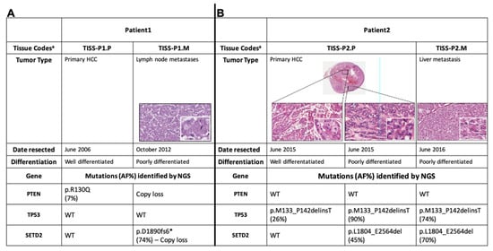
Hürthle cell carcinomas (HCC) are rare differentiated thyroid cancers that display low avidity for radioactive iodine and respond poorly to kinase inhibitors. Here, using next-generation sequencing, we analyzed the mutational status of primary tissue and poorly differentiated metastatic tissue from two HCC patients. In both cases, metastatic tissues harbored a mutation of SETD2, each resulting in loss of the SRI and WW domains of SETD2, a methyltransferase that trimethylates H3K36 (H3K36me3) and also interacts with p53 to promote its stability. Functional studies of the novel p.D1890fs6* mutation (case 1) revealed significantly reduced H3K36me3 levels in SETD2-mutated tissue and primary cell cultures and decreased levels of the active form of p53. Restoration of SETD2-wildtype expression in the SETD2-mutant cells significantly reduced the expression of four well-known stemness markers (OCT-4, SOX2, IPF1, Goosecoid). These findings suggest potential roles for SETD2 loss-of-function mutations in HCC progression, possibly involving p53 destabilization and promotion of stemness. Their prevalence and potential treatment implications in thyroid cancer, especially HCC, require further study. Full article
(This article belongs to the Collection Cancer Biomarkers)
Show Figures

Figure 1

10 pages, 1054 KiB  
Article
Clinical, Hematologic, Biologic and Molecular Characteristics of Patients with Myeloproliferative Neoplasms and a Chronic Myelomonocytic Leukemia-Like Phenotype
by Sonja Heibl, Bettina Gisslinger, Eva Jäger, Agnes Barna, Michael Gurbisz, Maike Stegemann, Peter Bettelheim, Sigrid Machherndl-Spandl, Michael Pfeilstöcker, Thomas Nösslinger, Gökhan Uyanik, Gregor Hoermann, Reinhard Stauder, Josef Thaler, Rajko Kusec, Peter Valent, Heinz Gisslinger and Klaus Geissler
Cancers 2020, 12(7), 1891; https://doi.org/10.3390/cancers12071891 - 14 Jul 2020
Cited by 5 | Viewed by 3501
Abstract
Patients with a myeloproliferative neoplasm (MPN) sometimes show a chronic myelomonocytic leukemia (CMML)-like phenotype but, according to the 2016 WHO classification, a documented history of an MPN excludes the diagnosis of CMML. Forty-one patients with an MPN (35 polycythemia vera (PV), 5 primary [...] Read more.
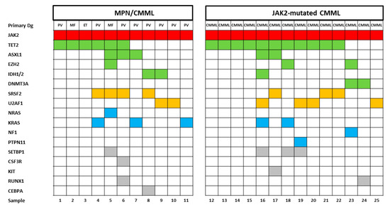
Patients with a myeloproliferative neoplasm (MPN) sometimes show a chronic myelomonocytic leukemia (CMML)-like phenotype but, according to the 2016 WHO classification, a documented history of an MPN excludes the diagnosis of CMML. Forty-one patients with an MPN (35 polycythemia vera (PV), 5 primary myelofibrosis, 1 essential thrombocythemia) and a CMML-like phenotype (MPN/CMML) were comprehensively characterized regarding clinical, hematologic, biologic and molecular features. The white blood cell counts in MPN/CMML patients were not different from CMML patients and PV patients. The hemoglobin values and platelet counts of these patients were higher than in CMML but lower than in PV, respectively. MPN/CMML patients showed myelomonocytic skewing, a typical in vitro feature of CMML but not of PV. The mutational landscape of MPN/CMML was not different from JAK2-mutated CMML. In two MPN/CMML patients, development of a CMML-like phenotype was associated with a decrease in the JAK2 V617F allelic burden. Finally, the prognosis of MPN/CMML (median overall survival (OS) 27 months) was more similar to CMML (JAK2-mutated, 28 months; JAK2-nonmutated 29 months) than to PV (186 months). In conclusion, we show that patients with MPN and a CMML-like phenotype share more characteristics with CMML than with PV, which may be relevant for their classification and clinical management. Full article
(This article belongs to the Special Issue New Insights into Myeloproliferative Neoplasms)
Show Figures

Figure 1

14 pages, 2329 KiB  
Article
Identification of Novel microRNA Prognostic Markers Using Cascaded Wx, a Neural Network-Based Framework, in Lung Adenocarcinoma Patients
by Jeong Seon Kim, Sang Hoon Chun, Sungsoo Park, Sieun Lee, Sae Eun Kim, Ji Hyung Hong, Keunsoo Kang, Yoon Ho Ko and Young-Ho Ahn
Cancers 2020, 12(7), 1890; https://doi.org/10.3390/cancers12071890 - 14 Jul 2020
Cited by 7 | Viewed by 3603
Abstract
The evolution of next-generation sequencing technology has resulted in a generation of large amounts of cancer genomic data. Therefore, increasingly complex techniques are required to appropriately analyze this data in order to determine its clinical relevance. In this study, we applied a neural [...] Read more.
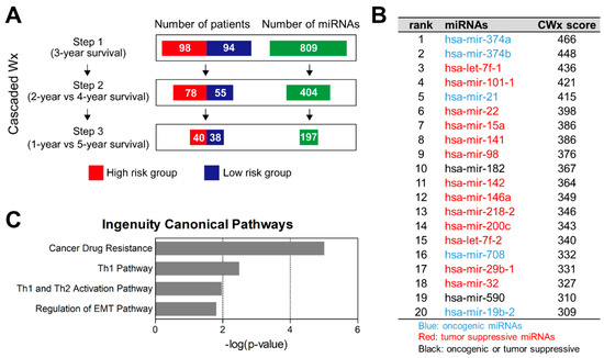
The evolution of next-generation sequencing technology has resulted in a generation of large amounts of cancer genomic data. Therefore, increasingly complex techniques are required to appropriately analyze this data in order to determine its clinical relevance. In this study, we applied a neural network-based technique to analyze data from The Cancer Genome Atlas and extract useful microRNA (miRNA) features for predicting the prognosis of patients with lung adenocarcinomas (LUAD). Using the Cascaded Wx platform, we identified and ranked miRNAs that affected LUAD patient survival and selected the two top-ranked miRNAs (miR-374a and miR-374b) for measurement of their expression levels in patient tumor tissues and in lung cancer cells exhibiting an altered epithelial-to-mesenchymal transition (EMT) status. Analysis of miRNA expression from tumor samples revealed that high miR-374a/b expression was associated with poor patient survival rates. In lung cancer cells, the EMT signal induced miR-374a/b expression, which, in turn, promoted EMT and invasiveness. These findings demonstrated that this approach enabled effective identification and validation of prognostic miRNA markers in LUAD, suggesting its potential efficacy for clinical use. Full article
(This article belongs to the Collection Cancer Biomarkers)
Show Figures

Figure 1

30 pages, 1406 KiB  
Review
Towards Clinical Implementation of Adeno-Associated Virus (AAV) Vectors for Cancer Gene Therapy: Current Status and Future Perspectives
by Ulrich T. Hacker, Martin Bentler, Dorota Kaniowska, Michael Morgan and Hildegard Büning
Cancers 2020, 12(7), 1889; https://doi.org/10.3390/cancers12071889 - 14 Jul 2020
Cited by 55 | Viewed by 10496
Abstract
Adeno-associated virus (AAV) vectors have gained tremendous attention as in vivo delivery systems in gene therapy for inherited monogenetic diseases. First market approvals, excellent safety data, availability of large-scale production protocols, and the possibility to tailor the vector towards optimized and cell-type specific [...] Read more.
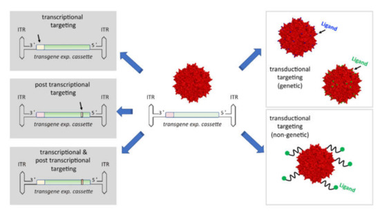
Adeno-associated virus (AAV) vectors have gained tremendous attention as in vivo delivery systems in gene therapy for inherited monogenetic diseases. First market approvals, excellent safety data, availability of large-scale production protocols, and the possibility to tailor the vector towards optimized and cell-type specific gene transfer offers to move from (ultra) rare to common diseases. Cancer, a major health burden for which novel therapeutic options are urgently needed, represents such a target. We here provide an up-to-date overview of the strategies which are currently developed for the use of AAV vectors in cancer gene therapy and discuss the perspectives for the future translation of these pre-clinical approaches into the clinic. Full article
(This article belongs to the Special Issue Antigens and Cancer Therapy)
Show Figures

Figure 1

19 pages, 4034 KiB  
Review
Relevance of Regulatory T Cells during Colorectal Cancer Development
by Jonadab E. Olguín, Itzel Medina-Andrade, Tonathiu Rodríguez, Miriam Rodríguez-Sosa and Luis I. Terrazas
Cancers 2020, 12(7), 1888; https://doi.org/10.3390/cancers12071888 - 14 Jul 2020
Cited by 47 | Viewed by 6163
Abstract
In recent years, there has been a significant increase in the study of own and foreign human factors favoring the development of different types of cancer, including genetic and environmental ones. However, the fact that the immune response plays a fundamental role in [...] Read more.
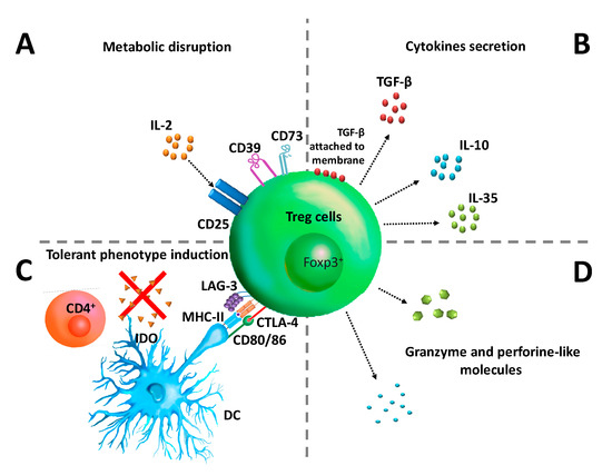
In recent years, there has been a significant increase in the study of own and foreign human factors favoring the development of different types of cancer, including genetic and environmental ones. However, the fact that the immune response plays a fundamental role in the development of immunity and susceptibility to colorectal cancer (CRC) is much stronger. Among the many cell populations of the immune system that participate in restricting or favoring CRC development, regulatory T cells (Treg) play a major role in orchestrating immunomodulation during CRC. In this review, we established concrete evidence supporting the fact that Treg cells have an important role in the promotion of tumor development during CRC, mediating an increasing suppressive capacity which controls the effector immune response, and generating protection for tumors. Furthermore, Treg cells go through a process called “phenotypic plasticity”, where they co-express transcription factors that promote an inflammatory profile. We reunited evidence that describes the interaction between the different effector populations of the immune response and its modulation by Treg cells adapted to the tumor microenvironment, including the mechanisms used by Treg cells to suppress the protective immune response, as well as the different subpopulations of Treg cells participating in tumor progression, generating susceptibility during CRC development. Finally, we discussed whether Treg cells might or might not be a therapeutic target for an effective reduction in the morbidity and mortality caused by CRC. Full article
(This article belongs to the Special Issue Targeting Therapy for Colon Cancer)
Show Figures

Figure 1

28 pages, 2127 KiB  
Review
The Role of Cancer-Associated Fibroblasts in Prostate Cancer Tumorigenesis
by Francesco Bonollo, George N. Thalmann, Marianna Kruithof-de Julio and Sofia Karkampouna
Cancers 2020, 12(7), 1887; https://doi.org/10.3390/cancers12071887 - 13 Jul 2020
Cited by 107 | Viewed by 8909
Abstract
Tumors strongly depend on their surrounding tumor microenvironment (TME) for growth and progression, since stromal elements are required to generate the optimal conditions for cancer cell proliferation, invasion, and possibly metastasis. Prostate cancer (PCa), though easily curable during primary stages, represents a clinical [...] Read more.
Tumors strongly depend on their surrounding tumor microenvironment (TME) for growth and progression, since stromal elements are required to generate the optimal conditions for cancer cell proliferation, invasion, and possibly metastasis. Prostate cancer (PCa), though easily curable during primary stages, represents a clinical challenge in advanced stages because of the acquisition of resistance to anti-cancer treatments, especially androgen-deprivation therapies (ADT), which possibly lead to uncurable metastases such as those affecting the bone. An increasing number of studies is giving evidence that prostate TME components, especially cancer-associated fibroblasts (CAFs), which are the most abundant cell type, play a causal role in PCa since the very early disease stages, influencing therapy resistance and metastatic progression. This is highlighted by the prognostic value of the analysis of stromal markers, which may predict disease recurrence and metastasis. However, further investigations on the molecular mechanisms of tumor–stroma interactions are still needed to develop novel therapeutic approaches targeting stromal components. In this review, we report the current knowledge of the characteristics and functions of the stroma in prostate tumorigenesis, including relevant discussion of normal prostate homeostasis, chronic inflammatory conditions, pre-neoplastic lesions, and primary and metastatic tumors. Specifically, we focus on the role of CAFs, to point out their prognostic and therapeutic potential in PCa. Full article
(This article belongs to the Section Tumor Microenvironment)
Show Figures

Figure 1

33 pages, 2812 KiB  
Review
Cancer Nanomedicine Special Issue Review Anticancer Drug Delivery with Nanoparticles: Extracellular Vesicles or Synthetic Nanobeads as Therapeutic Tools for Conventional Treatment or Immunotherapy
by Maria Raffaella Zocchi, Francesca Tosetti, Roberto Benelli and Alessandro Poggi
Cancers 2020, 12(7), 1886; https://doi.org/10.3390/cancers12071886 - 13 Jul 2020
Cited by 24 | Viewed by 5634
Abstract
Both natural and synthetic nanoparticles have been proposed as drug carriers in cancer treatment, since they can increase drug accumulation in target tissues, optimizing the therapeutic effect. As an example, extracellular vesicles (EV), including exosomes (Exo), can become drug vehicles through endogenous or [...] Read more.
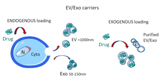
Both natural and synthetic nanoparticles have been proposed as drug carriers in cancer treatment, since they can increase drug accumulation in target tissues, optimizing the therapeutic effect. As an example, extracellular vesicles (EV), including exosomes (Exo), can become drug vehicles through endogenous or exogenous loading, amplifying the anticancer effects at the tumor site. In turn, synthetic nanoparticles (NP) can carry therapeutic molecules inside their core, improving solubility and stability, preventing degradation, and controlling their release. In this review, we summarize the recent advances in nanotechnology applied for theranostic use, distinguishing between passive and active targeting of these vehicles. In addition, examples of these models are reported: EV as transporters of conventional anticancer drugs; Exo or NP as carriers of small molecules that induce an anti-tumor immune response. Finally, we focus on two types of nanoparticles used to stimulate an anticancer immune response: Exo carried with A Disintegrin And Metalloprotease-10 inhibitors and NP loaded with aminobisphosphonates. The former would reduce the release of decoy ligands that impair tumor cell recognition, while the latter would activate the peculiar anti-tumor response exerted by γδ T cells, creating a bridge between innate and adaptive immunity. Full article
(This article belongs to the Collection Cancer Nanomedicine)
Show Figures

Figure 1

2 pages, 469 KiB  
Correction
Correction: Ordoñez, et al.; DNA Methylation of Enhancer Elements in Myeloid Neoplasms: Think Outside the Promoters? Cancers 2019, 11, 1424
by Raquel Ordoñez, Nicolás Martínez-Calle, Xabier Agirre and Felipe Prosper
Cancers 2020, 12(7), 1885; https://doi.org/10.3390/cancers12071885 - 13 Jul 2020
Viewed by 2123
Abstract
The authors would like to make a correction to their published paper [...] Full article
Show Figures

Figure 1

20 pages, 3693 KiB  
Article
Current Trends of Artificial Intelligence for Colorectal Cancer Pathology Image Analysis: A Systematic Review
by Nishant Thakur, Hongjun Yoon and Yosep Chong
Cancers 2020, 12(7), 1884; https://doi.org/10.3390/cancers12071884 - 13 Jul 2020
Cited by 62 | Viewed by 8145
Abstract
Colorectal cancer (CRC) is one of the most common cancers requiring early pathologic diagnosis using colonoscopy biopsy samples. Recently, artificial intelligence (AI) has made significant progress and shown promising results in the field of medicine despite several limitations. We performed a systematic review [...] Read more.
Colorectal cancer (CRC) is one of the most common cancers requiring early pathologic diagnosis using colonoscopy biopsy samples. Recently, artificial intelligence (AI) has made significant progress and shown promising results in the field of medicine despite several limitations. We performed a systematic review of AI use in CRC pathology image analysis to visualize the state-of-the-art. Studies published between January 2000 and January 2020 were searched in major online databases including MEDLINE (PubMed, Cochrane Library, and EMBASE). Query terms included “colorectal neoplasm,” “histology,” and “artificial intelligence.” Of 9000 identified studies, only 30 studies consisting of 40 models were selected for review. The algorithm features of the models were gland segmentation (n = 25, 62%), tumor classification (n = 8, 20%), tumor microenvironment characterization (n = 4, 10%), and prognosis prediction (n = 3, 8%). Only 20 gland segmentation models met the criteria for quantitative analysis, and the model proposed by Ding et al. (2019) performed the best. Studies with other features were in the elementary stage, although most showed impressive results. Overall, the state-of-the-art is promising for CRC pathological analysis. However, datasets in most studies had relatively limited scale and quality for clinical application of this technique. Future studies with larger datasets and high-quality annotations are required for routine practice-level validation. Full article
(This article belongs to the Section Systematic Review or Meta-Analysis in Cancer Research)
Show Figures

Figure 1

13 pages, 2303 KiB  
Article
Fast, In Vivo Model for Drug-Response Prediction in Patients with B-Cell Precursor Acute Lymphoblastic Leukemia
by Anton Gauert, Nadine Olk, Helia Pimentel-Gutiérrez, Kathy Astrahantseff, Lasse D. Jensen, Yihai Cao, Angelika Eggert, Cornelia Eckert and Anja I.H. Hagemann
Cancers 2020, 12(7), 1883; https://doi.org/10.3390/cancers12071883 - 13 Jul 2020
Cited by 19 | Viewed by 4786
Abstract
Only half of patients with relapsed B-cell precursor (BCP) acute lymphoblastic leukemia (ALL) currently survive with standard treatment protocols. Predicting individual patient responses to defined drugs prior to application would help therapy stratification and could improve survival. With the purpose to aid personalized [...] Read more.
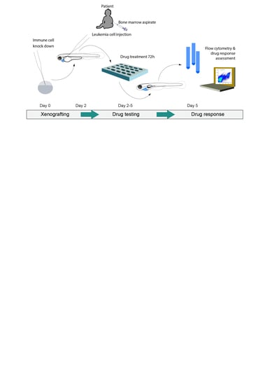
Only half of patients with relapsed B-cell precursor (BCP) acute lymphoblastic leukemia (ALL) currently survive with standard treatment protocols. Predicting individual patient responses to defined drugs prior to application would help therapy stratification and could improve survival. With the purpose to aid personalized targeted treatment approaches, we developed a human–zebrafish xenograft (ALL-ZeFiX) assay to predict drug response in a patient in 5 days. Leukemia blast cells were pericardially engrafted into transiently immunosuppressed Danio rerio embryos, and engrafted embryos treated for the test case, venetoclax, before single-cell dissolution for quantitative whole blast cell analysis. Bone marrow blasts from patients with newly diagnosed or relapsed BCP-ALL were successfully expanded in 60% of transplants in immunosuppressed zebrafish embryos. The response of BCP-ALL cell lines to venetoclax in ALL-ZeFiX assays mirrored responses in 2D cultures. Venetoclax produced varied responses in patient-derived BCP-ALL grafts, including two results mirroring treatment responses in two refractory BCP-ALL patients treated with venetoclax. Here we demonstrate proof-of-concept for our 5-day ALL-ZeFiX assay with primary patient blasts and the test case, venetoclax, which after expanded testing for further targeted drugs could support personalized treatment decisions within the clinical time window for decision-making. Full article
(This article belongs to the Special Issue Patient-Derived Xenograft-Models in Cancer Research)
Show Figures

Graphical abstract

19 pages, 7040 KiB  
Article
CCR2 of Tumor Microenvironmental Cells Is a Relevant Modulator of Glioma Biology
by Matthäus Felsenstein, Anne Blank, Alexander D. Bungert, Annett Mueller, Adnan Ghori, Irina Kremenetskaia, Olga Rung, Thomas Broggini, Kati Turkowski, Lea Scherschinski, Jonas Raggatz, Peter Vajkoczy and Susan Brandenburg
Cancers 2020, 12(7), 1882; https://doi.org/10.3390/cancers12071882 - 13 Jul 2020
Cited by 24 | Viewed by 5156
Abstract
Glioblastoma multiforme (GBM) shows a high influx of tumor-associated macrophages (TAMs). The CCR2/CCL2 pathway is considered a relevant signal for the recruitment of TAMs and has been suggested as a therapeutic target in malignant gliomas. We found that TAMs of human GBM specimens [...] Read more.
Glioblastoma multiforme (GBM) shows a high influx of tumor-associated macrophages (TAMs). The CCR2/CCL2 pathway is considered a relevant signal for the recruitment of TAMs and has been suggested as a therapeutic target in malignant gliomas. We found that TAMs of human GBM specimens and of a syngeneic glioma model express CCR2 to varying extents. Using a Ccr2-deficient strain for glioma inoculation revealed a 30% reduction of TAMs intratumorally. This diminished immune cell infiltration occurred with augmented tumor volumes likely based on increased cell proliferation. Remaining TAMs in Ccr2-/- mice showed comparable surface marker expression patterns in comparison to wildtype mice, but expression levels of inflammatory transcription factors (Stat3, Irf7, Cox2) and cytokines (Ifnβ, Il1β, Il12α) were considerably affected. Furthermore, we demonstrated an impact on blood vessel integrity, while vascularization of tumors appeared similar between mouse strains. The higher stability and attenuated leakiness of the tumor vasculature imply improved sustenance of glioma tissue in Ccr2-/- mice. Additionally, despite TAMs residing in the perivascular niche in Ccr2-/- mice, their pro-angiogenic activity was reduced by the downregulation of Vegf. In conclusion, lacking CCR2 solely on tumor microenvironmental cells leads to enhanced tumor progression, whereby high numbers of TAMs infiltrate gliomas independently of the CCR2/CCL2 signal. Full article
Show Figures

Figure 1

25 pages, 2202 KiB  
Review
Nitric Oxide (NO) and NO Synthases (NOS)-Based Targeted Therapy for Colon Cancer
by Hao Wang, Liye Wang, Zuoxu Xie, Shuang Zhou, Yan Li, Yue Zhou and Meiyan Sun
Cancers 2020, 12(7), 1881; https://doi.org/10.3390/cancers12071881 - 13 Jul 2020
Cited by 63 | Viewed by 7735
Abstract
Colorectal cancer (CRC) is one of the most lethal malignancies worldwide and CRC therapy remains unsatisfactory. In recent decades, nitric oxide (NO)—a free-radical gas—plus its endogenous producer NO synthases (NOS), have attracted considerable attention. NO exerts dual effects (pro- and anti-tumor) in cancers. [...] Read more.
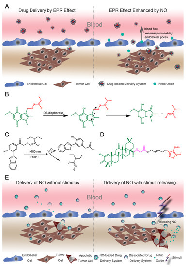
Colorectal cancer (CRC) is one of the most lethal malignancies worldwide and CRC therapy remains unsatisfactory. In recent decades, nitric oxide (NO)—a free-radical gas—plus its endogenous producer NO synthases (NOS), have attracted considerable attention. NO exerts dual effects (pro- and anti-tumor) in cancers. Endogenous levels of NO promote colon neoplasms, whereas exogenously sustained doses lead to cytotoxic functions. Importantly, NO has been implicated as an essential mediator in many signaling pathways in CRC, such as the Wnt/β-catenin and extracellular-signal-regulated kinase (ERK) pathways, which are closely associated with cancer initiation, metastasis, inflammation, and chemo-/radio-resistance. Therefore, NO/NOS have been proposed as promising targets in the regulation of CRC carcinogenesis. Clinically relevant NO-donating agents have been developed for CRC therapy to deliver a high level of NO to tumor sites. Notably, inducible NOS (iNOS) is ubiquitously over-expressed in inflammatory-associated colon cancer. The development of iNOS inhibitors contributes to targeted therapies for CRC with clinical benefits. In this review, we summarize the multifaceted mechanisms of NO-mediated networks in several hallmarks of CRC. We review the clinical manifestation and limitations of NO donors and NOS inhibitors in clinical trials. We also discuss the possible directions of NO/NOS therapies in the immediate future. Full article
(This article belongs to the Special Issue Targeting Therapy for Colon Cancer)
Show Figures

Figure 1

25 pages, 4691 KiB  
Article
Leucine-Rich Diet Modulates the Metabolomic and Proteomic Profile of Skeletal Muscle during Cancer Cachexia
by Bread Cruz, André Oliveira, Lais Rosa Viana, Leisa Lopes-Aguiar, Rafael Canevarolo, Maiara Caroline Colombera, Rafael Rossi Valentim, Fernanda Garcia-Fóssa, Lizandra Maia de Sousa, Bianca Gazieri Castelucci, Sílvio Roberto Consonni, Daniel Martins-de-Souza, Marcelo Bispo de Jesus, Steven Thomas Russell and Maria Cristina Cintra Gomes-Mardondes
Cancers 2020, 12(7), 1880; https://doi.org/10.3390/cancers12071880 - 13 Jul 2020
Cited by 17 | Viewed by 5245
Abstract
Background: Cancer-cachexia induces a variety of metabolic disorders, including skeletal muscle imbalance. Alternative therapy, as nutritional supplementation with leucine, shows a modulatory effect over tumour damage in vivo and in vitro. Method: Adult rats distributed into Control (C), Walker tumour-bearing (W), control fed [...] Read more.
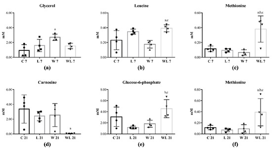
Background: Cancer-cachexia induces a variety of metabolic disorders, including skeletal muscle imbalance. Alternative therapy, as nutritional supplementation with leucine, shows a modulatory effect over tumour damage in vivo and in vitro. Method: Adult rats distributed into Control (C), Walker tumour-bearing (W), control fed a leucine-rich diet (L), and tumour-bearing fed a leucine-rich diet (WL) groups had the gastrocnemius muscle metabolomic and proteomic assays performed in parallel to in vitro assays. Results: W group presented an affected muscle metabolomic and proteomic profile mainly related to energy generation and carbohydrates catabolic processes, but leucine-supplemented group (WL) recovered the energy production. In vitro assay showed that cell proliferation, mitochondria number and oxygen consumption were higher under leucine effect than the tumour influence. Muscle proteomics results showed that the main affected cell component was mitochondria, leading to an impacted energy generation, including impairment in proteins of the tricarboxylic cycle and carbohydrates catabolic processes, which were modulated and improved by leucine treatment. Conclusion: In summary, we showed a beneficial effect of leucine upon mitochondria, providing information about the muscle glycolytic pathways used by this amino acid, where it can be associated with the preservation of morphometric parameters and consequent protection against the effects of cachexia. Full article
(This article belongs to the Special Issue The Cancer Proteome)
Show Figures

Figure 1

16 pages, 2937 KiB  
Article
MiR-216b/Smad3/BCL-2 Axis Is Involved in Smoking-Mediated Drug Resistance in Non-Small Cell Lung Cancer
by Trung Vu, Shanzhong Yang and Pran K. Datta
Cancers 2020, 12(7), 1879; https://doi.org/10.3390/cancers12071879 - 13 Jul 2020
Cited by 13 | Viewed by 3572
Abstract
Epidemiologic studies have shown that vast majority of lung cancers (85–90%) are causally linked to tobacco smoking. Although much information has been gained about the effects of smoking on various signaling pathways, little is known about how deregulation of miRNAs leads to activation [...] Read more.
Epidemiologic studies have shown that vast majority of lung cancers (85–90%) are causally linked to tobacco smoking. Although much information has been gained about the effects of smoking on various signaling pathways, little is known about how deregulation of miRNAs leads to activation of oncogenes and inhibition of tumor suppressor genes in non-small cell lung cancer (NSCLC). Our previous study showed that smoking inhibits TGF-β-induced tumor suppressor functions through downregulation of Smad3 in lung cancer cells. In order to understand the upstream mechanism of downregulation of Smad3 by smoking, we performed miRNA microarray analyses after treating human lung adenocarcinoma A549 and immortalized peripheral lung epithelial HPL1A cells with cigarette smoke condensate (CSC). We identified miR-216b as being upregulated in CSC treated cells. MiR-216b overexpression decreases Smad3 protein expression by binding to its 3′-UTR, and attenuates transforming growth factor beta (TGF-β) signaling and target gene expression. MiR-216b increases B-cell lymphoma 2 (BCL-2) expression and promotes chemoresistance of NSCLC cells by decreasing apoptosis. Increased acetylation of histones H3 and H4 in miR-216b gene promoter plays a role in CSC induced miR-216b expression. Taken together, these results suggest that smoking-mediated upregulation of miR-216b increases NSCLC cell growth by downregulating Smad3 and inhibiting TGF-β-induced tumor suppressor function, and induces resistance to platinum-based therapy. Full article
(This article belongs to the Section Molecular Cancer Biology)
Show Figures

Figure 1