Article

Phenotype-Driven Diagnostic of PTEN Hamartoma Tumor Syndrome: Macrocephaly, But Neither Height nor Weight Development, Is the Important Trait in Children

Pediatric Endocrinology and Diabetology Division, Children’s Hospital, University of Bonn, Adenauerallee 119, 53113 Bonn, Germany
*
Authors to whom correspondence should be addressed.
Cancers 2019, 11(7), 975; https://doi.org/10.3390/cancers11070975
Submission received: 7 May 2019 / Revised: 4 July 2019 / Accepted: 8 July 2019 / Published: 11 July 2019
(This article belongs to the Special Issue PTEN: A Multifaceted Tumor Suppressor)

Abstract

:
PTEN hamartoma tumor syndrome (PHTS) encompasses different syndromic disorders which are associated with autosomal-dominant mutations of the tumor suppressor gene PTEN. Patients are at high risk to develop benign and malignant tumors. Macrocephaly is a diagnostic feature, but there is a paucity of data on auxological development during childhood. Growth charts for height, weight and head circumference for PHTS do not exist yet. In this study, patient data for height, weight and head circumferences (HC) were collected from repeated medical exams or prevention check-up visits starting at birth. Growth charts were generated and compared to German reference data. Standard deviation scores (SDS) of HC, height and body mass index (BMI) were calculated. We included 23 pediatric patients (8 female, 15 male) with molecular proven PTEN gene mutation. Most male patients already demonstrated macrocephaly at birth (73%), whereas only one female patient had documented congenital macrocephaly. By the age of two years all patients exhibited a head circumference above the 97th percentile. Stratified for different age groups the median HC-SDSs were between +3.3 and +5.5 in male patients and between +2.9 and +4.1 in female patients. Height, weight and BMI measurements for both sexes were mostly within the normal range. We conclude that macrocephaly, but not height, weight or BMI, is useful in the identification of PHTS patients. The increased HC in PHTS patients develops early in life and is more pronounced in males than in females, which might explain the finding of a higher percentage of male PHTS patients diagnosed during childhood.

1. Introduction

PTEN hamartoma tumor syndrome (PHTS) encompasses different syndromic disorders (Cowden syndrome, Bannayan Riley Ruvalcaba syndrome, Lhermitte Duclos syndrome, Proteus and Proteus-like syndrome, juvenile polyposis of infancy, autism spectrum disorders with macrocephaly or children with macrocephaly and developmental delay/cognitive impairment), which are all associated with germline autosomal-dominant mutations of the tumor suppressor gene PTEN.
Bannayan-Riley-Ruvalcaba syndrome (BRRS) and Cowden syndrome (CS) can be seen as one condition with variable expression and age-related penetrance [1,2]. Patients with Cowden syndrome (CS) especially are at a significantly increased risk for the development of benign and malignant tumors. The most frequently affected organs are the breast, endometrium and thyroid, but colorectal, renal, central nervous system, and skin tumors have also been reported [3]. It is noteworthy however, that in addition to CS and BRRS, all individuals with PHTS have an increased risk of malignancy and benefit from cancer surveillance strategies.
In the diagnostic workup of subjects with CS [4,5,6] and BRRS [1,7], macrocephaly (MC) is an important diagnostic feature. Whereas several forms of cancer and polyps are common in adult patients, macrocephaly with or without neurodevelopmental disorders is frequently the only or the most evident symptom in childhood, thereby prompting a diagnostic workup. However, to date there is limited data available on prevalence, degree and longitudinal development of head circumference and other auxological parameters during childhood [8,9,10]. Syndrome-specific longitudinal data and growth charts for length/height, weight and head circumference do not exist yet.

2. Methods

We conducted a single-centered study on children and adolescents with molecularly proven PTEN hamartoma tumor syndrome, who presented in our pediatric endocrine clinic between the years 2013 and 2019. We included 23 patients (15 male, 8 female) with a current age range between two and 17 years (age range in boys from two to 17 years of age; in girls from three to ten years of age).
All available auxological data (length/height, weight and head circumference) were collected from medical reports and from the standardized German prevention check-up exams. Data were collected retrospectively starting at patient’s birth and prospectively from the first visit in our clinic until 2019. The collection of clinical data and cerebral magnetic resonance imaging (MRI) was performed prior to a diagnosis of PHTS or after confirmation of PHTS as part of a symptom-related workup. Growth charts were generated for height, weight and head circumference (HC) of each patient and compared to German reference data [11]. Standard deviation scores (SDS) for head circumference, height and BMI were calculated. Familial target height was calculated according to Tanner [12].
Macrocephaly was defined as a head circumference above the 97th percentile of German reference data from the KiGGS surveillance program [11]. Overweight was defined as a BMI above the 90th percentile, and obesity as a BMI above the 97th percentile [11].
Standard deviation scores (z-scores) were calculated by using the LMS method. It was chosen as it summarizes the data in terms of three age-specific curves called lambda (L), mu (M) and sigma (S) base on German population- specific data [13]. The calculation is based on the following formula:
Z = ((X/M)L − 1)/(S × L), for L not 0.
Because of the relatively small number of cases at different timepoints, patient data in growth charts are displayed as complete years. Patient’s age was defined as X years for patients between X ± 0.5 year (e.g., a patient’s age would be defined as 5 years for children aged 4.5 to 5.4 years).

3. Results

We included longitudinal auxological data from a total of 23 patients (8 female and 15 male patients) with molecularly proven PTEN gene mutation.

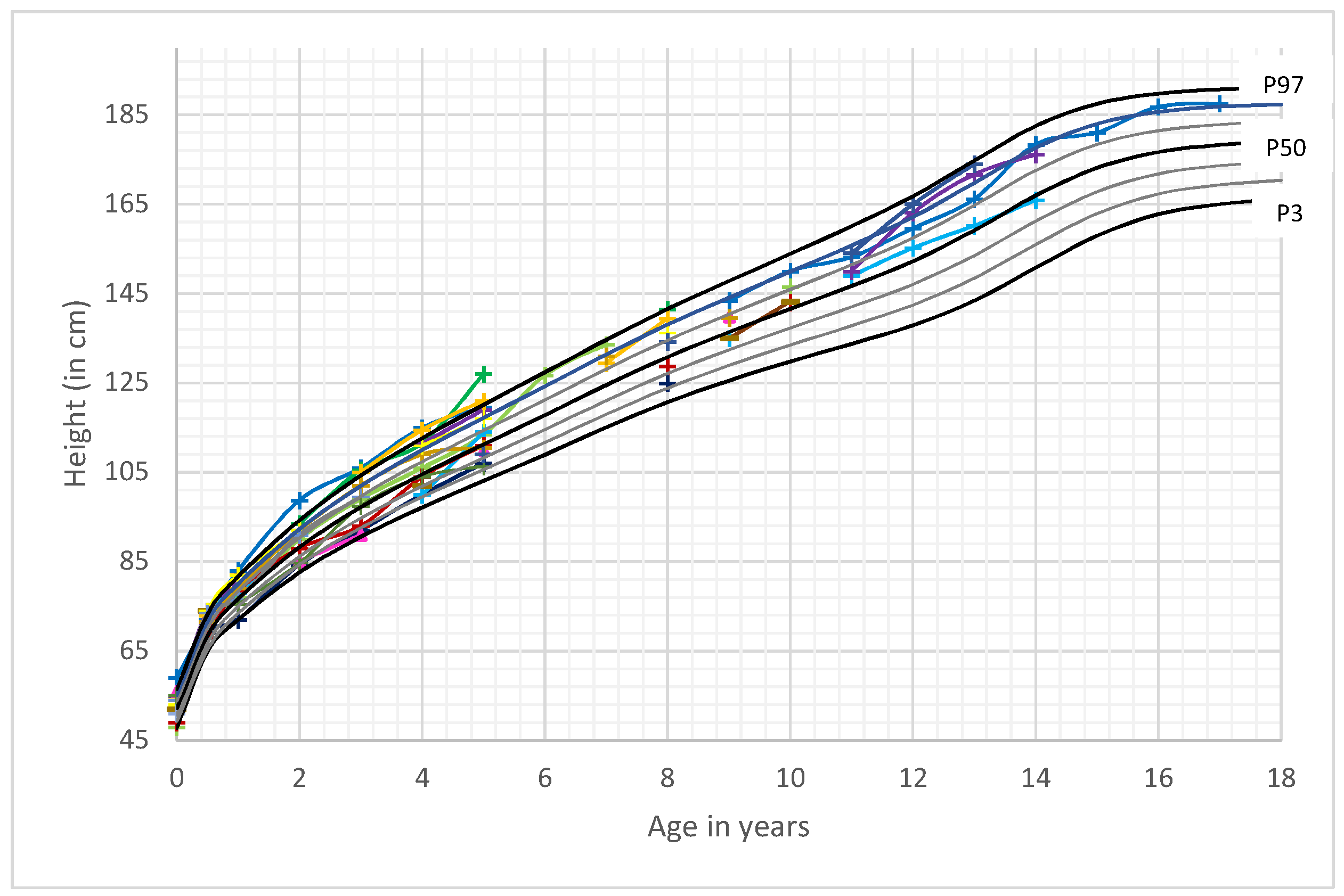
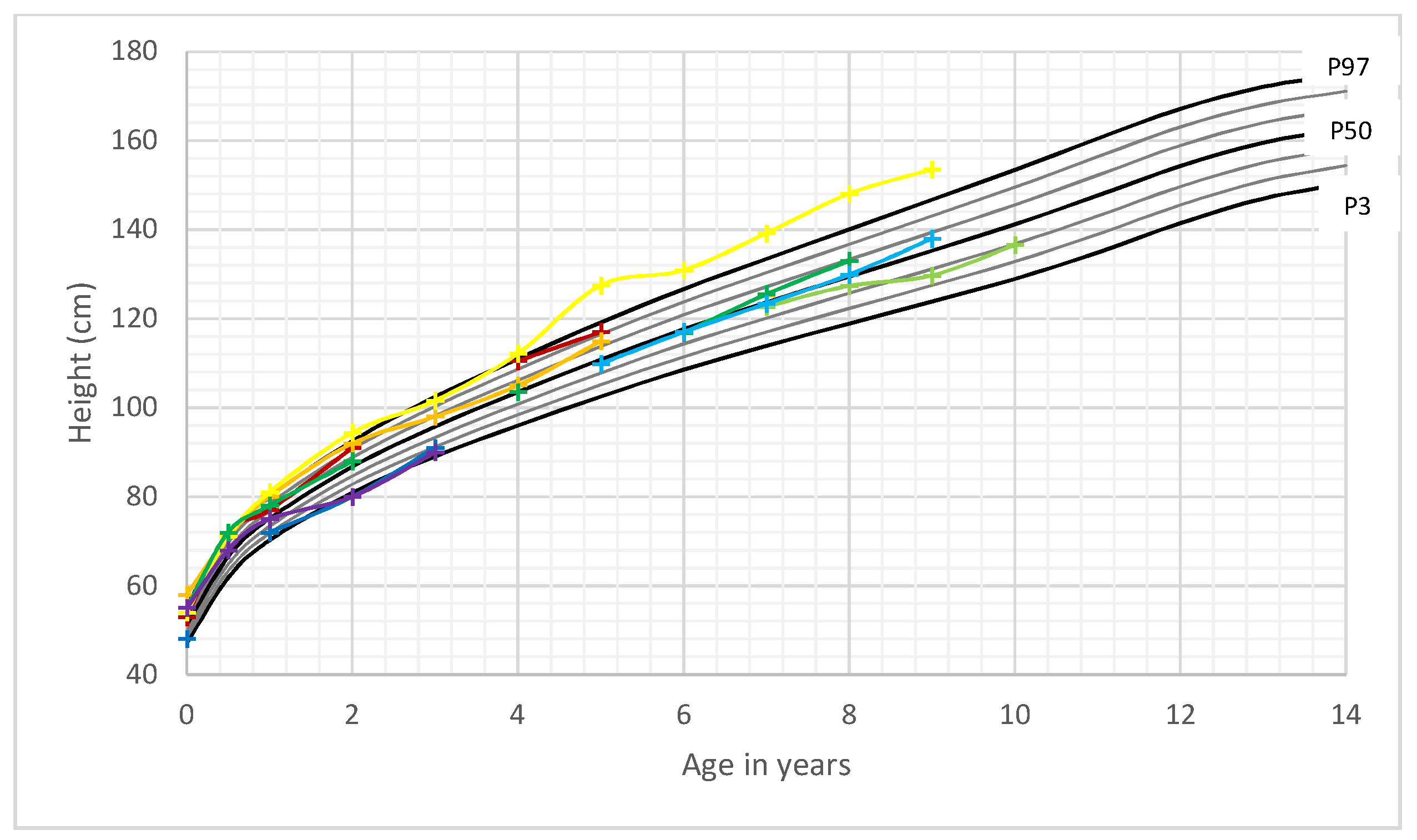
3.1. Head Circumference
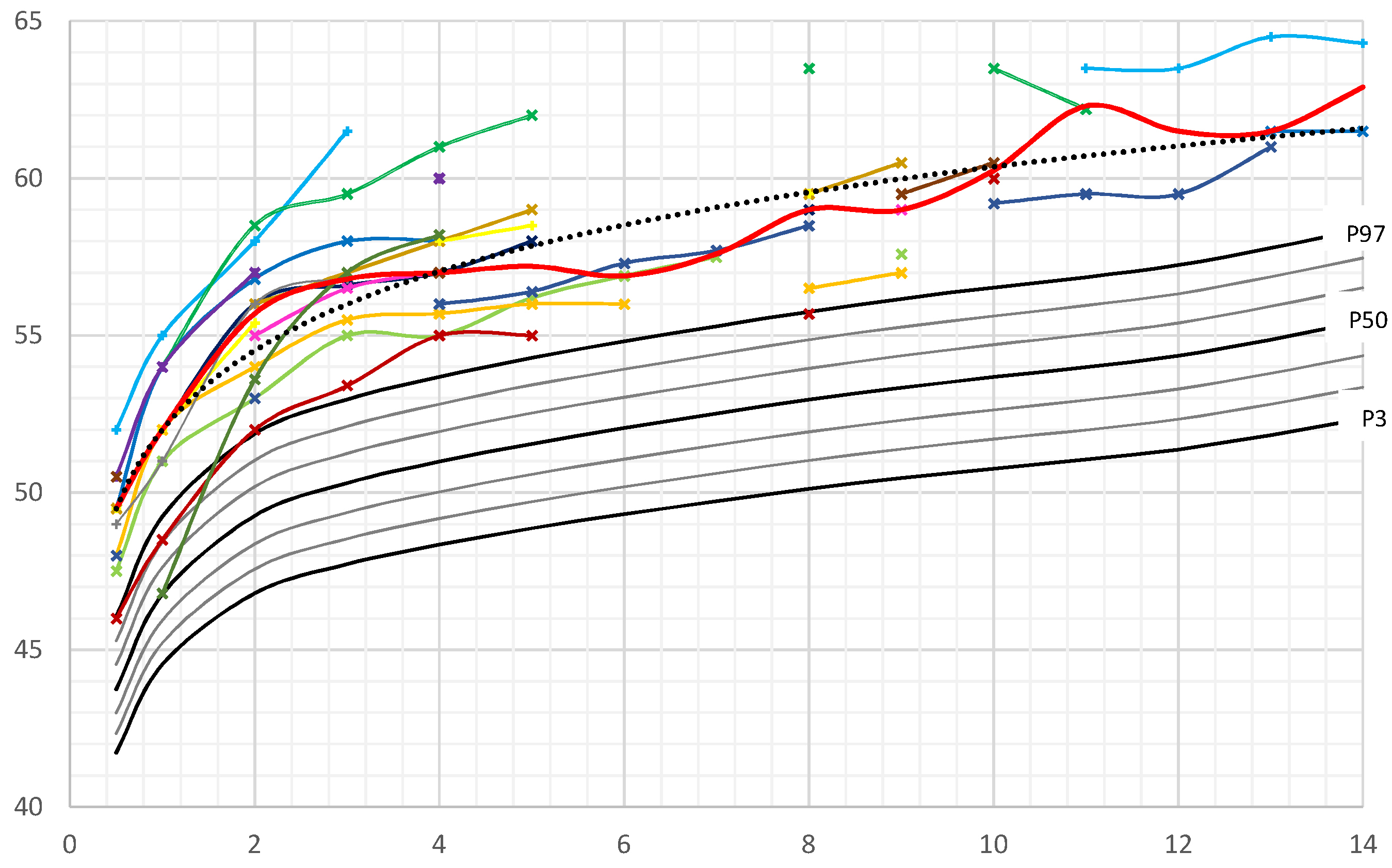
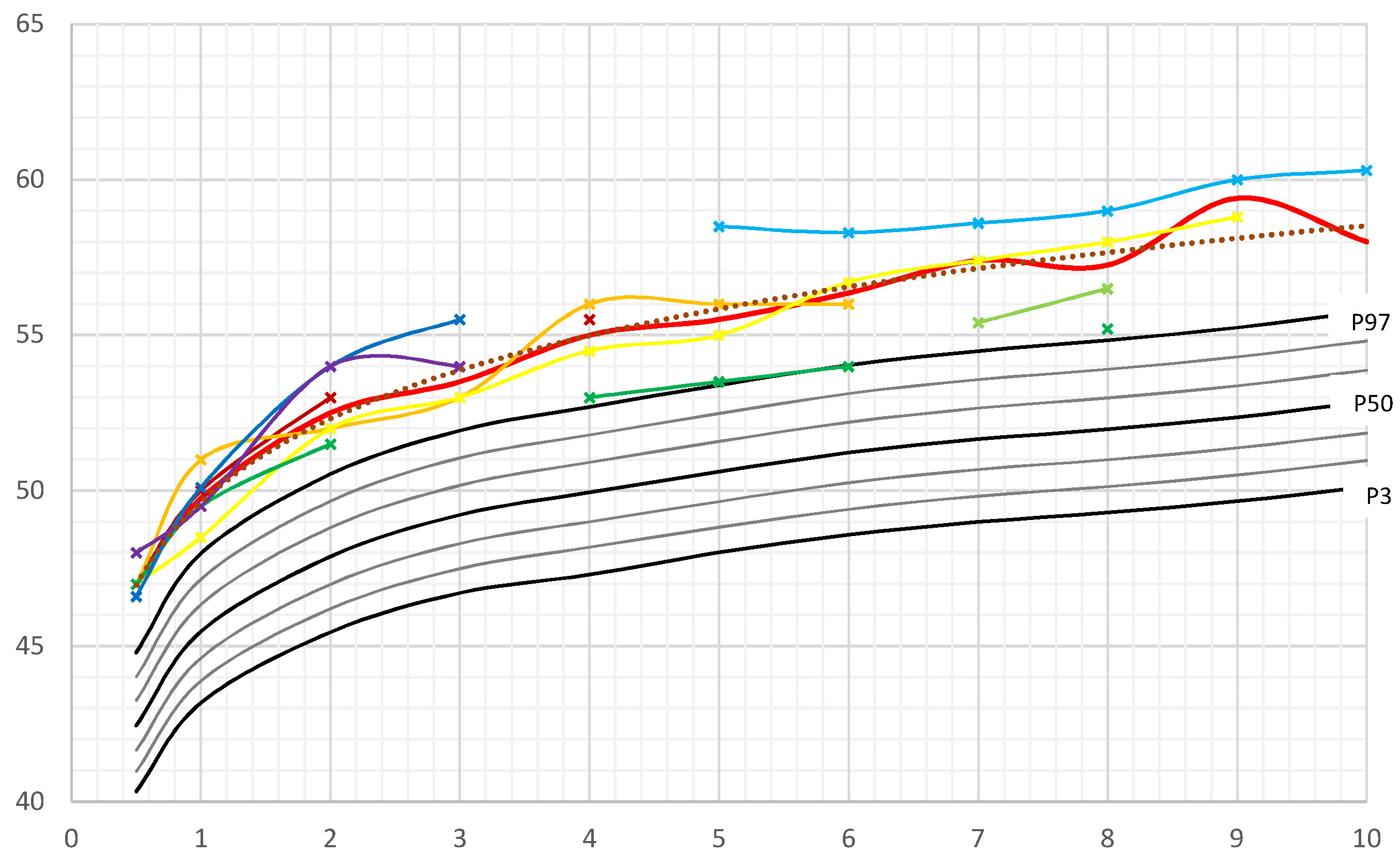
Birth data for all male patients were available. The majority of male patients exhibited macrocephaly already at birth (11/15, corresponding to 73%). Even though two patients with a head circumference below the 97th percentile were born preterm (34 + 5 and 35 + 0 weeks of gestation), there was still no difference in the percentage of macrocephaly when corrected for gestational age.
At the age of two years all male patients exhibited a head circumference above the 97th percentile (Figure 1).
Birth data from our female patients were available for 7/8 patients and corrected for gestational age. Only one female patient had documented congenital macrocephaly (14%), whereas all other female patients presented a head circumference between the 75th and 97th percentile. However, at the age of 0.5 years, HC was above the 97th percentile in all patients with data available (Figure 2).
12 out of 22 patients (55%) were born with macrocephaly. The method of delivery could be elicited for 20 patients. 14 of those 20 patients were born by Caesarean section (70%).
Stratified for different age groups, the median HC-SDSs of boys were between +3.3 and +5.5 (mean 4.2 SDS) (Figure S1). Median HC-SDS was lowest at the age of six and seven years and highest at the age of eleven. Outliers can be explained by the paucity of data for these age groups, with measurements of HC being available only in two patients (at seven years) respectively three patients (at six and eleven years).
Female median HC-SDSs were between 2.9 and 4.1 (mean 3.3) (Figure S2).
In our study, mean and median HC-SDS of male patients was almost one SDS higher than those of girls with PHTS. This finding was consistently present in all age groups (except for the few outliners described before).

3.2. Length/Height

Height development of boys and girls was within the normal range for most time points. All female patients grew within the adequate range for their individually calculated target height. Three out of the 15 boys (20%) demonstrated a growth pattern with a height above their respective familial target height range. All remaining boys (80%) exhibited a growth pattern corresponding to their parent’s heights (Figure 3 and Figure 4).

3.3. Weight and BMI Development

The majority of patients had a BMI-SDS above the 50th percentile (Figures S3 and S4). Especially in the younger age groups, the percentage of overweight and obese patients was higher compared to German reference data (Table 1).

3.4. Additional Brain MRI and Clinical Characteristics of the Cohort

3.4.1. Brain MRI Scans

15 of the 23 patients underwent a cerebral MRI scan. In all cases, the first MRI scan was performed within the diagnostic work-up of macrocephaly. 13 of 15 patients exhibited noticeable variations in the MRI; seven patients presented with white matter abnormalities. In three patients, enlarged perivascular spaces were found (Virchow Robin spaces). Additional pathologies included a Chiari malformation type I, arachnoid cysts, focal cortical dysplasia, a cavernoma and a clinical diagnosis of pseudotumor cerebri in one patient, respectively (Table 2).

3.4.2. Clinical Features

14 of the 23 (61%) patients exhibited thyroid pathology, whereas nine patients had no pathologies detected so far with ultrasound screening. One third of those without pathologies were less than three years, one third were between three and ten years of age, the last third were aged between eleven and thirteen years. Of those patients exhibiting pathology with ultrasound screening, seven patients (50%) underwent thyroid surgery due to highly suspicious lesions (age range six to thirteen years). Histopathology included nodular goiter, follicular adenoma, papillary microcarcinoma in a six-year-old-boy and follicular carcinoma in a 13-year-old girl. Additional pathologies comprised autoimmune thyroid disease, thyroid nodules, which are still under regular observation, and thyroid cysts (Table 2). Some of these findings have already been published [14]. Other features in PHTS patients of this cohort were lipoma, hemangioma, trichilemmoma and penile freckling. Until now colonoscopy has been performed in three patients. All of them revealed multiple polyps of the gastrointestinal tract. One patient carried an additional BMPR1A deletion and exhibited severe gastrointestinal symptoms. He underwent colectomy for disease control. In most cases, parents reported a relatively late motor development or a motoric clumsiness, as well as muscle hypotonia. Developmental delay and autism were documented in some cases (Table 2). Most of these patients fulfilled the criteria for BRRS. However, in several of these patients it was difficult to distinguish between a diagnosis of BRRS and CS, due to the difficulty when dealing with pediatric cases.

4. Discussion

In this study macrocephaly was found in all PHTS patients. Macrocephaly is a required criterion for diagnosis of PTEN hamartoma syndrome in childhood. A prevalence of 100% has been reported in several publications concerning pediatric patients [15,16,17,18]. This is also consistent with previous studies, which included adult patients with molecularly proven PTEN gene mutation, reporting macrocephaly prevalence between of 93% [19] and 100% [2,8,15,20]. One previous study reported a lower frequency of MC (80%) in patients with a clinical diagnosis of CS. However, this study was undertaken before PTEN was identified as the predisposing gene of CS [9]. In our study we observed a more pronounced degree of macrocephaly in male children and adolescents (mean HC-SDS + 4.2) compared to female patients (mean HC-SDS + 3.3). This sex discordance for the degree of HC-SDS might explain why more boys than girls presented with a diagnosis of a PTEN gene mutation in our institution. In contrast, Mester et al. found no sex difference in the degree of macrocephaly in pediatric patients and described a high percentage of patients with an occipital-frontal circumference (OFC) beyond + 5 standard deviation (SD) in childhood [8]. Kato et al. identified six patients with PTEN mutation who showed macrocephaly with a degree of +3.2 to 6.0 SD [17].
Although macrocephaly is a well-known required criterion for PTEN testing in children, the natural history of head circumference growth is still largely unknown. Our study reports longitudinal data of children with PHTS, showing that the increase in head circumference occurs early in infancy. Frequently macrocephaly is already present prenatally, which probably explains the high rate of Caesarean sections in our cohort. More male (73%) than female newborns (14%) showed macrocephaly at birth. By the age of two years all children exhibited macrocephaly, even those who were born preterm. Some authors reported that macrocephaly persisted from birth into adulthood [21,22,23]. Others described a progression in the degree of head circumference and macrocephaly. Tan et al. [16] reported a 13% incidence of macrocephaly in newborns, Ciaccio et al. [10] presented two of 16 patients (14%) who exhibited macrocephaly at birth. Busa et al. [18] reported a series of six patients, two of whom had a diagnosis of macrocephaly during pregnancy and four presented macrocephalic at birth.
At a very young age, macrocephaly often seems to be the only noticeable feature of PHTS. Other symptoms, such as skin manifestation (lipoma, penile freckling), thyroid disease, hamartomatous polyps of the gastrointestinal tract or associated tumor diseases usually develop later in life. Considering that MC is a diagnostic feature in several syndromal disorders, diagnosis of PHTS in childhood is especially challenging. Therefore, in our view, which is also reflected in the German national PHTS guideline for children [24], genetic testing for PTEN gene mutation should be less restrictive in childhood [16,24] than recommended in the National comprehensive cancer network (NCCN) Guidelines, which addresses adult patients [5]. Based on our findings, we recommend genetic testing in children and adolescents with macrocephaly (defined as occipitofrontal circumference > 2 standard deviations over the populations mean, or 97th percentile) and at least one of the following additional features (modified after Tan), [16]: autism or developmental delay, skin abnormalities (like lipoma, hemangioma, penile freckling or others), vascular malformations, gastrointestinal hamartoma or thyroid disease, in particular adenoma and carcinoma. However, to date there are still few reports on patients with tumor disease and proven PTEN mutation [8] who did not demonstrate macrocephaly, which underlines the importance to initiate genetic testing not only in patients with macrocephaly, but also in those who fulfill other major and minor criteria for disease diagnosis.
Early diagnosis is important, for several reasons. First a diagnosis aids in explaining the child’s peculiarity and associated symptoms, such as developmental delay, autism, and muscle weakness [25]. Secondly, a delayed and/or misleading diagnostic work up can be avoided. But most importantly a surveillance program for early tumor detection should be installed immediately after diagnosis due to the increased risk of development of thyroid carcinoma and melanoma in early childhood [3,14,19,20,26].
Pavone et al. divides the occurrence of a head circumference of + 2 SDS into categories of megalencephaly and macrocephaly. Megalencephaly defines an increased growth of cerebral structures related to dysfunctional neuronal proliferaton and/or migration phases. Macrocephaly is linked to various events that can result in an increase of orbito-frontal head circumference. PHTS is assigned to the group with anatomic megalencephaly, which is linked to a single gene mutation. The differential diagnosis of combined megalencephaly and gigantism includes Sotos syndrome, Weaver syndrome and Simpson-Golabi-Behmel syndrome [27]. Besides megalencephaly, several patients in this cohort exhibited variable white matter abnormalities (Table 2). In addition to analyzing frequency and longitudinal development of head circumference in PHTS subjects, we speculated that length and weight development in combination with macrocephaly could improve an early identification of pediatric patients with PTEN gene mutation. However, in this study of 23 patients, height development was mostly within the normal range and within the familial target height of the child. Even though the percentage of overweight and obese patients was relatively high in the younger age group, and most patients exhibited a BMI above average (>50th percentile for sex and age), this was not a constant attribute and therefore could not help to distinguish PHTS patients from other children. Thus, our data do not support using an auxological screening for these patients apart from focusing on abnormal head circumference. A limitation of our study is the relatively small number of patients and that most of our patients have not yet reached adulthood. Therefore, it is not possible to build specific growth charts for PHTS at this point in time. To create growth charts for this rare disease, it would be helpful to initiate national and international registers with standardized measurements of head circumference, height and weight during childhood and adolescence.

5. Conclusions

Macrocephaly, but no other auxological parameter (length/height, weight, BMI), is helpful in the early identification of PHTS patients. The increase in HC in PHTS patients develops early in life and is more pronounced in males than in females, which might explain the fact that a higher percentage of male PHTS patients are diagnosed during childhood.

Supplementary Materials

The following are available online at https://www.mdpi.com/2072-6694/11/7/975/s1, Figure S1: Male head circumference-SDS (Median and Mean), Figure S2: Female head circumference-SDS (Median and Mean), Figure S3: BMI-SDS of male PHTS patients in comparison to German reference charts [11], Figure S4: BMI-SDS of female PHTS patients in comparison to German reference charts [11].

Author Contributions

Conceptualization, M.P. and J.W.; Data curation, M.P., F.S. and J.W.; Formal analysis, M.P.; Investigation, J.W.; Methodology, M.P.; Resources, J.W.; Software, M.P.; Supervision; B.G.; Writing–original draft, M.P.; Writing—review & editing, M.P., B.G., F.S. and J.W.

Funding

This research received no external funding.

Conflicts of Interest

The authors declare no conflict of interest.

References

  1. Marsh, D.J.; Kum, J.B.; Lunetta, K.L.; Bennett, M.J.; Gorlin, R.J.; Ahmed, S.F.; Bodurtha, J.; Crowe, C.; Curtis, M.A.; Dasouki, M.; et al. PTEN mutation spectrum and genotype-phenotype correlations in Bannayan-Riley-Ruvalcaba syndrome suggest a single entity with Cowden syndrome. Hum. Mol Genet. 1999, 8, 1461–1472. [Google Scholar] [CrossRef] [PubMed]
  2. Lachlan, K.L.; Lucassen, A.M.; Bunyan, D.; Temple, I.K. Cowden syndrome and Bannayan Riley Ruvalcaba syndrome represent one condition with variable expression and age-related penetrance: Results of a clinical study of PTEN mutation carriers. J. Med. Genet. 2007, 44, 579–585. [Google Scholar] [CrossRef] [PubMed]
  3. Tan, M.H.; Mester, J.L.; Ngeow, J.; Rybicki, L.A.; Orloff, M.S.; Eng, C. Lifetime cancer risks in individuals with germline PTEN mutations. Clin. Cancer Res. 2012, 18, 400–407. [Google Scholar] [CrossRef] [PubMed]
  4. Pilarski, R.; Burt, R.; Kohlman, W.; Pho, L.; Shannon, K.M.; Swisher, E. Cowden syndrome and the PTEN hamartoma tumor syndrome: Systematic review and revised diagnostic criteria. J. Natl. Cancer Inst. 2013, 105, 1607–1616. [Google Scholar] [CrossRef] [PubMed]
  5. Daly, M.B.; Pilarski, R.; Axilbund, J.E.; Buys, S.S.; Crawford, B.; Friedman, S.; Garber, J.E.; Horton, C.; Kaklamani, V.; Klein, C.; et al. Genetic/Familial High-Risk Assessment: Breast and Ovarien, Version 1.2014. J. Natl. Compr. Cancer Netw. 2014, 12, 1326–1338. [Google Scholar] [CrossRef]
  6. Pilarski, R.; Eng, C. Will the real Cowden syndrome please stand up (again)? Expanding mutational and clinical spectra of the PTEN hamartoma tumour syndrome. J. Med. Genet. 2004, 41, 323–326. [Google Scholar] [CrossRef] [PubMed]
  7. Parisi, M.A.; Dinulos, M.B.; Leppig, K.A.; Sybert, V.P.; Eng, C.; Hudgins, L. The spectrum and evolution of phenotypic findings in PTEN mutation positive cases of Bannayan-Riley-Ruvalcaba syndrome. J. Med. Genet. 2001, 38, 52–58. [Google Scholar] [CrossRef]
  8. Mester, J.L.; Tilot, A.K.; Rybicki, L.A.; Frazier, T.W.; Eng, C. Analysis of prevalence and degree of macrocephaly in patients with germline PTEN mutations and of brain weight in Pten knock-in murine model. Eur. J. Hum. Genet. 2011, 19, 763–768. [Google Scholar] [CrossRef]
  9. Starnik, T.M.; Van der Veen, J.P.; Arwert, F.; de Waal, L.P.; de Lange, G.G.; Gille, J.J.; Eriksson, A.W. The Cowden syndrome: A clinical and genetic study in 21 patients. Clin. Genet. 1986, 29, 222–233. [Google Scholar] [CrossRef]
  10. Ciaccio, C.; Saletti, V.; D’Arrigo, S.; Esposito, S.; Alfei, E.; Moroni, I.; Tonduti, D.; Chiapparini, L.; Pantaleoni, C.; Milani, D. Clinical spectrum of PTEN mutation in prediatric patients. A bicenter experience. Eur. J. Med. Genet. 2018. [Google Scholar] [CrossRef]
  11. Neuhauser, H.; Schienkiewitz, A.; Schaffrath Rosario, A.; Dortschy, R.; Kurth, B.M. Referenzperzentilen für anthropometrische Maßzahlen und Blutdruck aus der Studie zur Gesundheit von Kindern und Jugendlichen in Deutschland (KiGGS). In Beiträge zur Gesundheitsberichterstattung des Bundes, 2nd ed.; Robert Koch Institut: Berlin, Germany, 2013; ISBN 978-3-89606-218-5. [Google Scholar]
  12. Tanner, J.M.; Goldsteni, H.; Whitehouse, R.H. Standards for children’s height at ages 2–9 years allowing for heights of parents. Arch. Dis. Child. 1970, 45, 755–762. [Google Scholar] [CrossRef] [PubMed]
  13. Stolzenberg, H.; Khal, H.; Bergmann, K.E. Body measurements of children and adolescents in Germany. Results of the German health interview and examination survey for children and adolescents (KiGGS). Bundesgesundheitsblatt Gesundheitsforschung Gesundheitsschutz 2007, 50, 659–669. [Google Scholar] [CrossRef] [PubMed]
  14. Plamper, M.; Schreiner, F.; Gohlke, B.; Kionke, J.; Korsch, E.; Kirkpatrick, J.; Born, M.; Aretz, S.; Woelfle, J. Thyroid disease in children and adolescents with PTEN hamartoma tumor syndrome (PHTS). Eur. J. Pediatr. 2017, 177, 429–435. [Google Scholar] [CrossRef] [PubMed]
  15. Tan, W.H.; Baris, H.N.; Burrows, P.E.; Robson, C.D.; Alomari, A.I.; Mulliken, J.B.; Fishman, S.J.; Irons, M.B. The spectrum of vascular anomalies in patients with PTEN mutations: Implications for diagnosis and management. J. Med. Genet. 2007, 44, 594–602. [Google Scholar] [CrossRef] [PubMed]
  16. Tan, M.H.; Mester, J.; Peterson, C.; Yang, Y.; Chen, J.L.; Rybicki, L.A.; Milas, K.; Pederson, H.; Remzi, B.; Orloff, M.S.; et al. A clinical scoring system for selection of patients for PTEN mutation testing is proposed on the basis of a prospective study of 3042 probands. Am. J. Hum. Genet. 2011, 88, 42–56. [Google Scholar] [CrossRef]
  17. Kato, K.; Mizuno, S.; Inaba, M.; Fukumura, S.; Kurahashi, N.; Maruyama, K.; Ieda, D.; Ohashi, K.; Hori, I.; Negishi, Y.; et al. Distinctive facies, macrocephaly, and developmental delay are signs of PTEN mutation in childhood. Brain Dev. 2018, 40, 678–684. [Google Scholar] [CrossRef]
  18. Busa, T.; Milh, M.; Degardin, N.; Girard, N.; Sigaudy, S.; Longy, M.; Olshchwang, S.; Sobol, H.; Chabrol, B.; Philip, N. Clicical presentations of PTEN mutations in childhood in the absence of family history of Cowden syndrome. Eur. J. Paediatr. Neurol. 2015, 19, 188–192. [Google Scholar] [CrossRef]
  19. Bubien, V.; Bonnet, F.; Brouste, V.; Hoppe, S.; Barouk-Simonet, E.; David, A.; Edery, P.; Bottani, A.; Layet, V.; Caron, O.; et al. French Cowden Disease Network. High cumulative risks of cancer in patients with PTEN hamartoma tumour syndrome. J. Med. Genet. 2013, 50, 255–263. [Google Scholar] [CrossRef]
  20. Smpokou, P.; Fox, V.L.; Tan, W.H. PTEN hamartoma tumour syndrome: Early tumour development in children. Arch. Dis. Child. 2015, 100, 34–37. [Google Scholar] [CrossRef]
  21. Piccione, M.; Fragapane, T.; Antona, V.; Giachino, D.; Cupido, F.; Corsello, G. PTEN hamartoma tumor syndromes in childhood: Description of two cases and a proposal for follow-up protocol. Am. J. Med. Genet. A 2013, 161, 2902–2908. [Google Scholar] [CrossRef]
  22. DiLiberti, J.H. Inherited macrocephaly-hamartoma syndromes. Am. J. Med. Genet. 1998, 79, 284–290. [Google Scholar] [CrossRef]
  23. Hansen-Kiss, E.; Beinkampen, S.; Adler, B.; Frazier, T.; Prior, T.; Erdman, S.; Eng, C.; Herman, G. A retrospective chart review of the features of PTEN hamartoma tumour syndrome in children. J. Med. Genet. 2017, 54, 471–478. [Google Scholar] [CrossRef] [PubMed]
  24. Plamper, M.; Aretz, S.; Buderus, S.; Kionke, J.; Schneider, D.; Kraegeloh-Mann, I.; Woelfe, J. AWMF Leitlinie: Diagnostik und Management von Patienten mit PTEN Hamartom Tumor Syndrom (PHTS) im Kindes- und Jugendalter. Available online: http://www.AWMF.org (accessed on 10 July 2019).
  25. Eng, C. PTEN Hamartoma Tumor Syndrome. 2001 Nov 29 [Updated 2016 Jun 2]. In GeneReviews® [Internet]; Adam, M.P., Ardinger, H.H., Pagon, R.A., Eds.; University of Washington: Seattle, WA, USA, 2016; ISSN 2372-0697. [Google Scholar]
  26. Smith, J.R.; Marqusee, E.; Webb, S.; Nose, V.; Fishman, S.J.; Shamberger, R.C.; Frates, M.C.; Huang, S.A. Thyroid nodules and cancer in children with PTEN hamartoma tumor syndrome. J. Clin. Endocrinol. Metab. 2011, 96, 34–37. [Google Scholar] [CrossRef] [PubMed]
  27. Pavone, P.; Pratico, A.D.; Rizzo, R.; Corsello, G.; Ruggieri, M.; Parano, E.; Falsaperla, R. A clinical review on megalencephaly: A large brain as a possible sign of cerebral impairment. Medicine 2017, 96, e6814. [Google Scholar] [CrossRef] [PubMed]
Figure 1. Head circumference (HC) of male PTEN hamartoma tumor syndrome (PHTS) patients in comparison to German reference growth charts [11]. Legend: ___ (red): Median, ……: Trendline, ___: P3-P97. Colored lines: individual patient’s HC curves.
Cancers 11 00975 g001
Figure 2. Head circumference (HC) of female PHTS patients in comparison to German reference growth charts [11]. Legend: ___(red): Median, ……: Trendline, ___: P3–P97. Colored lines: individual patient’s HC curves.
Cancers 11 00975 g002
Figure 3. Height development of male PHTS patients in comparison to German reference growth charts [11]. Legend: ___: P3–P97. Colored lines: individual patient’s length curves.
Cancers 11 00975 g003
Figure 4. Height of female PHTS patients in comparison to German reference growth charts [11]. Legend: ___: P3–P97. Colored lines: individual patient’s length curves.
Cancers 11 00975 g004
Table 1. Rate of overweight and obesity in pediatric patients with PHTS.
Age in YearsnOverweight n (%)Obesity n (%)
0.5164 (25%)1 (6.25%)
1172 (11.8%)2 (11.8%)
21810 (55.5%)3 (16.7%)
3134 (30.8%)0
4167 (43.8%)6 (37.5%)
5174 (23.5%)4 (23.5%)
641 (25%)0
783 (37.5%)0
892 (22.2%)1 (11.1%)
981 (12.5%)0
1052 (40%)0
11400
12400
13400
Table 2. Additional clinical characteristics, magnetic resonance imaging (MRI) features and molecular findings of pediatric PHTS patients. n.d. (not detected)
Patient No (Male)Mutation/Deletion in PTEN Gene (Localisation)MRI FeaturesAdd. Clinical Features (Except Macrocephaly)
1c.389G>A; Arg130 Gln (exon 5)n.d.Follicular adenoma (thyroid), multiple GI polyps, lipoma, hemangioma
2c.389G>A; Arg130 Gln (exon 5)Virchow-Robin spaces (enlarged perivascular spaces)
white matter abnormalities
Papillary microcarcinoma in follicular adenoma (thyroid) [14], autism, developmental delay, lipoma, penile freckling,
3c.540C>A; p.Y180X (exon 6) Virchow-Robin spacesNodular goiter, lipoma, penile freckling
4c.737C>T.p.Pro246Leu (exon 7)n.d.Autoimmune thyroid disease, haemangioma, penile freckling
5c.209+5G>A (Intron 3)White matter abnormalitiesDevelopmental delay
6c.445C>T; Gln149X (exon 5)White matter abnormalitiesAutoimmune thyroid disease, penile freckling, trichilemmoma
7c.509G>A; pSer170Asn (exon 6)n.d.Penile freckling
8heterozygous deletion (exon 1–2)Pseudotumor cerebri, Ventriculoperitoneal shuntMultiple gastrointestinal polyps, lipoma, hydronephrosis
9partial deletion (exon 6) White matter abnormalitiesPenile freckling, muscle hypotonia, lipoma
10c.697C>T; pArg233 *(exon 7)n.d.Two follicular adenoma (thyroid), lipoma
11c.959T>G (p.Leu320 *)CavernomaThyroid adenoma, penile freckling, lipoma, developmental delay
12c.987dup T (p.Lys330 *) (exon 8)n.d.Colloid cysts of thyroid, lipoma, haemangioma
13c.(492+1_493-1)_(1026+1_1027-1)del Focal cortical dysplasiasModerate developmental delay, lipoma
14heterozygous deletion PTEN and BMPR1A Gene Arachnoid cysts left and right of the pineal regionLittle lesions of left thyroidal lobe. Additional BMPR1A deletion, severe gastrointestinal disease, juvenile polyposis requiring colectomy, penile freckling, dilatative cardiomyopathy
15c.800_801delAG (exon 7)Enlarged periventricular spaces, Virchow-Robin spaces Moderately reduced IQ, scoliosis,
Patient No. (Female)Mutation in PTEN Gene (Localisation)MRI FeaturesAdd. Clinical Features (Except MC)
1c.741dupA; p.Pro248Thrfs*5 (exon 7)n.d.Follicular carcinoma and microfollicular adenoma of thyroid, lipoma
2c.302T>C; p.Ile101Thr (exon 5)n.d.Suspicious lesion in ultrasound screening of thyroid, developmental delay, trichilemmoma
3c.762dupA;p.Val255Serfs*43 (exon 7)White matter abnormalitiesLipoma, developmental delay, precocious puberty
4c.49C>T;p.Gln17* (exon1)Normal MRI ScanNodular goiter, lipoma
5c.1008C>G;p.Tyr336* (exon 8)n.d.Suspicious lesion (hyper-perfusion, microcalcific.) of the thyroid, lipoma, developmental delay
6c.492delG;p.Gly165Glufs*2 (exon 5)Normal MRI ScanFollicular nodule (thyroid), lipoma
7c.1133_1136del.pArg378ilefs*37 (exon 9)Dysmyelinisation, microgyria, Chiari malformation IDevelopmental delay, muscle hypotonia, diarrhea
8c.389G>A; p.(Arg130 Gln) (exon 5)White matter abnormalitiesFamily history, muscle hypotonia, cutis laxa, developmental delay

