Article

Audencel Immunotherapy Based on Dendritic Cells Has No Effect on Overall and Progression-Free Survival in Newly Diagnosed Glioblastoma: A Phase II Randomized Trial

by
Johanna Buchroithner
1,†,
Friedrich Erhart
2,3,†,
Josef Pichler
4,
Georg Widhalm
2,
Matthias Preusser
5,
Günther Stockhammer
6,
Martha Nowosielski
6,
Sarah Iglseder
6,
Christian F. Freyschlag
7,
Stefan Oberndorfer
8,
Karin Bordihn
9,
Gord Von Campe
10,
Markus Hoffermann
10,
Reinhard Ruckser
11,
Karl Rössler
12,
Sabine Spiegl-Kreinecker
1,
Michael B. Fischer
13,
Thomas Czech
2,
Carmen Visus
14,
Günther Krumpl
14,
Thomas Felzmann
14 and
Christine Marosi
5,*
add Show full author list remove Hide full author list
1
University Clinic for Neurosurgery, Kepler University Hospital, Johannes Kepler University, Wagner-Jauregg-Weg 15, 4020 Linz, Austria
2
Department of Neurosurgery, Medical University of Vienna, Währinger Gürtel 18-20, 1090 Vienna, Austria
3
Institute of Neurology, Medical University of Vienna, Währinger Gürtel 18-20, 1090 Vienna, Austria
4
Department of Internal Medicine and Neurooncology, Kepler University Hospital, Johannes Kepler University, Wagner-Jauregg-Weg 15, 4020 Linz, Austria
5
Clinical Division of Medical Oncology, Department for Internal Medicine I, Medical University of Vienna, Währinger Gürtel 18-20, 1090 Vienna, Austria
6
Department of Neurology, Medical University of Innsbruck, Christoph-Probst-Platz 1, Innrain 52, 6020 Innsbruck, Austria
7
Department of Neurosurgery, Medical University of Innsbruck, Christoph-Probst-Platz 1, Innrain 52, 6020 Innsbruck, Austria
8
Department of Neurology, University Clinic St. Pölten, Karl Landsteiner Privat Universität, Dunant-Platz 1, 3100 St. Pölten, Austria
9
Department of Neurosurgery, Landeskrankenhaus Salzburg, University Clinic of the Paracelsus Private Medical University, Müllner Hauptstraße 48, 5020 Salzburg, Austria
10
Department of Neurosurgery, Medical University of Graz, Auenbruggerplatz 29, 8036 Graz, Austria
11
Department of Internal Medicine 2, Donauspital, Langobardenstraße 122, 1220 Vienna, Austria
12
Department of Neurosurgery, Friedrich-Alexander-Universität Erlangen-Nürnberg, Schwabachanlage 6, 91054 Erlangen, Germany
13
Department of Blood Group Serology and Transfusion Medicine, Medical University of Vienna, Währinger Gürtel 18-20, 1090 Vienna, Austria
14
Activartis Biotech GmbH, Wilhelminenstraße 91/IIf, 1160 Vienna, Austria
*
Author to whom correspondence should be addressed.
These authors contributed equally to this work.
Cancers 2018, 10(10), 372; https://doi.org/10.3390/cancers10100372
Submission received: 24 August 2018 / Revised: 22 September 2018 / Accepted: 30 September 2018 / Published: 5 October 2018

Abstract

:
Dendritic cells (DCs) are antigen-presenting cells that are capable of priming anti-tumor immune responses, thus serving as attractive tools to generate tumor vaccines. In this multicentric randomized open-label phase II study, we investigated the efficacy of vaccination with tumor lysate-charged autologous DCs (Audencel) in newly diagnosed glioblastoma multiforme (GBM). Patients aged 18 to 70 years with histologically proven primary GBM and resection of at least 70% were randomized 1:1 to standard of care (SOC) or SOC plus vaccination (weekly intranodal application in weeks seven to 10, followed by monthly intervals). The primary endpoint was progression-free survival at 12 months. Secondary endpoints were overall survival, safety, and toxicity. Seventy-six adult patients were analyzed in this study. Vaccinations were given for seven (3–20) months on average. No severe toxicity was attributable to vaccination. Seven patients showed flu-like symptoms, and six patients developed local skin reactions. Progression-free survival at 12 months did not differ significantly between the control and vaccine groups (28.4% versus 24.5%, p = 0.9975). Median overall survival was similar with 18.3 months (vaccine: 564 days, 95% CI: 436–671 versus control: 568 days, 95% CI: 349–680; p = 0.89, harzard ratio (HR) 0.99). Hence, in this trial, the clinical outcomes of patients with primary GBM could not be improved by the addition of Audencel to SOC.

1. Introduction

Patients with glioblastoma multiforme (GBM) represent an unmet need for effective treatment. Current standard of care (SOC) achieves a rate of 27.2% of patients alive after two years, with radiotherapy and concomitant adjuvant temozolomide (TMZ) [1]. The results of recent trials showed that after 24 months, 12.5% to 45% of newly diagnosed GBM patients were still alive, e.g., 12.5% of patients with unmethylated O-6-methylguanine-DNA methyltransferase (MGMT) promoters treated with cilengitide in addition to SOC [2], as compared to 45% of patients with methylated MGMT promoter [3]. In two trials using bevacizumab in addition to SOC, 16.1% and 25.4% of enrolled patients survived for two years [4,5] and 15% survived with dose-dense TMZ [6]. Recently, the phase III trial using rindopepimut, a vaccine against the mutated form of the epithelial growth factor receptor variant III (EGFRvIII), was stopped because of futility. In all of these trials, no significant improvement compared to the SOC was obtained. In contrast, the most recent trial used a device generating electromagnetic fields to the tumor bed for maintenance therapy after concomitant chemoradiation therapy [7], and slightly more than 30% of patients survived for two years. These results demonstrate the great need to develop novel effective therapies against malignant gliomas. Most recently, immunotherapies have shown activity in a number of various cancers, including hematological diseases and solid tumors, in particular malignant melanoma, renal carcinoma, and non-small cell lung cancer. In 2010, Sipuleucel-T was the first dendritic cell (DC)-based vaccine approved by the US Food and Drug Administration (FDA). It has shown consistently significant survival benefit in patients with castrate resistant prostate cancer. Most of the benefit was observed in patients with low tumor burden [8,9,10,11]. Also, against GBM, a number of DC-based therapeutic vaccines are currently being developed [12]. So far, clinical trials indicated the safety and feasibility of DC vaccines against GBM [12,13,14]. Despite recent promising interim results from a phase III trial, efficacy has not been shown yet [15].
The brain has unique features in interaction with the immune system, whose structures and functions have been discovered only recently [16,17,18]. GBM is known to grow in a highly immunosuppressive microenvironment that is favored by tumor hypoxia and the presence of immunosuppressive cytokines produced partly by the glioma cells, but also by the immune system [19]. Tumor-infiltrating immune cells e.g., by T helper cells and cytotoxic T cells, DCs, monocytes, and macrophages [20] may make up 15% of the tumor mass. Macrophages infiltrating the tumor bulk are mainly of the immunosuppressive M2 phenotype [21,22]. The hypoxic environment stimulates the activation of signal transducers and activators of transcription 3 (STAT 3), which induce the formation of tumor-associated macrophages (TAMS), resulting in neoangiogenesis and the invasive growth of GBMs [23]. Within the glioma microenvironment, antigen presentation by microglia is compromised. Moreover, in the systemic circulation, patients show a decreased proportion of CD4 positive T cells and an increased proportion of CD8 positive T cells in peripheral blood, illustrating their vulnerability to nosocomial infections. This shortened overview lists only partly the mechanisms by which gliomas escape immunosurveillance and promote the survival of tumor cells.
DCs are antigen-presenting cells that guide immune reactions [24,25]. Contact with pathogen-associated microbial pattern molecules, which are also known as “danger molecules”, such as lipopolysaccharides (LPS), switches DCs into a potent immune stimulatory mode of action, “maturation”, characterized by the release of interleukin (IL)-12 [26,27,28]. This distinguishes “Audencel”, the DC-based cancer immunotherapy technology (CIT) that is used in this study, from other DC-CIT concepts [29,30]. IL-12-secreting DCs trigger robust helper T-lymphocyte type 1 and cytotoxic T-lymphocyte dominated immune responses in vitro [30,31,32] as well as in vivo [29,33]. This unique immunostimulatory capability of DCs has been investigated as a way to combat cancers through counteracting their immunosuppressive features for several years [31,34,35].
We report on a randomized controlled phase II trial with a fixed starting point for vaccination therapy within one week after the end of chemoradiation, an active immunotherapy based on autologous DCs charged with autologous tumor lysate (“Audencel”) for patients with newly diagnosed GBM in addition to standard radiochemotherapy treatment.

2. Results

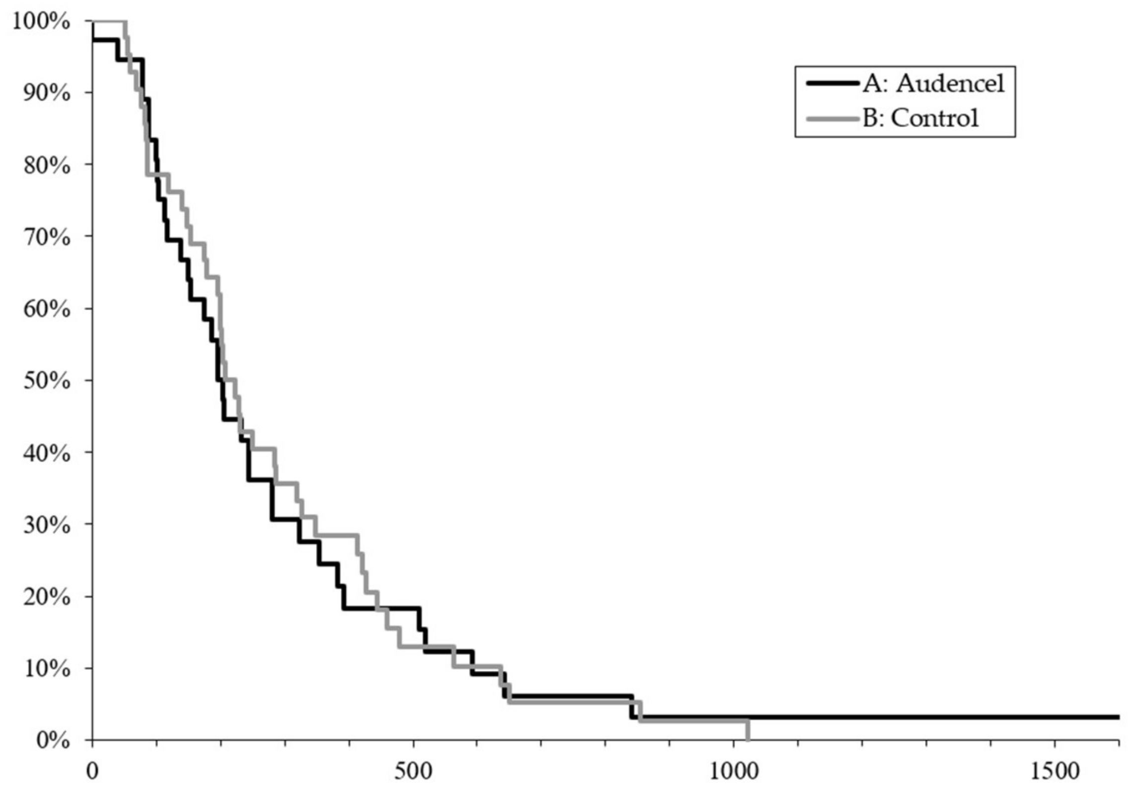
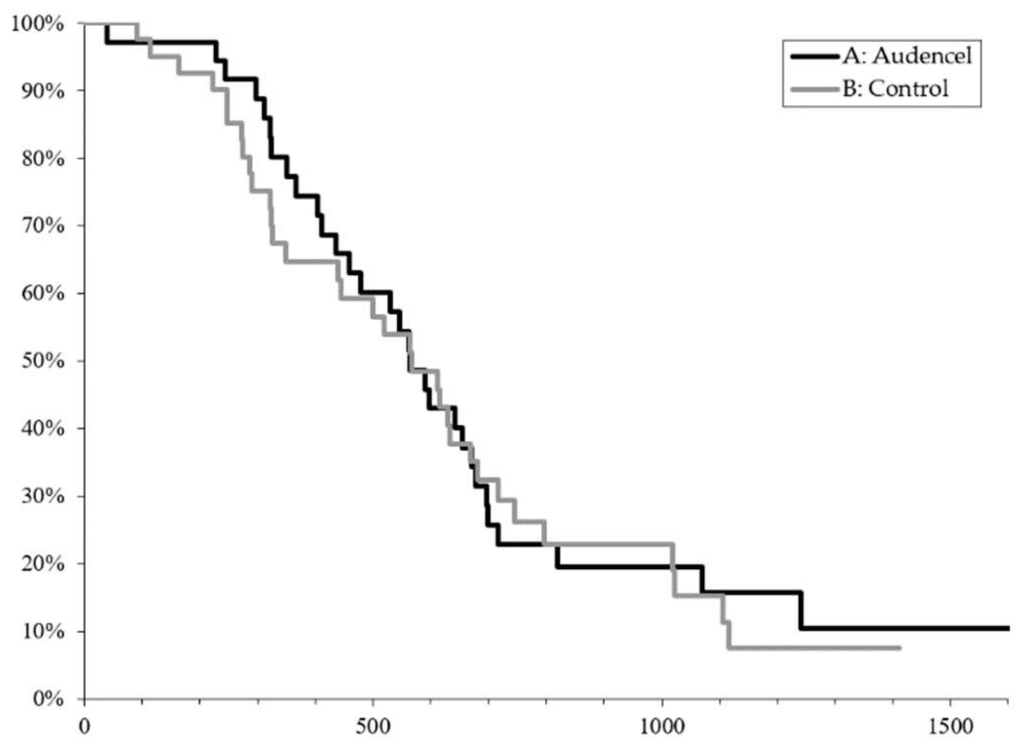
2.1. Survival Outcome

A total of 76 patients were enrolled and analyzed in this study: 34 in the experimental arm with Audencel, and 42 in the control arm. Initially, after treatment center-specific 1:1 randomization, 39 patients had been formally included in the experimental arm and 42 had been formally included in the control arm. Five patients of the experimental arm were later excluded because they had been double-counted (due to switching from one treatment center to another) or because they did not actually meet the formal inclusion criteria (e.g., age). As a result, the experimental cohort for analysis consisted of 34 patients. Mutations in the isocitrate dehydrogenase 1 (IDH1) gene were not detected in any patient, confirming that all of the patients suffered from primary GBM. Their baseline characteristics and demographics are given in Table 1. Twenty-five female and 51 male patients entered the study, aged from 19 to 70 years, with a median age of 54 years. The median duration from surgery to the start of radiochemotherapy was four weeks in both groups.
Patients randomized to the vaccination arm underwent leucocyte apheresis before the start of radiotherapy, and subsequently received vaccine treatment by intranodal application starting in week 7 after the treatment start, as detailed previously (Figure 1). The duration of vaccination was seven months on average (3–20 months). No patient stopped vaccination due to side effects.
All of the patients were analyzed according to intention-to-treat. The main reason for treatment discontinuation was disease progression in both arms. One female patient aged 65 years and randomized to the experimental arm experienced status epilepticus on the day after leucocyte apheresis; she was admitted to the hospital where the status was terminated, but disease progression was diagnosed. She died without receiving postsurgical anti-tumor treatment. All of the other randomized patients could be treated according to the SOC, all of the patients were able to receive radiotherapy up to 60 gray (Gy), and concomitant and adjuvant therapy with TMZ, respectively.
Patients randomized into the experimental arm received four to 15 vaccinations, with 10 being the average.
Time to progression was between six and eight months, e.g., 204 days, 95% CI: (138; 280) in the vaccine arm versus 6.9 months e.g., 210 days, 95% CI (179; 286) (p = 0.83) in the control arm, as shown in Figure 2. Pseudoprogression occurred in three patients of the Audencel group and eight patients of the control group. For details regarding the definition of progression and pseudoprogression, see the Materials and Methods section.
Overall, survival was similar in both groups: vaccination group: 564 days, 95% CI: (436; 671) versus control group: 568 days, 95% CI (349; 680) (p = 0.99, Figure 3). The survival of patients with methylated MGMT promoter was 800 days on average, whereas it reached 340 days in patients who had the unmethylated MGMT promoter, irrespective of whether patients received an add-on vaccination or not (for details on MGMT, see below). One vaccinated patient survived beyond five years.

2.2. Adverse Events

There were more adverse events recorded in the treatment group as compared to the control group; also, events related to the alkylating drug TMZ were more frequently observed in the Audencel group than in the control group. However, except for severe thrombocytopenia, these differences did not reach significance. There was no increase of intracerebral bleedings observed in the vaccine group (Table 2).
Adverse events that were related to the vaccine therapy consisted mainly of local pain and local reactions; these were mostly mild to moderate, and affected six out of 34 patients (18%). Seven vaccinated patients reacted with fever up to 39 °C and general weakness and joint pain on the day of vaccine administration, which resolved spontaneously within 12 h. Of note, there were no signs of autoimmune toxicity in the central nervous system (CNS) observed in vaccinated patients.

2.3. Treatment after Relapse

Six patients in the Audencel group and 12 patients in the control group received no active treatment in relapse. Four patients in the Audencel group and three patients in the control group underwent a second surgical resection in relapse.
The most common treatment for relapse consisted of bevacizumab (10 mg/kg, given every two weeks) administered to 20 patients in the Audencel group and 23 in the control group, followed by TMZ rechallenge (nine patients in the Audencel group and 12 patients in the control group).

2.4. Extent of Resection

In glioblastoma, the extent of resection (EOR) is known to have an influence on survival [36]. In the specific context of DC immunotherapy, it has been discussed for a number of malignant entities that DC immunotherapy might be most applicable to patients with minimal residual disease [37,38]. Thus, we investigated a potential influence of EOR on outcome in our clinical trial. Patients of the Audencel and the SOC cohort were stratified into three groups, with each based on EOR (gross total resection of 100% versus partial resection of 90–99% versus partial resection of 70–90%). No patient was below the threshold of at least 70% EOR, as this was an inclusion criterion. Exploratory Kaplan–Meier analysis showed that EOR did not have an influence on the OS outcome of the Audencel cohort compared to the SOC cohort for all three groups (p = 0.762, Figure S1).

2.5. DC Vaccine Quality

DC vaccine quality was ensured via stringent phenotypical and functional release criteria for each vaccine batch produced (for details, see the Supplementary Material). Functionally, interleukin-12 (IL-12) production capacity and T-cell stimulation capacity (via mixed leukocyte reactions, MLR) were measured in analogy to the DC trials performed by others [39,40]. Release criteria were >100 pg/mL IL-12, and at least 30% T-cell proliferation upon DC co-culture in relation to reference stimulation with staphylococcal enterotoxin A/B (SEA/SEB). These criteria ensured standard quality for all of the batches, but still left room for variation. Thus, we tested whether vaccine quality had a relation to outcome. Exploratory Pearson correlation analysis showed that neither IL-12 production (p = 0.891, Figure S2) nor T-cell stimulation capacity at various DC:T-cell ratios (1:5 p = 0.603, 1:10 p = 0.488, 1:20 p = 0.330, Figure S2) had an impact on survival (OS).

2.6. DC Vaccine Quantity

As mentioned, depending on their individual course of disease, all of the Audencel-treated patients received four to 15 vaccinations, with the exception of the one patient who died prior to vaccination. While four vaccinations were the minimum, again, there was variation in the final number of vaccinations given. Hence, we also explored a potential relation of the number of vaccines received and patient outcome. In Pearson correlation analysis, a trend toward a positive association was seen, but without reaching statistical significance (p = 0.081, Figure S3).

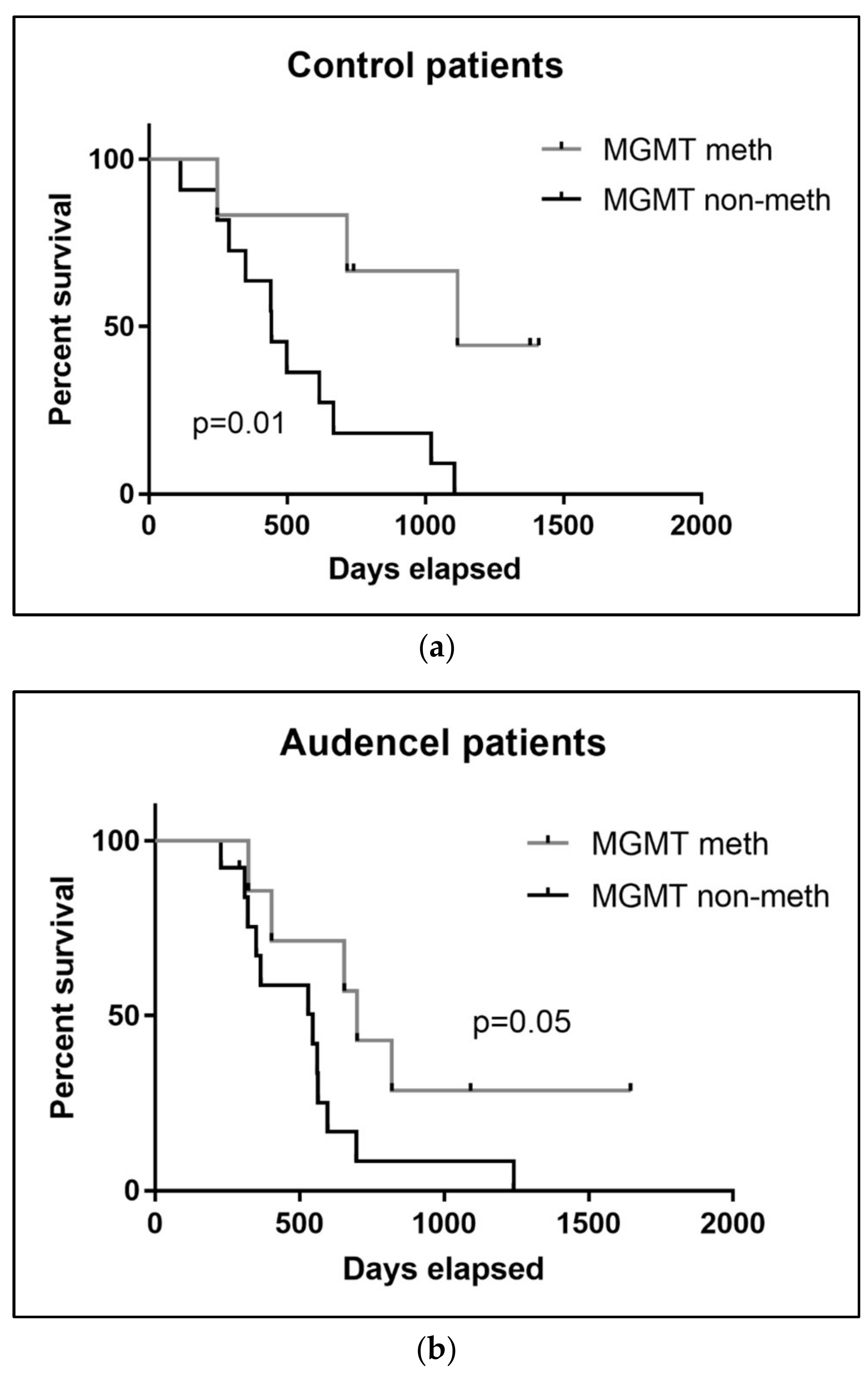
2.7. MGMT Promoter Methylation

Thirty-seven tumor samples were available for MGMT promoter methylation testing by pyrosequencing. Out of these, 35% (13/37) harbored a methylated MGMT promoter. Over half (65%) of the tumors investigated were shown to have a mean methylation percentage below 8%, and were therefore designated as unmethylated.
The fraction of patients with a methylated MGMT promoter was identical for the Audencel group (35%, seven out of 20) and the control/SOC group (35%, six out of 17). In the control/SOC group, the MGMT status had a significant impact on overall survival: control patients with a methylated MGMT promoter had a significantly better outcome (p = 0.01, Figure 4). For Audencel patients, a similar trend was visible; however, it did not reach statistical significance (p = 0.05, Figure 4). It is well-known that MGMT methylation is a predictor for the effectiveness of temozolomide chemotherapy [41], which was in line with our observation in the control/SOC group. Why the effect was not significant in the Audencel group remains to be determined. One overall caveat of the exploratory MGMT analysis presented here is the limited sample availability, as MGMT measurement was only retrospectively introduced to the study (see Section 4. Material and Methods). Validation in larger patient cohorts will be necessary.

3. Discussion

The addition of Audencel, an autologous DC-based vaccine with tumor peptides, to SOC in patients with newly diagnosed GBM failed to prolong progression-free survival (PFS) and OS. However, vaccinated patients developed an active immune response, as shown by the associated in vitro surveillance [42], although this was not sufficient to avoid tumor recurrence. The median OS of 18.3 months compares to the actually reported treatment results for GBM patients [4,6,7]. Of note, one of the vaccinated patients became a long-term survivor for more than five years.
Previous trials using DC-based vaccinations did not enable anticipating the lack of efficacy that was experienced in this trial. In a review published in 2013, Bregy et al. [43] evaluated 19 studies and two case reports using DC-based vaccinations for 403 glioma patients; currently, more than 20 trials are ongoing or have recently been terminated. Thirteen of the trials summarized by Bregy et al. also included patients with recurrent gliomas; nine trials included patients with WHO III gliomas, only three trials focused on patients with newly diagnosed GBM; among them was Ardon et al. with 77 patients [44]. Most notably, 11 out of 12 studies in newly diagnosed GBM showed an increase of survival duration, with seven studies reporting a median survival duration that was longer than two years [14,39,45,46,47,48,49], and one of them was even more than three years [50]. However, in most of the trials, the starting point of vaccination therapy is reported, and most noticeably, only in one study was an early start of vaccination within one to two months following surgery reported [46]. In five studies, the start of vaccination was delayed: from between six and seven weeks after the termination of radiotherapy [39], nine weeks after surgery [44], or even as long as seven to 30 weeks after surgery [14], 18 weeks (in median) after surgery [47] and even after tumor recurrence [48]. In one study, non-progressing patients after chemoradiation were included [50], which also implied a certain delay, allowing for an magnetic resonance imaging (MRI) assessment prior to vaccination start. The delay before initiating vaccination therapy avoids the inclusion of early progressing patients, and is an effective (statistical) mechanism for prolonging survival. Of note, similar to in our study, in none of these studies were autoimmune reactions reported in vaccinated patients. In one study, an inoculation of a cutaneous GBM at the site of a cutaneous injection of irradiated tumor cells for delayed-type hypersensitivity (DTH) testing was mentioned [51].
An alternative approach to the use of autologous tumor lysate to load dendritic cells is the use of selected peptide antigens, either glioma-associated antigens [52,53], or as in several ongoing trials, antigens associated with glioma stem cells [54] or mRNA derived from autologous neurospheres [40]. Furthermore, a recent phase II study using ICT-107, which is a DC vaccine of autologous DCs pulsed with 20 μg/mL of each of six synthetic peptides that are associated with glioma stem cell antigens, enrolled 124 patients with newly diagnosed GBMs. These patients were treated first with standard chemoradiation and underwent four weekly vaccinations during the following month, and further vaccinations during adjuvant treatment. There was a survival benefit in vaccinated patients with the human leukocyte antigen (HLA) haplotype A2, particularly for MGMT promoter methylated and vaccinated patients. A phase III was launched, but it was then suspended, which was apparently due to insufficient funding [12].
Several reasons may explain the failure of improving the outcome of glioblastoma patients that was observed in this study.
Firstly, the GBM tumor antigens that were presented by DCs might not have been effective triggers of an immune response to inhibit glioma growth. The vast majority of protein that is taken up, processed, and presented by the DCs might be normal, healthy, and not show any mutation that contributes to the malignant phenotype of tumor cells. Moreover, tumor lysate-loaded DCs not only contain autoantigens that are expressed by nervous tissue alone. A plethora of proteins will be the same as in any other cells, similar to the proteins that are involved in metabolism, the cytoskeleton, gene expression, and many more. Other cells in the tumor tissue will end up in DCs as well, such as blood vessel cells, and cells from the tumor’s connective tissue even infiltrating immune cells. Furthermore, not only intracellular proteins will be present in the tumor material, but also extracellular matrix proteins. Therefore, only a minority of proteins that are loaded onto DCs might have actually been immunogenic. Interestingly, in this study—similar to other DC vaccination studies reported previously—no event related to autoimmune toxicity, neither in CNS nor systemically, was observed. This can support the argument that only insufficient immune reactions were triggered by the vaccine, against both malignant as well as healthy tissue antigens. In other immunotherapeutic modalities, such as chimeric antigen receptor (CAR) T cells, fulminant immune reactions (e.g., the cytokine release syndrome) have been observed [55]. Such phenomena can be an indicator for anti-tumor efficacy. None of these fulminant reactions were observed in the present trial. As a consequence of the apparently low immunogenicity, future studies could explore an augmentation of anti-tumor immune reactions, e.g., via the combination of DC vaccination with checkpoint inhibitors.
A second potential reason for the failure of our treatment concept could be found in the hematotoxic effect of TMZ impeding the multiplication of effector T cells that should reverse the immune tolerance of the tumor. In the case of DC-CIT, the parallel application of chemotherapy might prevent an effective anti-tumor immunity from developing by killing the majority of primed cells during their proliferative phase. It is likely that this was the case in this study, which would have contributed significantly to the lack of survival benefit in patients receiving Audencel.
Thirdly, even if a number of groups are working on similar DC vaccination technologies, the production protocols are never exactly the same. One driver is the complexity of the production process that involves multiple steps in Good Manufacturing Practice (GMP) cell processing facilities. Thus, in light of encouraging prior data by other groups [14,15,47,56], the disappointing results of this trial might stem from DC production differences. For instance, DCs in this study were matured with LPS/IFNγ, while other groups do not have this step in their production protocol [47,51,56]. Future studies should compare different DC production techniques to clarify the possible relevance of technological specifics. DC production quality per se—following our predefined production protocols and technologies mentioned above—did not seem to be an influencing factor based on the functional DC vaccine analyses that we made (see Section 2. Results).
A fourth potential reason could be an insufficient number of DCs administered by cycle. In fact, nine out of the 12 studies that were reported by Bregy et al. and the (recently suspended) ICT-107 trial used at least twice the number of dendritic cells per dose. Based on a prior clinical study with Audencel [31], and for the regulatory reasons that resulted, less DCs per dose than those reported by others were given in this study. That quantity might generally matter is supported by our observation of a non-significant, trend-level association between the number of vaccines given and the outcome. While the number of vaccines was dependent on the individual patient’s course of disease, and can thus not be changed, future studies of the Audencel technology could consider higher doses per vaccination to increase the total quantity of DCs delivered.

4. Materials and Methods

The Audencel trial was a national randomized multicenter open label phase II trial including nine active sites in Austria that was performed between 2010–2015 (first patient in April 2010, last patient out June 2015). The trial ended as planned when all of the patients reached the predefined observation period of at least 12 months (see primary objective below). Eligible patients were aged 18 to 70 years, with newly diagnosed, histologically confirmed primary GBM. Tumor samples were tested for IDH mutation and MGMT promoter methylation according to previously reported methods [57,58]—for details, see below.
Patients were eligible if their tumor had been resected for at least 70% as confirmed by a postsurgical MRI scan. Additional inclusion criteria were written informed consent, the availability of tumor tissue for vaccine preparation, as well as adequate hematological, renal, and hepatic function. Exclusion criteria were a lack of sufficient amount of tumor protein (100 µg) for Audencel production, anti-neoplastic chemotherapy or radiotherapy for four weeks before entering the study, participation in another therapeutic clinical trial, a positive pregnancy test or breast-feeding, patients unwilling to perform a safe method of birth control, and known hypersensitivity to TMZ.
The primary objective of the study was the improvement of progression-free survival (PFS) at 12 months, which was measured as the percentage of non-progressive patients one year after a post-operative MRI scan and treated according to SOC, consisting of maximal safe surgical resection, irradiation, oral chemotherapy with TMZ, and Audencel as add-on therapy, in comparison to the control group of patients receiving SOC alone. Secondary objectives included the improvement of PFS measured as the percentage of non-progressive patients with newly diagnosed GBM at 18 and 24 months. Further, median overall survival (OS), as well as quality of life (QOL), and the safety and feasibility of Audencel immunotherapy were analyzed.
The study was conducted according to the Helsinki Declaration and approved by the ethics committees of all of the participating centers (TRX 2/P-II-018). All of the patients gave oral and written informed consent. The study was registered with EudraCT number 2009-015979-27. The full trial protocol is available from the authors upon reasonable request.

4.1. Randomization

Patients were stratified according to site, age, sex, and performance status and centrally randomized with the use of an interactive voice respond system in a 1:1 ratio into the treatment groups. Randomization was performed for each of the nine treatment centers individually.

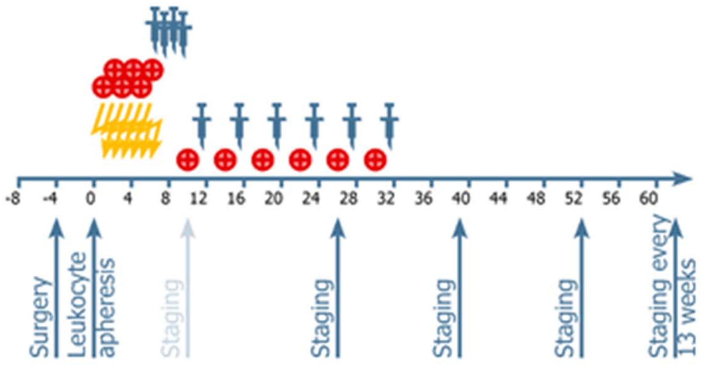
4.2. Procedures: Treatments Administered

All of the patients were treated according to the current SOC for GBM, using conformal radiation therapy with two Gy/fraction up to 60 Gy, with concomitant treatment with 75 mg/m2 TMZ followed by six cycles of adjuvant TMZ at a dose of 150–200 mg/m2 for five days of each 28-day cycle. For safety evaluations, blood samples were taken weekly during radiochemotherapy and the following four weeks, then at the beginning of each adjuvant cycle and whenever clinically indicated. Radiation therapy had to start within four weeks after the postoperative MRI scan. For patients in the vaccine group, Audencel was produced individually as a personalized cellular medicine. Production followed prior research on Audencel [20,30,31,59]. Leucocyte apheresis had to be done before initiation of radiation therapy. Vaccine preparation and quality control are described in detail in the Supplementary Material.

4.3. Vaccine Administration

Audencel was administered by injection into an inguinal lymph node using visual ultrasound control. Each single dose consisted of 1–5 × 10⁶ autologous DCs in 100 µL. The rationale for this dosing regimen were regulatory (toxicity) considerations, as a prior Audencel clinical trial [34] had also used this dosage for intranodal injections, and no toxicity had been observed in that earlier study. Vaccinations were started in week seven, just after chemoradiation, with three boosting doses applied weekly to week 10, which was the start of adjuvant therapy with TMZ (Figure 1). Subsequently, vaccinations were given in monthly intervals, two weeks after start of adjuvant TMZ treatment, in weeks 12, 16, 20, 24, and 32. After the completion of six adjuvant cycles with TMZ, vaccination was pursued in a three-month interval, as long as autologous vaccine was available.
Local tolerability was assessed in all of the patients following each Audencel application. Before administering a subsequent vaccine, the injection site of the previous vaccination was inspected and assessed. Local tolerability including pain, itching, induration, edema, and erythema were evaluated. Adverse events (AEs) were recorded and graded according to CTC 4.0.

4.4. Neuroimaging and Response Assessment

Neuroradiological examinations were carried out using brain MRI scans with and without gadolinium enhancement before surgery and within the first 48 hours after surgery. The postoperative MRI scan represented the baseline for the evaluation of disease progression. The next follow-up MRI scan was performed 10 weeks after start of chemoradiation ± two weeks, and then every three months ± two weeks. In case of clinical deterioration, an MRI scan was performed as soon as possible to allow decisions about disease progression.
The response assessment was based on the MRI response assessment criteria that were currently valid at the time of study protocol approval, namely the MacDonald criteria [60]. According to these, progression is defined as a ≥25% increase in known contrast-enhancing lesions or any new lesion. However, prior studies taught us that a therapy-induced immune reaction might lead to inflammation and consequently to a new (therapy-induced) disruption of the blood–brain barrier with new MRI contrast enhancement. To avoid the premature discontinuation of treatment, therapy was applied according to the study protocol as long as pseudoprogression was suspected and the MRI interval was shortened. When clinical worsening occurred and the follow-up MRI showed a further increase of contrast enhancement, true tumor progression was assumed and therapy was discontinued. If the follow-up MRI showed stable or even decreasing contrast enhancement with a stable or improving clinical condition, pseudoprogression was still assumed, and treatment was continued. Progression-free survival in our study was defined as the survival from study entry until true tumor progression was confirmed by a second MRI scan (in case of suspected pseudoprogression).

4.5. Treatment after Relapse

Treatment after relapse was left to the investigator’s choice.

4.6. DNA Extraction and MGMT Promoter Methylation Analysis

In the initial clinical trial protocol that was developed in the year 2009, MGMT testing was not established as standard procedure for patients included in the trial; it was added ex post. A retrospective collection of samples for MGMT analysis was thus only successful in a fraction of patients (see Section 2. Results). Depending on availability, DNA was extracted from either tumor tissue or formalin-fixed paraffin-embedded tissue (FFPE) sections using the QiAmp DNA Mini-Kit and FFPE tissue kit (both Qiagen, Hilden, Germany) according to the manufacturer’s instructions. Subsequently, DNA modification was performed with the EpiTect Bisulfite Kit (Qiagen). Using the Therascreen MGMT Pyro Kit (Qiagen), bisulfite converted genomic DNA was amplified by PCR and sequenced on a Pyromark Q24 (Qiagen, Germany) system. Following previous research [58], a mean value of the methylation percentage of the four CpG sites investigated was calculated. Accordingly, a mean methylation >8% was considered as methylated.

4.7. IDH1 Mutation Analysis

IDH1 mutation status was registered via a mutation-specific antibody (Anti-IDH1 R132H) that was used for immunohistochemistry according to the previously published standard “Vienna” protocol [61].

4.8. Statistical Analysis

All of the statistical analyses were performed using SAS version 9.3 or GraphPad Prism 7.04.
Descriptive analyses of continuous variables (summary statistics) were described with the number of non-missing observations, arithmetic mean, standard deviation (±SD), median, quartiles (Q1 and Q3), and range (minimum and maximum). Categorical variables (frequency statistics) were described with the number of non-missing observations and percentages. Percentages were calculated within each stratum on the total number of non-missing observations.
Progression-free survival and overall survival were estimated using the Kaplan–Meier method. Treatment groups were compared using a log-rank test. A Cox proportional hazards model with stratification according to randomization strata was used for the calculation of hazard ratios and 95% confidence intervals. All of the outcome tests were done with the intention to treat the population, and safety was assessed for all patients randomized into the study.
Statistical tests were two-sided with a significance level of 5%. All of the p-values were rounded to four decimal places and were presented as nominal p-values with no multiplicity adjustment.
The study sample size was calculated in order to detect a doubling of surviving patients at 12 months with 80% power.

5. Conclusions

To summarize, the Audencel trial did not meet our expectations. However, the challenging multicentric design was feasible in terms of logistics and the practicability of delivering a personalized, custom-made cellular therapy. The vaccination was well-tolerated, and no signs of autoimmunity were detected. Vaccinated patients developed tumor directed immune responses [42], so it seems justified to further develop vaccination strategies that take into account the failure of this trial in the future speculatively by combining DC vaccination with the administration of immune check point inhibitors, and by increasing the dose or other strategies.

Supplementary Materials

The following are available online at https://www.mdpi.com/2072-6694/10/10/372/s1, Figure S1: Influence of extent-of-resection (EOS) on overall survival (OS); Figure S2: Relation of DC vaccine quality control and overall survival (OS); Figure S3: Analysis of potential influence of number of vaccinations received on overall survival (OS); Figure S4: CONSORT 2010 Flow Diagram. Table S1: CONSORT 2010 checklist of information to include when reporting a randomised trial.

Author Contributions

J.B., C.M., T.F., G.K. designed the study, interpreted results and wrote the manuscript. J.B., J.P., M.P., G.S., M.N., S.I., S.O., K.B., G.v.C., M.H., T.C. and R.R. recruited and treated patients. G.W., C.F.F. and K.R. operated on patients and provided material for vaccine production. S.S.-K., F.E. and C.V. performed MGMT analyses, interpreted results and wrote parts of the manuscript. M.B.F. performed leukocyte apheresis procedures. All authors approved the final version of the manuscript.

Funding

This study was funded by Activartis Biotech GmbH. The study was performed within the network of the Austrian Society of Neurooncology (SANO).

Acknowledgments

We thank the patients and their families at all the participating study sites. Also, we are grateful for the excellent technical support in the production of the vaccine by Activartis technicians.

Conflicts of Interest

F.E. was formerly a (part-time) employee of Activartis Biotech. The funding organization (Activartis Biotech GmbH) participated in the design of the study. The funder had no role in the collection, analyses, or interpretation of data; in the writing of the manuscript, and in the decision to publish the results.

References

  1. Stupp, R.; Mason, W.P.; van den Bent, M.J.; Weller, M.; Fisher, B.; Taphoorn, M.J.B.; Belanger, K.; Brandes, A.A.; Marosi, C.; Bogdahn, U.; et al. National Cancer Institute of Canada Clinical Trials Group Radiotherapy plus Concomitant and Adjuvant Temozolomide for Glioblastoma. N. Engl. J. Med. 2005, 352, 987–996. [Google Scholar] [CrossRef] [PubMed]
  2. Nabors, L.B.; Fink, K.L.; Mikkelsen, T.; Grujicic, D.; Tarnawski, R.; Nam, D.H.; Mazurkiewicz, M.; Salacz, M.; Ashby, L.; Zagonel, V.; et al. Two cilengitide regimens in combination with standard treatment for patients with newly diagnosed glioblastoma and unmethylated MGMT gene promoter: Results of the open-label, controlled, randomized phase II CORE study. Neuro Oncol. 2015, 17, 708–717. [Google Scholar] [CrossRef] [PubMed]
  3. Stupp, R.; Hegi, M.E.; Gorlia, T.; Erridge, S.C.; Perry, J.; Hong, Y.-K.; Aldape, K.D.; Lhermitte, B.; Pietsch, T.; Grujicic, D.; et al. CENTRIC study team Cilengitide combined with standard treatment for patients with newly diagnosed glioblastoma with methylated MGMT promoter (CENTRIC EORTC 26071-22072 study): a multicentre, randomised, open-label, phase 3 trial. Lancet Oncol. 2014, 15, 1100–1108. [Google Scholar] [CrossRef]
  4. Chinot, O.L.; Wick, W.; Cloughesy, T. Bevacizumab for newly diagnosed glioblastoma. N. Engl. J. Med. 2014, 370, e2049. [Google Scholar] [CrossRef] [PubMed]
  5. Gilbert, M.R.; Dignam, J.J.; Armstrong, T.S.; Wefel, J.S.; Blumenthal, D.T.; Vogelbaum, M.A.; Colman, H.; Chakravarti, A.; Pugh, S.; Won, M.; et al. A Randomized Trial of Bevacizumab for Newly Diagnosed Glioblastoma. N. Engl. J. Med. 2014, 370, 699–708. [Google Scholar] [CrossRef] [PubMed] [Green Version]
  6. Gilbert, M.R.; Wang, M.; Aldape, K.D.; Stupp, R.; Hegi, M.E.; Jaeckle, K.A.; Armstrong, T.S.; Wefel, J.S.; Won, M.; Blumenthal, D.T.; et al. Dose-Dense Temozolomide for Newly Diagnosed Glioblastoma: A Randomized Phase III Clinical Trial. J. Clin. Oncol. 2013, 31, 4085–4091. [Google Scholar] [CrossRef] [PubMed] [Green Version]
  7. Stupp, R.; Taillibert, S.; Kanner, A.A.; Kesari, S.; Steinberg, D.M.; Toms, S.A.; Taylor, L.P.; Lieberman, F.; Silvani, A.; Fink, K.L.; et al. Maintenance Therapy With Tumor-Treating Fields Plus Temozolomide vs Temozolomide Alone for Glioblastoma: A Randomized Clinical Trial. JAMA 2015, 314, 2535–2543. [Google Scholar] [CrossRef] [PubMed]
  8. Pieczonka, C.M.; Telonis, D.; Mouraviev, V.; Albala, D. Sipuleucel-T for the treatment of patients with metastatic castrate-resistant prostate cancer: considerations for clinical practice. Rev. Urol. 2015, 17, 203–210. [Google Scholar] [PubMed]
  9. Kantoff, P.W.; Higano, C.S.; Shore, N.D.; Berger, E.R.; Small, E.J.; Penson, D.F.; Redfern, C.H.; Ferrari, A.C.; Dreicer, R.; Sims, R.B.; et al. Sipuleucel-T immunotherapy for castration-resistant prostate cancer. N. Engl. J. Med. 2010, 363, 411–422. [Google Scholar] [CrossRef] [PubMed]
  10. Tanimoto, T.; Hori, A.; Kami, M. Sipuleucel-T immunotherapy for castration-resistant prostate cancer. N. Engl. J. Med. 2010, 363, 1967–1968. [Google Scholar]
  11. Nabhan, C. Sipuleucel-T immunotherapy for castration-resistant prostate cancer. N. Engl. J. Med. 2010, 363, 1966–1967. [Google Scholar] [PubMed]
  12. Lim, M.; Xia, Y.; Bettegowda, C.; Weller, M. Current state of immunotherapy for glioblastoma. Nat. Rev. Clin. Oncol. 2018, 15, 422–442. [Google Scholar] [CrossRef] [PubMed]
  13. Inogés, S.; Tejada, S.; de Cerio, A.L.-D.; Gállego Pérez-Larraya, J.; Espinós, J.; Idoate, M.A.; Domínguez, P.D.; de Eulate, R.G.; Aristu, J.; Bendandi, M.; et al. A phase II trial of autologous dendritic cell vaccination and radiochemotherapy following fluorescence-guided surgery in newly diagnosed glioblastoma patients. J. Transl. Med. 2017, 15, e104. [Google Scholar] [CrossRef] [PubMed]
  14. Prins, R.M.; Soto, H.; Konkankit, V.; Odesa, S.K.; Eskin, A.; Yong, W.H.; Nelson, S.F.; Liau, L.M. Gene expression profile correlates with T-cell infiltration and relative survival in glioblastoma patients vaccinated with dendritic cell immunotherapy. Clin. Cancer Res. 2011, 17, 1603–1615. [Google Scholar] [CrossRef] [PubMed]
  15. Liau, L.M.; Ashkan, K.; Tran, D.D.; Campian, J.L.; Trusheim, J.E.; Cobbs, C.S.; Heth, J.A.; Salacz, M.; Taylor, S.; D’Andre, S.D.; et al. First results on survival from a large Phase 3 clinical trial of an autologous dendritic cell vaccine in newly diagnosed glioblastoma. J. Transl. Med. 2018, 16, e142. [Google Scholar] [CrossRef] [PubMed]
  16. Aspelund, A.; Antila, S.; Proulx, S.T.; Karlsen, T.V.; Karaman, S.; Detmar, M.; Wiig, H.; Alitalo, K. A dural lymphatic vascular system that drains brain interstitial fluid and macromolecules. J. Exp. Med. 2015, 212, 991–999. [Google Scholar] [CrossRef] [PubMed] [Green Version]
  17. Louveau, A. Cerebral lymphatic drainage: implication in the brain immune privilege. Med. Sci. (Paris) 2015, 31, 953–956. [Google Scholar] [CrossRef] [PubMed]
  18. Louveau, A.; Harris, T.H.; Kipnis, J. Revisiting the Mechanisms of CNS Immune Privilege. Trends Immunol. 2015, 36, 569–577. [Google Scholar] [CrossRef] [PubMed] [Green Version]
  19. Razavi, S.-M.; Lee, K.E.; Jin, B.E.; Aujla, P.S.; Gholamin, S.; Li, G. Immune Evasion Strategies of Glioblastoma. Front. Surg. 2016, 3, 1–9. [Google Scholar] [CrossRef] [PubMed]
  20. Van Meir, E.G. Cytokines and tumors of the central nervous system. GLIA 1995, 15, 264–288. [Google Scholar] [CrossRef] [PubMed]
  21. Komohara, Y.; Ohnishi, K.; Kuratsu, J.; Takeya, M. Possible involvement of the M2 anti-inflammatory macrophage phenotype in growth of human gliomas. J. Pathol. 2008, 216, 15–24. [Google Scholar] [CrossRef] [PubMed]
  22. Wu, A.; Wei, J.; Kong, L.-Y.; Wang, Y.; Priebe, W.; Qiao, W.; Sawaya, R.; Heimberger, A.B. Glioma cancer stem cells induce immunosuppressive macrophages/microglia. Neuro. Oncol. 2010, 12, 1113–1125. [Google Scholar] [CrossRef] [PubMed] [Green Version]
  23. Doucette, T.A.; Kong, L.-Y.; Yang, Y.; Ferguson, S.D.; Yang, J.; Wei, J.; Qiao, W.; Fuller, G.N.; Bhat, K.P.; Aldape, K.; et al. Signal transducer and activator of transcription 3 promotes angiogenesis and drives malignant progression in glioma. Neuro. Oncol. 2012, 14, 1136–1145. [Google Scholar] [CrossRef] [PubMed] [Green Version]
  24. Steinman, R.M.; Cohn, Z.A. Identification of a novel cell type in peripheral lymphoid organs of mice. I. Morphology, quantitation, tissue distribution. J. Exp. Med. 1973, 137, 1142–1162. [Google Scholar] [CrossRef] [PubMed]
  25. Steinman, R.M.; Cohn, Z.A. Pillars Article: Identification of a novel cell type in peripheral lymphoid organs of mice. I. Morphology, quantitation, tissue distribution. J. Immunol. 2007, 178, 5–25. [Google Scholar] [PubMed]
  26. Langenkamp, A.; Messi, M.; Lanzavecchia, A.; Sallusto, F. Kinetics of dendritic cell activation: impact on priming of TH1, TH2 and nonpolarized T cells. Nat. Immunol. 2000, 1, 311–316. [Google Scholar] [CrossRef] [PubMed]
  27. Luger, R.; Valookaran, S.; Knapp, N.; Vizzardelli, C.; Dohnal, A.M.; Felzmann, T. Toll-like receptor 4 engagement drives differentiation of human and murine dendritic cells from a pro- into an anti-inflammatory mode. PLoS ONE 2013, 8, e54879. [Google Scholar] [CrossRef] [PubMed]
  28. Medzhitov, R.; Janeway, C.A. Decoding the Patterns of Self and Nonself by the Innate Immune System. Science 2002, 296, 298–300. [Google Scholar] [CrossRef] [PubMed] [Green Version]
  29. Hüttner, K.G.; Breuer, S.K.; Paul, P.; Majdic, O.; Heitger, A.; Felzmann, T. Generation of potent anti-tumor immunity in mice by interleukin-12-secreting dendritic cells. Cancer Immunol. Immunother. 2005, 54, 67–77. [Google Scholar] [CrossRef] [PubMed]
  30. Felzmann, T.; Hüttner, K.G.; Breuer, S.K.; Wimmer, D.; Ressmann, G.; Wagner, D.; Paul, P.; Lehner, M.; Heitger, A.; Holter, W. Semi-mature IL-12 secreting dendritic cells present exogenous antigen to trigger cytolytic immune responses. Cancer Immunol. Immunother. 2005, 54, 769–780. [Google Scholar] [CrossRef] [PubMed]
  31. Dohnal, A.M.; Witt, V.; Hügel, H.; Holter, W.; Gadner, H.; Felzmann, T. Phase I study of tumor Ag-loaded IL-12 secreting semi-mature DC for the treatment of pediatric cancer. Cytotherapy 2007, 9, 755–770. [Google Scholar] [CrossRef] [PubMed]
  32. Kaiser, A.; Bercovici, N.; Abastado, J.-P.; Nardin, A. Naive CD8+ T cell recruitment and proliferation are dependent on stage of dendritic cell maturation. Eur. J. Immunol. 2003, 33, 162–171. [Google Scholar] [CrossRef] [PubMed]
  33. Camporeale, A.; Boni, A.; Iezzi, G.; Degl’Innocenti, E.; Grioni, M.; Mondino, A.; Bellone, M. Critical impact of the kinetics of dendritic cells activation on the in vivo induction of tumor-specific T lymphocytes. Cancer Res. 2003, 63, 3688–3694. [Google Scholar] [PubMed]
  34. Sabado, R.L.; Bhardwaj, N. Cancer immunotherapy: dendritic-cell vaccines on the move. Nature 2015, 519, 300–311. [Google Scholar] [CrossRef] [PubMed]
  35. Schuler, G.; Schuler-Thurner, B.; Steinman, R.M. The use of dendritic cells in cancer immunotherapy. Curr. Opin. Immunol. 2003, 15, 138–147. [Google Scholar] [CrossRef]
  36. Stummer, W.; Reulen, H.-J.; Meinel, T.; Pichlmeier, U.; Schumacher, W.; Tonn, J.-C.; Rohde, V.; Oppel, F.; Turowski, B.; Woiciechowsky, C.; et al. ALA-Glioma Study Group extent of resection and survival in glioblastoma multiforme. Neurosurgery 2008, 62, 564–576. [Google Scholar] [PubMed]
  37. Subklewe, M.; Geiger, C.; Lichtenegger, F.S.; Javorovic, M.; Kvalheim, G.; Schendel, D.J.; Bigalke, I. New generation dendritic cell vaccine for immunotherapy of acute myeloid leukemia. Cancer Immunol. Immunother. 2014, 63, 1093–1103. [Google Scholar] [CrossRef] [PubMed]
  38. Reinis, M.; Indrová, M.; Mendoza, L.; Mikysková, R.; Bieblová, J.; Bubeník, J.; Símová, J. HPV16-associated tumours: therapy of surgical minimal residual disease with dendritic cell-based vaccines. Int. J. Oncol. 2004, 25, 1165–1170. [Google Scholar] [PubMed]
  39. Fadul, C.E.; Fisher, J.L.; Hampton, T.H.; Lallana, E.C.; Li, Z.; Gui, J.; Szczepiorkowski, Z.M.; Tosteson, T.D.; Rhodes, C.H.; Wishart, H.A.; et al. Immune response in patients with newly diagnosed glioblastoma multiforme treated with intranodal autologous tumor lysate-dendritic cell vaccination after radiation chemotherapy. J. Immunother. 2011, 34, 382–389. [Google Scholar] [CrossRef] [PubMed]
  40. Vik-Mo, E.O.; Nyakas, M.; Mikkelsen, B.V.; Moe, M.C.; Due-Tønnesen, P.; Suso, E.M.I.; Sæbøe-Larssen, S.; Sandberg, C.; Brinchmann, J.E.; Helseth, E.; et al. Therapeutic vaccination against autologous cancer stem cells with mRNA-transfected dendritic cells in patients with glioblastoma. Cancer Immunol. Immunother. 2013, 62, 1499–1509. [Google Scholar] [CrossRef] [PubMed] [Green Version]
  41. Hegi, M.E.; Diserens, A.-C.; Gorlia, T.; Hamou, M.-F.; de Tribolet, N.; Weller, M.; Kros, J.M.; Hainfellner, J.A.; Mason, W.; Mariani, L.; et al. MGMT Gene Silencing and Benefit from Temozolomide in Glioblastoma. N. Engl. J. Med. 2005, 352, 997–1003. [Google Scholar] [CrossRef] [PubMed]
  42. Erhart, F.; Buchroithner, J.; Reitermaier, R.; Fischhuber, K.; Klingenbrunner, S.; Sloma, I.; Hibsh, D.; Kozol, R.; Efroni, S.; Ricken, G.; et al. Immunological analysis of phase II glioblastoma dendritic cell vaccine (Audencel) trial: immune system characteristics influence outcome and Audencel up-regulates Th1-related immunovariables. Acta Neuropath. Comm. Submitted for publication. 2018. [Google Scholar]
  43. Bregy, A.; Wong, T.M.; Shah, A.H.; Goldberg, J.M.; Komotar, R.J. Active immunotherapy using dendritic cells in the treatment of glioblastoma multiforme. Cancer Treat. Rev. 2013, 39, 891–907. [Google Scholar] [CrossRef] [PubMed]
  44. Ardon, H.; Van Gool, S.W.; Verschuere, T.; Maes, W.; Fieuws, S.; Sciot, R.; Wilms, G.; Demaerel, P.; Goffin, J.; Van Calenbergh, F.; et al. Integration of autologous dendritic cell-based immunotherapy in the standard of care treatment for patients with newly diagnosed glioblastoma: results of the HGG-2006 phase I/II trial. Cancer Immunol. Immunother. 2012, 61, 2033–2044. [Google Scholar] [CrossRef] [PubMed]
  45. Chang, C.-N.; Huang, Y.-C.; Yang, D.-M.; Kikuta, K.; Wei, K.-J.; Kubota, T.; Yang, W.-K. A phase I/II clinical trial investigating the adverse and therapeutic effects of a postoperative autologous dendritic cell tumor vaccine in patients with malignant glioma. J. Clin. Neurosci. 2011, 18, 1048–1054. [Google Scholar] [CrossRef] [PubMed]
  46. Cho, D.-Y.; Yang, W.-K.; Lee, H.-C.; Hsu, D.-M.; Lin, H.-L.; Lin, S.-Z.; Chen, C.-C.; Harn, H.-J.; Liu, C.-L.; Lee, W.-Y.; et al. Adjuvant Immunotherapy with Whole-Cell Lysate Dendritic Cells Vaccine for Glioblastoma Multiforme: A Phase II Clinical Trial. World Neurosurg. 2012, 77, 736–744. [Google Scholar] [CrossRef] [PubMed]
  47. Liau, L.M.; Prins, R.M.; Kiertscher, S.M.; Odesa, S.K.; Kremen, T.J.; Giovannone, A.J.; Lin, J.-W.; Chute, D.J.; Mischel, P.S.; Cloughesy, T.F.; et al. Dendritic cell vaccination in glioblastoma patients induces systemic and intracranial T-cell responses modulated by the local central nervous system tumor microenvironment. Clin. Cancer Res. 2005, 11, 5515–5525. [Google Scholar] [CrossRef] [PubMed]
  48. Yamanaka, R.; Homma, J.; Yajima, N.; Tsuchiya, N.; Sano, M.; Kobayashi, T.; Yoshida, S.; Abe, T.; Narita, M.; Takahashi, M.; et al. Clinical evaluation of dendritic cell vaccination for patients with recurrent glioma: Results of a clinical phase I/II trial. Clin. Cancer Res. 2005, 11, 4160–4167. [Google Scholar] [CrossRef] [PubMed]
  49. Yu, J.S.; Black, K.L.; Liu, G.; Wheeler, C.J.; Wagenberg, M.; Das, A.; Mindlin, E.; Chu, R.M.; Luptrawan, A.; Badruddoja, M.A. 843 Results of a Phase II Trial of Tumor Lysate-pulsed Dendritic Cell Vaccination for Malignant Glioma. Neurosurgery 2005, 57, 409–410. [Google Scholar] [CrossRef]
  50. Phuphanich, S.; Wheeler, C.J.; Rudnick, J.D.; Mazer, M.; Wang, H.; Nuño, M.A.; Richardson, J.E.; Fan, X.; Ji, J.; Chu, R.M.; et al. Phase I trial of a multi-epitope-pulsed dendritic cell vaccine for patients with newly diagnosed glioblastoma. Cancer Immunol. Immunother. 2013, 62, 125–135. [Google Scholar] [CrossRef] [PubMed]
  51. Wheeler, C.J.; Black, K.L.; Liu, G.; Mazer, M.; Zhang, X.-X.; Pepkowitz, S.; Goldfinger, D.; Ng, H.; Irvin, D.; Yu, J.S. Vaccination Elicits Correlated Immune and Clinical Responses in Glioblastoma Multiforme Patients. Cancer Res. 2008, 68, 5955–5964. [Google Scholar] [CrossRef] [PubMed] [Green Version]
  52. Pallini, R.; Ricci-Vitiani, L.; Montano, N.; Mollinari, C.; Biffoni, M.; Cenci, T.; Pierconti, F.; Martini, M.; De Maria, R.; Larocca, L.M. Expression of the stem cell marker CD133 in recurrent glioblastoma and its value for prognosis. Cancer 2011, 117, 162–174. [Google Scholar] [CrossRef] [PubMed]
  53. Zeppernick, F.; Ahmadi, R.; Campos, B.; Dictus, C.; Helmke, B.M.; Becker, N.; Lichter, P.; Unterberg, A.; Radlwimmer, B.; Herold-Mende, C.C. Stem Cell Marker CD133 Affects Clinical Outcome in Glioma Patients. Clin. Cancer Res. 2008, 14, 123–129. [Google Scholar] [CrossRef] [PubMed] [Green Version]
  54. Esparza, R.; Azad, T.D.; Feroze, A.H.; Mitra, S.S.; Cheshier, S.H. Glioblastoma stem cells and stem cell-targeting immunotherapies. J. Neurooncol. 2015, 123, 449–457. [Google Scholar] [CrossRef] [PubMed]
  55. Bonifant, C.L.; Jackson, H.J.; Brentjens, R.J.; Curran, K.J. Toxicity and management in CAR T-cell therapy. Mol. Ther. Oncolytics 2016, 3, e16011. [Google Scholar] [CrossRef] [PubMed]
  56. Yu, J.S.; Liu, G.; Ying, H.; Yong, W.H.; Black, K.L.; Wheeler, C.J. Vaccination with tumor lysate-pulsed dendritic cells elicits antigen-specific, cytotoxic T-cells in patients with malignant glioma. Cancer Res. 2004, 64, 4973–4979. [Google Scholar] [CrossRef] [PubMed]
  57. Balss, J.; Meyer, J.; Mueller, W.; Korshunov, A.; Hartmann, C.; von Deimling, A. Analysis of the IDH1 codon 132 mutation in brain tumors. Acta Neuropathol. 2008, 116, 597–602. [Google Scholar] [CrossRef] [PubMed]
  58. Von Deimling, A.; Korshunov, A.; Hartmann, C. The Next Generation of Glioma Biomarkers: MGMT Methylation, BRAF Fusions and IDH1 Mutations. Brain Pathol. 2011, 21, 74–87. [Google Scholar] [CrossRef] [PubMed]
  59. Felzmann, T.; Witt, V.; Wimmer, D.; Ressmann, G.; Wagner, D.; Paul, P.; Hüttner, K.; Fritsch, G. Monocyte enrichment from leukapharesis products for the generation of DCs by plastic adherence, or by positive or negative selection. Cytotherapy 2003, 5, 391–398. [Google Scholar] [CrossRef] [PubMed]
  60. Macdonald, D.R.; Cascino, T.L.; Schold, S.C.; Cairncross, J.G. Response criteria for phase II studies of supratentorial malignant glioma. J. Clin. Oncol. 1990, 8, 1277–1280. [Google Scholar] [CrossRef] [PubMed]
  61. Preusser, M.; Capper, D.; Hartmann, C.; Euro-CNS Research, Committee. IDH testing in diagnostic neuropathology: Review and practical guideline article invited by the Euro-CNS research committee. Clin. Neuropathol. 2011, 30, 217–230. [Google Scholar] [CrossRef] [PubMed]
Figure 1. Treatment schedule.
Cancers 10 00372 g001
Figure 2. Progression-free survival in days. Kaplan–Meier analysis by treatment group indicates no difference between the vaccine and the control arm (p = 0.83).
Cancers 10 00372 g002
Figure 3. Overall survival in days. Kaplan–Meier analysis by treatment group indicates no difference between the vaccine and the control arm (p = 0.99).
Cancers 10 00372 g003
Figure 4. Impact of MGMT promoter methylation status on overall survival (a) in the control group (p = 0.01) and (b) the Audencel group (p = 0.05).
Cancers 10 00372 g004
Table 1. Patient characteristics and demographics at baseline.
PatientsAudencel GroupControl Group
N 3442
Sex
female1213
male2229
Age (years)54.654
ECOG
014 (41%)14 (33%)
120 (59%)21 (50%)
207 (17%)
>200
Surgery
Gross total resection24 (71%)35 (83%)
Partial resection10 (29%)7 (17%)
Number of target lesions at post OP screening
024 (71%)35 (83%)
110 (29%)7 (17%)
Number of non-target lesions at post-OP screening
03442
>100
Anti-epileptic drugs
Non enzyme-inducing AEDs1118
Enzyme inducing AEDs00
MGMT promoter methylationN = 20 samples measuredN = 17 samples measured
methylated7/20 35%6/17 35%
unmethylated13/20 65%11/17 65%
ECOG: Eastern Cooperative Oncology Group; OP: operation; AED: anit-epileptic drugs; MGMT: O-6-methylguanine-DNA methyltransferase.
Table 2. Audencel toxicity assessment according to WHO guidelines.
ToxicityControl n = 42 Audencel n = 34
WHOII%III-IV%All GradesII%III-IV%All Grades
Haematologic toxicity
Anemia0000038.2003
Leucopenia37.125526264
Lymphopenia24.800226133
Thrombopenia9212511617720.5013
Non-haematologic toxicity
Fatigue921121015453818
Headache1126251313382615
Nausea71700713381314
Vomiting49.5004412004
VTE event0037300000
Intracranial bleeding0012100000
Rash0012100252
Influenza like illness12.400138.2003
Fever24.8002721007
Vaccination site reactionnot relevant
erythema 618006
pruritus 618006
Pain, induration 618006
VTE: venous thromboembolism.

