Article

Light-Addressable Electrodeposition of Magnetically-Guided Cells Encapsulated in Alginate Hydrogels for Three-Dimensional Cell Patterning

1
Department of Mechanical and Mechatronic Engineering, National Taiwan Ocean University, Keelung 202-24, Taiwan
2
Center for Marine Mechatronic Systems (CMMS), National Taiwan Ocean University, Keelung 202-24, Taiwan
*
Author to whom correspondence should be addressed.
Micromachines 2014, 5(4), 1173-1187; https://doi.org/10.3390/mi5041173
Submission received: 3 September 2014 / Revised: 5 November 2014 / Accepted: 13 November 2014 / Published: 18 November 2014
(This article belongs to the Special Issue Biomedical Microdevices)

Abstract

:
This paper describes a light-addressable electrolytic system used to perform an electrodeposition of magnetically-guided cells encapsulated in alginate hydrogels using a digital micromirror device (DMD) for three-dimensional cell patterning. In this system, the magnetically-labeled cells were first manipulated into a specific arrangement by changing the orientation of the magnetic field, and then a patterned light illumination was projected onto a photoconductive substrate serving as a photo-anode to cause gelation of calcium alginate through sol-gel transition. By controlling the illumination pattern on the DMD, we first successfully produced cell-encapsulated multilayer alginate hydrogels with different shapes and sizes in each layer via performing multiplexed micropatterning. By combining the magnetically-labeled cells, light-addressable electrodeposition, and orientation of the magnetic fields, we have successfully demonstrated to fabricate two layers of the cell-encapsulated alginate hydrogels, where cells in each layer can be manipulated into cross-directional arrangements that mimic natural tissue. Our proposed method provides a programmable method for the spatiotemporally controllable assembly of cell populations into three-dimensional cell patterning and could have a wide range of biological applications in tissue engineering, toxicology, and drug discovery.

1. Introduction

Many complex human organs consist of multiple types of cells orderly organized in a complex pattern to meet specific functional needs of the organ [1,2]. Manipulation of cell patterns in three dimensions (3D) in a manner that mimics natural tissue organization and function is likely essential for successfully regenerating tissues or organ. Therefore, development of a technology that can arrange cells in the right place and provide proper microenvironments is one of the major trends in current tissue engineering research.
The cell manipulation technologies typically require the ability to manipulate cells combined with a method for immobilizing and maintaining the pattern. Recently, a biocompatible polysaccharide of sodium alginate, which can form a calcium alginate gel in the presence of calcium ions (Ca2+), is widely used to entrap and immobilize prokaryotes and eukaryotic cells in 3D hydrogel structures [3,4,5]. Among these fabrication methods to form alginate hydrogels, electroaddressing is an attractive technique due to its ability to deposit alginate hydrogels [6,7,8,9,10,11,12,13] at specific addresses and in specific shapes with a controllable pattern on the preformed metal thin-film electrode surfaces. Payne et al. [8,11] reported the electroaddressing of calcium alginate hydrogels, with the ability to entrap viable cell populations within the electrodeposited films. However, gel patterns involving locations, shapes and dimensions are completely subject to pre-defined configurations of microelectrodes. Changing the pattern requires the re-design and re-fabrication of the photo-masks, microelectrodes and sometimes the chip structures themselves. Instead, our previous work [14] proposed an alternative approach to produce cell-encapsulated alginate hydrogels by utilizing a light-addressable electrolytic system to perform an electrodeposition of calcium alginate hydrogels using a digital micromirror device (DMD). The DMD provides spatiotemporal illumination pattern switching, which is used to achieve flexible electrode patterning for the dynamical and multiplexed electrodeposition of calcium alginate hydrogels. Changing the patterns desired do not require the re-design and re-fabrication of the photo-masks. Although we had successfully performed micropatterning of cell-encapsulated alginate hydrogels, the organization of the encapsulated cells inside alginate hydrogels was still uncontrollable, i.e., cells irregularly distributed and encapsulated inside alginate hydrogels. Besides, formation of cell-encapsulated multilayer alginate hydrogels with different shapes and sizes in each layer via performing multiplexed micropatterning was also not demonstrated in our previous work.
Recently, magnetic force-based cell manipulation has been applied in cell patterning [15,16,17,18,19]. These methods utilize magnetic force to manipulate cells to form a desired pattern and reconstruct 3D multilayer tissues from cell sheets, demonstrating the feasibility of using magnetic force in tissue engineering. The important issue is that these methods need to respectively fabricate each cell sheet with specific patterns and later stack them manually layer by layer into a 3D structure, which can cause misalignment to significantly decrease spatial resolution when multilayers are needed be stacked. The procedures are also laborious and time-consuming to align each layer. Besides, these methods cannot freely manipulate the magnetically-labeled cells at arbitrary angles in plane or out of plane before positioning them in place to form a desired pattern. Grogan et al. [20] reported the cell patterning technique to produce multi-directional cell arrangements within 3D alginate hydrogels by arranging iron oxide-labeled cells using magnetic fields. Although they can control the shape and orientation of magnetic fields to form desired cell patterns, they are still not able to pattern arbitrary shapes and sizes of the formed alginate hydrogel to encapsulate the cells with specific organization. To create spatially varying patterns of cells, they formed two separate alginate gels-sheet/slabs with differently organized magnetically-labeled cells, and then fused these gels together using filter paper presoaked with sodium citrate. This procedure to assemble cell-encapsulated alginate hydrogel layer by layer is complicated and can cause unnecessary interface (filter paper) between each layer.
Here, we proposed a light-addressable electrolytic system used to perform an electrodeposition of magnetically-guided cells encapsulated in alginate hydrogels using a DMD for 3D cell patterning. This method relies on the binding of nano-sized iron oxide particles to cells, which are mixed into alginate hydrogels, and crosslinked by a patterned light illumination, while subjected to specifically orientated external magnetic fields. A patterned light illumination is projected onto a photoconductive substrate serving as a photo-anode to cause gelation of calcium alginate through sol-gel transition. The DMD provides spatiotemporal illumination pattern switching, which is used to achieve flexible electrode patterning for the dynamical and multiplexed electrodeposition of calcium alginate hydrogels. By combining the magnetically-labeled cells, light-addressable electrodeposition, and orientation of the magnetic field, we can fabricate cell-encapsulated multilayer alginate hydrogels, where cells in each layer can be manipulated into multi-directional arrangements (such as parallel, diagonal, vertical, and perpendicular alignments) that mimic natural tissue. Our proposed technique provides some advantages over previously mentioned literatures. First, we can freely manipulate the magnetically-labeled cells at arbitrary angles in plane or out of plane into a desired arrangement before performing light-addressable electrodeposition to immobilize them in place. Second, we can produced cell-encapsulated alginate hydrogels with different shapes and sizes ranging from μm to mm scales by controlling the illumination pattern. Finally, we can fabricate cell-encapsulated multilayer alginate hydrogels layer by layer with different shapes and sizes without the need of manually stacking them, where each layer can contain different types of cell lines that are manipulated into multi-directional arrangement.

2. Experimental Section

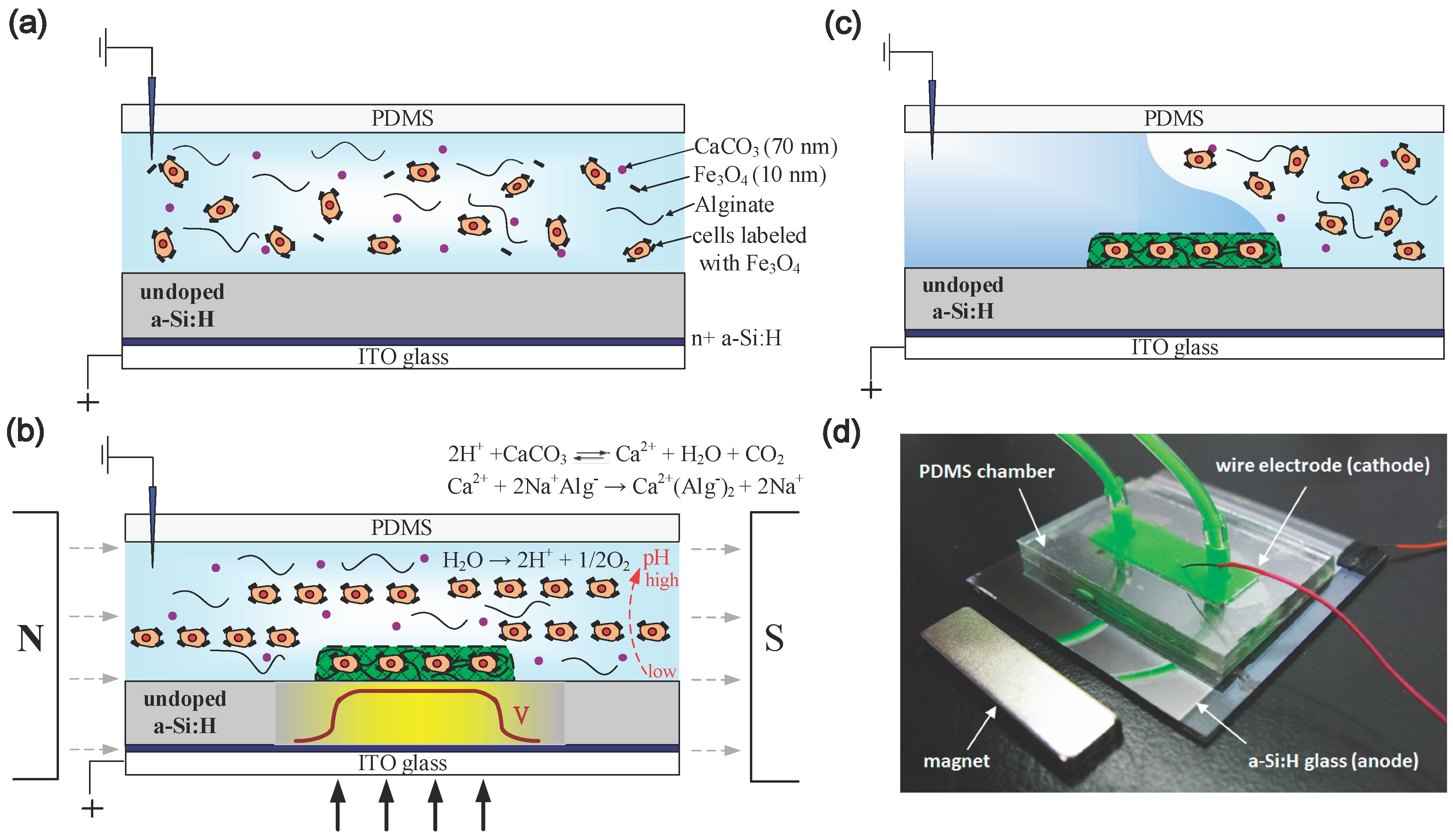
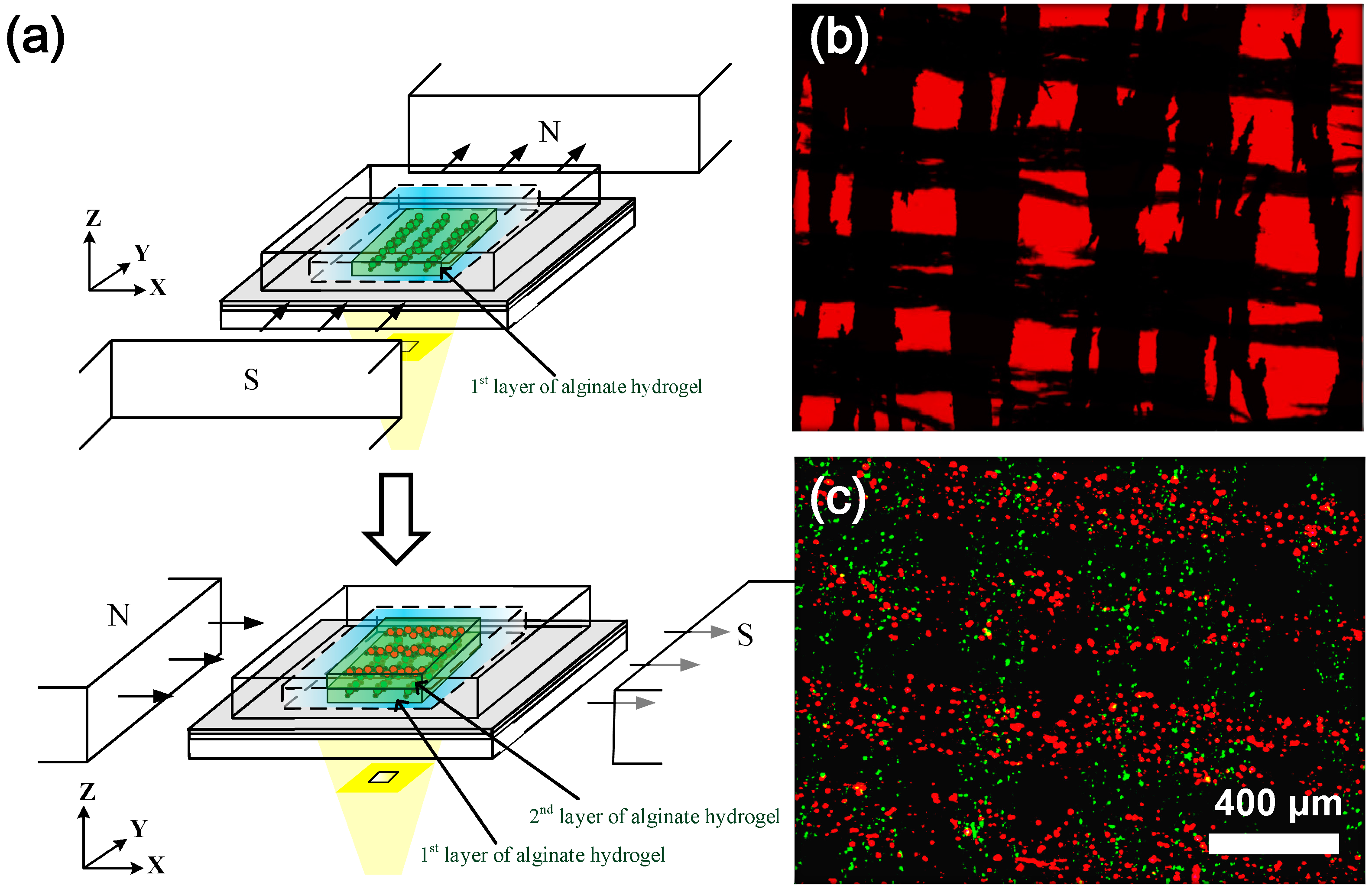
2.1. Principle of Operation

Figure 1a–c shows schematic diagrams of a light-addressable electrolytic system used to perform an electrodeposition of magnetically-guided cells encapsulated in alginate hydrogels using a DMD. A photoconductive substrate, which consists of 0.2 μm of heavily doped hydrogenated amorphous silicon (n+ a-Si:H) and 1 μm of undoped a-Si:H on a 700-μm indium tin oxide (ITO) glass substrate, serves as a light-addressable electrode. A microchamber with 1 to 5 mm in height was fabricated of polydimethylsiloxane (PDMS) and then bonded to the photoconductive substrate. The deposition solution, which contains soluble sodium alginate (80–120 mPa·S, Sigma-Aldrich), suspended cells labeled with magnetic particles (Fe3O4; 10 nm, Sigma-Aldrich), and insoluble calcium carbonate (CaCO3) nanoparticles (70 nm, Specialty Minerals, UK), was introduced into the microchamber (Figure 1a). We used CaCO3 nanoparticles (70 nm in diameter) as the calcium complex to drastically increase the sedimentation time to over 10 min and to ensure a homogeneous dispersion within the microchamber for the production of calcium alginate hydrogels with high shape fidelity [14]. The concentration of 2 wt % alginate solution was chosen for this study because they are optimal concentrations with suitably low viscosities for facilitating the homogeneous dispersion of the suspended cells and CaCO3 nanoparticles within the deposition solutions. An anodic voltage was applied to the photoconductive substrate via the ITO layer, and metallic or platinum wire was inserted into the microchamber to serve as the cathode.
Figure 1. (ac) Schematic diagrams of a light-addressable electrolytic system used to perform an electrodeposition of magnetically-guided cells encapsulated in alginate hydrogels using a digital micromirror device (DMD). (d) The image of the polydimethylsiloxane (PDMS) microchamber bonded to the photoconductive substrate with a metallic wire inserted into the microchamber as the cathode (green dye was introduced into the microchamber for visibility purposes).
Micromachines 05 01173 g001
To align the cells into a specific arrangement, a pair of permanent neodymium iron boron (NdFeB) magnets were simultaneously placed at left and right side of the PDMS microchamber to align the magnetically-labeled cells from irregular arrangement into parallel alignment (Figure 1b). Before performing electrodeposition to encapsulate cells inside alginate hydrogels, we can rotate a pair of NdFeB magnets horizontally or perpendicularly at arbitrary angles to freely manipulate the magnetically-labeled cells into a specific arrangement. Next, when a DC voltage is applied, a patterned light illumination is concurrently projected onto a photoconductive substrate to acts as a photo-anode, which can electrolytically produce protons to result in a decreased pH gradient (Figure 1b). The low pH generated at the anode can locally release calcium ions (Ca2+) from insoluble CaCO3 nanoparticles (Equation (1)) and cause gelation of the calcium alginate through sol-gel transition (Equation (2)).
2H+ + CaCO3 → Ca2+ + H2O + CO2
Ca2+ + 2Na+ Alg → Ca2+ (Alg)2 + 2Na+
During this process, the hydrogel network provides a suitable matrix for encapsulation of the magnetically-labeled cells, which has been manipulated into a specific arrangements within the deposition solution. After few seconds, we turned off the light pattern and DC power supply and immediately introduced the fresh culture medium into the microchamber to remove the residual deposition solution (Figure 1c). By combining the magnetically-labeled cells, light-addressable electrodeposition, and orientation of the magnetic field, we can produce the cell-encapsulated alginate hydrogels, where cells can be freely manipulated into a specific arrangement. Figure 1d shows the image of the PDMS microchamber bonded to the photoconductive substrate with a metallic wire inserted into the microchamber as the cathode.

2.2. Magnetically-Labeled Cells and Chemical Compounds

All cell culture media, sera, and reagents were purchased from Invitrogen (Grand Island, NY, USA). Other chemicals were purchased from Sigma-Aldrich (St. Louis, MO, USA) unless indicated otherwise. Permanent neodymium iron boron (NdFeB) magnets were obtained from Super Electronics (Taiwan).
Baby hamster kidney-21 (BHK-21) fibroblast cells were used in this study. The BHK-21 cells were cultured in 75 cm2 tissue-culture Petri dishes at 37 °C in a humidified atmosphere of 5% CO2/95% air. The BHK-21 cells were incubated in RPMI-1640 medium (1X liquid), which was supplemented with 5% bovine calf serum, 200 U/mL penicillin, and 200 μg/mL streptomycin. The nano-sized iron oxide particles (Fe3O4; 10 nm avg. part. size, 5 mg/mL in H2O) were used to bind to the cell membrane of the BHK-21 cells. The Fe3O4 particles were first washed with phosphate-buffered saline (PBS) and sterilized by an autoclave. After dispersion by vortex, no clumping or aggregation was observed in the absence of a magnetic field. The BHK-21 cells were seeded in monolayer culture at a density of 5 × 10⁴ cells/ cm2 and cultured for 24 h. The Fe3O4 magnetic particles (0.1 to 1 mg/mL) was added to the cells in RPMI-1640 (Grand Island, NY, USA) medium and incubated for 24 h before washing cells with PBS to remove unbound particles. Particles were either engulfed by the cell or remain associated with the cell surface. The cells were grown to preconfluence and detached by trypsinization in a 0.5% trypsin/0.01% EDTA solution in PBS for 5 min at 37 °C. After resuspension, the BHK-21 cells (1 × 10⁶ cell/mL) were homogeneously suspended in deposition solutions of 2 wt % sodium alginate solution with the insoluble CaCO3 powder (70-nm particles; 1 wt %). Fluorescent marker dyes used in this study were DiO (green) and DiI (red) at working concentrations of 5–25 mM.

2.3. Experimental Setup for a Light-Addressed Electrolytic System

Figure 2 shows an experimental setup for a light-addressed electrolytic system for magnetically-guided cells encapsulated in alginate hydrogels using a modified commercial DMD projector. To observe the illumination pattern and fluorescence dye-labeled cell images, a standard upright BX51 fluorescence microscope (Olympus, Tokyo, Japan) was used with a charge coupled device (CCD) camera (INFINITY2-1, Lumenera, Ottawa, Canada). Excitation light for fluorescence was projected onto the photoconductive substrate by a 10× objective lens through a suitable filter unit for fluorescent marker dyes of DiO (green) and DiI (red). A pair of NdFeB magnets was simultaneously placed at left and right side of the PDMS microchamber to align the magnetically-labeled cells into parallel alignment. Before performing electrodeposition, a pair of NdFeB magnets can be rotated horizontally (in X-Y plane) or perpendicularly (in Y-Z plane) at arbitrary angles to freely manipulate the magnetically-labeled cells into a specific arrangement. The fluorescence dye-labeled cell images were observed via a computer (C2). A DMD-based light-addressing projection optical system was set up under the microscope stage. This setup uses a modified commercial MP525P DMD projector (BENQ, Hsinchu, Taiwan) that is equipped with a mercury lamp unit as the light source. We modified the commercial DMD projector by simply removing the projection lens and the color filter wheel [14,21]. The structured light patterns of the DMD were controlled by a computer (C1), such as a rectangular shape. The continuous light illuminated the DMD uniformly through the built-in condensing lens within the DMD projector and then spatially projected it onto the photoconductive substrate through a focus lens (L1 and L2), a 50/50 beam splitter (BS), and an objective lens with 10× magnification. A DC voltage via a 50 W 6613C DC power source (HP/Agilent, Santa Clara, CA, USA) was applied between the photoconductive substrate and metallic wire. The voltage application was synchronized with the projection of an illumination pattern by the DMD. Focal light illumination locally increases the conductivity of the photoconductive substrate and thus creates a virtual photo-electrode with flexible addressability and patternability, which is a substitute candidate for conventional metal microelectrodes. We can control the illumination pattern on the DMD, which enables the performance of an electrodeposition of alginate hydrogel with different shapes and sizes and multiplexed micropatterning.
Figure 2. Schematic diagram of an experimental setup for a light-addressed electrolytic system for magnetically-guided cells encapsulated in alginate hydrogels using a modified commercial DMD projector. A modified commercial DMD projector that is equipped with a mercury lamp unit as the light source. L1 and L2: focus lens; BS: beam splitter.
Micromachines 05 01173 g002

3. Results and Discussion

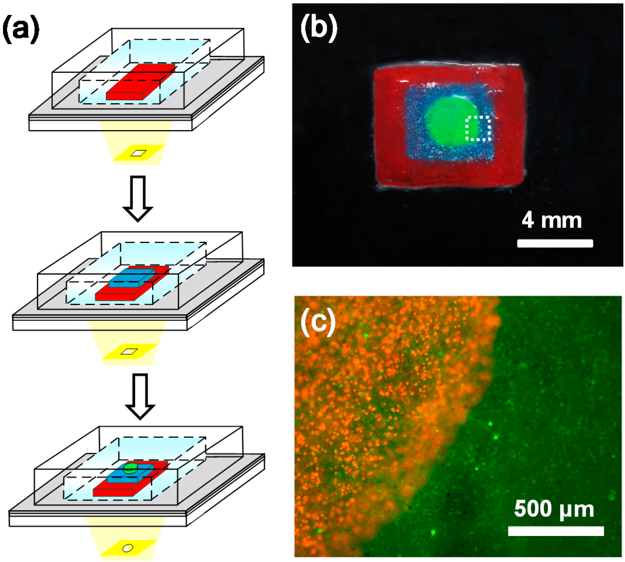
3.1. Formation of Cell-Encapsulated Multilayer Alginate Hydrogels

Our previous work [14] had successfully performed light-addressable micropatterning of cell-encapsulated alginate hydrogels and experimentally examined the effect of the illumination time of the light pattern, the concentration of the alginate and CaCO3 solutions on shape fidelity between the illumination pattern and the produced alginate hydrogels. However, formation of cell-encapsulated multilayer alginate hydrogels with different shapes and sizes in each layer via performing multiplexed micropatterning was not demonstrated yet. Here, we have successfully demonstrated the ability to produce three layers of cell-encapsulated alginate hydrogels with different rectangular sizes for first and second layers and circular shape for the third layer by controlling the illumination pattern on the DMD as shown in Figure 3. In these experiments, the deposition solution, which contains soluble sodium alginate, suspended BHK-21 cells, and insoluble CaCO3 nanoparticles, was introduced into the microchamber. A DC voltage was applied between the photoconductive substrate and metallic wire for 15 s (typical voltage was about 10 V). The illumination patterns on the DMD such as rectangular shape (Figure 3a) was then projected onto the photoconductive substrate to electrolytically produce protons for the electrodeposition of calcium alginate hydrogels. After 15 s, we turned off the light pattern and DC power supply and immediately introduced fresh culture medium into the microchamber to flush away the deposition solution. The procedures of the light-addressable electrodepositions can be sequentially repeated to produce the cell-encapsulated multilayer alginate hydrogels with different shapes and sizes in each layer. In this experiment, the first layer of the alginate hydrogel with 300 μm in thickness was performed at 20 V for 20 s, the second layer with 150 μm in thickness at 20 V for 35 s, and the third layer with 80 μm in thickness at 20 V for 55 s.
Figure 3. (a) Schematic diagrams of performing multiplexed micropatterning for formation of multilayer alginate hydrogels by controlling different illumination patterns in sequence. (b) Image of three layers of alginate hydrogels with different rectangular sizes for first and second layers and circular shape for the third layer. Three different colors of watercolor pigments were blended into the deposition solution for the purpose of easy observation. (c) The merged fluorescence image of the BHK-21 cells encapsulated in the second layer of rectangular alginate hydrogel (green, cells labeled with DiO dye) and the third layer of circular alginate hydrogel (red, cells labeled with DiI dye) at the regional area marked by dotted-line in (b) via performing multiplexed micropatterning.
Micromachines 05 01173 g003
Figure 3b shows three layers of alginate hydrogels with different rectangular sizes for first and second layers and circular shape for the third layer by controlling the illumination pattern on the DMD. We first performed an electrodeposition of alginate hydrogels with a red color using an illumination pattern with rectangular shape, and then we repeated the procedures of the light-addressable electrodeposition twice to form the second layer of rectangular alginate hydrogel with a blue color and the third layer of circular alginate hydrogel with a green color. Figure 3c shows the merged fluorescence image of the BHK-21 cells encapsulated in the second layer of rectangular alginate hydrogel (green, cells labeled with DiO dye) and the third layer of circular alginate hydrogel (red, cells labeled with DiI dye) at the regional area marked by dotted-line in Figure 3b via performing multiplexed micropatterning. It was noted that when multiple layers of cell-encapsulated alginate hydrogels were assembled, the first layer of cell-encapsulated alginate hydrogels might have been experienced with multiple times of electrolysis during multiplex micropatterning. The long period for encapsulated cells to be exposed with low pH and Ca2+ ions at an anode can decrease cell viability. Our previous work showed that the cell viability for a single layer of the cell-encapsulated alginate hydrogel was over 95% at an applied voltage of 10 V for 15 s [14], but gradually decreased with increasing the illumination time of the light pattern, i.e., ~75% live cells at 10 V for 120 s and only ~35% live cells at 10 V for 240 s. Increasing the applied voltage from 20 to 40 V significantly decreased the cell viability, e.g., only ~35% live cells at 40 V for 15 s. To possess an acceptable cell viability for the first layer of cell-encapsulated alginate hydrogels, we need to control the overall illumination time of the light pattern at a preset voltage for the multiplex micropatterning. To avoid this issue, we can simply produce the first layer of the alginate hydrogel without cell encapsulation serving as the buffer layer.
The dimensional characterization of the second or third layer of the alginate hydrogel mostly depends on the thickness of the preformed alginate hydrogel beneath. Table 1 shows the least illumination time (t*) required for alginate hydrogel formation, dimensional resolution (D/Do) and thickness (h2) of alginate hydrogels produced at an applied voltage of Vo for illumination time (t) as well as the smallest feature size (Ds) enable to be produced at different thicknesses (h1) of the preformed alginate hydrogel beneath, respectively. The Do and D denotes an illumination pattern of circular shapes with Do = 600 μm in diameter and the corresponding diameter (D) of the alginate hydrogel produced by the illumination pattern, respectively. The data were rounded up to indicate the average value of the three experiments at the same operating conditions. For a single layer of alginate hydrogels (h1 = 0 μm), we can successfully produce the 56-μm-thick (h2) alginate hydrogel with dimensional resolution of D/Do = 1.05 at an applied voltage of Vo = 10 V for the least illumination time of t* = 5 s. Increasing illumination time (t = 30 s) for an applied voltage (10 V) results in an increase in the Ca2+ ions released from the insoluble CaCO3 nanoparticles. The Ca2+ ions diffuse and then react with the alginate solution to produce thicker (h2 = 280 μm) and larger (D/Do = 1.08) alginate hydrogels. Increasing an applied voltage (20 V) can also produce a higher generation rate of H+ to cause a higher release rate of Ca2+ ions, which leads to a decrease of the least illumination time (t* = 3 s), but an increases of the dimensional resolution (D/Do = 1.12) due to diffusion of Ca2+ ions. The details of the effect of the illumination time of the light pattern on the dimensional resolution (D/Do) with different applied voltages have been described in our previous work [14]. Cheng et al. utilized the transparent fluidic channel with built-in sidewall electrodes to observe the gelation process of alginate hydrogels during electrodeposition [6]. They found that the thickness and the volume of alginate hydrogels linearly increase with the deposition time at the beginning and later saturate at a higher value with longer deposition time. To demonstrate the dimension limitation of the produced alginate hydrogels, we projected an illumination pattern of circular shapes with different diameters ranging from 50 to 1000 μm with 50 μm increment. We can successfully produce 102-μm-sized (Ds) alginate hydrogels on the photoconductive substrate after flushing with DI water. The alginate hydrogels smaller than 102 μm (Ds) were easily flushed away due to uncompleted gelation process of alginate hydrogels.
When the first layer of 200-μm-thick (h1) alginate hydrogels was preformed, we found that the least illumination time (t*) required to produce the second layer of alginate hydrogels increased up to 34 s at an applied voltage of Vo = 10 V, and the dimensional resolution (D/Do) and smallest feature (Ds) increased to 1.14 and 155 μm, respectively. This might be due to the fact that the H+ ions, generated from the anode surface, require more time to diffuse further passing through the preformed alginate hydrogel in order to react with CaCO3 nanoparticles to release Ca2+ ions for gelation. A longer illumination time results in an increase of the dimensional resolution (D/Do) and smallest feature size (Ds). Contrast to a single layer of the 280 μm-thick alginate hydrogel produced at Vo = 10 V for t = 30 s, we need to increase the illumination time up to 70 s to form a 275 μm-thick alginate hydrogel at the same applied voltage. Increasing an applied voltage (20 V) leads to a decrease of the least illumination time (t* = 21 s) and an increases of the dimensional resolution (D/Do = 1.23). Besides, a thicker preformed alginate hydrogel beneath (h2 = 400 μm) would lead to a longer illumination time (t* = 82 s) required for alginate hydrogel formation, and also the increase of the D/Do and Ds.
Table 1. The dimensional characterization of the second layer of the alginate hydrogel produced at different thicknesses (h1) of the preformed alginate hydrogel beneath. t*: the least illumination time required for alginate hydrogel formation; t: illumination time; D/Do: dimensional resolution; h2: thickness of the second layer of alginate hydrogels; Vo: applied voltages; Ds: the smallest feature size.
h1 (μm)Vo (V)t* (s)t (s)h2 (μm)D/DoDs (μm) Micromachines 05 01173 i001
0105556 ± 1.81.05 ± 0.11102 ± 2.2
30280 ± 3.41.08 ± 0.16
203354 ± 2.51.12 ± 0.13
200 ± 3.610343463 ± 3.91.14 ± 0.17155 ± 3.7
70275 ± 5.81.18 ± 0.12Do: the illumination pattern of circular shapes.
D: the corresponding diameter of the alginate hydrogels produced by the illumination pattern.
20212171 ± 4.21.23 ± 0.15
400 ± 5.910828269 ± 5.11.27 ± 0.14213 ± 4.2
150415 ± 9.41.34 ± 0.12
20474775 ± 4.41.39 ± 0.18

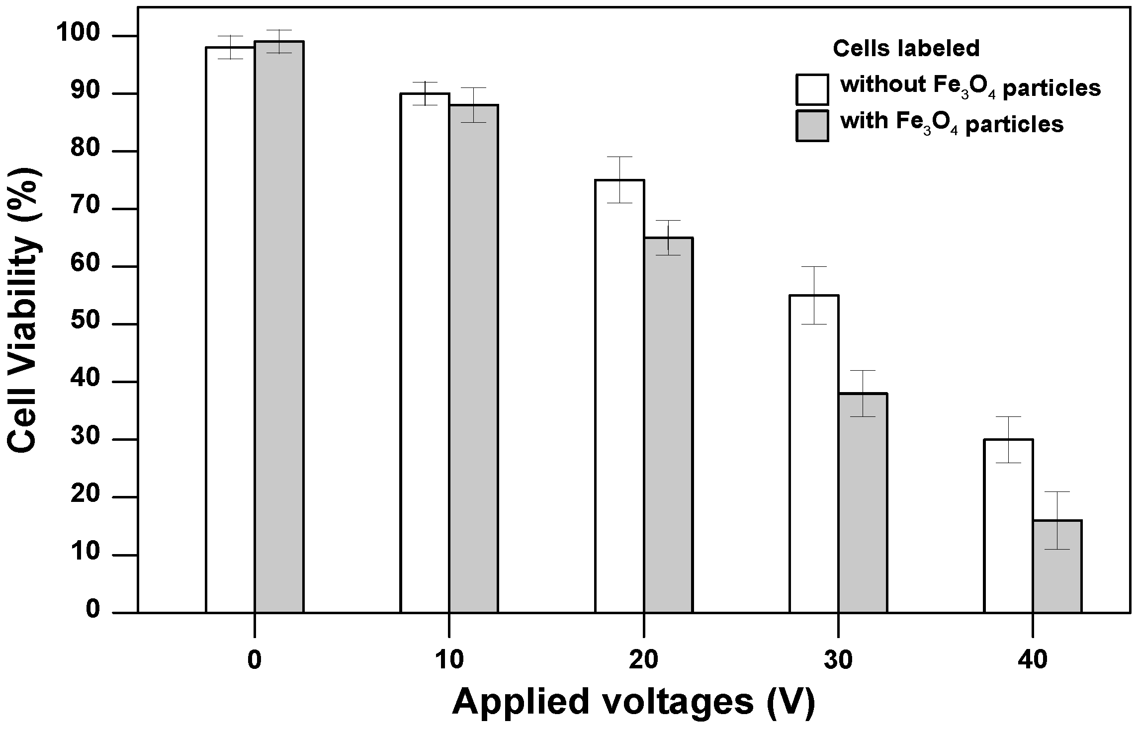
3.2. Cell Viability for Magnetically-Labeled Cells after Electrodeposition

Figure 4 shows the average percentage of viable cells for BHK-21 cells labeled with or without Fe3O4 particles inside alginate hydrogels after performing light-addressable electrodeposition at applied voltages of 0–40 V for 15 s. Cell viability was determined using a live/dead assay (Invitrogen, Grand Island, NY, USA) containing calcein AM (acetoxymethyl ester) (live cells, green) and ethidium homodimer (dead cells, red). Cell-encapsulated alginate hydrogels were stained by incubation with the live/dead assay agents for 10 min to allow the stain to diffuse into the hydrogels. The stain was removed by washing the hydrogels with culture media before the hydrogels were imaged. The average cell viability percentage was calculated by counting the number of pixels in the green (living cells) channel from three different experiments.
Iron oxide is well known to have allowable biocompatibility with most of mammalian cells [22]. Our viability assessments of BHK-21 cells labeled with Fe3O4 particles (1 mg/mL) confirm this observation showing no adverse effect on cell viability (98% live cells) when mixed and cultured in alginate over 4 days before performing light-addressable electrodeposition (for V = 0) as shown in Figure 4. The Fe3O4 particles are either engulfed by the cell or remain associated with the cell surface. Cell viability for BHK-21 cells labeled with or without Fe3O4 particles was found to be high (>90%) at 10 V, but significantly decreased for both cell conditions with increasing the applied voltage from 20 to 40 V. Increasing the applied voltage can prompt the electrolysis to cause a decreased pH and induces more Ca2+ ions to be released from the insoluble CaCO3 nanoparticles. The low pH and the increase of Ca2+ ions can decrease cell viability [14]. At high applied voltages from 20 to 40 V, the cell viability for BHK-21 cells labeled with Fe3O4 particles was found to be lower than those labeled without Fe3O4 particles. We attribute this phenomenon to the heating effect of Fe3O4 particles. The Fe3O4 nanoparticles, a good thermal conductor to absorb heat from surroundings or generate heat by applying an alternating magnetic field, have been extensively used in the clinical cancer therapy [23]. Increasing the applied voltage not only causes low pH and the increase of Ca2+ ions, but also leads to the slight rise in medium temperature. The BHK-21 cells labeled with Fe3O4 particles on the cell surface can absorb heat from surroundings easier than those labeled without Fe3O4 particles to cause cell membrane damage and further decrease cell viability.
Figure 4. The average percentage of viable cells for BHK-21 cells labeled with or without Fe3O4 particles inside alginate hydrogels after performing light-addressable electrodeposition at applied voltages of 0–40 for 15 s.
Micromachines 05 01173 g004

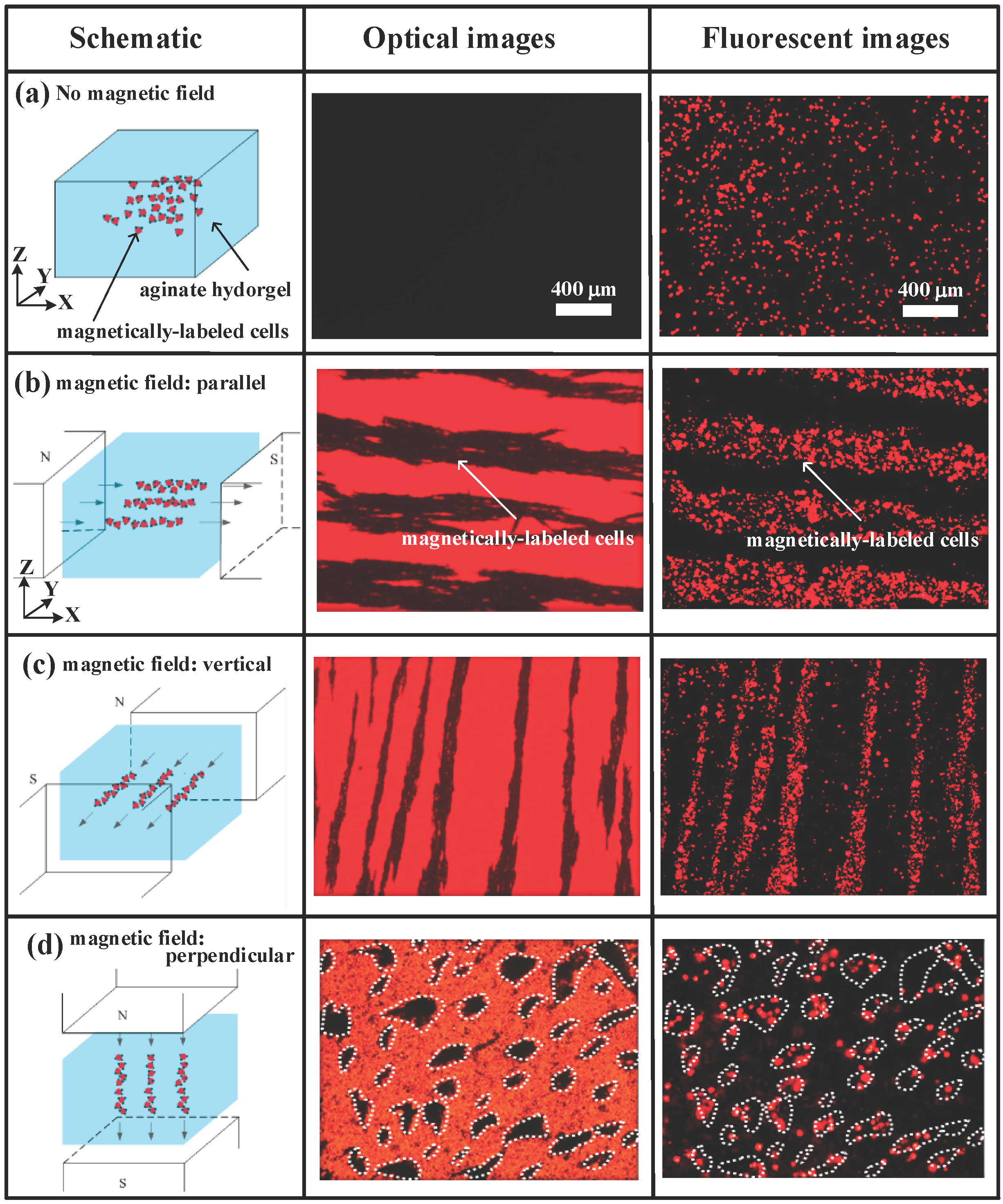
3.3. Light-Addressable Electrodeposition of Magnetically-Guided Cells Encapsulated in Alginate Hydrogels

Figure 5 shows the optical and fluorescent images of magnetically-guided cells encapsulated in rectangular alginate hydrogels where cells were aligned into a specific arrangement after performing light-addressable electrodeposition. For the magnetically-labeled cells with no applied magnetic field, we observed the optical image completely dark and discretely distributed red fluorescence image for DiI-labeled BHK-21 cells in Figure 5a due to random distribution of the encapsulated cells inside alginate hydrogels. To align the cells into a specific arrangement, we can simultaneously place a pair of NdFeB magnets at left and right side of the PDMS microchamber along X axis to align the magnetically-labeled cells into parallel alignment. After performing light-addressable electrodepostion, the aligned magnetically-labeled cells were encapsulated inside the rectangular alginate hydrogels (Figure 5b). In contrast with the case in Figure 5a, we can clearly observe that the magnetically-labeled cells were magnetically manipulated to aggregate into the parallel black and red stripes in the optical and fluorescence images, respectively. Similarly, we can rotate a pair of NdFeB magnets horizontally (in XY plane) or perpendicularly (in XZ plane) at arbitrary angles to freely manipulate the magnetically-labeled cells into a specific arrangements and subsequently encapsulated inside the rectangular alginate hydrogels by performing electrodepostion as shown in Figure 5c,d, respectively. When rotating a pair of NdFeB magnets to form the magnetic field along Z axis (Figure 5d), we observed the magnetically-labeled cells aggregated to form discrete clusters (denoted by dotted lines) where the magnetically-labeled cells were aligned perpendicular to the observation plane.
By combining the magnetically-labeled cells, light-addressable electrodeposition, and orientation of the magnetic fields, we can fabricate two layers of the cell-encapsulated alginate hydrogels, where cells in each layer can be manipulated into cross-directional arrangements as shown in Figure 6. Two layers of cell-encapsulated alginate hydrogels were fabricated by performing light-addressable electrodepositions sequentially. Briefly, after introducing the deposition solution, we first place a pair of NdFeB magnets at two sides of the PDMS microchamber (alone Y axis) to align the magnetically-labeled cells and subsequently encapsulated inside the first layer of the rectangular alginate hydrogel by performing electrodepostion using an illumination pattern with rectangular shape (Figure 6a). Then we repeated the procedures to encapsulate the magnetically-labeled cells inside the second layer of the alginate hydrogel, except we rotated a pair of NdFeB magnets horizontally (in XY plane) by 90° before performing second light-addressable electrodeposition. Figure 6b,c show the optical and merged fluorescent images of the magnetically-labeled BHK-21 cells encapsulated in the two layers of rectangular alginate hydrogels where cells in each layer were manipulated into cross-directional arrangements via performing multiplexed micropatterning.
Cell patterning is a key step before generation of cell sheets for fabrication of complex organs through the layer-by-layer assembly strategy. In the tissue engineering field, cell patterning technologies may have potential applicability to fabricate tissue constructs with complex cell arrangements such as capillary blood vessels, cartilage tissues, and neural networks [15,17]. Our proposed technique provides the advantage that we can fabricate cell-encapsulated multilayer alginate hydrogels layer by layer with different shapes and sizes without the need of manually stacking them, where each layer can contain different types of cell lines that are manipulated into multi-directional arrangement. Besides, we can freely manipulate the magnetically-labeled cells at arbitrary angles in plane or out of plane into a desired arrangement before performing light-addressable electrodeposition to immobilize them in place. By controlling the illumination pattern, we can produced cell-encapsulated alginate hydrogels with different shapes and sizes ranging from μm to mm scales in each layer. In some practical applications, the cell-encapsulated multilayer alginate hydrogels could be cut and peeled off from substrates, and subsequently to be implanted to repair the damaged cartilage tissues for the patients suffering osteoarthritis diseases. Grogan et al. [20] reported the cell patterning technique to produce multi-directional cell arrangements within 3D alginate hydrogels by arranging magnetically-labeled cells using magnetic fields. They also implanted this 3D alginate hydrogel into an in vitro bovine osteochondral defect model showing that the repair tissue formed by magnetically-labeled and arranged cells (vertically aligned), after 2 weeks of culture, appeared to be of higher quality compared to control formed by unarranged cells (randomly distributed). Akiyama et al. [15] created line patterning of human umbilical vein endothelial cells (HUVECs) on artificial skin tissues (C2C12 cells) using micrometer-thick steel plates under magnetic fields. They also fabricated the multilayered cell sheets into 3D tissue constructs by stacking the cross-directional line arrangements of HUVECs on C2C12 cell sheets layer by layer. Because of its capability to generating cell-encapsulated multilayer alginate hydrogels with different thickness and with different cell lines in each layer, we can use it to study cell migration and cell–cell interactions to mimic the in vivo conditions and therefore it is useful for drug screening. As magnetic tissue assembly technology matures and more data are accumulated, we believe that our proposed technology may be applicable for the fabrication of complex tissue architectures required in tissue engineering and find many applications in biomedical research in the near future.
Figure 5. The optical and fluorescent images of magnetically-guided cells encapsulated in rectangular alginate hydrogels after performing light-addressable electrodeposition, where the magnetically-labeled cells were randomly distributed with no applied magnetic field in (a) or manipulated into (b) parallel, (c) vertical, and (d) perpendicular alignments by rotating a pair of NdFeB magnets horizontally (in XY plane) or perpendicularly (in XZ plane). The undoped a-Si:H ITO glass substrate showed red color in optical images; DiI-labeled BHK-21 cells showed red fluorescence in fluorescent images.
Micromachines 05 01173 g005
Figure 6. (a) Schematic diagrams of performing multiplexed micropatterning and adjusting orientation of the magnetic fields for formation of two layers of the cell-encapsulated alginate hydrogels, where cells in each layer can be manipulated into cross-directional arrangements. (b) The optical and (c) merged fluorescent images of the magnetically-labeled BHK-21 cells encapsulated in the two layers of rectangular alginate hydrogels. Cells with green fluorescence in the first layer; cells with red fluorescence in the second layer.
Micromachines 05 01173 g006

4. Conclusions

In this study, we proposed a light-addressable electrolytic system used to perform an electrodeposition of magnetically-guided cells encapsulated in alginate hydrogels using a DMD for 3D cell patterning. Flexible patternability of the photo-electrodes and multiplexed micropatterning provides a great advantage to fabricate multilayer alginate hydrogels with different shapes and sizes in each layer. The magnetically-labeled cells allow to be freely manipulate into a specific arrangements by adjusting orientation of the magnetic fields. By combining the magnetically-labeled cells, light-addressable electrodeposition, and orientation of the magnetic fields, we have successfully demonstrated to fabricate two layers of the cell-encapsulated alginate hydrogels, where cells in each layer were manipulated into cross-directional arrangements. Now we are moving forward to produce cell-encapsulated multilayer alginate hydrogels with different shapes and sizes, where each layer can contain different types of cell lines that are manipulated into multi-directional arrangements to mimic natural tissue. We anticipate that this simple, rapid, and flexible method for light-addressable electrodeposition of cell populations to systematically assemble the cell lines layer by layer into 3D tissues could have a wide range of applications in drug discovery, toxicology, stem cell research, and potentially therapy.

Acknowledgments

This work was partially supported by National Science Council, Taiwan, through the grant NSC 102-2221-E-019-015-MY2.

Author Contributions

Hsiao-Tzu Chu, Yan-Min Liou, and Kuo-Sheng Huang performed the design, microfabrication, experiments, and analyzed the data. Shih-Hao Huang contributed to the original idea of this study, supervised the experiments and wrote the manuscript.

Conflicts of Interest

The authors declare no conflict of interest.

References

  1. Griffith, L.G.; Swartz, M.A. Capturing complex 3D tissue physiology in vitro. Nat. Rev. Mol. Cell. Bio. 2006, 7, 211–224. [Google Scholar] [CrossRef]
  2. Rivron, N.C.; Rouwkema, J.; Truckenmuller, R.; Karperien, M.; De Boer, J.; van Blitterswijk, C.A. Tissue assembly and organization: Developmental mechanisms in microfabricated tissues. Biomaterials 2009, 30, 4851–4858. [Google Scholar] [CrossRef] [PubMed]
  3. Fernandes, T.G.; Kwon, S.-J.; Bale, S.S.; Lee, M.-Y.; Diogo, M.M.; Clark, D.S.; Cabral, J.M.S.; Dordick, J.S. Three-dimensional cell culture microarray for high-throughput studies of stem cell fate. Biotechnol. Bioeng. 2010, 106, 106–118. [Google Scholar] [PubMed]
  4. Lee, M.-Y.; Kumar, R.A.; Sukumaran, S.M.; Hogg, M.G.; Clark, D.S.; Dordick, J.S. Three-dimensional cellular microarray for high-throughput toxicology assays. Proc. Natl. Acad. Sci. USA 2008, 105, 59–63. [Google Scholar] [CrossRef] [PubMed]
  5. Tan, W.H.; Takeuchi, S. Monodisperse alginate hydrogel microbeads for cell encapsulation. Adv. Mater. 2007, 19, 2696–2701. [Google Scholar] [CrossRef]
  6. Cheng, Y.; Luo, X.L.; Betz, J.; Payne, G.F.; Bentley, W.E.; Rubloff, G.W. Mechanism of anodic electrodeposition of calcium alginate. Soft Matter 2011, 7, 5677–5684. [Google Scholar] [CrossRef]
  7. Cheng, Y.; Luo, X.L.; Payne, G.F.; Rubloff, G.W. Biofabrication: Programmable assembly of polysaccharide hydrogels in microfluidics as biocompatible scaffolds. J. Mater. Chem. 2012, 22, 7659–7666. [Google Scholar] [CrossRef]
  8. Cheng, Y.; Luo, X.L.; Tsao, C.Y.; Wu, H.C.; Betz, J.; Payne, G.F.; Bentley, W.E.; Rubloff, G.W. Biocompatible multi-address 3D cell assembly in microfluidic devices using spatially programmable gel formation. Lab Chip 2011, 11, 2316–2318. [Google Scholar] [CrossRef] [PubMed]
  9. Cheng, Y.; Tsao, C.Y.; Wu, H.C.; Luo, X.L.; Terrell, J.L.; Betz, J.; Payne, G.F.; Bentley, W.E.; Rubloff, G.W. Electroaddressing functionalized polysaccharides as model biofilms for interrogating cell signaling. Adv. Funct. Mater. 2012, 22, 519–528. [Google Scholar] [CrossRef]
  10. Luo, X.L.; Wu, H.C.; Tsao, C.Y.; Cheng, Y.; Betz, J.; Payne, G.F.; Rubloff, G.W.; Bentley, W.E. Biofabrication of stratified biofilm mimics for observation and control of bacterial signaling. Biomaterials 2012, 33, 5136–5143. [Google Scholar] [CrossRef] [PubMed]
  11. Shi, X.W.; Tsao, C.Y.; Yang, X.H.; Liu, Y.; Dykstra, P.; Rubloff, G.W.; Ghodssi, R.; Bentley, W.E.; Payne, G.F. Electroaddressing of cell populations by co-deposition with calcium alginate hydrogels. Adv. Funct. Mater. 2009, 19, 2074–2080. [Google Scholar] [CrossRef]
  12. Shi, X.W.; Yang, X.H.; Liu, Y.; Dykstra, P.; Rubloff, G.W.; Ghodssi, R.; Bentley, W.E.; Payne, G.F. Stimulus responsive biopolymers for protein and cell electroaddressing. In Presented at The 238th ACS National Meeting, Washington, DC, USA, 16–20 August 2009; 2009. [Google Scholar]
  13. Yang, X.H.; Kim, E.; Liu, Y.; Shi, X.W.; Rubloff, G.W.; Ghodssi, R.; Bentley, W.E.; Pancer, Z.; Payne, G.F. In-film bioprocessing and immunoanalysis with electroaddressable stimuli-responsive polysaccharides. Adv. Funct. Mater. 2010, 20, 1645–1652. [Google Scholar] [CrossRef]
  14. Huang, S.H.; Hsueh, H.J.; Jiang, Y.L. Light-addressable electrodeposition of cell-encapsulated alginate hydrogels for a cellular microarray using a digital micromirror device. Biomicrofluidics 2011, 5, 034109. [Google Scholar] [CrossRef]
  15. Akiyama, H.; Ito, A.; Kawabe, Y.; Kamihira, M. Fabrication of complex three-dimensional tissue architectures using a magnetic force-based cell patterning technique. Biomed. Microdevices 2009, 11, 713–721. [Google Scholar] [CrossRef] [PubMed]
  16. Akiyama, H.; Ito, A.; Kawabe, Y.; Kamihira, M. Cell-patterning using poly (ethylene glycol)-modified magnetite nanoparticles. J. Biomed. Mater. Res. A 2010, 92A, 1123–1130. [Google Scholar] [PubMed]
  17. Fu, C.Y.; Lin, C.Y.; Chu, W.C.; Chang, H.Y. A simple cell patterning method using magnetic particle-containing photosensitive poly (ethylene glycol) hydrogel blocks: A technical note. Tissue Eng. Part. C Methods 2011, 17, 871–877. [Google Scholar] [CrossRef] [PubMed]
  18. Ino, K.; Ito, A.; Honda, H. Cell patterning using magnetite nanoparticles and magnetic force. Biotechnol. Bioeng. 2007, 97, 1309–1317. [Google Scholar] [CrossRef] [PubMed]
  19. Ino, K.; Okochi, M.; Konishi, N.; Nakatochi, M.; Imai, R.; Shikida, M.; Ito, A.; Honda, H. Cell culture arrays using magnetic force-based cell patterning for dynamic single cell analysis. Lab Chip 2008, 8, 134–142. [Google Scholar] [CrossRef] [PubMed]
  20. Grogan, S.P.; Pauli, C.; Chen, P.; Du, J.; Chung, C.B.; Kong, S.D.; Colwell, C.W.; Lotz, M.K.; Jin, S.G.; D’Lima, D.D. In situ tissue engineering using magnetically guided three-dimensional cell patterning. Tissue Eng. Part C Methods 2012, 18, 496–506. [Google Scholar] [CrossRef] [PubMed]
  21. Huang, S.H.; Wei, L.S.; Chu, H.T.; Jiang, Y.L. Light-addressed electrodeposition of enzyme-entrapped chitosan membranes for multiplexed enzyme-based bioassays using a digital micromirror device. Sens. Basel 2013, 13, 10711–10724. [Google Scholar] [CrossRef]
  22. Dobson, J.; Bowtell, R.; Garcia-Prieto, A.; Pankhurst, Q. Safety implications of high-field MRI: Actuation of endogenous magnetic iron oxides in the human body. PLoS One 2009, 4, e5431. [Google Scholar] [CrossRef] [PubMed]
  23. Silva, A.C.; Oliveira, T.R.; Mamani, J.B.; Malheiros, S.M.F.; Malavolta, L.; Pavon, L.F.; Sibov, T.T.; Amaro, E.; Tannus, A.; Vidoto, E.L.G.; Martins, M.J.; Santos, R.S.; Gamarra, L.F. Application of hyperthermia induced by superparamagnetic iron oxide nanoparticles in glioma treatment. Int. J. Nanomed. 2011, 6, 591–603. [Google Scholar]

