Article

Molecular Characterization of Three Novel Phospholipase A2 Proteins from the Venom of Atheris chlorechis, Atheris nitschei and Atheris squamigera

1
School of Integrative Medicine, Fujian University of Traditional Chinese Medicine, No.1 Qiu Yang Road, Shangjie Town, Fuzhou 350122, Fujian, China
2
School of Pharmacy, Fujian Medical University, No.1 Xueyuan Road, Shangjie Town, Fuzhou 350004, Fujian, China
3
Natural Drug Discovery Group, School of Pharmacy, Queen’s University Belfast, University Road, Belfast BT7 1NN, UK
*
Authors to whom correspondence should be addressed.
Toxins 2016, 8(6), 168; https://doi.org/10.3390/toxins8060168
Submission received: 24 February 2016 / Revised: 12 May 2016 / Accepted: 20 May 2016 / Published: 1 June 2016
(This article belongs to the Section Animal Venoms)

Abstract

:
Secretory phospholipase A2 (sPLA2) is known as a major component of snake venoms and displays higher-order catalytic hydrolysis functions as well as a wide range of pathological effects. Atheris is not a notoriously dangerous genus of snakes although there are some reports of fatal cases after envenomation due to the effects of coagulation disturbances and hemorrhaging. Molecular characterization of Atheris venom enzymes is incomplete and there are only a few reports in the literature. Here, we report, for the first time, the cloning and characterization of three novel cDNAs encoding phospholipase A2 precursors (one each) from the venoms of the Western bush viper (Atheris chlorechis), the Great Lakes bush viper (Atheris nitschei) and the Variable bush viper (Atheris squamigera), using a “shotgun cloning” strategy. Open-reading frames of respective cloned cDNAs contained putative 16 residue signal peptides and mature proteins composed of 121 to 123 amino acid residues. Alignment of mature protein sequences revealed high degrees of structural conservation and identity with Group II venom PLA2 proteins from other taxa within the Viperidae. Reverse-phase High Performance Liquid Chromatography (HPLC) profiles of these three snake venoms were obtained separately and chromatographic fractions were assessed for phospholipase activity using an egg yolk suspension assay. The molecular masses of mature proteins were all identified as approximately 14 kDa. Mass spectrometric analyses of the fractionated oligopeptides arising from tryptic digestion of intact venom proteins, was performed for further structural characterization.

Graphical Abstract

1. Introduction

As major components of snake venoms, proteins of the secretory phospholipase A2 (sPLA2) family have been structurally-defined, characterized and their catalytic mechanisms have been elucidated [1]. Traditionally, these snake venom enzymes have been divided into two main groups, Group I (GI) and Group II (GII), used to distinguish between molecules based on their amino acid sequences, disulfide-bridging patterns, unique functional loops and extension amino acids [2]. Phospholipase A2 catalyzes the hydrolysis of the sn-2 fatty acyl bond of phospholipids in a calcium-dependent manner [3]. A broad range of physiological molecules, such as long-chain fatty acid phospholipids, short-chain fatty acid-containing oxidized phospholipids and platelet-activating factors, act as the natural substrates of phospholipase A2 [4]. The higher-order catalytic hydrolysis functions of these snake enzymes play a central role in lipid metabolism of numerous cells and tissues, contributing to prey digestion. In addition, much is known about the wide variety of pathological effects exhibited by the snake venom phospholipase A2s including their neurotoxicity, myotoxicity, cytotoxicity, anticoagulant effects, cardiotoxicity, hypotension induction, platelet aggregation/inhibition and anti-bacterial activities [5,6,7,8,9,10], which in many cases, are fundamental to the toxicity of snake venoms [11,12].
Of the 400 species of snakes in Africa, bush vipers (Atheris, Viperidae) are not considered to be highly-dangerous in terms of the numbers of documented bites and deaths. This may in part be due to their arboreal behavior and their distribution in often inhospitable habitats [13]. A full envenomation by Atheris is likely to be fatal, however, due to the injection of a large quantity of toxic venom through their long and sharp fangs [14]. Envenomations involving Atheris vipers are clinically-characterized by coagulation disturbances and hemorrhage, local pain, edema, bleeding and a prolongation of coagulation time and non-clotting blood [15,16]. Some victims develop acute renal failure and hypertension [17,18]. However, studies on characterization of venom enzymes to explain these sequelae, are scarce and are incomplete [19]. In fact, even superficial reports on the components of whole venoms are equally rare. Thus, hunting for and describing the toxic polypeptides and/or proteins in Atheris venoms became a focus of our interest. Novel disintegrins had been isolated and identified in a previous study [20], and in this work, cloning of precursor-encoding cDNAs and characterization of the mature phospholipase A2 proteins from Atheris chlorechis (A. chlorechis), Atheris nitschei (A. nitschei) and Atheris squamigera (A. squamigera) venoms, were carried out in an attempt to explain the toxic effects of the venom of Atheris snakes in more depth.

2. Results

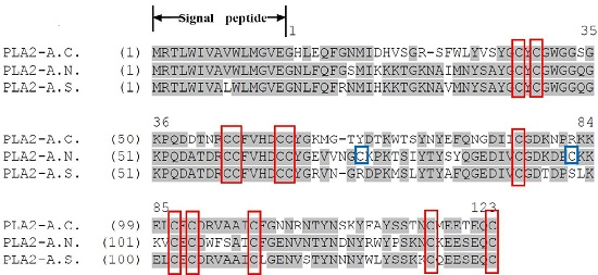
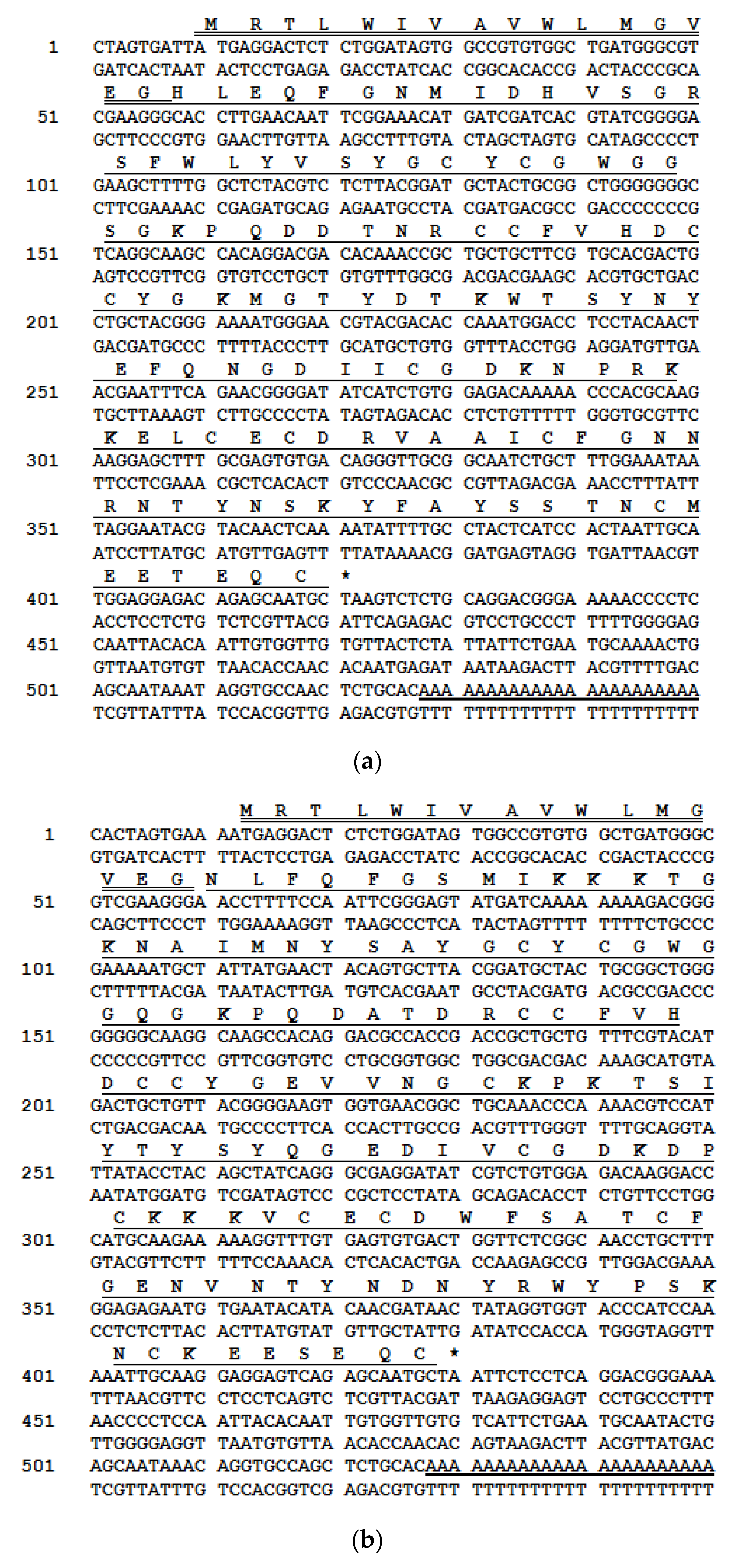
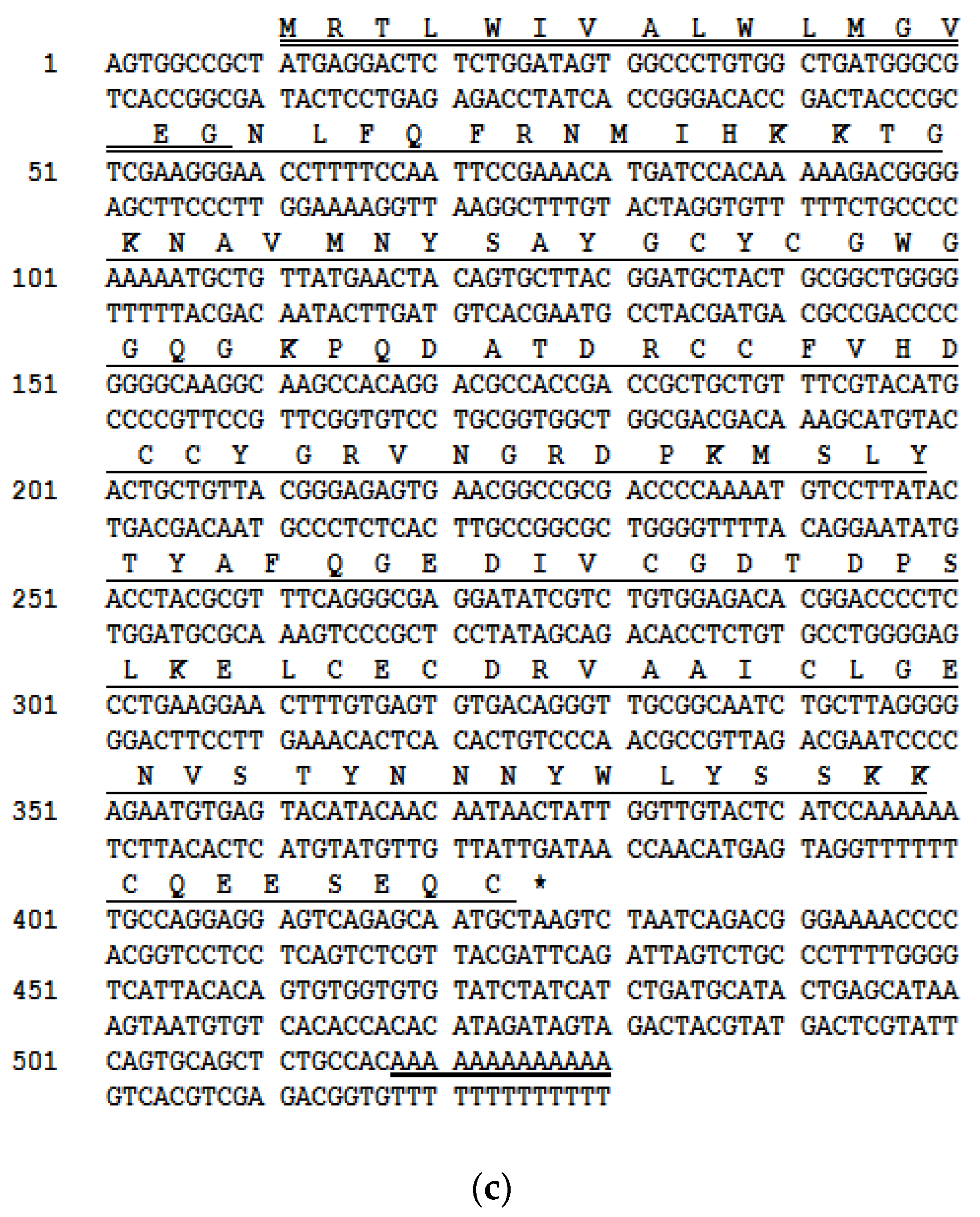
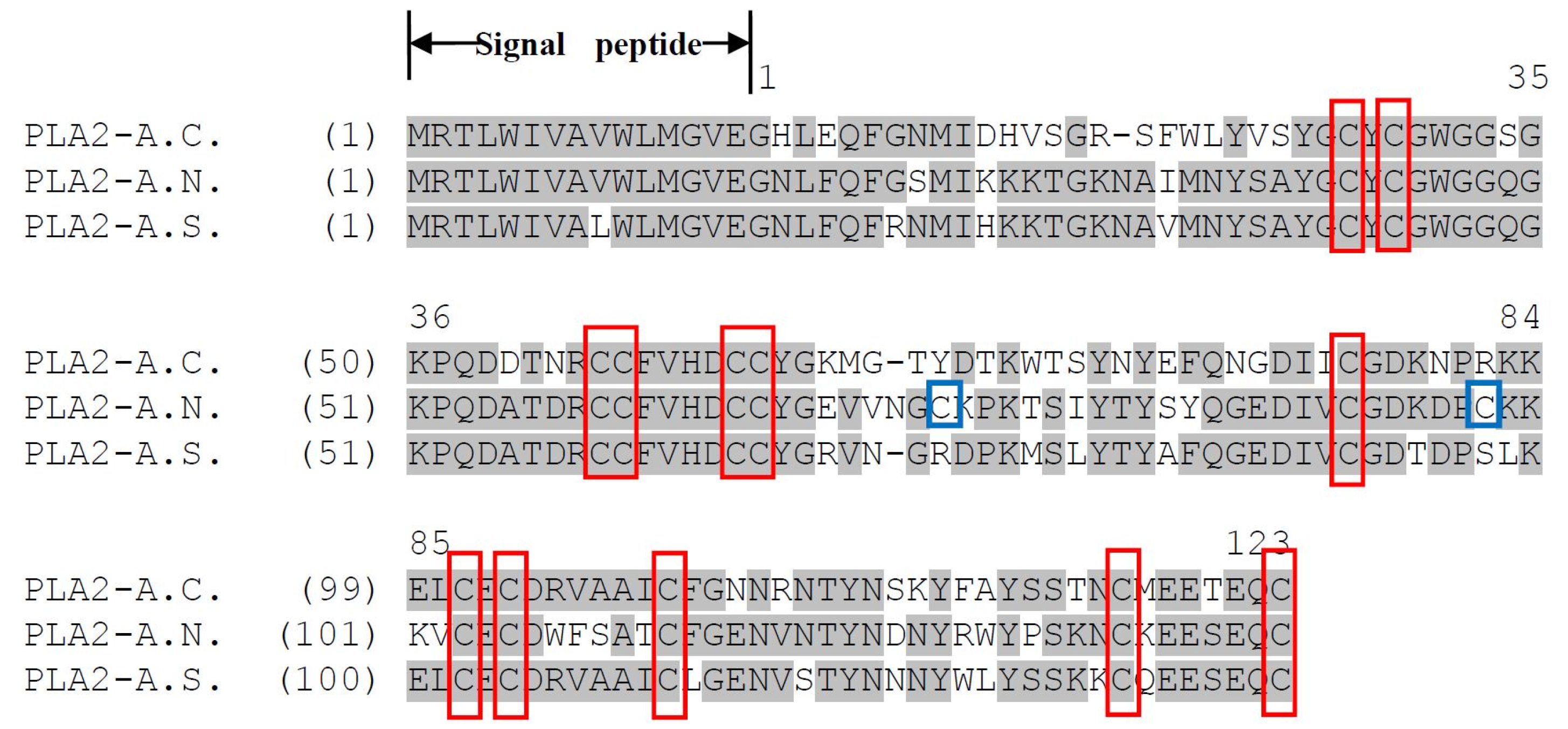
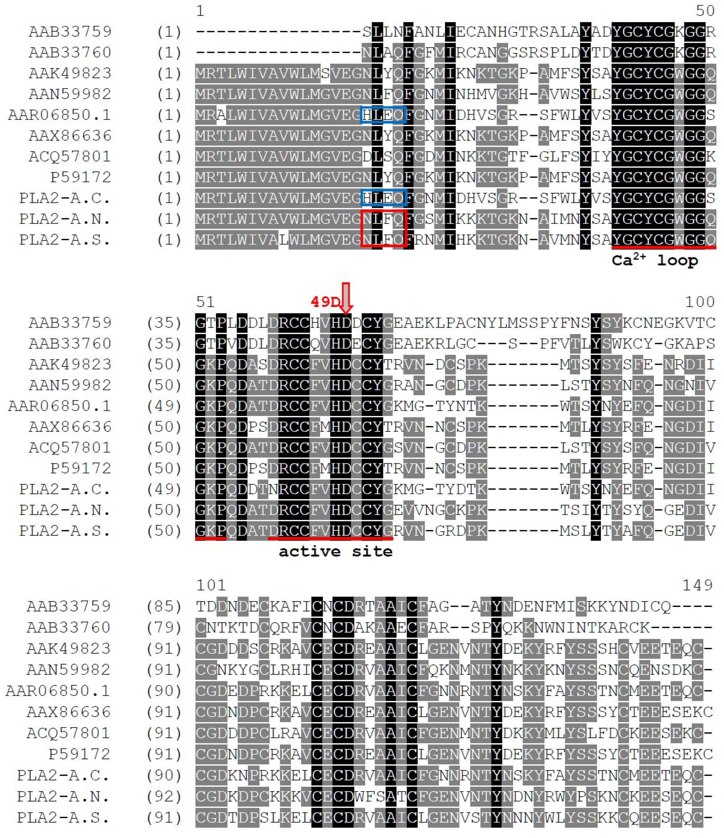
2.1. cDNAs Encoding PLA2 Precursors and Bioinformatic Analyses

A single novel unique full-length phospholipase-encoding precursor was cloned from each of the venom-derived libraries of A. chlorechis, A. nitschei and A. squamigera and each encoded a single copy of PLA2 (Figure 1). Here, the first genomic sequences for the three cDNAs encoding phospholipase A2 precursors from Atheris bush viper venoms, are reported and were named PLA2-A.C., PLA2-A.N. and PLA2-A.S., respectively. These data have been deposited in the Genbank Nucleotide Sequence Database under the accession codes, KP11968, KP119683 and KP119684, respectively. The open-reading frame amino acid sequences of these proteins exhibited a similar structural pattern: all contained a putative 16-residue signal peptide sequence and a mature protein sequence consisting of 123 residues for A. nitschei, 121 residues for A. chlorechis and 122 residues for A. squamigera. All contained a conserved Histidine/Aspartic acid dyad-motif and a cysteine-rich sequence that defines the PLA2 hydrolytic function.
Comparison of these three precursors, established by deduction from cloned cDNA, is shown in the alignment in Figure 2. They exhibit a remarkable degree of identity: the signal peptides differ by only one residue, with the mature proteins showing high similarity especially in the features of the catalytic network and the primary metal binding site. Except for PLA2-A.N., with two additional cysteine residues, the positions of the 12 cysteines are homologous in the three precursors, meaning that this uniform character of disulfide bridging pattern should lead to similar crystal structures of those three PLA2s.
The open-reading frames of the three PLA2 proteins were employed in Basic Local Alignment Search Tool (BLAST) searches using the National Center for Biotechnological Information (NCBI) on-line portal. The results indicated that they had a relatively high identity with Group II phospholipase A2s from Viperidae snake venoms (Table 1, Figure 2). These studies revealed some very interesting characteristics of the three novel enzymes: PLA2-A.C. and PLA2-A.S., which both possessed 12 cysteines, showed a disparate alignment map. PLA2-A.S. showed some unexpected identities with PLA2-A.N. that has 14 cysteines, and they were both obviously most similar to the phospholipases A2 classified as Group IIA—the majority cluster. However, PLA2-A.C. is nearly identical to phospholipase A2 AAR06850.1 from Bitis gabonica [21], which falls within the extraordinary Group IIB PLA2s that lack one disulfide-bridge conserved in the Group IIA as mentioned in the work of Six et al. [2].

2.2. Identification and Structural Analysis of PLA2 Proteins

A large range of components was successfully resolved following reverse phase High Performance Liquid Chromatography (HPLC) fractionation of the crude lyophilized venoms from A. chlorechis, A. nitschei and A. squamigera. Initial PLA2 activity screening of eluted HPLC fractions was performed separately by agarose/egg yolk suspension plates (Supplementary materials Figure S1, Table S1). A number of fractions were identified as possessing phospholipase A2 activity and were displayed as isolated peaks in each chromatogram (Figure 3).
The molecular masses of each protein were determined by Matrix-assisted laser desorption/ionization-Time of Flight (MALDI-TOF) mass spectrometry and single major ions with m/z ratios around 14 KDa were resolved which are in accordance with data from the cDNA-deduced amino acid sequences. The primary structures were subsequently unequivocally confirmed by Liquid chromatography–tandem mass spectrometry (LC/MS/MS) fragmentation sequencing (Table 2).

3. Discussion

Whole venoms from A. chlorechis, A. nitschei and A. squamigera were subjected to reverse phase HPLC fractionation, followed by mass spectrometric analysis of each fraction, resulting in consistent identification of PLA2 proteins with the predicted masses of mature proteins deduced from cDNA-encoded precursors. It can be seen from Figure 3 that active PLA2 fractions from A. squamigera venom seems to contain more than one component. However, as shown in Table 2, the spectrum of the peaks at 106 and 113 min differed by only six mass units from the theoretical molecular mass of 13841 Da. The discrepant masses of these two peaks probably could be accounted for by several amino-acid substitutions in the sequences of related isoenzymes which often occur in surface exposed residues which play a key role in recognition of target proteins [22]. Although the spectrum of A. squamigera exhibited the probable existence of PLA2 enzyme isoforms in the venom, the tiny mass difference of the two peaks indicated a very low possibility. Therefore, we suppose it could just be due to slight differences in hydrophobic properties or the isoelectric points of the two subunits constituting the PLA2 homodimer (related to the elutropic theory of reverse-phase chromatography); even external conditions, such as room temperature, stability of ionization, etc., could also be responsible. However, as a consequence of intrinsic molecular diversity, single snake venom can contain up to 16 discrete PLA2 homologs with phospholipase activity [23], we cannot affirm that the precursors obtained by molecular cloning in our study represent the only PLA2 proteins occurring in these snake venoms.
Comparative sequence analyses have revealed a high conservation of functionally important domains and disulfide bonding patterns among the different groups of phospholipase A2s. This can be seen clearly by our alignment of Groups I and II PLA2s from Genbank and PLA2-A.C., PLA2-A.N. and PLA2-A.S., from our original experimental data, as shown in Figure 4. The sequence identity as well as the BLAST data shown in Table 1 proves our hypothesis on the relationship of PLA2-A.C., PLA2-A.N. and PLA2-A.S. PLA2-A.N. and PLA2-A.S. share the same N-terminal sequence NLFQ-, show higher similarity in functional amino-acid sequence and both belong to Group IIA. In contrast, PLA2-A.C. has a different N-terminal amino acid sequence, HLEQ-, to the previous two PLA2s and was identical to AAR 06850.1—the PLA2 from Gaboon viper venom [21], which was identified as a Group IIB PLA2. Structural comparisons provide a basis for speculation as to the novel phospholipase A2 protein metal binding sites, disulfide-bond assignments and residues participating in the catalytic site. These three novel phospholipase A2s share, in common with the other PLA2s, the Ca2+ binding loop, active site, and pivotal amino-acid residues such as the His 48/Asp 49 core and calcium-binding assistants, Tyr 28, Gly 30 and Gly 32 (shown in Figure 4). Many of the similar pre-determinations of former newly-discovered PLA2s, derived from such homologies, have been confirmed by enzyme activity assays and by X-ray crystallographic analysis. It is thus justifiable to consider the relationships between primary structures and enzyme properties of these three PLA2s and the effects of their key structural frameworks as well as the location and types of residues involved in activity or toxicity.
Little is known about the toxicity of enzymes from venoms of the Atheris genus, although the coagulation disturbances, hemorrhaging, acute renal failure and hypertension following snakebite, can all be fatal. Our group has characterized the disintegrin proteins in a previous study [20] and these could explain the platelet aggregation inhibition and blood clotting blocking toxicity of the Atheris venoms. It would thus be interesting to predict pharmacological effects and look for the experimental evidence for the involvement of these novel phospholipase A2s. As direct experimental proof is not yet available, it is not possible to predict the properties of these proteins at the moment, and thus the question remains open.

4. Experimental

4.1. Materials

Lyophilized venoms from the Western bush viper (A. chlorechis), the Great Lakes bush viper (A. nitschei) and the Variable bush viper (A. squamigera), were obtained from a commercial source (Latoxan, Valence, France).

4.2. Molecular Cloning of the Phospholipase A2 Precursor-Encoding cDNAs

Five milligrams of each venom sample were separately dissolved in 1 mL of lysis/binding buffer (Dynal Biotech, Wirral, UK); the polyadenylated mRNA was extracted by oligo-dT Dynabeads (Dynal Biotech, Wirral, UK) and was isolated as described by the manufacturer. A SMARTTM Rapid Amplification of cDNA ends (RACE) cDNA Amplification kit (BD Clontech, Basingstoke, UK) was then employed. Both 5'-and 3' cDNA ends were synthesized using SMART (Switching Mechanism At 5' end of RNA Transcription) cDNA synthesis technology. The reaction was performed by using a nested universal primer (NUP) and a sense primer (S1: 5'-ATGAGGACTCTCTGGATAGTGGCCG-3') that was designed to be complementary to a highly-conserved domain of the 5'-untranslated region of previously-characterized phospholipase A2 cDNAs from related snake species(Accession Nos.: GU012263, AY430405, DQ288157, AM114013 and DQ295886). The DNA fragments of expected size were purified and inserted into the pGEM-T vector system (Promega, Southampton, UK). Plasmid DNAs selected after transformation by Escherichia coli (E. coli) cells, were amplified and then purified by use of a Rapid HiYield™ Gel/PCR DNA Extraction Kit (RBC Bioscience, Duren, Germany). Sequences of the products were obtained use of an automated 3730 capillary DNA sequencer (Applied Biosystems, Wamington, UK).

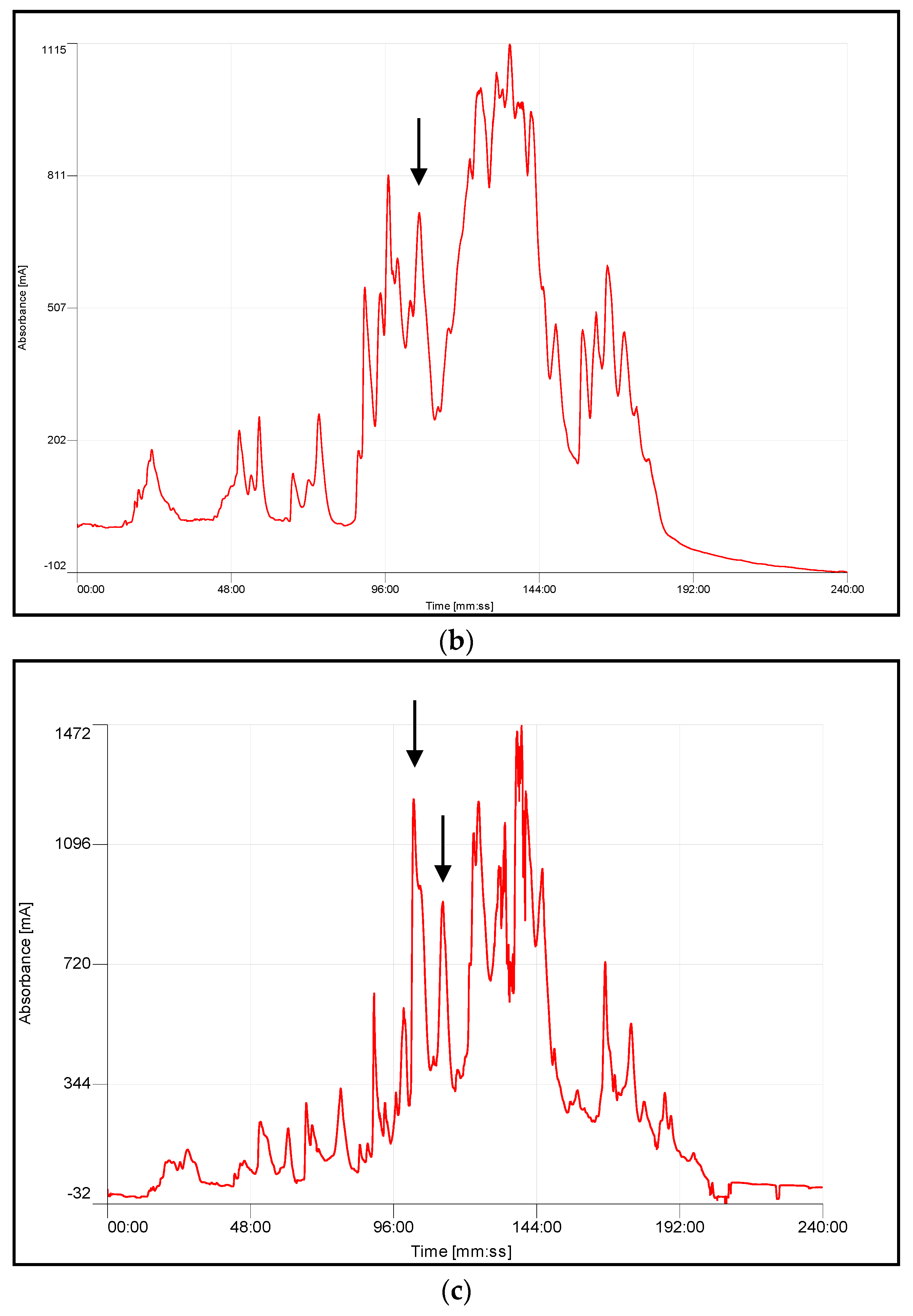
4.3. Chromatographic Fractionation and Activity Determination

Fractionation of components of all three snake venoms was achieved by gradient reverse-phase HPLC (Cecil CE 4200 Adept, Cambridge, UK), using a Phenomenex C5 (300 A, 250 × 10 mm) column. Five milligram samples from each venom were separately reconstituted in 0.05% (v/v) trifluoroacetic acid (TFA)/water, injected onto the column and eluted with a linear gradient formed from 0.05/99.5 (v/v) TFA/water to 0.05/19.95/80.0 (v/v/v) TFA/water/acetonitrile, by increasing the percentage of the latter buffer gradually over 240 min at a flow rate of 1 mL/min. The fractions were collected at minute intervals and samples (200 μL) were removed from each fraction in triplicate, concentrated and stored at 4 °C prior to functional studies.
The determination of phospholipase A2 (PLA2) activity in fractions was achieved by use of agarose/egg yolk suspension plates containing phosphatidylcholine as substrate. A 20% portion of HPLC fractions from each of the snake venoms were concentrated and reconstituted in 0.1% Albumin from bovine serum/phosphate buffer saline (BSA/PBS). The plates were prepared with 4% egg yolk emulsion (Oxoid, Basingstoke, UK) in agarose solution in the presence of calcium, punched with 8 wells per plate and each well was loaded with 10 μL sample. The diameters of cleared egg yolk zones after the 20 h incubation (37 °C) were measured to locate the fractions that possessed PLA2 activity.

4.4. Identification and Structural Investigations

Reverse phase High Performance Liquid Chromatography (RP-HPLC) fractions possessing PLA2 activity were subjected to matrix-assisted laser desorption/ionization, time-of-flight mass spectrometry (MALDI–TOF MS) on a linear time-of-flight Voyager DE mass spectrometer (Perseptive Biosystems, Warrington, UK) in positive detection mode using α-cyano-4-hydroxycinnamic acid as the matrix. Internal mass calibration of the instrument with known standards established the accuracy of mass determination as ±0.1%. Those fractions with masses of approximately 14 kDa coincident with those deduced from cloned cDNAs were digested using trypsin and the resultant tryptic peptides were subjected to primary structural analysis by use of an LCQ Fleet electrospray ion-trap mass spectrometer (Thermo, Cheshire, UK).

4.5. Ethical Statement

Venoms used in this study were obtained non-invasively from captive snakes.

Supplementary Materials

The following are available online at www.mdpi.com/2072-6651/8/6/168/s1, Figure S1: Dose response curve of diameter of PLA2 clearance zones against log10 enzyme concentration of the standard PLA2 from honey bee venom. Linear regression analysis was employed, and the regression equation (Y = 0.7039X − 0.34) was obtained to analyse the equivalent PLA2 content of sample venoms. The clearance zones of samples in the agarose/egg yolk suspension plate assay were used to estimate content of PLA2s in sample venom fractions. The results are summarised in Table S1. Table S1: Clearance zones and corresponding estimated quantities of PLA2 in sample venoms from A. chlorechis, A. nitschei and A. squamigera.

Acknowledgments

This project was supported by the National Science Foundation, China (Grant No. 81402842) and the Natural Science Foundation of Fujian Province, China (Grant No. 2015J05162).

Author Contributions

He Wang, Xiaole Chen and Mei Zhou conceived and designed the experiments; He Wang performed the experiments. He Wang and Chris Shaw analyzed the data. Tianbao Chen and Lei Wang contributed reagents/materials/analysis tools. He Wang wrote the manuscript. Chris Shaw and Xiaole Chen revised the manuscript.

Conflicts of Interest

The authors declare that they have no conflicts of interest.

References

  1. Dennis, E.A. Diversity of group types, regulation, and function of phospholipase A2. J. Biol. Chem. 1994, 269, 13057–13060. [Google Scholar] [PubMed]
  2. Six, D.A.; Dennis, E.A. The expanding superfamily of phospholipase A2 enzymes: Classification and characterization. Biochim. Biophys. Acta 2000, 1488, 1–19. [Google Scholar] [CrossRef]
  3. Dufton, M.J.; Hider, R.C. Classification of phospholipases A2 according to sequence. Evolutionary and pharmacological implications. Eur. J. Biochem. 1983, 137, 545–551. [Google Scholar] [CrossRef] [PubMed]
  4. Gutiérrez, J.M.; Lomonte, B. Phospholipases A2: Unveiling the secrets of a functionally versatile group of snake venom toxins. Toxicon 2013, 62, 27–39. [Google Scholar] [CrossRef] [PubMed]
  5. Botes, D.P.; Viljoen, C.C. Bitis gabonica venom. The amino acid sequence of phospholipase A. J. Biol. Chem. 1974, 249, 3827–3835. [Google Scholar] [PubMed]
  6. Buckland, A.G.; Wilton, D.C. The antibacterial properties of secreted phospholipases A2. Biochim. Biophys. Acta 2000, 1488, 71–82. [Google Scholar] [CrossRef]
  7. Jan, V.M.; Guillemin, I.; Robbe-Vincent, A.; Choumet, V. Phospholipase A2 diversity and polymorphism in European viper venoms: Paradoxical molecular evolution in Viperinae. Toxicon 2007, 50, 1140–1161. [Google Scholar] [CrossRef] [PubMed]
  8. Leiguez, E.; Giannotti, K.C.; Moreira, V.; Matsubara, M.H.; Gutiérrez, J.M.; Lomonte, B.; Rodríguez, J.P.; Balsinde, J.; Teixeira, C. Critical role of TLR2 and Myd88 for functional response of macrophages to a group IIa-secreted phospholipase A2 from snake venom. PLoS ONE 2014, 9. [Google Scholar] [CrossRef] [PubMed]
  9. Saikia, D.; Mukherjee, A.K. Anticoagulant and membrane damaging properties of snake venom phospholipase A2 enzymes. Toxinology 2015. [Google Scholar] [CrossRef]
  10. Sudarshan, S.; Dhananjaya, B.L. The antimicrobial activity of an acidic phospholipase A2 (NN-XIa-PLA2) from the venom of Naja naja naja (Indian Cobra). Appl. Biochem. Biotechnol. 2015, 176, 2027–2038. [Google Scholar] [CrossRef] [PubMed]
  11. Adamich, M.; Dennis, E.A. Specificity reversal in phospholipase A2 hydrolysis of lipid mixtures. Biochem. Biophys. Res. Commun. 1987, 80, 424–428. [Google Scholar] [CrossRef]
  12. Berger, M.; Santi, L.; Beysdasilva-da-Silva, W.O.; Oliveira, F.M.S.; Caliari, M.V.; Yates, J.R.Y., III; Ribeiro Vieira, M.A.; Guimarães, J.A. Mechanisms of acute kidney injury induced by experimental Lonomia obliqua envenomation. Arch. Toxicol. 2015, 89, 459–483. [Google Scholar] [CrossRef] [PubMed]
  13. Christensen, P.A. South African Snake Venoms and Antivenoms; South African Institute for Medical Research: Johannesburg, South African, 1955. [Google Scholar]
  14. Branch, W.R.; Haagner, G.V.; Morgan, D.R.; Lanoie, L.O. Venoms and snakebite. J. Herpetol. Assoc. Afr. 1991, 39, 28–29. [Google Scholar] [CrossRef]
  15. Favreau, P.; Chenval, O.; Menin, L.; Michalet, S.; Gerner, H.; Principaud, F.; Thai, R.; Stöcklin, R. The venom of the snake genus Atheris contains a new class of peptides with clusters of histidine and glycine residues. Rapid 2007, 21, 406–412. [Google Scholar]
  16. Mebs, D.; Holada, K.; Kornalik, F.; Simak, J. Severe coagulopathy after a bite of a green bush viper (Atheris squamiger): Severe coagulopathy after a bite case report and biochemical analysis of the venom. Toxicon 1998, 36, 1333–1340. [Google Scholar] [CrossRef]
  17. Top, L.J.; Tulleken, J.E.; Ligtenberg, J.J.; Meertens, J.H.; van de Werf, T.S.; Zijlstra, J.G. Serious envenomation after a snakebite by a Western bush viper (Atheris chlorechis) in The Netherlands: A case report. Neth. J. Med. 2006, 64, 153–156. [Google Scholar] [PubMed]
  18. Valenta, J.; Stach, Z.; Fricova, D.; Zak, J.; Balik, M. Envenoming by the viperid snake Proatheris superciliaris: A case report. Toxicon 2008, 52, 392–394. [Google Scholar] [CrossRef] [PubMed]
  19. Mebs, D.; Fach, A.; Herrmann, H.W. Enzyme analysis of Atheris snake venom. Toxicon 1997, 35, 813. [Google Scholar] [CrossRef]
  20. He, W.; Chen, X.; Lei, W.; Wei, C.; Mei, Z.; Chen, T.; Shaw, C. Cloning and characterisation of three novel disintegrin precursors from the venoms of three Atheris species: Atheris chlorechis, Atheris nitschei and Atheris squamigera. Toxicon Off. J. Int. Soc. Toxinol. 2013, 71, 31–40. [Google Scholar]
  21. Francischetti, I.M.B.; My-Pham, V.; Harrison, J.; Garfield, M.K.; Ribeiro, J.M.C. Bitis gabonica (gaboon viper) snake venom gland: Toward a catalog for the full-length transcripts (cDNA) and proteins. Gene 2004, 337, 55–69. [Google Scholar] [CrossRef] [PubMed]
  22. Ghazaryan, N.A.; Ghulikyan, L.; Kishmiryan, A.; Andreeva, T.V.; Utkin, Y.N.; Tsetlin, V.I.; Lomonte, B.; Ayvazyan, N.M. Phospholipases a2 from Viperidae snakes: Differences in membranotropic activity between enzymatically active toxin and its inactive isoforms. Biochim. Biophys. Acta (BBA) Biomembr. 2015, 1848, 463–468. [Google Scholar] [CrossRef] [PubMed]
  23. Valentin, E.; Lambeau, G. What can venom phospholipases A2 tell us about the functional diversity of mammalian secreted phospholipases A2? Biochimie 2000, 82, 815–831. [Google Scholar] [CrossRef]
Figure 1. Nucleotide and translated amino acid sequences of the three Phospholipase A2 (PLA2) precursors: (a) PLA2-A.C. from A. chlorechis; (b) PLA2-A.N. from A. nitschei; and (c) PLA2-A.S. from A. squamigera. The putative signal sequences are double-underlined, the mature proteins are single-underlined and stop codons are indicated by asterisks. Polyadenylation sites are underlined.
Toxins 08 00168 g001aToxins 08 00168 g001b
Figure 2. Alignment of phospholipase A2 proteins from the venoms of A. chlorechis, PLA2-A.C.; A. nitschei, PLA2-A.N.; and A. squamigera, PLA2-A.S. Identical amino acid residues are shaded gray. Putative signal peptides are marked and positionally-conserved cysteines in all of the proteins are outlined in red boxes, with the two additional cysteine residues outlined by blue boxes. The numbering of amino acid residues is indicated above the first sequence, and gaps are shown as dashes.
Toxins 08 00168 g002
Figure 3. Region of reverse phase High Performance Liquid Chromatography (HPLC) profiles of venoms from (a) A. chlorechis; (b) A. nitschei; and (c) A. squamigera, indicating (arrow) elution position/retention times of PLA2-A.C., PLA2-A.N. and PLA2-A.S., respectively.
Toxins 08 00168 g003aToxins 08 00168 g003b
Figure 4. Alignments of PLA2-A.C., PLA2-A.N., and PLA2-A.S. structures and the selected PLA2s from the Genbank database. Identical residues are shaded with black and conserved residues with gray. The functional regions are underlined and the highly-conserved active center is exhibited using an arrow. The number of amino acids is illustrated above the first sequence, and the accession numbers of each protein at the National Center for Biotechnology Information (NCBI) Genbank is marked at the front of the array. The N-terminal sequences of PLA2-A.N. and PLA2-A.S. are indicated by a red box; PLA2-A.C. and the similar PLA2 AAR06850.1 are indicated by a blue box.
Toxins 08 00168 g004
Table 1. Comparison of the results of similarity searches by use of the Basic Local Alignment Search Tool (BLAST) tool on the Phospholipase A2 (PLA2) precursor sequences obtained from the venoms of A. chlorechis (PLA2-A.C.), A. nitschei (PLA2-A.N.), and A. squamigera (PLA2-A.S.). The accession numbers shown here are unique identifiers of the recorded protein sequences archived in Genbank.
ProteinAccession NumberOrganismIdentitiesGroup
PLA2-A.C.AAR06850.1Bitis gabonica96%GIIB
PLA2-A.N.AAK49823.1Echis coloratus71%GIIA
ACQ57801.1Macrovipera lebetina71%GIIA
PLA2-A.S.AAK49822Echis coloratus73%GIIA
ACQ57801.1Macrovipera lebetina72%GIIA
Table 2. Assignment of the Liquid chromatography–tandem mass spectrometry (LC/MS/MS) identified Atheris venom protein fragments (A. chlorechis, A. nitschei and A. squamigera) from fractions shown in Figure 3.
SpeciesRetention Time (min)Average Mass ObservationAverage Mass CalculationMS/MS-Derived Sequence
A. chlorechis103–10413,960.5 Da13,964 DaHLEQFGNMIDHVSGR
CCFVHDCCYGK
MGTYDTK
ELCECDR
VAAICFGNNR
NTYNSK
A. nitschei106–10913,975 Da13,979 DaNLFQFGSMIK
NAIMNYSAYGCYCGWGGQGKPQDATDR
DKDPCK
VNTYNDNYR
WYPSK
A. squamigera105–10713,840 Da13,841 DaNLFQFR
NMIHK
NAVMNYSAYGCYCGWGGQGKPQDATDR
113 13,847 DaNLFQFR CCFVHDCCYGR
ELCECDR
CQEESEQC

