Article

In Vitro Sensitization of Erythrocytes to Programmed Cell Death Following Baicalein Treatment

1
Department of Physiology, University of Tübingen, Gmelinstr. 5, 72076 Tuebingen, Germany
2
Department of Biochemistry, University of Agriculture, 38040 Faisalabad, Pakistan
*
Author to whom correspondence should be addressed.
Toxins 2014, 6(9), 2771-2786; https://doi.org/10.3390/toxins6092771
Submission received: 9 May 2014 / Revised: 3 September 2014 / Accepted: 4 September 2014 / Published: 18 September 2014

Abstract

:
The polyphenolic flavonoid Baicalein has been shown to trigger suicidal death or apoptosis of tumor cells and is thus considered for the prevention and treatment of malignancy. Similar to apoptosis of nucleated cells, erythrocytes may enter eryptosis, the suicidal erythrocyte death characterized by cell shrinkage and cell membrane scrambling with phosphatidylserine translocation to the erythrocyte surface. Stimulators of eryptosis include increase of cytosolic Ca2+-activity ([Ca2+]i) and ceramide. The present study explored whether Baicalein stimulates eryptosis. To this end, forward scatter was taken for measurement of cell volume, annexin-V-binding for phosphatidylserine-exposure, Fluo3 fluorescence for [Ca2+]i and fluorescent antibodies for ceramide abundance. As a result, a 48 h exposure of human erythrocytes to Baicalein was followed by significant decrease of forward scatter (≥10 µM), significant increase of the percentage of annexin-V-binding cells (≥25 µM), significant increase of [Ca2+]i (50 µM) and significant increase of ceramide abundance (50 µM). The effect of Baicalein (50 µM) on annexin-V-binding was significantly blunted but not abrogated by removal of extracellular Ca2+. In conclusion, at the concentrations employed, Baicalein stimulates suicidal erythrocyte death or eryptosis, an effect at least in part due to the combined effects of Ca2+ entry and ceramide formation.

1. Introduction

Baicalein, a major polyphenolic flavonoid from dried roots of Scutellaria baicalensis [1], has been shown to protect against a wide variety of malignancies [2,3,4,5,6,7,8]. Baicalein is effective at least in part by triggering apoptosis [2,4,7,9,10,11,12,13,14]. On the other hand, Baicalein may protect against apoptosis [15,16,17,18]. Along those lines, Baicalein is a potent free radical scavenger and xanthine oxidase inhibitor supporting endothelial function and protecting against oxidative stress-induced cell injury [1]. Cellular mechanisms involved in the effects of Baicalein include suppression of the transcription factor NF-κB [11,19,20], modulation of the PI3K/Akt pathway [3,12,21] and mitochondria-dependent caspase activation [9].
Similar to apoptosis of nucleated cells, erythrocytes may enter eryptosis, a suicidal erythrocyte death characterized by cell shrinkage and cell membrane scrambling with translocation of phosphatidylserine to the cell surface [22]. Eryptosis is stimulated by increase of cytosolic Ca2+ concentration ([Ca2+]i), which is followed by activation of Ca2+-sensitive K+ channels with subsequent K+ exit, hyperpolarization, Cl exit and thus cellular loss of KCl and water with subsequent cell shrinkage [23] as well as by cell membrane scrambling with subsequent phosphatidylserine exposure at the erythrocyte surface [22]. Stimulators of eryptosis further include ceramide formation [24], caspase activation [25,26,27,28,29] and deranged activities of AMP activated kinase AMPK [30], casein kinase 1α [31,32], cGMP-dependent protein kinase [26], Janus-activated kinase JAK3 [33], protein kinase C [34], p38 kinase [35], PAK2 kinase [36], sorafenib sensitive kinases [37] and sunitinib sensitive kinases [38].
Eryptosis is elicited by a wide variety of xenobiotics [24,37,38,39,40,41,42,43,44,45,46,47,48,49,50,51,52,53,54,55,56,57,58,59,60,61,62,63,64,65,66,67,68] and is observed in several clinical conditions including sepsis, malaria, sickle cell disease, Wilson’s disease, iron deficiency, malignancy, metabolic syndrome, diabetes, renal insufficiency, hemolytic uremic syndrome, hyperphosphatemia and phosphate depletion [22,69]. However, to the best of our knowledge, experiments exploring an effect of the polyphenolic flavonoid Baicalein on eryptosis have never been reported.
The present study thus tested whether Baicalein stimulates eryptosis. To this end, human erythrocytes were incubated in Ringer with or without presence of Baicalein and cell volume, phosphatidylserine abundance at the cell surface, [Ca2+]i, as well as ceramide abundance determined utilizing flow cytometry.

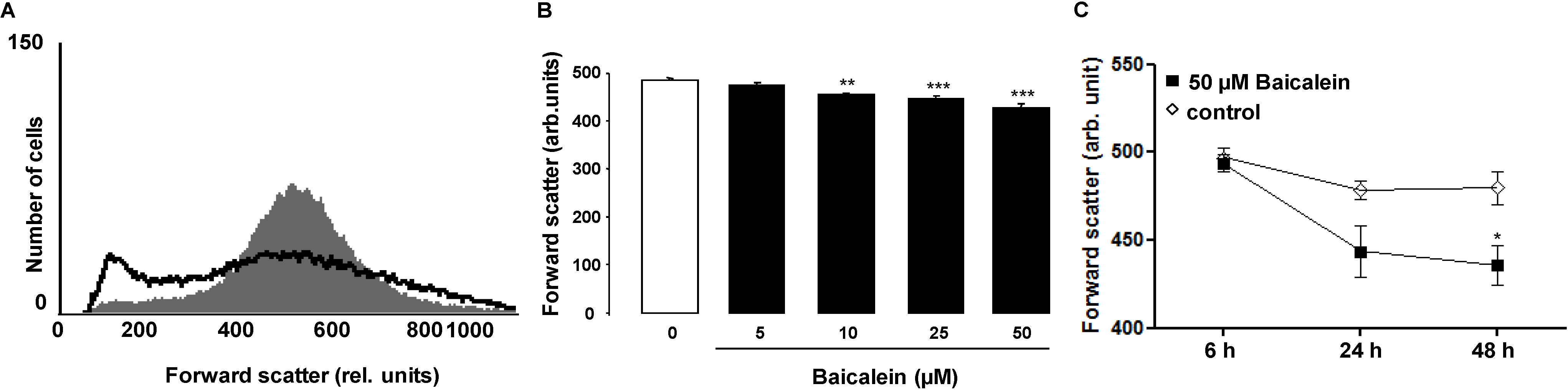
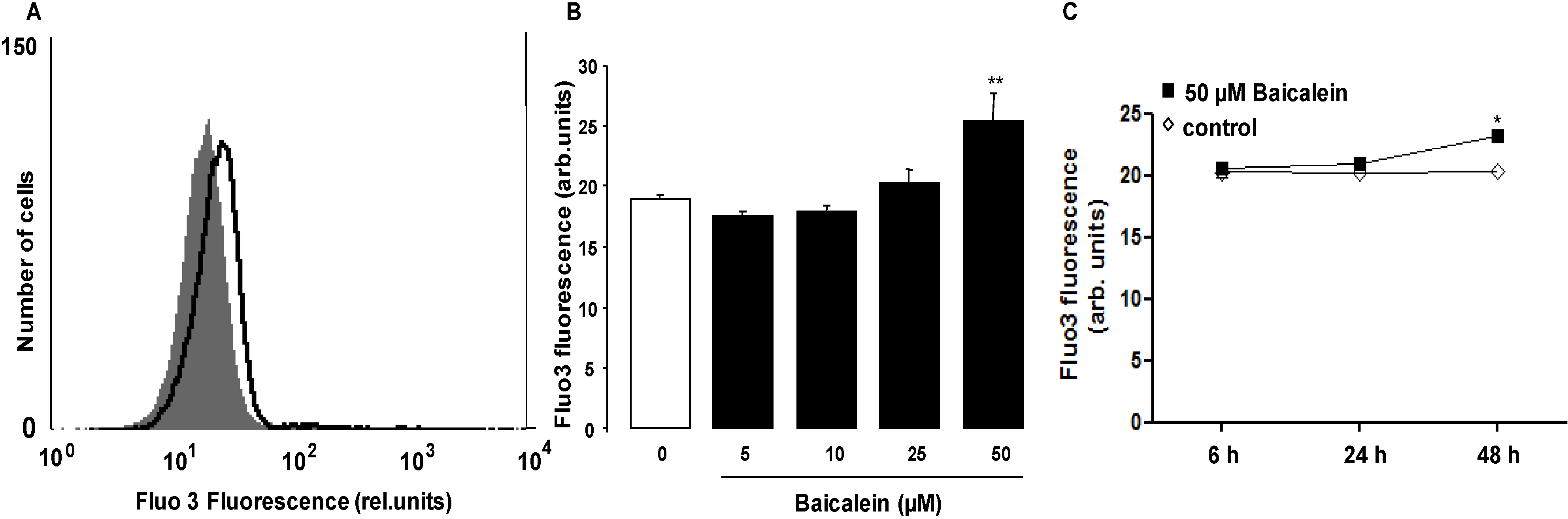
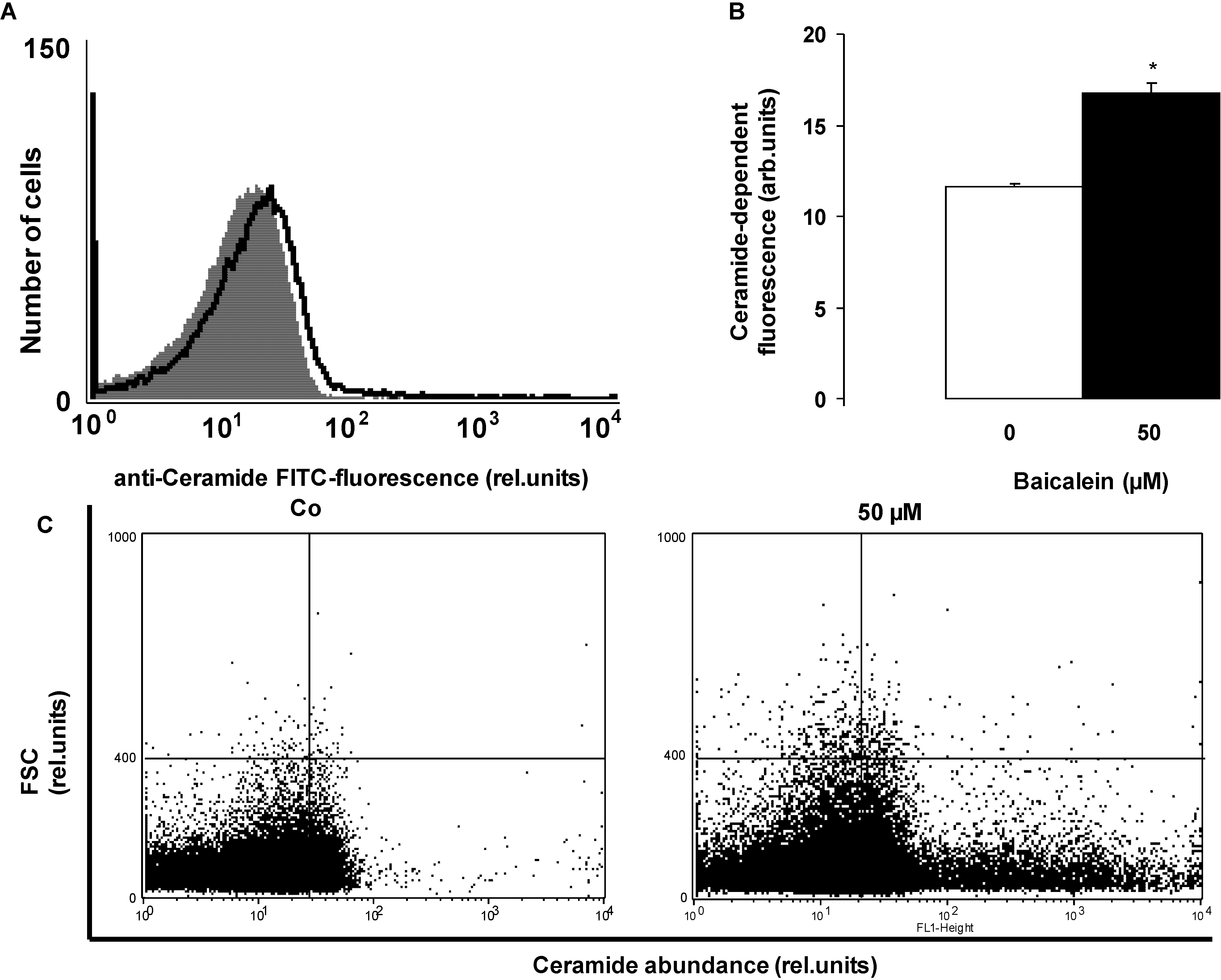
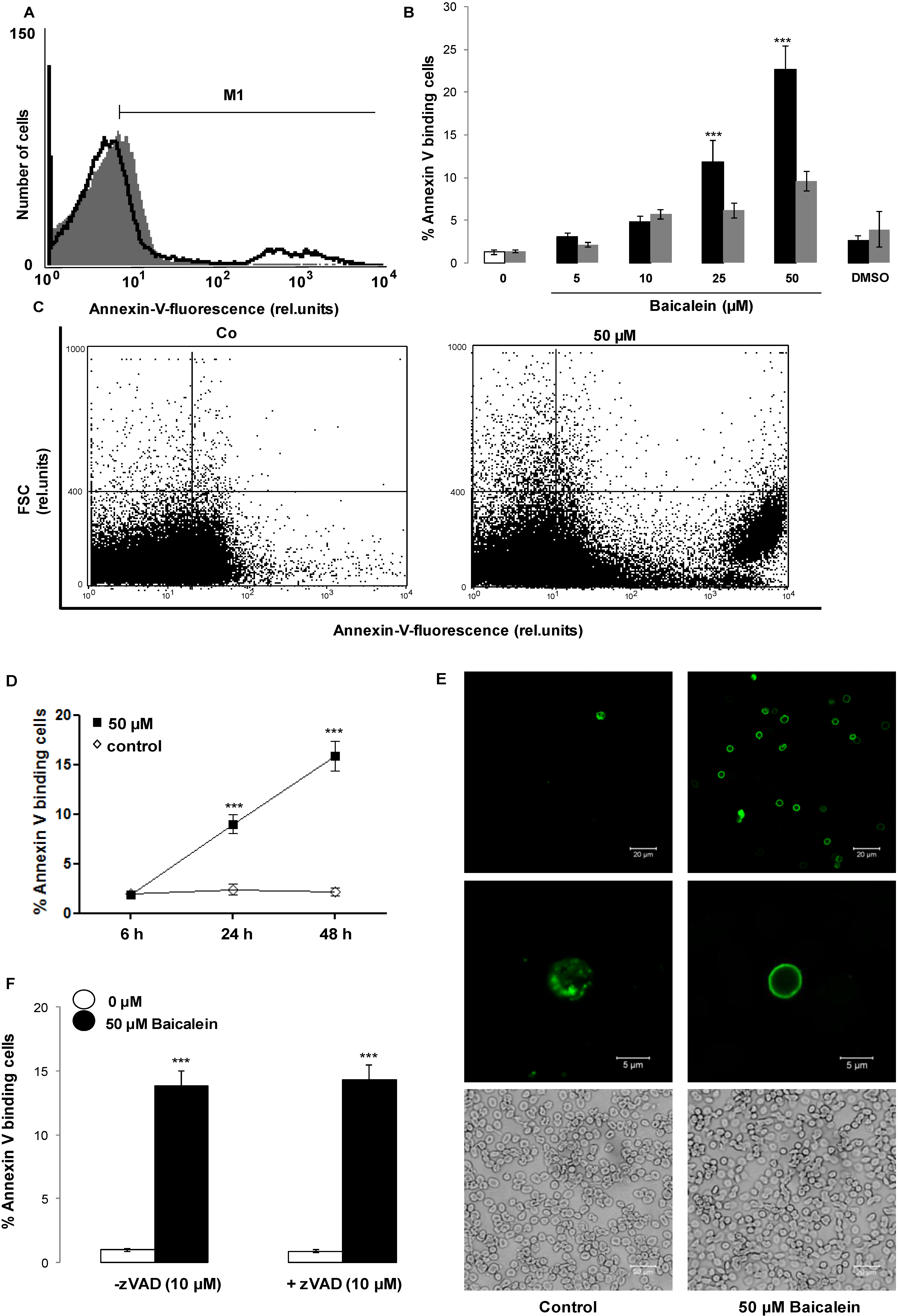
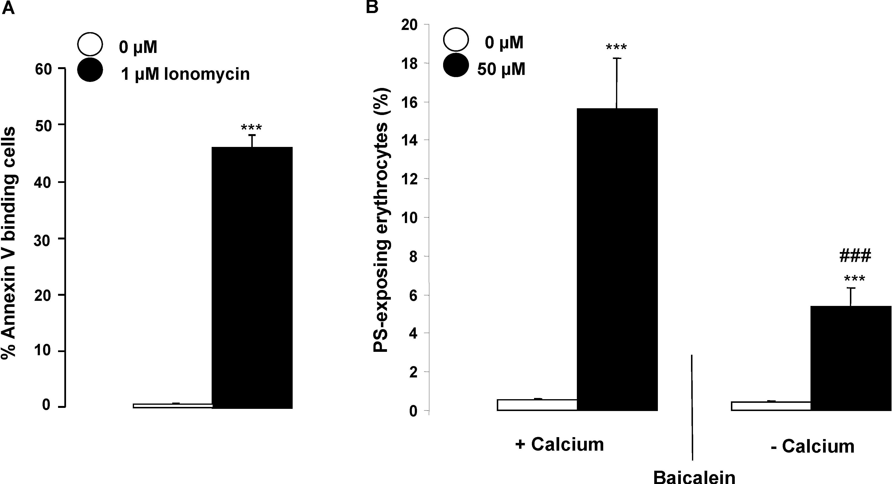
2. Results and Discussion

The present study explored the influence of the polyphenolic flavonoid Baicalein on eryptosis, the suicidal erythrocyte death characterized by cell shrinkage and phosphatidylserine translocation to the cell surface.
In a first step, cell volume was estimated from forward scatter determined in flow cytometry following an incubation of human erythrocytes for 48 h in Ringer solution without or with Baicalein (5–50 µM). As shown in Figure 1, Baicalein treatment was followed by a decrease of average erythrocyte forward scatter reflecting cell shrinkage, an effect reaching statistical significance at 10 µM Baicalein concentration. The histogram reveals that Baicalein increases forward scatter in a subpopulation of erythrocytes.
Figure 1. Effect of Baicalein on erythrocyte forward scatter. (A) Original histogram of forward scatter of erythrocytes following exposure for 48 h to Ringer solution without (grey area) and with (black line) presence of 50 µM Baicalein; (B) Arithmetic means ± SEM (n = 12) of the normalized erythrocyte forward scatter (FSC) following incubation for 48 h to Ringer solution without (white bar) or with (black bars) Baicalein (5–50 µM). ** (p < 0.01), *** (p < 0.001) indicate significant difference from the absence of Baicalein (ANOVA); (C) Arithmetic means ± SEM (n = 5) of forward scatter (arbitrary units) in erythrocytes exposed for 6–48 h to Ringer solution without (white squares) or with 50 µM Baicalein (black squares). * (p < 0.05) indicates significant difference from the absence of Baicalein.
Toxins 06 02771 g001
In a second step, cell membrane phospholipid scrambling with phosphatidylserine translocation to the erythrocyte surface was quantified utilizing annexin-V-binding in flow cytometry following a 48 h incubation in Ringer solution without or with Baicalein (5–50 µM). As illustrated in Figure 2, a 48 h treatment with Baicalein increased the percentage of annexin-V-binding erythrocytes, an effect reaching statistical significance at 25 µM Baicalein concentration.
The effect of Baicalein on phosphatidylserine exposure is paralleled by hemolysis, which, however, affects fewer erythrocytes than cell membrane scrambling (Figure 2). The phosphatidylserine exposure was not modified by inhibition of caspases with the pancaspase inhibitor zVAD (10 µM).
Cell shrinkage and cell membrane scrambling with phosphatidylserine translocation to the cell surface are both known to be stimulated by increase of cytosolic Ca2+ activity ([Ca2+]i). Thus, a further series of experiments was performed to elucidate the effect of Baicalein on [Ca2+]i. Erythrocytes were loaded with Fluo3-AM and the Fluo3 fluorescence determined by flow cytometry following incubation for 48 h in Ringer solution without or with Baicalein (5–50 µM). As illustrated in Figure 3, exposure of the erythrocytes to Baicalein increased the Fluo3 fluorescence, an effect reaching statistical significance at 50 µM Baicalein concentration.
Figure 2. Effect of Baicalein on phosphatidylserine exposure. (A) Original histogram of annexin-V-binding erythrocytes following exposure for 48 h to Ringer solution without (grey area) and with (black line) presence of 50 µM Baicalein; (B) Arithmetic means ± SEM of erythrocyte annexin-V-binding (n = 12) following incubation for 48 h to Ringer solution without (white bar) or with (black bars) presence of Baicalein (5–50 µM). *** (p < 0.001) indicates significant difference from the absence of Baicalein (ANOVA). For comparison, the arithmetic means ± SEM of hemolysis (n = 4) following incubation for 48 h to Ringer solution without or with presence of Baicalein is shown (grey bars); (C) Original dot blots of forward scatter as a function of annexin-V-binding following exposure for 48 h to Ringer solution without and with presence of 50 µM Baicalein; (D) Arithmetic means ± SEM (n = 5) of annexin-V-binding erythrocytes (arbitrary units) following exposure for 6–48 h to Ringer solution without (white squares) or with 50 µM Baicalein (black squares). *** (p < 0.001) indicates significant difference from the absence of Baicalein; (E) Confocal images of FITC dependent fluorescence (upper panels) and light microscopy (lower panels) of human erythrocytes stained with FITC-conjugated annexin-V following a 48 h exposure to Ringer without (left panels) or with (right panels) 50 µM Baicalein; (F) Arithmetic means ± SEM (n = 5) of the percentage of annexin-V-binding erythrocytes after a 48 h treatment with Ringer solution without (white bars) or with 50 µM Baicalein (black bars) in the absence (left panels) and presence (right panels) of 10 µM pancaspase inhibitor zVAD. *** (p < 0.001) indicates significant difference from the absence of Baicalein (ANOVA).
Toxins 06 02771 g002
Figure 3. Effect of Baicalein on erythrocyte cytosolic Ca2+ concentration. (A) Original histogram of Fluo3 fluorescence in erythrocytes following exposure for 48 h to Ringer solution without (grey area) and with (black line) presence of 50 µM Baicalein; (B) Arithmetic means ± SEM (n = 12) of the Fluo3 fluorescence (arbitrary units) in erythrocytes exposed for 48 h to Ringer solution without (white bar) or with (black bars) Baicalein (5–50 µM). ** (p < 0.01) indicates significant difference from the absence of Baicalein (ANOVA); (C) Arithmetic means ± SEM (n = 5) of Fluo 3 fluorescence (arbitrary units) in erythrocytes exposed for 6–48 h to Ringer solution without (white squares) or with 50 µM Baicalein (black squares). * (p < 0.05) indicates significant difference from the absence of Baicalein.
Toxins 06 02771 g003
Exposure of the erythrocytes to the Ca2+ ionophore ionomycin was followed by a strong increase of annexin-V-binding (Figure 4). In order to test, whether the Baicalein-induced cell membrane scrambling required entry of extracellular Ca2+, erythrocytes were exposed for 48 h to 50 µM Baicalein in the presence or nominal absence of extracellular Ca2+. As illustrated in Figure 4, the effect of Baicalein on annexin-V-binding was significantly blunted in the nominal absence of Ca2+. Nevertheless, even in the nominal absence of extracellular Ca2+, the percentage of annexin-V-binding erythrocytes was significantly higher in the presence than in the absence of Baicalein. Thus, Baicalein was effective partially, but not exclusively, through stimulation of Ca2+ entry.
Figure 4. (A) Effect of ionomycin on phosphatidylserine exposure. Arithmetic means ± SEM (n = 5) of the percentage of annexin-V-binding erythrocytes following incubation for 1 h in the absence (white bar) or presence (black bar) of 1 µM ionomycin. *** (p < 0.001) indicates significant difference from the absence of 1 µM ionomycin (ANOVA); (B) Effect of Ca2+ withdrawal on Baicalein- induced annexin-V-binding. Arithmetic means ± SEM (n = 5) of the percentage of annexin-V-binding erythrocytes after a 48 h treatment with Ringer solution without (white bars) or with (black bars) 50 µM Baicalein in the presence (left bars, +Calcium) and absence (right bars, −Calcium) of calcium. *** (p < 0.001) indicates significant difference from the respective values in the absence of Baicalein, ### (p < 0.001) indicates significant difference from the respective value in the presence of Ca2+ (ANOVA).
Toxins 06 02771 g004
In search for an additional mechanism triggering eryptosis following Baicalein treatment, further experiments were performed to possibly disclose an effect of Baicalein on ceramide formation. Ceramide abundance at the erythrocyte surface was quantified utilizing an anti-ceramide antibody. As illustrated in Figure 5, exposure of erythrocytes to 50 µM Baicalein significantly increased the ceramide abundance at the erythrocyte surface.
Figure 5. Effect of Baicalein on ceramide formation. (A) Original histogram of ceramide surface abundance of erythrocytes following exposure for 48 h to Ringer solution without (grey shadow) and with (black line) presence of 50 µM Baicalein; (B) Arithmetic means ± SEM (n = 5) of ceramide abundance after a 48 h incubation in Ringer solution without (white bar) or with 50 µM Baicalein (black bar). * (p < 0.05) indicates significant difference from the absence of Baicalein (t-test); (C) Original dot blots of forward scatter as a function of ceramide abundance following exposure for 48 h to Ringer solution without and with the presence of 50 µM Baicalein.
Toxins 06 02771 g005
The present study discloses a novel effect of Baicalein, i.e., stimulation of eryptosis, the suicidal death of erythrocytes. Incubation of human erythrocytes with Baicalein is followed by cell membrane scrambling with phosphatidylserine translocation to the erythrocyte surface, the most important hallmark of eryptosis. The Baicalein induced cell membrane scrambling affected only a subpopulation of the erythrocytes, an observation pointing to variable vulnerability of the erythrocytes. According to an earlier study, the susceptibility against several triggers of eryptosis is enhanced in aged erythrocytes [44].
On average, Baicalein further decreases cell volume, another hallmark of eryptosis. However, Baicalein exposure increases cell volume of an erythrocyte subpopulation. This observation again points to heterogeneity among the erythrocytes, which may be similarly due to differences in erythrocyte age. The Baicalein concentrations (10–25 µM) required for those effects were in the range of the peak concentrations reported in Baicalein treated rats [70]. However, Baicalein was not immediately effective but a 24 h exposure of erythrocytes to the substance was required in order to trigger eryptosis. Whether or not those high concentrations could be maintained in vivo for 24 h remains uncertain.
Baicalein increased cytosolic Ca2+ activity ([Ca2+]i), an effect presumably due to stimulation of cation channels in the cell membrane. Earlier studies revealed that the erythrocyte cation channels involve TRPC6 [22].
The cell shrinkage following Baicalein treatment was presumably the result of Ca2+ entry with subsequent increase of [Ca2+]i, activation of Ca2+ sensitive K+ channels, K+ exit, cell membrane hyperpolarisation, Cl exit and thus cellular loss of KCl accompanied by osmotically driven water [23]. The cellular loss of KCl with water serves to counteract the swelling and subsequent hemolysis of injured erythrocytes. Hemolysis leads to release of hemoglobin, which is subject to glomerular filtration with subsequent precipitation in the acidic lumen of renal tubules [71]. The swelling of some erythrocytes following Baicalein exposure may result from Na+ entry through the unselective cation channel.
The stimulation of cell membrane scrambling by Baicalein is similarly in part due to increase of [Ca2+]i. Accordingly, the effect of Baicalein on phosphatidylserine translocation is in part dependent on entry of extracellular Ca2+.
However, even in the absence of extracellular Ca2+, Baicalein treatment is still followed by a significant increase of phosphatidylserine exposure. The residual effect is in part due to stimulation of ceramide formation. Ceramide is a well-known stimulator of eryptosis [22].
Similar to what has been shown for several other stimulators of eryptosis [22], the effect of Baicalein was not sensitive to the pancaspase inhibitor zVAD and thus did not require activation of caspases.
Consequences of excessive eryptosis include anemia, since phosphatidylserine exposing eryptotic erythrocytes are phagocytosed and thus rapidly cleared from circulating blood [22]. Anemia is prevented as long as accelerated clearance of erythrocytes during stimulated eryptosis is compensated by a similarly accelerated formation of new erythrocytes [22].
At least in theory, phosphatidylserine exposing erythrocytes may further interfere with microcirculation [72,73,74,75,76,77], as phosphatidylserine exposing erythrocytes adhere to endothelial CXCL16/SR-PSO [73], stimulate blood clotting and trigger thrombosis [72,78,79]. Baicalein has, however, been shown to counteract thrombosis and to inhibit thrombin-induced production of plasminogen activator inhibitor-1, and endothelial adhesion molecule expression [1]. Accordingly, Baicalein and its analogs have been proposed for the treatment of arteriosclerosis and hypertension [1].
Elimination of phosphatidylserine exposing erythrocytes may protect against untoward effects of hemolysis [22]. The removal of phosphatidylserine exposing erythrocytes further impacts on the clinical course of malaria [80]. Infected erythrocytes undergo eryptosis [80], since the intraerythrocytic pathogen activates ion channels including the Ca2+-permeable erythrocyte cation channels [81,82]. Subsequent clearance of phosphatidylserine exposing infected erythrocytes from circulating blood decreases parasitemia and by the same token precedes and thus prevents hemolysis of the parasitized erythrocytes [80]. Accordingly, the clinical course of malaria is ameliorated by genetic disorders sensitizing erythrocytes to eryptosis, such as sickle-cell trait, beta-thalassemia-trait, homozygous Hb-C and G6PD-deficiency, [22,83,84,85], by conditions with enhanced eryptosis, such as iron deficiency [86], and by eryptosis stimulating xenobiotics, such as lead [87], chlorpromazine [88] or NO synthase inhibitors [89]. In theory, Baicalein may similarly decrease parasitemia in malaria.

3. Experimental Section

3.1. Erythrocytes, Solutions and Chemicals

Leukocyte-depleted erythrocytes were kindly provided by the blood bank of the University of Tübingen. The study is approved by the ethics committee of the University of Tübingen (184/2003V). Erythrocytes were incubated in vitro at a hematocrit of 0.4% in Ringer solution containing (in mM) 125 NaCl, 5 KCl, 1 MgSO4, 32 N-2-hydroxyethylpiperazine-N-2-ethanesulfonic acid (HEPES), 5 glucose, 1 CaCl2; pH 7.4 at 37 °C for 48 h. Where indicated, erythrocytes were exposed to Baicalein (Enzo Life Sciences, Lörrach, Germany) at the indicated concentrations. In Ca2+-free Ringer solution, 1 mM CaCl2 was substituted by 1 mM glycol-bis(2-aminoethylether)-N,N,N',N'-tetraacetic acid (EGTA).

3.2. Analysis of Annexin-V-Binding and Forward Scatter

After incubation under the respective experimental condition, 50 µL cell suspension was washed in Ringer solution containing 5 mM CaCl2 and then stained with Annexin-V-FITC (1:200 dilution; ImmunoTools, Friesoythe, Germany) in this solution at 37 °C for 20 min under protection from light. In the following, the forward scatter (FSC) of the cells was determined, and annexin-V fluorescence intensity was measured with an excitation wavelength of 488 nm and an emission wavelength of 530 nm on a FACS Calibur (BD, Heidelberg, Germany).

3.3. Measurement of Intracellular Ca2+

After incubation, erythrocytes were washed in Ringer solution and then loaded with Fluo-3/AM (Biotium, Hayward, CA, USA) in Ringer solution containing 5 mM CaCl2 and 5 µM Fluo-3/AM. The cells were incubated at 37 °C for 30 min and washed twice in Ringer solution containing 5 mM CaCl2. The Fluo-3/AM-loaded erythrocytes were resuspended in 200 µL Ringer. Then, Ca2+-dependent fluorescence intensity was measured with an excitation wavelength of 488 nm and an emission wavelength of 530 nm on a FACS Calibur (BD, Heidelberg, Germany).

3.4. Determination of Ceramide Formation

For the determination of ceramide abundance, a monoclonal antibody-based assay was used. After incubation, cells were stained for 1 h at 37 °C with 1 µg/mL anti ceramide antibody (clone MID 15B4, Alexis, Grünberg, Germany) in PBS containing 0.1% bovine serum albumin (BSA) at a dilution of 1:5. The samples were washed twice with PBS-BSA. Subsequently, the cells were stained for 30 min with polyclonal fluorescein isothiocyanate (FITC) conjugated goat anti-mouse IgG and IgM specific antibody (Pharmingen, Hamburg, Germany) diluted 1:50 in PBS-BSA. Unbound secondary antibody was removed by repeated washing with PBS-BSA. The samples were then analyzed by flow cytometric analysis with an excitation wavelength of 488 nm and an emission wavelength of 530 nm.

3.5. Statistics

Data are expressed as arithmetic means ± SEM. As indicated in the figure legends, statistical analysis was made using ANOVA with Tukey’s test as post-test and t-test as appropriate. n denotes the number of different erythrocyte specimens studied. Since different erythrocyte specimens used in distinct experiments are differently susceptible to triggers of eryptosis, the same erythrocyte specimens have been used for control and experimental conditions.

4. Conclusions

In conclusion, the polyphenolic flavonoid Baicalein stimulates Ca2+ entry and ceramide formation thus leading to subsequent erythrocyte shrinkage and erythrocyte cell membrane scrambling. Accordingly, Baicalein stimulates eryptosis, the suicidal erythrocyte death. The concentrations required for those effects are 10–50 µM.

Acknowledgments

The authors acknowledge the meticulous preparation of the manuscript by Tanja Loch. The study was supported by the Deutsche Forschungsgemeinschaft and the Open Access Publishing Fund of Tuebingen University.

Author Contributions

Rosi Bissinger, Abaid Malik, Sabina Honisch and Jamshed Warsi performed experiments and evaluated the data; Kashif Jilani designed the experiments; Florian Lang drafted the manuscript and all authors corrected and approved the manuscript.

Conflicts of Interest

The authors declare no conflict of interest.

References

  1. Huang, Y.; Tsang, S.Y.; Yao, X.; Chen, Z.Y. Biological properties of baicalein in cardiovascular system. Curr. Drug Targets Cardiovasc. Haematol. Disord. 2005, 5, 177–184. [Google Scholar] [CrossRef]
  2. Donald, G.; Hertzer, K.; Eibl, G. Baicalein—An intriguing therapeutic phytochemical in pancreatic cancer. Curr. Drug Targets 2012, 13, 1772–1776. [Google Scholar] [CrossRef]
  3. Kim, S.J.; Kim, H.J.; Kim, H.R.; Lee, S.H.; Cho, S.D.; Choi, C.S.; Nam, J.S.; Jung, J.Y. Antitumor actions of baicalein and wogonin in HT-29 human colorectal cancer cells. Mol. Med. Rep. 2012, 6, 1443–1449. [Google Scholar]
  4. Li, H.L.; Zhang, S.; Wang, Y.; Liang, R.R.; Li, J.; An, P.; Wang, Z.M.; Yang, J.; Li, Z.F. Baicalein induces apoptosis via a mitochondrial-dependent caspase activation pathway in T24 bladder cancer cells. Mol. Med. Rep. 2013, 7, 266–270. [Google Scholar]
  5. Li-Weber, M. New therapeutic aspects of flavones: The anticancer properties of Scutellaria and its main active constituents Wogonin, Baicalein and Baicalin. Cancer Treat. Rev. 2009, 35, 57–68. [Google Scholar] [CrossRef]
  6. Luo, R.; Wang, J.; Zhao, L.; Lu, N.; You, Q.; Guo, Q.; Li, Z. Synthesis and biological evaluation of baicalein derivatives as potent antitumor agents. Bioorg. Med. Chem. Lett. 2014, 24, 1334–1338. [Google Scholar] [CrossRef]
  7. Ma, G.Z.; Liu, C.H.; Wei, B.; Qiao, J.; Lu, T.; Wei, H.C.; Chen, H.D.; He, C.D. Baicalein inhibits DMBA/TPA-induced skin tumorigenesis in mice by modulating proliferation, apoptosis, and inflammation. Inflammation 2013, 36, 457–467. [Google Scholar] [CrossRef]
  8. Nipun Babu, V.; Kannan, S. Enhanced delivery of baicalein using cinnamaldehyde cross-linked chitosan nanoparticle inducing apoptosis. Int. J. Biol. Macromol. 2012, 51, 1103–1108. [Google Scholar] [CrossRef]
  9. Chen, Y.J.; Wu, C.S.; Shieh, J.J.; Wu, J.H.; Chen, H.Y.; Chung, T.W.; Chen, Y.K.; Lin, C.C. Baicalein Triggers Mitochondria-Mediated Apoptosis and Enhances the Antileukemic Effect of Vincristine in Childhood Acute Lymphoblastic Leukemia CCRF-CEM Cells. Evid. Based Complement. Altern. Med. 2013, 2013, 124747. [Google Scholar] [CrossRef]
  10. Helmerick, E.C.; Loftus, J.P.; Wakshlag, J.J. The effects of baicalein on canine osteosarcoma cell proliferation and death. Vet. Comp. Oncol. 2012. [Google Scholar] [CrossRef]
  11. Kim, D.H.; Hossain, M.A.; Kang, Y.J.; Jang, J.Y.; Lee, Y.J.; Im, E.; Yoon, J.H.; Kim, H.S.; Chung, H.Y.; Kim, N.D.; et al. Baicalein, an active component of Scutellaria baicalensis Georgi, induces apoptosis in human colon cancer cells and prevents AOM/DSS-induced colon cancer in mice. Int. J. Oncol. 2013, 43, 1652–1658. [Google Scholar]
  12. Zhang, H.B.; Lu, P.; Guo, Q.Y.; Zhang, Z.H.; Meng, X.Y. Baicalein induces apoptosis in esophageal squamous cell carcinoma cells through modulation of the PI3K/Akt pathway. Oncol. Lett. 2013, 5, 722–728. [Google Scholar]
  13. Zhang, Y.; Song, L.; Cai, L.; Wei, R.; Hu, H.; Jin, W. Effects of baicalein on apoptosis, cell cycle arrest, migration and invasion of osteosarcoma cells. Food Chem. Toxicol. 2013, 53, 325–333. [Google Scholar] [CrossRef]
  14. Dong, Q.H.; Zheng, S.; Xu, R.Z.; Lu, Q.H. Baicalein selectively induce apoptosis in human leukemia K562 cells. Yao Xue Xue Bao 2003, 38, 817–820. [Google Scholar]
  15. Chang, W.T.; Li, J.; Vanden Hoek, M.S.; Zhu, X.; Li, C.Q.; Huang, H.H.; Hsu, C.W.; Zhong, Q.; Li, J.; Chen, S.J.; et al. Baicalein preconditioning protects cardiomyocytes from ischemia-reperfusion injury via mitochondrial oxidant signaling. Am. J. Chin. Med. 2013, 41, 315–331. [Google Scholar] [CrossRef]
  16. Chao, H.M.; Chuang, M.J.; Liu, J.H.; Liu, X.Q.; Ho, L.K.; Pan, W.H.; Zhang, X.M.; Liu, C.M.; Tsai, S.K.; Kong, C.W.; et al. Baicalein protects against retinal ischemia by antioxidation, antiapoptosis, downregulation of HIF-1alpha, VEGF, and MMP-9 and upregulation of HO-1. J. Ocul. Pharmacol. Ther. 2013, 29, 539–549. [Google Scholar] [CrossRef]
  17. Jung, E.B.; Lee, C.S. Baicalein attenuates proteasome inhibition-induced apoptosis by suppressing the activation of the mitochondrial pathway and the caspase-8- and Bid-dependent pathways. Eur. J. Pharmacol. 2014, 730, 116–124. [Google Scholar] [CrossRef]
  18. Mabalirajan, U.; Ahmad, T.; Rehman, R.; Leishangthem, G.D.; Dinda, A.K.; Agrawal, A.; Ghosh, B.; Sharma, S.K. Baicalein reduces airway injury in allergen and IL-13 induced airway inflammation. PLoS One 2013, 8, e62916. [Google Scholar]
  19. Chen, S.S.; Michael, A.; Butler-Manuel, S.A. Advances in the treatment of ovarian cancer: A potential role of antiinflammatory phytochemicals. Discov. Med. 2012, 13, 7–17. [Google Scholar]
  20. Song, L.; Yang, H.; Wang, H.X.; Tian, C.; Liu, Y.; Zeng, X.J.; Gao, E.; Kang, Y.M.; Du, J.; Li, H.H.; et al. Inhibition of 12/15 lipoxygenase by baicalein reduces myocardial ischemia/reperfusion injury via modulation of multiple signaling pathways. Apoptosis 2014, 19, 567–580. [Google Scholar] [CrossRef]
  21. Zhang, Z.; Cui, W.; Li, G.; Yuan, S.; Xu, D.; Hoi, M.P.; Lin, Z.; Dou, J.; Han, Y.; Lee, S.M.; et al. Baicalein protects against 6-OHDA-induced neurotoxicity through activation of Keap1/Nrf2/HO-1 and involving PKCalpha and PI3K/AKT signaling pathways. J. Agric. Food Chem. 2012, 60, 8171–8182. [Google Scholar] [CrossRef]
  22. Lang, E.; Qadri, S.M.; Lang, F. Killing me softly—Suicidal erythrocyte death. Int. J. Biochem. Cell Biol. 2012, 44, 1236–1243. [Google Scholar] [CrossRef]
  23. Lang, P.A.; Kaiser, S.; Myssina, S.; Wieder, T.; Lang, F.; Huber, S.M. Role of Ca2+-activated K+ channels in human erythrocyte apoptosis. Am. J. Physiol. Cell Physiol. 2003, 285, C1553–C1560. [Google Scholar] [CrossRef]
  24. Abed, M.; Towhid, S.T.; Mia, S.; Pakladok, T.; Alesutan, I.; Borst, O.; Gawaz, M.; Gulbins, E.; Lang, F. Sphingomyelinase-induced adhesion of eryptotic erythrocytes to endothelial cells. Am. J. Physiol. Cell Physiol. 2012, 303, C991–C999. [Google Scholar] [CrossRef]
  25. Bhavsar, S.K.; Bobbala, D.; Xuan, N.T.; Foller, M.; Lang, F. Stimulation of suicidal erythrocyte death by alpha-lipoic acid. Cell. Physiol. Biochem. 2010, 26, 859–868. [Google Scholar] [CrossRef]
  26. Foller, M.; Feil, S.; Ghoreschi, K.; Koka, S.; Gerling, A.; Thunemann, M.; Hofmann, F.; Schuler, B.; Vogel, J.; Pichler, B.; et al. Anemia and splenomegaly in cGKI-deficient mice. Proc. Natl. Acad. Sci. USA 2008, 105, 6771–6776. [Google Scholar] [CrossRef] [Green Version]
  27. Foller, M.; Mahmud, H.; Gu, S.; Wang, K.; Floride, E.; Kucherenko, Y.; Luik, S.; Laufer, S.; Lang, F. Participation of leukotriene C(4) in the regulation of suicidal erythrocyte death. J. Physiol. Pharmacol. 2009, 60, 135–143. [Google Scholar]
  28. Lau, I.P.; Chen, H.; Wang, J.; Ong, H.C.; Leung, K.C.; Ho, H.P.; Kong, S.K. In vitro effect of CTAB- and PEG-coated gold nanorods on the induction of eryptosis/erythroptosis in human erythrocytes. Nanotoxicology 2012, 6, 847–856. [Google Scholar] [CrossRef]
  29. Maellaro, E.; Leoncini, S.; Moretti, D.; del Bello, B.; Tanganelli, I.; de Felice, C.; Ciccoli, L. Erythrocyte caspase-3 activation and oxidative imbalance in erythrocytes and in plasma of type 2 diabetic patients. Acta Diabetol. 2013, 50, 489–495. [Google Scholar] [CrossRef]
  30. Foller, M.; Sopjani, M.; Koka, S.; Gu, S.; Mahmud, H.; Wang, K.; Floride, E.; Schleicher, E.; Schulz, E.; Munzel, T.; et al. Regulation of erythrocyte survival by AMP-activated protein kinase. FASEB J. 2009, 23, 1072–1080. [Google Scholar] [CrossRef]
  31. Kucherenko, Y.; Zelenak, C.; Eberhard, M.; Qadri, S.M.; Lang, F. Effect of casein kinase 1alpha activator pyrvinium pamoate on erythrocyte ion channels. Cell. Physiol. Biochem. 2012, 30, 407–417. [Google Scholar] [CrossRef]
  32. Zelenak, C.; Eberhard, M.; Jilani, K.; Qadri, S.M.; Macek, B.; Lang, F. Protein kinase CK1alpha regulates erythrocyte survival. Cell. Physiol. Biochem. 2012, 29, 171–180. [Google Scholar] [CrossRef]
  33. Bhavsar, S.K.; Gu, S.; Bobbala, D.; Lang, F. Janus kinase 3 is expressed in erythrocytes, phosphorylated upon energy depletion and involved in the regulation of suicidal erythrocyte death. Cell. Physiol. Biochem. 2011, 27, 547–556. [Google Scholar] [CrossRef]
  34. Klarl, B.A.; Lang, P.A.; Kempe, D.S.; Niemoeller, O.M.; Akel, A.; Sobiesiak, M.; Eisele, K.; Podolski, M.; Huber, S.M.; Wieder, T.; et al. Protein kinase C mediates erythrocyte “programmed cell death” following glucose depletion. Am. J. Physiol. Cell Physiol. 2006, 290, C244–C253. [Google Scholar]
  35. Gatidis, S.; Zelenak, C.; Fajol, A.; Lang, E.; Jilani, K.; Michael, D.; Qadri, S.M.; Lang, F. p38 MAPK activation and function following osmotic shock of erythrocytes. Cell. Physiol. Biochem. 2011, 28, 1279–1286. [Google Scholar] [CrossRef]
  36. Zelenak, C.; Foller, M.; Velic, A.; Krug, K.; Qadri, S.M.; Viollet, B.; Lang, F.; Macek, B. Proteome analysis of erythrocytes lacking AMP-activated protein kinase reveals a role of PAK2 kinase in eryptosis. J. Proteome Res. 2011, 10, 1690–1697. [Google Scholar] [CrossRef]
  37. Lupescu, A.; Jilani, K.; Zelenak, C.; Zbidah, M.; Qadri, S.M.; Lang, F. Hexavalent chromium-induced erythrocyte membrane phospholipid asymmetry. Biometals 2012, 25, 309–318. [Google Scholar] [CrossRef]
  38. Shaik, N.; Lupescu, A.; Lang, F. Sunitinib-sensitive suicidal erythrocyte death. Cell. Physiol. Biochem. 2012, 30, 512–522. [Google Scholar] [CrossRef]
  39. Abed, M.; Towhid, S.T.; Shaik, N.; Lang, F. Stimulation of suicidal death of erythrocytes by rifampicin. Toxicology 2012, 302, 123–128. [Google Scholar] [CrossRef]
  40. Bottger, E.; Multhoff, G.; Kun, J.F.; Esen, M. Plasmodium falciparum-infected erythrocytes induce granzyme B by NK cells through expression of host-Hsp70. PLoS One 2012, 7, e33774. [Google Scholar] [CrossRef]
  41. Firat, U.; Kaya, S.; Cim, A.; Buyukbayram, H.; Gokalp, O.; Dal, M.S.; Tamer, M.N. Increased caspase-3 immunoreactivity of erythrocytes in STZ diabetic rats. Exp. Diabetes Res. 2012, 2012, 316384. [Google Scholar] [CrossRef]
  42. Ganesan, S.; Chaurasiya, N.D.; Sahu, R.; Walker, L.A.; Tekwani, B.L. Understanding the mechanisms for metabolism-linked hemolytic toxicity of primaquine against glucose 6-phosphate dehydrogenase deficient human erythrocytes: Evaluation of eryptotic pathway. Toxicology 2012, 294, 54–60. [Google Scholar] [CrossRef]
  43. Gao, M.; Cheung, K.L.; Lau, I.P.; Yu, W.S.; Fung, K.P.; Yu, B.; Loo, J.F.; Kong, S.K. Polyphyllin D induces apoptosis in human erythrocytes through Ca2+ rise and membrane permeabilization. Arch. Toxicol. 2012, 86, 741–752. [Google Scholar] [CrossRef]
  44. Ghashghaeinia, M.; Cluitmans, J.C.; Akel, A.; Dreischer, P.; Toulany, M.; Koberle, M.; Skabytska, Y.; Saki, M.; Biedermann, T.; Duszenko, M.; et al. The impact of erythrocyte age on eryptosis. Br. J. Haematol. 2012, 157, 606–614. [Google Scholar] [CrossRef]
  45. Jilani, K.; Lupescu, A.; Zbidah, M.; Abed, M.; Shaik, N.; Lang, F. Enhanced Apoptotic Death of Erythrocytes Induced by the Mycotoxin Ochratoxin A. Kidney Blood Press. Res. 2012, 36, 107–118. [Google Scholar] [CrossRef]
  46. Jilani, K.; Qadri, S.M.; Lang, F. Geldanamycin-induced phosphatidylserine translocation in the erythrocyte membrane. Cell. Physiol. Biochem. 2013, 32, 1600–1609. [Google Scholar]
  47. Kucherenko, Y.V.; Lang, F. Inhibitory Effect of Furosemide on Non-Selective Voltage-Independent Cation Channels in Human Erythrocytes. Cell. Physiol. Biochem. 2012, 30, 863–875. [Google Scholar] [CrossRef]
  48. Lupescu, A.; Jilani, K.; Zbidah, M.; Lang, E.; Lang, F. Enhanced Ca2+ entry, ceramide formation, and apoptotic death of erythrocytes triggered by plumbagin. J. Nat. Prod. 2012, 75, 1956–1961. [Google Scholar] [CrossRef]
  49. Lupescu, A.; Jilani, K.; Zbidah, M.; Lang, F. Induction of apoptotic erythrocyte death by rotenone. Toxicology 2012, 300, 132–137. [Google Scholar] [CrossRef]
  50. Polak-Jonkisz, D.; Purzyc, L. Ca Influx versus efflux during eryptosis in uremic erythrocytes. Blood Purif. 2012, 34, 209–210. [Google Scholar] [CrossRef]
  51. Qian, E.W.; Ge, D.T.; Kong, S.K. Salidroside protects human erythrocytes against hydrogen peroxide-induced apoptosis. J. Nat. Prod. 2012, 75, 531–537. [Google Scholar] [CrossRef]
  52. Shaik, N.; Zbidah, M.; Lang, F. Inhibition of Ca2+ entry and suicidal erythrocyte death by naringin. Cell. Physiol. Biochem. 2012, 30, 678–686. [Google Scholar] [CrossRef]
  53. Vota, D.M.; Maltaneri, R.E.; Wenker, S.D.; Nesse, A.B.; Vittori, D.C. Differential erythropoietin action upon cells induced to eryptosis by different agents. Cell Biochem. Biophys. 2013, 65, 145–157. [Google Scholar] [CrossRef]
  54. Weiss, E.; Cytlak, U.M.; Rees, D.C.; Osei, A.; Gibson, J.S. Deoxygenation-induced and Ca2+ dependent phosphatidylserine externalisation in red blood cells from normal individuals and sickle cell patients. Cell Calcium 2012, 51, 51–56. [Google Scholar] [CrossRef]
  55. Zappulla, D. Environmental stress, erythrocyte dysfunctions, inflammation, and the metabolic syndrome: Adaptations to CO2 increases? Cardiometab. Syndr. 2008, 3, 30–34. [Google Scholar] [CrossRef]
  56. Zbidah, M.; Lupescu, A.; Jilani, K.; Lang, F. Stimulation of suicidal erythrocyte death by fumagillin. Basic Clin. Pharmacol. Toxicol. 2013, 112, 346–351. [Google Scholar] [CrossRef]
  57. Zbidah, M.; Lupescu, A.; Shaik, N.; Lang, F. Gossypol-induced suicidal erythrocyte death. Toxicology 2012, 302, 101–105. [Google Scholar] [CrossRef]
  58. Zelenak, C.; Pasham, V.; Jilani, K.; Tripodi, P.M.; Rosaclerio, L.; Pathare, G.; Lupescu, A.; Faggio, C.; Qadri, S.M.; Lang, F.; et al. Tanshinone IIA stimulates erythrocyte phosphatidylserine exposure. Cell. Physiol. Biochem. 2012, 30, 282–294. [Google Scholar] [CrossRef]
  59. Abed, M.; Herrmann, T.; Alzoubi, K.; Pakladok, T.; Lang, F. Tannic Acid induced suicidal erythrocyte death. Cell. Physiol. Biochem. 2013, 32, 1106–1116. [Google Scholar] [CrossRef]
  60. Ahmed, M.S.; Langer, H.; Abed, M.; Voelkl, J.; Lang, F. The uremic toxin acrolein promotes suicidal erythrocyte death. Kidney Blood Press. Res. 2013, 37, 158–167. [Google Scholar] [CrossRef]
  61. Ghashghaeinia, M.; Cluitmans, J.C.; Toulany, M.; Saki, M.; Koberle, M.; Lang, E.; Dreischer, P.; Biedermann, T.; Duszenko, M.; Lang, F.; et al. Age Sensitivity of NFκB Abundance and Programmed Cell Death in Erythrocytes Induced by NFκB Inhibitors. Cell. Physiol. Biochem. 2013, 32, 801–813. [Google Scholar] [CrossRef]
  62. Abed, M.; Feger, M.; Alzoubi, K.; Pakladok, T.; Frauenfeld, L.; Geiger, C.; Towhid, S.T.; Lang, F. Sensitization of erythrocytes to suicidal erythrocyte death following water deprivation. Kidney Blood Press. Res. 2013, 37, 567–578. [Google Scholar]
  63. Alzoubi, K.; Honisch, S.; Abed, M.; Lang, F. Triggering of Suicidal Erythrocyte Death by Penta-O-galloyl-β-d-glucose. Toxins 2014, 6, 54–65. [Google Scholar] [CrossRef]
  64. Jilani, K.; Lang, F. Carmustine-induced phosphatidylserine translocation in the erythrocyte membrane. Toxins 2013, 5, 703–716. [Google Scholar] [CrossRef]
  65. Jilani, K.; Enkel, S.; Bissinger, R.; Almilaji, A.; Abed, M.; Lang, F. Fluoxetine induced suicidal erythrocyte death. Toxins 2013, 5, 1230–1243. [Google Scholar] [CrossRef]
  66. Lupescu, A.; Bissinger, R.; Jilani, K.; Lang, F. Triggering of suicidal erythrocyte death by celecoxib. Toxins 2013, 5, 1543–1554. [Google Scholar] [CrossRef]
  67. Lupescu, A.; Jilani, K.; Zbidah, M.; Lang, F. Patulin-induced suicidal erythrocyte death. Cell. Physiol. Biochem. 2013, 32, 291–299. [Google Scholar] [CrossRef]
  68. Lang, E.; Qadri, S.M.; Jilani, K.; Zelenak, C.; Lupescu, A.; Schleicher, E.; Lang, F. Carbon monoxide-sensitive apoptotic death of erythrocytes. Basic Clin. Pharmacol. Toxicol. 2012, 111, 348–355. [Google Scholar]
  69. Voelkl, J.; Alzoubi, K.; Mamar, A.K.; Ahmed, M.S.; Abed, M.; Lang, F. Stimulation of Suicidal Erythrocyte Death by Increased Extracellular Phosphate Concentrations. Kidney Blood Press. Res. 2014, 38, 42–51. [Google Scholar]
  70. Wakui, Y.; Yanagisawa, E.; Ishibashi, E.; Matsuzaki, Y.; Takeda, S.; Sasaki, H.; Aburada, M.; Oyama, T. Determination of baicalin and baicalein in rat plasma by high-performance liquid chromatography with electrochemical detection. J. Chromatogr. 1992, 575, 131–136. [Google Scholar] [CrossRef]
  71. Harrison, H.E.; Bunting, H.; Ordway, N.K.; Albrink, W.S. The Pathogenesis of the Renal Injury Produced in the Dog by Hemoglobin or Methemoglobin. J. Exp. Med. 1947, 86, 339–356. [Google Scholar] [CrossRef]
  72. Andrews, D.A.; Low, P.S. Role of red blood cells in thrombosis. Curr. Opin. Hematol. 1999, 6, 76–82. [Google Scholar] [CrossRef]
  73. Borst, O.; Abed, M.; Alesutan, I.; Towhid, S.T.; Qadri, S.M.; Foller, M.; Gawaz, M.; Lang, F. Dynamic adhesion of eryptotic erythrocytes to endothelial cells via CXCL16/SR-PSOX. Am. J. Physiol. Cell Physiol. 2012, 302, C644–C651. [Google Scholar] [CrossRef]
  74. Closse, C.; Dachary-Prigent, J.; Boisseau, M.R. Phosphatidylserine-related adhesion of human erythrocytes to vascular endothelium. Br. J. Haematol. 1999, 107, 300–302. [Google Scholar] [CrossRef]
  75. Gallagher, P.G.; Chang, S.H.; Rettig, M.P.; Neely, J.E.; Hillery, C.A.; Smith, B.D.; Low, P.S. Altered erythrocyte endothelial adherence and membrane phospholipid asymmetry in hereditary hydrocytosis. Blood 2003, 101, 4625–4627. [Google Scholar] [CrossRef]
  76. Pandolfi, A.; di Pietro, N.; Sirolli, V.; Giardinelli, A.; di Silvestre, S.; Amoroso, L.; di Tomo, P.; Capani, F.; Consoli, A.; Bonomini, M.; et al. Mechanisms of uremic erythrocyte-induced adhesion of human monocytes to cultured endothelial cells. J. Cell. Physiol. 2007, 213, 699–709. [Google Scholar] [CrossRef]
  77. Wood, B.L.; Gibson, D.F.; Tait, J.F. Increased erythrocyte phosphatidylserine exposure in sickle cell disease: Flow-cytometric measurement and clinical associations. Blood 1996, 88, 1873–1880. [Google Scholar]
  78. Chung, S.M.; Bae, O.N.; Lim, K.M.; Noh, J.Y.; Lee, M.Y.; Jung, Y.S.; Chung, J.H. Lysophosphatidic acid induces thrombogenic activity through phosphatidylserine exposure and procoagulant microvesicle generation in human erythrocytes. Arterioscler. Thromb. Vasc. Biol. 2007, 27, 414–421. [Google Scholar]
  79. Zwaal, R.F.; Comfurius, P.; Bevers, E.M. Surface exposure of phosphatidylserine in pathological cells. Cell. Mol. Life Sci. 2005, 62, 971–988. [Google Scholar] [CrossRef]
  80. Foller, M.; Bobbala, D.; Koka, S.; Huber, S.M.; Gulbins, E.; Lang, F. Suicide for survival—Death of infected erythrocytes as a host mechanism to survive malaria. Cell. Physiol. Biochem. 2009, 24, 133–140. [Google Scholar] [CrossRef]
  81. Duranton, C.; Huber, S.; Tanneur, V.; Lang, K.; Brand, V.; Sandu, C.; Lang, F. Electrophysiological properties of the Plasmodium falciparum-induced cation conductance of human erythrocytes. Cell. Physiol. Biochem. 2003, 13, 189–198. [Google Scholar] [CrossRef]
  82. Kirk, K. Membrane transport in the malaria-infected erythrocyte. Physiol. Rev. 2001, 81, 495–537. [Google Scholar]
  83. Ayi, K.; Giribaldi, G.; Skorokhod, A.; Schwarzer, E.; Prendergast, P.T.; Arese, P. 16alpha-bromoepiandrosterone, an antimalarial analogue of the hormone dehydroepiandrosterone, enhances phagocytosis of ring stage parasitized erythrocytes: A novel mechanism for antimalarial activity. Antimicrob. Agents Chemother. 2002, 46, 3180–3184. [Google Scholar] [CrossRef]
  84. Ayi, K.; Turrini, F.; Piga, A.; Arese, P. Enhanced phagocytosis of ring-parasitized mutant erythrocytes: A common mechanism that may explain protection against falciparum malaria in sickle trait and beta-thalassemia trait. Blood 2004, 104, 3364–3371. [Google Scholar] [CrossRef]
  85. Cappadoro, M.; Giribaldi, G.; O’Brien, E.; Turrini, F.; Mannu, F.; Ulliers, D.; Simula, G.; Luzzatto, L.; Arese, P. Early phagocytosis of glucose-6-phosphate dehydrogenase (G6PD)-deficient erythrocytes parasitized by Plasmodium falciparum may explain malaria protection in G6PD deficiency. Blood 1998, 92, 2527–2534. [Google Scholar]
  86. Koka, S.; Foller, M.; Lamprecht, G.; Boini, K.M.; Lang, C.; Huber, S.M.; Lang, F. Iron deficiency influences the course of malaria in Plasmodium berghei infected mice. Biochem. Biophys. Res. Commun. 2007, 357, 608–614. [Google Scholar] [CrossRef]
  87. Koka, S.; Huber, S.M.; Boini, K.M.; Lang, C.; Foller, M.; Lang, F. Lead decreases parasitemia and enhances survival of Plasmodium berghei-infected mice. Biochem. Biophys. Res. Commun. 2007, 363, 484–489. [Google Scholar] [CrossRef]
  88. Koka, S.; Lang, C.; Boini, K.M.; Bobbala, D.; Huber, S.M.; Lang, F. Influence of chlorpromazine on eryptosis, parasitemia and survival of Plasmodium berghe infected mice. Cell. Physiol. Biochem. 2008, 22, 261–268. [Google Scholar] [CrossRef]
  89. Koka, S.; Lang, C.; Niemoeller, O.M.; Boini, K.M.; Nicolay, J.P.; Huber, S.M.; Lang, F. Influence of NO synthase inhibitor L-NAME on parasitemia and survival of Plasmodium berghei infected mice. Cell. Physiol. Biochem. 2008, 21, 481–488. [Google Scholar] [CrossRef]

