Article

Spectral Slope as an Indicator of Pasture Quality

1
Civil Engineering Faculty, Ariel University, Ariel 4070000, Israel
2
Department of Geography and Human Environment, Tel-Aviv University, Tel-Aviv 6997801, Israel
3
Department of Natural Resources, Agricultural Research Organization, Gilat Research Center, Mobile Post Negev 8528000, Israel
4
Department of Natural Resources and Agronomy, Institute of Field and Garden Crops, Agricultural Research Organization, The Volcani Center, Bet Dagan 5025000, Israel
5
Soil Erosion Research Station, Ministry of Agriculture, Bet-Dagan 5025000, Israel
*
Author to whom correspondence should be addressed.
Remote Sens. 2015, 7(1), 256-274; https://doi.org/10.3390/rs70100256
Submission received: 15 August 2014 / Accepted: 15 December 2014 / Published: 25 December 2014
(This article belongs to the Special Issue Remote Sensing of Land Degradation in Drylands)

Abstract

:
In this study, we develop a spectral method for assessment of pasture quality based only on the spectral information obtained with a small number of wavelengths. First, differences in spectral behavior were identified across the near infrared–shortwave infrared spectral range that were indicative of changes in chemical properties. Then, slopes across different spectral ranges were calculated and correlated with the changes in crude protein (CP), neutral detergent fiber (NDF) and metabolic energy concentration (MEC). Finally, partial least squares (PLS) regression analysis was applied to identify the optimal spectral ranges for accurate assessment of CP, NDF and MEC. Six spectral domains and a set of slope criteria for real-time evaluation of pasture quality were suggested. The evaluation of three level categories (low, medium, high) for these three parameters showed a success rate of: 73%–96% for CP, 72%–87% for NDF and 60%–85% for MEC. Moreover, only one spectral range, 1748–1764 nm, was needed to provide a good estimation of CP, NDF and MEC. Importantly, five of the six selected spectral regions were not affected by water absorbance. With some modifications, this rationale can be applied to further analyses of pasture quality from airborne sensors.

1. Introduction

Rangelands play a socioeconomic role in semiarid zones as they support the economy and culture of pastoral societies [1]. Furthermore, rangelands are under constant threat of encroachment by humans, invasion by noxious plants, degradation and erosion processes and drought. Therefore, it is particularly crucial to monitor pasture quality in these regions.
The quality of plants consumed by livestock in pastures is an important factor for their productivity. The potential food’s quality is assessed by indicators, such as: crude protein (CP) concentration, cell-wall components (NDF—neutral detergent fiber and ADF—acid detergent fiber), digestibility, and metabolic energy concentration (MEC). The most widely accepted method for assessing these indicators is chemical analysis [2,3,4]. However, chemical measurements are relatively expensive and time-consuming.
Near infrared reflectance spectroscopy (NIRS) is a rapidly developing analytical technology that can provide information on the composition and properties of materials or manufactured products such as polymers, drugs, agricultural products and beverages (e.g., [5,6,7,8,9]). The constituents and properties of soils and/or sediments which have been analyzed by reflectance spectroscopy (in the laboratory or in situ) and by remote sensing, combined with multivariate calibration modeling include, for example: moisture, iron oxides, contents of organic and clay matter, organic C, N, P, S, K, Ca, aggregate size and soil crust [10,11,12,13,14,15,16,17,18,19]. The method is based on the interaction between electromagnetic radiation and matter. The energy that is absorbed or reflected from vegetation is wavelength-dependent and affected by chemical and physical chromophores. A chemical chromophore is a component or chemical group in a certain material that absorbs incident radiation at discrete energy levels by three mechanisms: (1) overtones of the fundamental vibration that occurs at 2500–10,000 nm; (2) combination bands of the fundamental vibrations and/or overtones; (3) absorbed radiation due to electronic transitions from the ground state to higher excited levels that usually occur at ultraviolet (UV) wavelengths but sometimes also in the near infrared (NIR) region (780–1100 nm) [11,20,21,22]. Physical chromophores affect the intensity of the radiation reflected from an object, and in plants, depend on leaf structure, angle and intensity of the incident radiation, color and moisture [23,24]. Vegetation is spectrally active throughout the visible (VIS)–near infrared (NIR)–shortwave infrared (SWIR) domain (400–2500 nm), but most of the absorption bands occur in the NIR–SWIR spectral region. For example: absorption of C–H bonds (910, 930, 1040, 1120, 1200, 1420, 1450, 1690, 1730, 1780, 2200, 2300 nm), N–H bonds (1020, 1470–1490, 1500, 1980, 2010–2060, 2180, 2300 nm) and O–H bonds (1200, 1400, 1450, 1490, 1500–1600, 1900, 2000, 2250 nm) [20,21,22,25].
Due to the enormous complexity of organic materials, the effect of a plant’s composition on its spectrum makes it difficult to identify the contribution of each component, i.e., to relate specific absorption bands to specific components (protein, cellulose, etc.). In fact, today, NIRS is combined with numerous statistical approaches to identify the chemical composition of plants [8,25,26]. Moreover, high-precision spectroscopic instruments are required, and the technique is dependent on laborious calibration procedures, complex choice of data treatment and lack of sensitivity for minor constituents [27]. Nevertheless, NIRS offers a number of important advantages over traditional chemical methods: it is a non-destructive and multi-analytical technique, i.e., several determinations can be made simultaneously.
While the use of NIRS in feed analysis has gained recognition because its level of accuracy, Landau et al. [8] suggested that the definition of measurement accuracy with respect to the quality of grazing animals’ diets is trickier because NIRS is calibrated against values that are also estimates, rather than directly measured. The selective behavior of small ruminants, which are often stocked in heterogeneous environments, makes reference values undependable for calibration. Hence, they concluded that the bottleneck in developing this process is the establishment of reliable data sets, with adequate variability, for calibration [8].
In arid zones it is challenging to monitor pasture quality in situ. Specifically, the vegetation in the northern Negev Desert in Israel reaches the minimum threshold biomass that is viable for livestock pasture (10 kg per acre) in mid-February–beginning of March, which is the time that herds are turned out to pasture (until the end of May). Due to the short grazing period and because the chemical analysis results are obtained three to four weeks after submission of the vegetation samples to the laboratory, results only become available at a later stage of the grazing season. It might thus be too late to allocate the pasture lands among shepherds based not only on biomass, but also on pasture quality estimation. Another problem is the wide range and dynamic variability of values for each chemical property (e.g., quality indicators such as protein, MEC, NDF, etc.) in each plant species, as well as for plant diversity and abundance. Furthermore, grassland and pastures have higher spatial and temporal heterogeneity than other areas due to the diversity of species, natural environmental conditions and management practices. As such, they pose a challenge for conventional multispectral remote-sensing technologies. Hyperspectral (HS) sensors can overcome issues of spatial and temporal variability and provide much more accurate estimates of pasture composition rapidly and non-destructively. In-situ measurements, although spatially restricted, allow direct and highly accurate field-data analyses, being a main ground-truth reference for airborne HS measurements. Therefore, combining both, HS and in-situ, is highly potential for research. In fact, several successful applications of the combination of airborne HS sensor and in-situ measurements to assess the chemical composition of pasture quality have been reported [28,29,30,31,32,33,34,35,36,37,38]. For example, Schellberg et al. [28] suggested that a sensor with high spectral and spatial resolution is a basic requirement for high-precision estimation of pasture quality. Pullanagari et al. [29,30] showed the ability of HS sensors to predict pasture quality parameters in research carried out on New Zealand dairy pastures. Sanches [31] and Mutanga and Skidmore [32,33] found relationships between nitrogen concentration and in-situ spectral reflectance of the vegetation. Pimstein et al. [34] found relationships between potassium and phosphorus concentrations and in-situ reflectance of the vegetation, while Mutanga et al. [35] identified relationships for these components as well as for calcium and magnesium concentrations. However, several studies have highlighted the problem of water interference with some biochemical and other parameters [29,31], which impacts the accuracy of the model estimates.
Therefore, the main objective of the present study was to develop a spectral method for the qualitative assessment of pastures based only on spectral information obtained from a small number of wavelengths. Such a method would enable evaluating the quality of various types of vegetation in pastures, as well as measure and analyze a large number of samples in a short time. We assumed that changes in the spectral structure would reflect changes in the vegetation’s composition, enabling use of the spectral slope as an indicator of qualitative information.

2. Materials and Methods

2.1. Study Area

The study area was Patish basin (31°22'N, 34°40'E), spanning an area of 230 km2. This is a semiarid region located in the northern Negev Desert of Israel. One of its main land uses is as a natural pasture; others include grazing on wheat fields after harvesting, bare loess soil plains and planted forest. The climate is mostly Mediterranean, with rainfall from November to April averaging 200–300 mm per year (The Israel Meteorological Service [39]) Average daily minimum winter temperatures are 6 °C–8 °C and average daily maximum summer temperatures are 32 °C–34 °C [38]. The area is hilly, with an average height of 200 m above sea level. The soil on the slopes is 1-m deep loess with a sandy loam texture, consisting of 14% clay, 27% silt and 59% sand (USA classification: Calcixerollic, Xerochrepts) on Eocene bedrock [40].
In these shrublands, the gentle slopes are characterized by a continuous matrix of flat soil surface covered with a biological soil crust consisting of bacteria, cyanobacteria, algae, moss and lichen [41]. This crusted intershrub matrix is interspersed with patches associated with shrubs and other large perennials. The natural vegetation includes woody shrubs (<1 m high) and annuals. The dominant perennial species in the research area are the shrubs Atractylis serratuloides (Asteraceae), Noaea mucronata (Chenopodiaceae) and Thymelaea hirsute (Thymelaeaceae), and the geophyte Asphodelus ramosus (Liliaceae). The main annual species are Reboudia pinnata (Brassicaceae), Avena sativa (Poaceae), Stipa capensis (Poaceae) and Hordeum glaucum (Poaceae) [42]. The Patish basin site is moderately grazed by Bedouin-owned herds of Awasi sheep. The grazing season is from mid-February to mid-May, when the annuals are at their peak of growth, to mid-spring (March–April) when they are subjected to dry conditions [43].

2.2. In-Situ Sample Collection and Spectral Measurements

Chemical and spectral data for the vegetation samples from the pasture area were obtained from the Agricultural Research Organization (ARO) archives at the Volcani Center, Israel (courtesy of Dr. Serge Yan Landau). The samples had been collected during the grazing season in the years 2002–2011 from the natural pasture area in Patish basin and from sown pasture in experimental farms (Migda and Karei-Deshe’, located in the northern Negev and eastern Galilee, respectively) in Israel [44,45,46]. The samples were collected from the vegetative bulk of each species.
The samples were oven-dried for 72 h at 60 °C and ground to pass through a 1-mm sieve [47]. Spectral measurements and chemical analysis of CP, NDF and digestibility were performed. The samples were scanned using a Foss NIRS system model 5000 NIR reflectance monochromator spectrometer (Hoganas, Sweden) at 1104–2492 nm in 2-nm increments (700 bands), set to collect NIR spectra as log(1/R) where R is the reflectance [44]. Each spectral measurement represented an average of 25 spectral readings. The spectra of two replicates for each vegetation sample were averaged.

2.3. Chemical Reference

We used % CP, % NDF and MEC as indicators of pasture quality. The chemical analysis for CP was performed by automated Kjeldahl method, for NDF according to Goering and Van Soest [4,47] and for in vitro dry matter digestibility according to Tilley and Terry [46,48]; MEC was calculated as (4.4 × digestibility × 0.82)/100 [49,50].

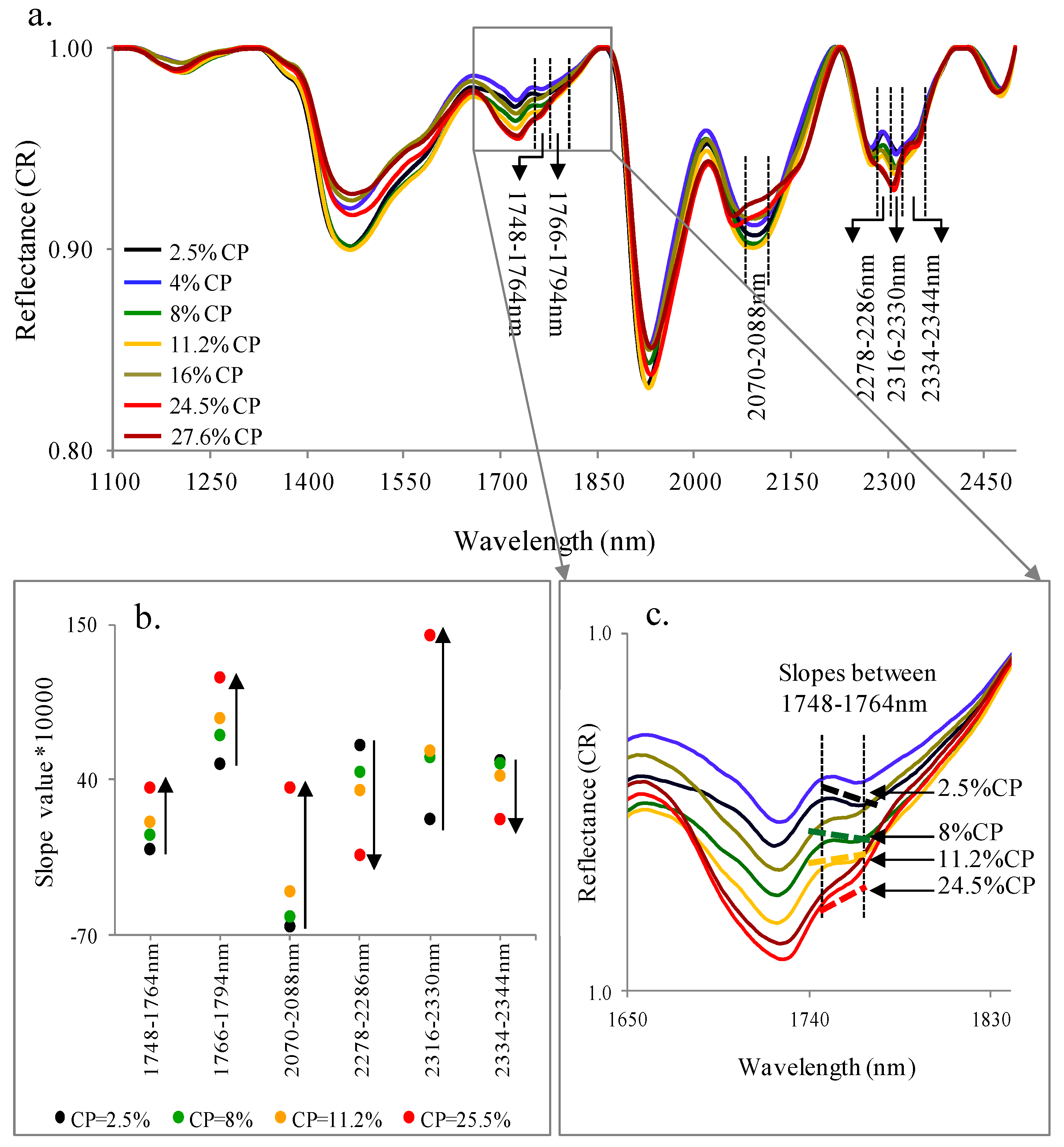
2.4. Slope Calculation and Data Analyses

To simplify the spectral signals, thereby ensuring stable calibration, and to improve the predictive ability of the final model, we applied different pretreatments to the spectral domain. The spectral data were considered in reflectance and absorbance (−log10R). In addition, continuum removal (CR) spectra were calculated. The commonly used CR technique [18,35,51,52,53] normalizes reflectance spectra and enhances the spectral differences, enabling the distinction and highlighting of individual absorption features from a baseline. On the one hand, it emphasizes the absorption features of the chemical chromophore and on the other, it reduces the physical chromophore effect [53]. In this algorithm, the monotonous spectra are characterized by a reflectance signal value of one, and absorption features are presented relative to a continuum of interpolated reflectance values that connect the two absorption edges. Then, slopes are calculated with the following equation:
m   =   y 2     y 1 x 2     x 1
where the x axis represents wavelength and the y axis represents CR data.
To develop an algorithm to assess pasture quality, we first used CR spectra to visually inspect the spectral behavior versus chemical information on the vegetation. Then, the differences in spectral behavior as a function of wavelength, which are indicative of changes in chemical properties, were identified, yielding six spectral ranges (see Section 3.2). The slopes across these spectral ranges, and their changes, were then calculated.

2.5. Data Processing and Analyses

At a first stage, calculated slopes were correlated with the changes in CP, NDF and MEC values. The data were analyzed for the distribution values of CP, NDF and MEC versus slope, to locate the thresholds for each parameter. Three categories were defined: high, medium and low, for each chemical reference and its corresponding slopes. The slope threshold for each spectral range was used as a criterion to evaluate pasture quality.
Next, Partial least Squares (PLS) regression analysis was used to identify which spectral ranges are optimal for predicting CP, NDF and MEC values. Finally, to investigate the feasibility of using the developed method for imaging data, all data set was resample from Foss NIRS system with 700 bands to AISA sensor. AISA, hyperspectral airborne sensor, with high special resolution (0.52 m from high of 1000 m), covers the full VIS-NIR-SWIR (400–2500 nm) range, which the SWIR region (970–2500 nm) include 109 bands [54].

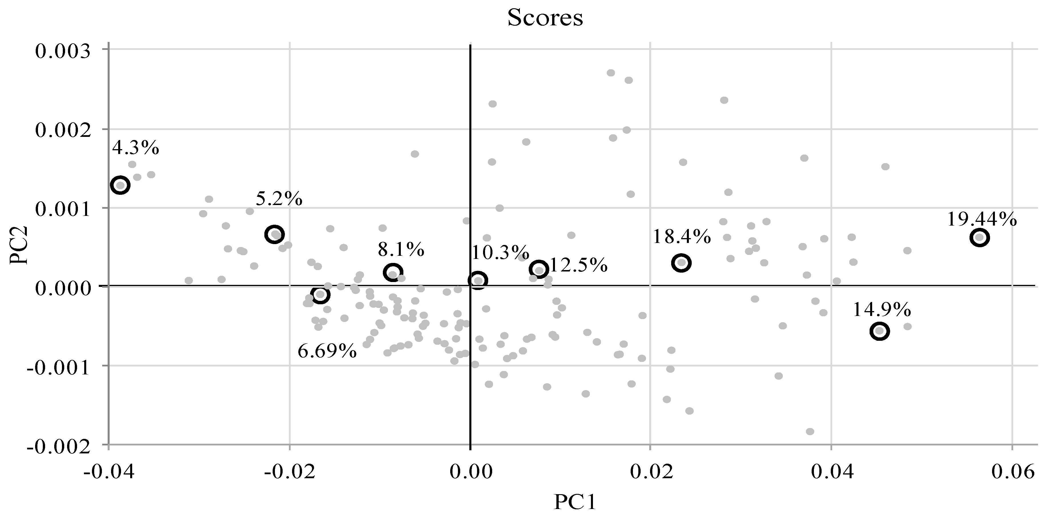
2.6. PLS Data Analyses

The PLS regression is generally based on latent variable decomposition of two blocks of variables, the X and Y matrices, which contain spectral data and any reference chemical variable, respectively. The objective of the regression is to locate small numbers of PLS components that efficiently predict Y when X is used [55]. PLS regression has advantages over other regression techniques, such as stepwise multilinear regression, principal component regression and multiple linear regression. It not only works with multicollinear variables, but also when the number of independent variables is greater than the number of observations, which is generally the case for NIRS analyses [56].
The ultimate goal of multivariate regression analysis is to create a calibration equation (or series of equations), which, when applied to data of “unknown” samples measured in the same manner, will accurately predict the quantities of the constituents of interest [55]. The multivariate calibration models were generated using PLS regression, with the goal of defining a relationship between the NIR–SWIR spectra of pasture samples and each chemical reference (i.e., CP, NDF and MEC values):
Y = A + A 1 X 1 + A 2 X 2 + A 3 X 3 + A n X n
where Y is the chemical reference of a sample, A is an empirical coefficient, and X is the spectral reflectance at a specific wavelength.
Different spectral ranges were identified based on the criteria described in Section 2.5. Numerous PLS regression models were constructed using two spectral criteria: (1) each spectral range was considered separately in the model, (2) the whole set of spectral ranges was considered in the model (53 bands, as will be explained in Section 3.3).
Cross validation was then performed. Statistical parameters for the calibration model were calculated by leave-one-out cross validation (only one sample at a time is kept out of the calibration and used for prediction). The performance and relevance of the PLS regression models were further evaluated by computing different statistics. The difference between the predicted values and measured chemical reference values was expressed as the root mean square error of prediction (RMSEP) or the root mean square error of cross validation (RMSECV). RMSEP is defined as the square root of the average of the squared differences between the predicted and measured values of the validation objects [55]:
R M S E P = [ ( X m X P ) 2 n v ] 1 / 2
where Xm is the chemically measured value of a sample, Xp is the predicted value of the sample based on the spectral analysis, and nv is the number of samples in the calibration stage.

3. Results and Discussion

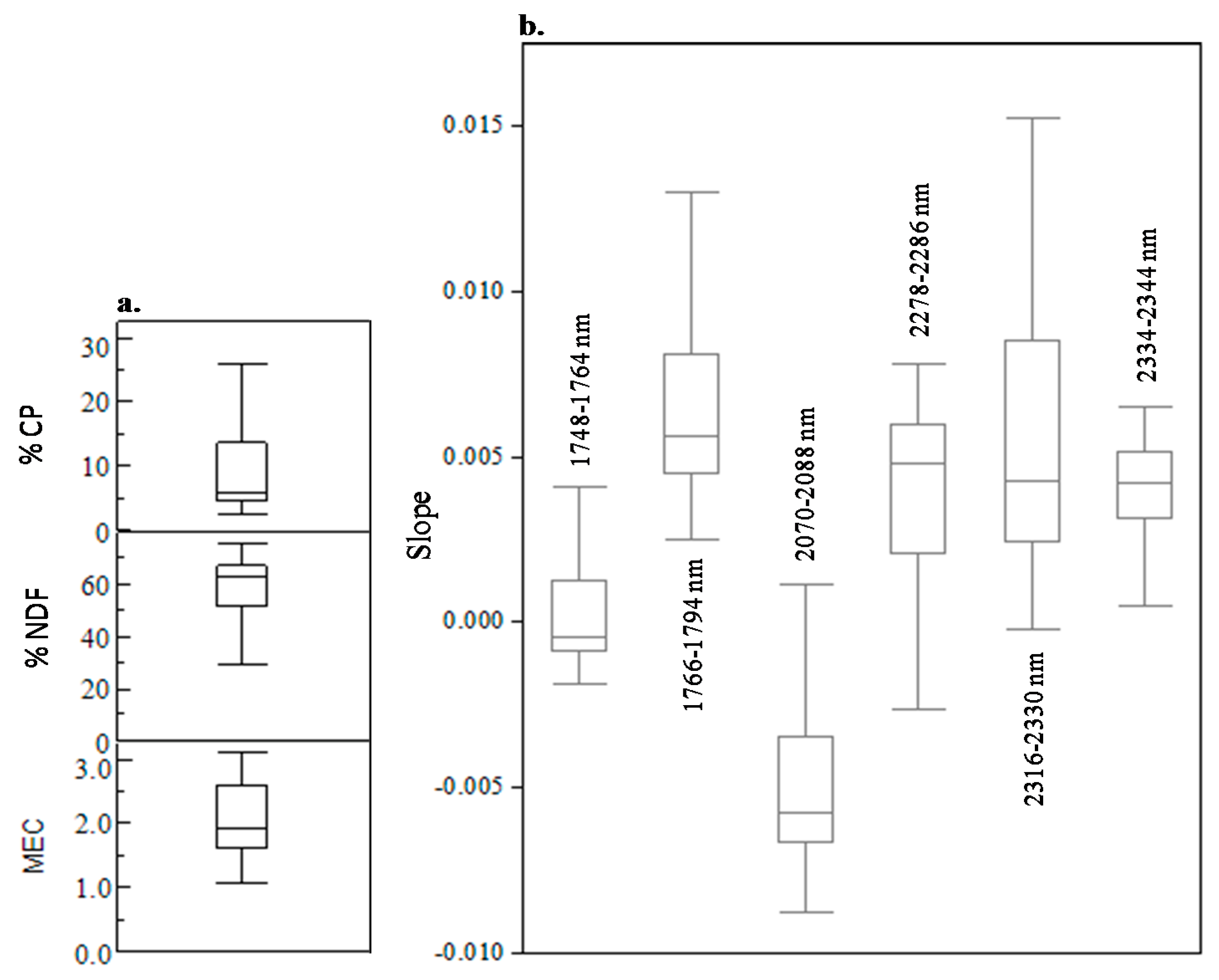
3.1. Chemical Reference: CP, NDF, MEC

The range of CP, NDF and MEC values was broad: 2.5%–32% (225 samples, average 9.46% and STD (standard deviation) 6.9%), 28%–76% (235 samples, average 59.24% and STD 10.95%) and 1.06–3.12 (166 samples, average 2.04 and STD 0.55), respectively.

3.2. Spectral Slope Analyses

Figure 1 shows the CR spectra of seven vegetation samples with different percentages of CP. Changes in slope across the different spectral ranges as a function of different CP contents could be seen at: 1748–1764 nm, 1766–1794 nm, 2070–2088 nm, 2278–2286 nm, 2316–2330 nm and 2334–2344 nm (Figure 1a,b). The spectral assignments for the main absorbance values of the vegetation were taken from Curran [20] and Schwanninger et al. [25]. Figure 1c shows an example of this relationship by zooming in on the 1748–1764 nm spectral range, where the slope is seen to increase with increasing CP content. Increases or decreases in spectral slope as function of CP content were also obtained for the other spectral ranges (Figure 1b).
Figure 1. (a) Continuum removal (CR) reflectance spectra of vegetation samples with different percentages of crude protein (CP). Note the variability in the slopes across the different spectral ranges: 1748–1764 nm, 1766–1794 nm, 2070–2088 nm, 2278–2286 nm, 2316–2330 nm, 2334–2344 nm. (b) Visualization of the slope’s tendency to increase or decrease as a function of different CP contents. Up-pointing arrow indicates a slope increase with CP content increase; down-pointing arrow indicates a slope decrease with CP content increase. (c) Zooming in on the 1748–1764 nm spectral range to demonstrate the changes in the slope with changes in CP concentration.
Remotesensing 07 00256 g001
The distribution of CP, NDF and MEC values is shown in Figure 2a, and that of the slopes for the six spectral ranges is shown in Figure 2b. Significant variability in the spectral slopes was obtained, presumably due to differences in chemical composition and the chromophores, which differ for each spectral range.
Figure 2. Distribution of (a) CP, neutral detergent fiber (NDF), metabolic energy concentration (MEC) and (b) slope values for six spectral ranges.
Remotesensing 07 00256 g002
To gain additional insight into the relationship between spectral slopes and chemical constituents, different ranges of CP conditional on slope ranges were divided into three main classes: slope less than zero (Figure 3a), between 0 and 0.001 (Figure 3b), and above 0.001 (Figure 3c). The negative slope was found to correspond to low CP values (Figure 3a). Thus, by knowing only the slope’s sign (negative/positive), it is possible to quantitatively assess the amount of CP in a sample. Specifically, slopes in the range of 0–0.001 corresponded to CP values of around 6%–15% (Figure 3b), whereas slopes above 0.001 corresponded to high CP values (around 15%–30%, Figure 3c).
In light of these results, the data distribution of CP, NDF and MEC versus slopes were analyzed and three main categories were found: (1) low, representing about 25% of the data, when CP ≤ 4.5%, NDF ≥ 67% and MEC ≤ 1.6; (2) medium, representing about 50% of the data, when 4.5% < CP < 13%, 53% < NDF < 67% and 1.6 < MEC < 2.5; (3) high, representing about 25% of the data, when CP ≥ 13%, NDF ≤ 53% and MEC ≥ 2.5. For each category of CP, NDF and MEC and for each spectral domain, the range of slope values was identified and defined to assess pasture quality according to the above three categories (summarized in Table 1). For example, the CP level was classified as low, medium or high when the slope value between 1748 and 1764 nm was ≤−0.0008, between −0.0008 and 0.001 or ≥0.001, respectively (Table 1).
Figure 3. Distribution of CP values for slopes (between 1748–1766 nm) of: (a) <0; (b) 0–0.001; (c) >0.001.
Remotesensing 07 00256 g003
Table 1. Slope-based criteria for assessment of pasture quality using three levels of crude protein (CP), neutral detergent fiber (NDF) and metabolic energy concentration (MEC).
%CP
Spectral RangeLow (≤4.5%)Medium (4.5%–13%)High (≥13%)
1748–1764 nmSlope ≤ −0.0008−0.0008 < Slope< 0.001Slope ≥ 0.001
1766–1794 nmSlope ≤ 0.0050.005 < Slope< 0.008Slope ≥ 0.008
2070–2088 nmSlope ≤ −0.0065−0.0065 < Slope< –0.003Slope ≥ −0.003
2278–2286 nmSlope ≥ 0.00580.0025 < Slope < 0.0058Slope ≤ 0.0025
2316–2330 nmSlope ≤ 0.0030.003 < Slope < 0.008Slope ≥ 0.008
2334–2344 nmSlope ≥ 0.00480.0034 < Slope < 0.0048Slope ≤ 0.0034
%NDF
Spectral RangeLow (≥67%)Medium (53%–67%)High (≤53%)
1748–1764 nmSlope ≤ −0.0008−0.0008 < Slope < 0.001Slope ≥0.001
1766–1794 nmSlope ≤ 0.00520.0052 < Slope < 0.0076Slope ≥ 0.0076
2070–2088 nmSlope ≤ −0.0065−0.0065 < Slope < −0.0032Slope ≥ −0.0032
2278–2286 nmSlope ≥ 0.00580.0026 < Slope < 0.0058Slope ≤ 0.0026
2316–2330 nmSlope ≤ 0.00320.0076 > Slope > 0.0032Slope ≥ 0.0076
2334–2344 nmSlope ≥ 0.0050.005 > Slope > 0.0033Slope ≤ 0.0033
MEC
Spectral RangeLow (≤1.6)Medium (1.6–2.5)High (≥2.5)
1748–1764 nmSlope ≤ −0.0005−0.0005 < Slope < 0.0016Slope ≥ 0.0016
1766–1794 nmSlope ≤ 0.00530.0053 < Slope < 0.0091Slope ≥ 0.0091
2070–2088 nmSlope ≤ −0.0062−0.0062 < Slope < −0.0022Slope ≥ −0.0022
2278–2286 nmSlope ≥ 0.00570.0018 < Slope < 0.0057Slope ≤ 0.0018
2316–2330 nmSlope ≤ 0.0030.003 < Slope < 0.0093Slope ≥ 0.0093
2334–2344 nmSlope ≥ 0.00480.0031 < Slope < 0.0048Slope ≤ 0.0031
The slope trends presented in Figure 2 and Figure 3 strongly suggest that the spectral reflectance properties of pastures with similar components can provide information regarding the amount of CP, NDF, MEC.
Assessing pasture quality using only slope-based criteria (Table 2), we found that in general, the total success rates were good for: (1) CP at 72%–80% (low), 50%–74% (medium) and 84%–98% (high); (2) NDF at 6%9–77% (low), 37%–72% (medium) and 79%–94% (high), (3) MEC at 56%–87% (low), 46%–60% (medium) and 79%–89% (high). Rating the overall success rate (“Total” row in Table 2) for the three categories in each spectral range showed that the best slope criteria for CP were in the 1748–1764 nm and 2278–2286 nm spectral ranges, for NDF in the 1748–1764 nm and 2278–2286 nm ranges, and for MEC in the 2316–2330 nm and 1748–1764 nm ranges. Importantly, the p-value was highly significant between number of identifications per category for all categories (p < 0.05), indicating that the slopes are highly associated with CP, NDF and MEC contents and their association is not due to random variation.
Table 2. Success rates of pasture-quality evaluations using slope algorithm. Note that chemical data were used as reference.
% CPTotal Per Category Based on Chemical DataTotal Per Category Based on Slope Algorithm
1748–1764 nm1764–1794 nm2070–2088 nm2278–2286 nm2316–2330 nm2334–2344 nm
Low (≤4.5%)544176%3972%4176%4176%4380%4380%
Medium (4.5%–13%)1148373%5750%8474%7969%7263%6860%
High (≥13%)575596%4884%4986%5698%5596%4986%
Total22517980%14464%17477%17678%17076%16071%
% NDF
Low (≥67%)644977%4977%4469%4875%4672%4570%
Medium (53%–67%)1097972%4037%7266%7165%5752%6459%
High (≤53%)625487%5182%4979%5690%5894%5081%
Total23518277%14060%16570%17574%16169%15968%
MEC
Low (≤1.6)393077%2667%2564%3077%3487%2256%
Medium (1.6%–2.5)804455%4151%4253%4860%4759%3746%
High (≥2.5)474289%4187%4187%4085%4187%3779%
Total16611670%10865%10865%11871%12273%9658%
Overgaard et al. [57] showed that one or two years of spectral measurement are insufficient to build fully operational models for cereal property predictions. In this regard, our study employed 10 years (2002–2011) of observations, and should thus adequately represent the expected range of conditions in our study area. Furthermore, Givens et al. [27] and Pullanagari [58] argued that high-precision spectroscopic instruments, spectral resolution and number of wavelengths are crucial for determining pasture quality with high accuracy. However, this study shows that highly accurate estimates of pasture quality using spectral slopes that require only two wavelengths are possible.
The chemometric approach essentially correlates the optical data with “reference” chemical constituents based on different statistical methods, enabling high-quality estimates of vegetation composition [8,9,26,59,60]. However, high-precision instruments with high spectral resolution are needed to use these methods, such as the Foss NIRS system model 5000 spectrometer. Furthermore, in HS remote sensing, it might not be possible to obtain such high-precision information due to atmospheric effects, a low signal-to-noise ratio, a varying field of view for every pixel, spectral instability and problems of spectral mixing [61]. Therefore, the developed method might overcome this limitation. Importantly, four of the six selected spectral regions were completely outside the spectral range of water absorbance (e.g., 1300–1650 nm, 1430–2230 nm). This result is encouraging, because it might expand the possibility of using the developed methodology with airborne HS or even multispectral sensors. Indeed, one needs only two wavelengths to calculate the slope data (e.g., the beginning of the spectral range and its last point), which are then incorporated into the model to assess the chemical constituents.
Next, to investigate the feasibility of our method for airborne HS sensor application, we resampled the spectral data from the Foss NIRS 5000 spectrometer (700 bands) to AISA, an airborne HS sensor with 109 bands between 1100 and 2363 nm. The procedure applied to the resampled data set was the same as that applied to the original data, i.e., CR spectra for visual inspection of spectral slope change vs. chemical data. Five spectral ranges (Table 3) were identified as candidates for the slope method, and a new set of slope criteria was defined (Table 3). The results are summarized in Table 4. Four of the five spectral ranges were similar to the four spectral ranges in the original data (Table 1 and Table 3) and an additional “AISA-fitted” spectral range (2306–2317 nm) was introduced. In general, the total success rates (Table 4) of the three categories were as good as those of the original data set (Table 2) for: (1) CP at 70%–91% (low), 58%–70% (medium) and 86%–100% (high); (2) NDF at 64%–95% (low), 33%–67% (medium) and 79%–92% (high); (3) MEC at 67%–85% (low), 46%–63% (medium) and 83%–94% (high).
Table 3. Slope-based criteria for assessment of pasture quality using three levels of CP, NDF and MEC, after resampling the spectral region of FOS-5000 (700 bands at 1100–2363 to spectral region of advanced imaging spectrometer for applications (AISA) (109 bands at 970–2500 nm).
% CP
Spectral RangeLow (≤4.5%)Medium (4.5%–13%)High (≥13%)
1747–1770nmSlope ≤ 0.000250.00025 < Slope< 0.003Slope ≥ 0.003
2061–2096 nmSlope ≤ −0.014−0.014 < Slope< −0.008Slope ≥ −0.008
2270–2293 nmSlope ≥ 0.010.01 > Slope> 0.0045Slope ≤ 0.0045
2306–2317 nmSlope ≤ −0.001−0.001 < Slope< 0.0025Slope ≥ 0.0025
2317–2328 nmSlope ≤ 0.00450.0045 < Slope < 0.008Slope ≥ 0.008
% NDF
Spectral RangeLow (≥67%)Medium (53%–67%)High (≤53%)
1747–1770nmSlope ≤ 0.00050.0005 < Slope< 0.003Slope ≥ 0.003
2061–2096 nmSlope ≤ −0.014−0.014 < Slope< −0.008Slope ≥ −0.008
2270–2293 nmSlope ≥ 0.010.01 > Slope> 0.0035Slope ≤ 0.0035
2306–2317 nmSlope ≤ 0.00040.0004 < Slope< 0.003Slope ≥ 0.003
2317–2328 nmSlope ≤ 0.00450.0045 < Slope < 0.008Slope ≥ 0.008
MEC
Spectral RangeLow (≤1)Medium (1.6–2.5)High (≥2.5)
1747–1770nmSlope ≤ 0.0010.001 < Slope< 0.004Slope ≥ 0.004
2061–2096 nmSlope ≤ −0.013−0.013 < Slope< −0.006Slope ≥ −0.006
2270–2293 nmSlope ≥ 0.010.01 > Slope> 0.001Slope ≤ 0.001
2306–2317 nmSlope ≤ −0.0005−0.0005 < Slope< 0.0035Slope ≥ 0.0035
2317–2328 nmSlope ≤ 0.00450.0045 < Slope < 0.009Slope ≥ 0.009
This “coarse” spectral resolution might be implemented in HS and/or multispectral remote-sensing sensors. In recent years, there has been an increase in the availability of images with high spatial resolution [1]. In this regard, our study might serve as a basic infrastructure for future directions in the use of this technology to determine pasture quality.
Table 4. Success rates of pasture-quality evaluations using slope algorithm. Note that chemical data were used as reference, after resampling the spectral region of FOS-5000 (700 bands at 1100–2363 to spectral region of AISA (109 bands at 970–2500 nm).
% CPTotal Per Category Based on Chemical DataTotal Per Category Based on Slope Algorithm
1747–1770 nm2061–2096 nm2270–2293 nm2306–2317 nm2317–2328 nm
Low (≤4.5%)544889%3870%4583%4991%4685%
Medium (4.5%–13%)1147263%8070%6658%7768%6658%
High (≥13%)575596%4986%57100%5596%5596%
Total22517578%16774%16875%18180%16774%
% NDF
Low (≥67%)644266%4164%5180%6195%4672%
Medium (53%–67%)1097367%6358%6358%3633%5752%
High (≤53%)624979%4979%5690%5385%5792%
Total23516470%15365%17072%15064%16068%
MEC
Low (≤1.6)393385%2667%2872%2974%3385%
Medium (1.6–2.5)803949%3746%5063%4050%4556%
High (≥2.5)474494%3983%4494%4289%4289%
Total16611670%10261%12273%11167%12072%

3.3. PLS Analyses

The results of PLS modeling of the CP, NDF and MEC values are presented in Table 5. Both the whole set of six spectral ranges (53 bands) and each individual spectral range was modeled, and both gave relatively accurate estimates of pasture quality. The best-fit model for the calibration set included all six spectral ranges (n = 53 wavelengths) in the model with R2 ranges from 0.83 for MEC to 0.96 for CP. Importantly, when only one spectral range was assessed in the model, the R2 was relatively high for all chemical constituents, with relatively high slope, and low RMSEP (Table 5). The best spectral domains for CP assessment were 1748–1764 nm and 1766–1794 nm (9 wavelengths and 15 wavelengths, respectively); for NDF assessment they were 2070–2088 nm and 2278–2286 nm (10 and 4 wavelengths, respectively), and for MEC assessment they were 2316–2330 nm and 2278–2286 nm (8 and 4 wavelengths, respectively).
A score plot of the samples from the PLS modeling demonstrated the excellent correlation between spectra and chemical constituents (CP, NDF and MEC), with an increase from left to right. The score plot for the CP model (Figure 4) indicated that most of the spectral variations observed in our field study were indeed related to the protein, as predicted by the PLS model (and were not influenced by unknown parameters). Furthermore, for the best CP, NDF and MEC models, the first two LV (latent variables) components in the PLS model explained 100% of the X variance (spectra), and 99%, 86%, and 79% (for CP, NDF and MEC, respectively) of the Y variance (chemical components). This indicated that most of the spectral variation in the six selected spectral domains is related to the CP, NDF and MEC components that were modeled by PLS.
Figure 4. A score plot for the partial least squares (PLS) CP model using the 1748–1764 nm spectral range.
Remotesensing 07 00256 g004
A comparison of the PLS and slope method results showed that the best PLS models for assessing CP, NDF and MEC are obtained with the spectral ranges of 1748–1764 nm, 2070–2088 and 2316–2330 nm, respectively, whereas using the slope method, only the 1748–1764 nm spectral range could be used to assess all parameters. Indeed, the PLS is a more accurate quantitative method, but requires chemical analysis to establish a calibration model; in contrast, the slope method provides a qualitative evaluation (three categories) with no need for chemical measurement of the samples.
Table 5. Partial least squares (PLS) regression model results of the correlations between different spectral regions and percentages of CP, NDF and MEC.
Spectral Range (nm) (Number of Bands) CP Model’s Statistical CharacteristicsCP Best ModelNDF Model’s Statistical CharacteristicsNDF Best ModelMEC Model’s Statistical CharacteristicsMEC Best Model
PredictionValidationPredictionValidationPredictionValidation
1748–1764
(n = 9)
Slope:0.9340.93320.8560.85040.7890.7925
Offset:0.60.6068.5088.8740.4290.423
RMSE:1.7031.7574.1834.3050.2520.263
R2:0.9340.9310.8560.8480.7890.769
1766–1794
(n = 15)
Slope:0.9130.90830.7970.78550.7940.7824
Offset:0.790.83412.04512.7500.4180.445
RMSE:1.9542.024.9775.0980.2480.259
R2:0.9130.9090.7970.7920.7940.783
2070–2088
(n = 10)
Slope:0.8970.89950.8650.85920.7160.7007
Offset:0.9320.9128.0198.3450.5760.623
RMSE:2.1222.2044.0624.1330.2910.324
R2:0.8970.8910.8650.8610.7160.654
2278–2286
(n = 4)
Slope:0.9030.89840.8560.85430.8050.7963
Offset:0.8710.98.4998.6770.3950.419
RMSE:2.0512.0964.1814.2170.2410.251
R2:0.9040.9020.8560.8550.8050.795
2316–2330
(n = 8)
Slope:0.8920.88960.7820.76170.8120.8022
Offset:0.9781.00412.91114.1570.3800.406
RMSE:2.1732.2155.1535.4200.2370.243
R2:0.8910.8880.7820.7600.8120.804
2334–2344
(n = 6)
Slope:0.780.7770.7870.78560.7150.7076
Offset:1.9872.0512.62912.7360.5790.594
RMSE:3.093.175.0975.2190.2920.302
R2:0.780.770.7870.7780.7150.703
All ranges
(n = 53)
Slope:0.9670.95610.8930.88210.8320.8231
Offset:0.320.396.3106.8500.3400.368
RMSE:1.2461.3553.6043.8580.2240.241
R2:0.9640.9580.8930.8780.8330.809

4. Conclusions

In this study, we hypothesized that changes in spectral slopes can be used as an indicator for qualitative assessment of plant composition. Six spectral regions across the NIR–SWIR region were identified: 1748–1764 nm, 1766–1794 nm, 2070–2088 nm, 2278–2286 nm, 2316–2330 nm, 2334–2344 nm. Slopes across these ranges were calculated and correlated with the changes in CP, NDF and MEC. The good success rate using these slope criteria allowed evaluating three level categories (low, medium and high) for CP, NDF and MEC. PLS analysis supported these findings, showing a high correlation between the six spectral regions and the chemical information. An important finding was the valid assessment obtained using only one spectral range, whereas two and three ranges will provide a good estimate for all three studied parameters. The slope method can therefore be further adopted to evaluate the quality of various types of vegetation in pasture areas and can be used to measure and analyze a large number of samples in a short time, providing a basis for future monitoring of pasture quality using remote sensing.
The results of our study are especially encouraging since the selected spectral regions, excluding 2070–2088 nm, are not directly affected by atmospheric water vapor or water in the plants. Current method is restricted to pasture quality estimates based only on dry samples’ spectra. In the future, by adjusting the method for fresh vegetation, it could be further applied for in-situ analyses using a relatively inexpensive instrument with a specific set of detectors.
Another point that should be considered in future studies is the difference between laboratory NIR spectra (i.e., stable conditions with an artificial light source) and in-situ field canopy reflectance (i.e., impacted by species composition, canopy structure, and atmospheric effects, etc.). While during PLS modeling, different sources of variability can be included in the calibration data set, we think that combining two data sets (laboratory and field measurements) in one calibration model is the most advantageous approach to start with.

Acknowledgments

This study was supported by the Chief Scientist of the Ministry of Agriculture, Israel, and the Jewish National Fund. The authors wish to thank to Serge Yan Landau from the Agricultural Research Organization, Volcani Center, Israel, for use of the archive data. We also appreciate comments of Graeme D. Batten from Charles Sturt University and Sydney University. The authors also wish to thank to the three anonymous reviewers for the constructive comments that improved our manuscript. This paper is publication No. 115/2014 of the Plant Science Institute, Agricultural Research Organization, Israel.

Author Contributions

Naftaly Goldshleger conceived the project and a project PI. Rachel Lugassi, Naftaly Goldshleger and Eli Zaady, designed, performed and supported the experiments. Rachel Lugassi analyzed the data. Rachel Lugassi and Alexandra Chudnovsky performed the statistical analysis, modeling and wrote the paper. Levana Dvash contributed materials. All authors contribute in paper writing.

Conflicts of Interest

The authors declare no conflict of interest.

References

  1. Svoray, T.; Perevolotsky, A.; Atkinson, P.M. Ecological sustainability in rangelands: The contribution of remote sensing. Int. J. Remote Sens. 2013, 34, 6216–6242. [Google Scholar] [CrossRef]
  2. Association of Official Analytical Chemists (AOAC). Official Methods of Analysis, 15th ed.; AOAC: Washington, DC, USA, 1990. [Google Scholar]
  3. Association of Official Analytical Chemists (AOAC), International. Official Methods of Analysis, 16th ed.; AOAC: Arlington, VA, USA, 1995. [Google Scholar]
  4. Goering, H.K.; van Soest, P.J. Forage fiber analysis (apparatus, reagents, procedures and some applications). In Agriculture Handbook; No. 379; Agriculture Research Service, United States Department of Agriculture: Washington, DC, USA, 1970. [Google Scholar]
  5. Blanco, M.; Villarroya, I. NIR spectroscopy: A rapid-response analytical tool. Trends Anal. Chem. 2002, 21, 240–250. [Google Scholar] [CrossRef]
  6. Dematte, J.A.M.; Campos, R.C.; Alves, M.C.; Fiorio, P.R.; Nanni, M.R. Visible-NIR reflectance: A new approach on soil evaluation. Geoderma 2004, 12, 95–112. [Google Scholar] [CrossRef]
  7. Goldshleger, N.; Chudnovsky, A.; Ben-Binyamin, R. Predicting salinity in tomatoes using soil reflectance spectra. Int. J. Remote Sens. 2013, 34, 6079–6093. [Google Scholar] [CrossRef]
  8. Landau, S.; Glasser, T.; Dvash, L. Monitoring nutrition in small ruminants by aids of near infrared spectroscopy (NIRS) technology: A review. Small Rumin. Res. 2006, 61, 1–11. [Google Scholar] [CrossRef]
  9. Norris, K.H.; Barners, R.F.; Moore, J.E.; Shenk, J.S. Predicting forage quality by infrared reflectance spectroscopy. J. Anim. Sci. 1976, 43, 889–897. [Google Scholar]
  10. Ben-Dor, E.; Banin, A. Near-infrared analysis as a method to simultaneously evaluate several soil properties. Soil Sci. Soc. Am. J. 1995, 59, 364–372. [Google Scholar] [CrossRef]
  11. Ben-Dor, E.; Inbar, Y.; Chen, Y. The reflectance spectra of organic matter in the visible near-infrared and short wave infrared region (400–2500 nm) during a controlled decomposition process. Remote Sens. Environ. 1997, 61, 1–15. [Google Scholar] [CrossRef]
  12. Chabrillat, S.; Ben-Dor, E.; Viscarra Rossel, R.A.; Demattê, J.A.M. Quantitative soil spectroscopy. Appl. Environ. Soil Sci. 2013. [Google Scholar] [CrossRef]
  13. Chudnovsky, A.; Ben-Dor, E.; Paz, E. Using NIRS for rapid assessment of sediment dust in the indoor environment. J. Near Infrared Spectrosc. 2007, 15, 59–70. [Google Scholar] [CrossRef]
  14. Karnieli, A. Development and implementation of spectral crust index over dune sands. Int. J. Remote Sens. 1997, 18, 1207–1220. [Google Scholar] [CrossRef]
  15. Karnieli, A.; Kidron, G.J.; Glaesser, C.; Ben-Dor, E. Spectral characteristics of cyanobacteria soil crust in semiarid environments. Remote Sens. Environ. 1999, 69, 67–75. [Google Scholar] [CrossRef]
  16. Kooistra, L.; Wehrens, R.; Leuven, W.; Buydens, L. Possibilities of visible-near-infrared spectroscopy for the assessment of soil contamination in river floodplains. Anal. Chim. Acta 2001, 446, 97–105. [Google Scholar] [CrossRef]
  17. Kooistra, L.; Wanders, G.; Epemac, R.; Leuven, W.; Wehrens, L.; Buydens, L. The potential of field spectroscopy for the assessment of sediment properties in river floodplains. Anal. Chim. Acta 2003, 484, 189–200. [Google Scholar] [CrossRef]
  18. Shoshany, M.; Svoray, T.; Curran, P.J.; Foody, G.M.; Perevolotsky, A. The relationship between ERS-2 SAR backscatter and soil moisture: Generalization from a humid to semi-arid transect. Int. J. Remote Sens. 2000, 21, 2337–2343. [Google Scholar] [CrossRef]
  19. Viscarra-Rossel, R.A.; Walvoort, D.J.J.; McBratney, A.B.; Janik, L.J.; Skjemstad, J.O. Visible, near infrared, mid infrared or combined diffuse reflectance spectroscopy for simultaneous assessment of various soil properties. Geoderma 2006, 131, 59–75. [Google Scholar] [CrossRef]
  20. Curran, P.J. Remote sensing of foliar chemistry. Remote Sens. Environ. 1989, 30, 271–278. [Google Scholar] [CrossRef]
  21. Murray, I.; Williams, P.C. Chemical principles of near infrared technology. In Near Infrared Technology in Agriculture and Food Industries; Wiliams, P.C., Noriss, K.H., Eds.; American Association of Cereal Chemistry Inc.: St. Paul, MN, USA, 1987; pp. 17–31. [Google Scholar]
  22. Workman, J., Jr.; Weyer, L. Practical Guide to Interpretive Near-Infrared Spectroscopy; CRC Press/Taylor & Francis Group: Boca Raton, FL, USA, 2008. [Google Scholar]
  23. Baumgardner, M.F.; Silva, L.F.; Biehl, L.L.; Stoner, E.R. Reflectance properties of soils. Adv. Agron. 1985, 38, 1–44. [Google Scholar]
  24. Dalal, R.C.; Henry, R.J. Simultaneous determination of moisture, organic carbon and total nitrogen by near infrared reflectance spectroscopy. Soil Sci. Soc. Am. J. 1986, 50, 120–123. [Google Scholar] [CrossRef]
  25. Schwanninger, M.; Rodrigues, J.C.; Fackler, K. A review of band assignments in near infrared spectra of wood and wood components. J. Near Infrared Spectrosc. 2011, 19, 287–308. [Google Scholar] [CrossRef]
  26. Decruyenaere, V.; Lecomte, P.; Demarquilly, C.; Aufrere, J.; Dardenne, P.; Stilmant, D.; Buldgen, A. Evaluation of green forage intake and digestibility in ruminants using near infrared reflectance spectroscopy (NIRS): Developing a global calibration. Anim. Feed Sci. Technol. 2009, 148, 138–156. [Google Scholar] [CrossRef]
  27. Givens, D.I.; de Boever, J.L.; Deaville, E.R. The principles, practices and some future applications of near infrared spectroscopy for predicting the nutritive value of foods for animals and humans. Nutr. Res. Rev. 1997, 10, 83–114. [Google Scholar] [CrossRef] [PubMed]
  28. Schellberg, J.; Hill, M.J.; Gerhards, R.; Rothmund, M.; Braun, M. Precision agriculture on grassland: Applications, perspectives and constraints. Eur. J. Agron. 2008, 29, 59–71. [Google Scholar] [CrossRef]
  29. Pullanagari, R.; Yule, I.; Tuohy, M.; Hedley, M.; Dynes, R.; King, W. In-field hyperspectral proximal sensing for estimating quality parameters of mixed pasture. Precis. Agric. 2012, 13, 351–369. [Google Scholar] [CrossRef]
  30. Pullanagari, R.; Yule, I.; Tuohy, M.; Hedley, M.; Dynes, R.; King, W. Multi-spectral radiometry to estimate pasture quality components. Precis. Agric. 2012, 13, 442–456. [Google Scholar] [CrossRef]
  31. Sanches, D.; Tuohy, M.P.; Hedley, M.J.; Bretherton, M.R. Large, durable and low-cost reflectance standard for field remote sensing applications. Int. J. Remote Sens. 2009, 30, 2309–2319. [Google Scholar] [CrossRef]
  32. Mutanga, O.; Skidmore, A.K. Narrow band vegetation indices overcome the saturation problem in biomass estimation. Int. J. Remote Sens. 2004, 25, 3999–4014. [Google Scholar] [CrossRef]
  33. Mutanga, O.; Skidmore, A.K. Integrating imaging spectroscopy and neural networks to map grass quality in the Kruger National Park, South Africa. Remote Sens. Environ. 2004, 90, 104–115. [Google Scholar] [CrossRef]
  34. Pimstein, A.; Karnieli, A.; Bansal, S.K.; Bonfil, D. Exploring remotely sensed technologies for monitoring wheat potassium and phosphorus using field spectroscopy. Field Crops Res. 2011, 121, 125–135. [Google Scholar] [CrossRef]
  35. Mutanga, O.; Skidmore, A.K.; Prins, H.H.T. Predicting in situ pasture quality in the Kruger National Park, South Africa, using continuum-removed absorption features. Remote Sens. Environ. 2004, 89, 393–408. [Google Scholar] [CrossRef]
  36. Lehnert, L.W.; Meyer, H.; Meyer, N.; Reudenbach, C.; Bendix, J. A hyperspectral indicator system for rangeland degradation on the Tibetan Plateau: A case study towards spaceborne monitoring. Ecol. Indic. 2014, 39, 54–64. [Google Scholar] [CrossRef]
  37. Kawamura, K.; Watanabe, N.; Sakanoue, S.; Yoshitoshi, R.; Odagawa, S. Herbage biomass and quality status assessment in a mixed sown pasture from airborne based hyperspectral imaging. In Proceedings of the 33rd Asian Conference on Remote Sensing, Pattaya, Thailand, 26–30 November 2012; pp. 2220–2228.
  38. Stern, A.; Gradus, Y.; Meir, A.; Krakover, S.; Tsoar, H. Atlas of the Negev; Ben Gurion University of the Negev: Beer-Sheva, Israel, 1986. [Google Scholar]
  39. The Israel Meteorological Service. Available online: http://www.ims.gov.il/IMSEng/ (accessed on 22 December 2014).
  40. Dan, J.; Yaalon, D.; Kundzimzinsky, H.; Raz, Z. The Soil of Israel; ARO Publication Bulletin No. 168 (in Hebrew). Volcani Center: Bet-Dagan, Israel, 1977. [Google Scholar]
  41. Zaady, E.; Levacov, R.; Shachak, M. Application of the herbicide, Simazine, and its effect on soil surface parameters and vegetation in a patchy desert landscape. Arid Land Res. Manag. 2004, 18, 397–410. [Google Scholar] [CrossRef]
  42. Feinbrun-Dothan, N.; Danin, A. Analytical Flora of Eretz-Israel; Cana Publishers: Jerusalem, Israel, 1991. [Google Scholar]
  43. Zaady, E.; Yonatan, R.; Shachak, M.; Perevolotsky, A. The effects of grazing on abiotic and biotic parameters in a semiarid ecosystem: A case study from the northern Negev desert, Israel. Arid Land Res. Manag. 2001, 15, 245–261. [Google Scholar] [CrossRef]
  44. Henkin, Z.; Landau, S.; Ungar, E.D.; Perevolotsky, A.; Yehuda, Y.; Sternberg, M. Effect of timing and intensity of grazing on the herbage quality of a Mediterranean rangeland. J. Anim. Feed Sci. 2007, 16, 318–322. [Google Scholar]
  45. Henkin, Z.; Ungar, E.D.; Dvash, L.; Perevolotsky, A.; Yehuda, Y.; Sternberg, M.; Voet, H.; Landau, S.Y. Effects of cattle grazing on herbage quality in a herbaceous Mediterranean rangeland. Grass Forage Sci. 2011, 66, 516–525. [Google Scholar] [CrossRef]
  46. Landau, S.; Friedman, S.; Brenner, S.; Bruckental, I.; Weinberg, Z.G.; Ashbell, G.; Hen, Y.; Dvash, L.; Leshem, Y. The value of safflower (Carthamus tinctorius) hay and silage grown under Mediterranean conditions as forage for dairy cattle. Livest. Prod. Sci. 2004, 88, 263–271. [Google Scholar] [CrossRef]
  47. Landau, S.; Giger-Reverdin, S.; Rapetti, L.; Dvash, L.; Dorleans, M.; Ungar, E.D. Data mining old digestibility trials for nutritional monitoring in confined goats with aids of fecal near infra-red spectrometry. Small Ruminant Res. 2008, 77, 146–158. [Google Scholar] [CrossRef]
  48. Tilley, J.M.A.; Terry, R.A. A two-stage technique for the in vitro digestion of forage crops. J. Br. Grassl. Soc. 1963, 18, 104–111. [Google Scholar] [CrossRef]
  49. Wagner, J.J.; Lusby, K.S.; Oltjen, J.W.; Rakestraw, J.; Wettemann, R.P.; Walters, L.E. Carcass composition in mature Hereford cows: Estimation and effect on daily metabolizable energy requirement during winter. J. Anim. Sci. 1988, 66, 603–612. [Google Scholar] [PubMed]
  50. Cook, C.W.; Stoddart, L.A.; Harris, L.E. Determining the digestibility and metabolizable energy of winter range plants by sheep. J. Anim. Sci. 1952, 11, 578–590. [Google Scholar]
  51. Noomen, M.F.; Skidmore, A.K.; van der Meer, F.D.; Prins, H.H.T. Continuum removed band depth analysis for detecting the effects of natural gas, methane and ethane on maize reflectance. Remote Sens. Environ. 2006, 105, 262–270. [Google Scholar] [CrossRef]
  52. Curcio, D.; Ciraolo, G.; D’Asaro, F.; Minacapilli, M. Prediction of soil texture distributions using VNIR-SWIR reflectance rpectroscopy. Procedia Environ. Sci. 2013, 19, 494–503. [Google Scholar] [CrossRef]
  53. Clark, R.N. Spectroscopy of rocks and minerals and principle of spectroscopy. In Manual of Remote Sensing; Rencz, A.N., Ed.; John Wiley & Sons: New York, NY, USA, 1999; pp. 3–59. [Google Scholar]
  54. SPECIM, Spectral Imaging Ltd. Available online: http://www.specim.fi (accessed on 22 December 2014).
  55. Esbensen, K. Multivariate data analyses. In Practice—An Introduction to Multivariate Data Analyses and Experimental Design; Aalborg University, CAMO: Esbjerg, Denmark, 2002. [Google Scholar]
  56. Wold, S.; Sjostrom, M.; Eriksson, L. PLS-regression: A basic tool of chemometrics. Chemom. Intell. Lab. Syst. 2001, 58, 109–130. [Google Scholar] [CrossRef]
  57. Øvergaard, S.I.; Isaksson, T.; Korsaeth, A. Prediction of wheat yield and protein using remote sensors on plots—Part I: Assessing near infrared model robustness for year and site variations. J. Near Infrared Spectrosc. 2013, 21, 117–131. [Google Scholar] [CrossRef]
  58. Pullanagari, R. Proximal Sensing Techniques to Monitor Pasture Quality and Quantity on Dairy Farms. Ph.D. Thesis, Massey University, Manawatu, New Zealand, 2011. [Google Scholar]
  59. Landau, S.; Nitzan, R.; Barkai, D.; Dvash, L. Excretal near infrared reflectance spectrometry to monitor the nutrient content of diets of grazing young ostriches (Struthio camelus). S. Afr. J. Anim. Sci. 2006, 36, 248–256. [Google Scholar]
  60. Swart, E.; Brand, T.S.; Engelbrecht, J. The use of near infrared spectroscopy (NIRS) to predict the chemical composition of feed samples used in ostrich total mixed ration. S. Afr. J. Anim. Sci. 2012, 42, 550–554. [Google Scholar]
  61. Ben-Dor, E.; Taylor, R.G.; Hill, J.; Dematte, J.A.M.; Whiting, M.L.; Chabrillat, S.; Sommer, S. Imaging spectrometry for soil applications. In Advances in Agronomy; Sparks, D.L., Ed.; Elsevier Inc.: Newark, DE, USA, 2008; Volume 97, pp. 321–392. [Google Scholar]

