
Microplastics in Glaciers: First Results from the Vatnajökull Ice Cap
by
 , , and
Hlynur Stefánsson
1,* ,
,
Mark Peternell
2,
Matthias Konrad-Schmolke
2,
Hrafnhildur Hannesdóttir
3,
Einar Jón Ásbjörnsson
1 and
Erik Sturkell
2 1
Department of Engineering, Reykjavik University, 102 Reykjavik, Iceland
2
Department of Earth Sciences, University of Gothenburg, 41320 Gothenburg, Sweden
3
Icelandic Meteorological Office, 105 Reykjavik, Iceland
*
Author to whom correspondence should be addressed.
Sustainability 2021, 13(8), 4183; https://doi.org/10.3390/su13084183
Submission received: 19 March 2021
/
Revised: 5 April 2021
/
Accepted: 7 April 2021
/
Published: 9 April 2021
Abstract
:Microplastic particles, as a second-phase material in ice, may contribute to the effect such particles have on the melting and rheological behaviour of glaciers, and thus influence the future meltwater contribution to the oceans and rising sea levels. Hence, it is of the utmost importance to map and understand the presence and dispersal of microplastics on a global scale. In this work, we identified microplastic particles in snow cores collected in a remote and pristine location on the Vatnajökull ice cap in Iceland. Utilising optical microscopy and µ-Raman spectroscopy, we visualised and identified microplastic particles of various sizes and materials. Our findings support that atmospheric transport of microplastic particles is one of the important pathways for microplastic pollution.
Keywords:
microplastics; glacier; snow; ice; µ-Raman spectroscopy; atmospheric transport; pollution; climate change1. Introduction
Plastic materials are some of the most important compounds used by modern societies, relying heavily on their use for healthcare, food supply chains, information technology, clothing, transportation, infrastructure, and other essential elements of societies. Plastic materials, made of synthetic or semi-synthetic organic compounds, have the valuable property of deforming irreversibly without breaking and can be moulded into various forms and shapes with different density, hardness, strength, resistance, and other physical properties. Commercial mass production of plastic materials from petrochemicals is relatively inexpensive and has grown massively since it started in around 1950 [1]. Most plastic materials are very durable and degrade slowly in nature, which has led to a tremendous accumulation of plastic waste [1]. The plastic pollutants can be debris of different types of plastic material, classified by size into nanoplastic (<1 µm), microplastic (1 µm to 5 mm), and macroplastic (>5 mm) [2]. The smaller the plastic debris, the more difficult it becomes to collect for recycling, and the easier they can spread in natural environments; hence, microplastic pollution is an increasing threat to the biosphere, including the food chain and human health [3]. Microplastic particles are usually categorised into primary particles, which are produced for specific purposes, and secondary microplastic particles, resulting from the disintegration of larger plastic elements [4]. The effects of plastic particles on the biosphere are not fully understood [5], although microplastics have been shown to affect animal and plant life and exist in the food chain. Furthermore, airborne plastic particles can accumulate in human lungs and can have various harmful effects on human health [6,7,8]. The immune system cannot remove the potentially toxic particles, which can affect the gut microbiota, lipid metabolism, and cause oxidative stress [9]. If the concentration is very high, microplastics may cause neurodegenerative diseases, immune disorders, and cancers, although further research is needed for an improved understanding of the effects on human health [10].
Microplastics in oceans have been rather widely studied in recent years, and it has been shown that microplastics are an emerging pollutant in the marine environment and aquatic life [11,12,13]. Several studies have also detected microplastics in other terrestrial ecosystems, such as sediments and soils, as well as freshwater ecosystems, including rivers and lakes [14]. Microplastic pollution has also been identified in bottled drinking water, tap water, and food [6,15,16]. Furthermore, microplastics have been detected in the human stool and it has been shown that they can enter the human body through dermal contact, ingestion, and inhalation [17]. Nevertheless, microplastic particles’ environmental transfer pathways are poorly understood [18]. It has been shown for the marine systems that microplastics are either directly discharged into the sea or transported in rivers, sewage, and by waste streams. In terrestrial systems, the highest concentration of microplastics is usually close to urban areas, although recently considerable microplastic pollution has been identified in remote areas far away from the known sources of microplastics [19]. The existence of microplastic in remote terrestrial areas is explained by convective atmospheric transfer of the small and light particles [19,20]. Microplastics are also believed to transfer through the hydrological cycle, which can carry plastic particles over long distances to receptor regions where they are distributed over marine or terrestrial areas with rain or snow. For example, microplastics have been identified in a remote, pristine mountain catchment in the French Pyrenees, where it was shown that particle transport occurs through the atmosphere over a distance of up to 95 km [21]. Microplastics have also been identified in seawater, sea ice, and sediments in Polar Regions [22,23,24,25,26,27], where long-range transport, including ocean currents and wind, is believed to play the key role [28].
Glaciers are a terrestrial system that has received little attention so far in the research literature regarding plastic pollution, even though glaciers cover around 10% of the Earth’s land surface [29]. Glaciers form over decades or centuries, where the accumulation rate of snow (and rain) exceeds the melting rate. Each year, a new layer of snow creates pressure on older layers, which gradually transforms the snow into ice. Microplastic particles have been shown in laboratory experiments to act as a nucleus for the creation of ice crystals in the atmosphere [30]. Snow and rain are effective mechanisms for scavenging particles from the atmosphere; hence, glaciers are significant sinks for atmospheric microplastics and other pollutants. Plastic materials should be well preserved in the glaciers due to the low temperatures and lack of sunlight resulting from fast burial beneath the snow column, which reduces particle degradation. Moreover, glaciers are often found in relatively remote locations, making them an important test site for understanding the airborne distribution of microplastics. Furthermore, drilling deep cores from ice sheets can provide important information on the historical development of microplastic contamination.
A limited number of studies have published results confirming the presence of plastic pollution in glaciers or snow samples. Ambrosini et al. [31] identified microplastic particles in supraglacial debris of the Furni Glacier in the Italian Alps. The identified types of plastic were polyesters, polyamide, polyethylene, and polypropylene and the quantified amount was similar to measurements in European marine and sediment samples. Bergmann et al. [32] identified microplastics in snow samples collected from ice floes drifting in Fram Strait (west of Svalbard) and on Svalbard. In their study, the measured microplastic concentration was lower than in snow samples collected in urban sites in Northern Europe, including the Alps. Finally, Cabrera et al. [33] identified microplastics in the remote Antisana Glacier in the Ecuadorian Andes, including fibres, films, fragments, and spheres of various colours and sizes from 60 to 2500 µm.
There is a gap in the knowledge regarding the presence, distribution, effect of microplastics, and in the understanding of the overall life-cycle of microplastics in the nature and the atmosphere [20]. In particular, it is largely unknown whether snow acts as a transmitter and glaciers as a sink for microplastics. The microplastic content has the potential of changing the light-absorbing, structural, and general rheological properties of glaciers and thereby potentially affect the melting of the sensitive cryosphere, which is the largest driver of rising sea levels [34].
In this paper, we present our first findings on the presence of microplastics in the central part of the Vatnajökull ice cap in southeast Iceland (see Figure 1), which is, by volume, the largest ice cap in Europe. Iceland is sparsely populated and the sampling location is remote. Hence, the ice cap represents one of the world’s best locations to study the long-distance transport mechanisms of microplastic, as well as to study its short and long-term effects on glacial dynamics. Herein, we report the findings of various types and differently sized microplastic particles within the uppermost 3 m of the Vatnajökull glacier that could be unambiguously identified utilising µ-Raman spectroscopy and optical microscopy.
2. Methods
2.1. Sampling
2.1.1. Location of Sampling
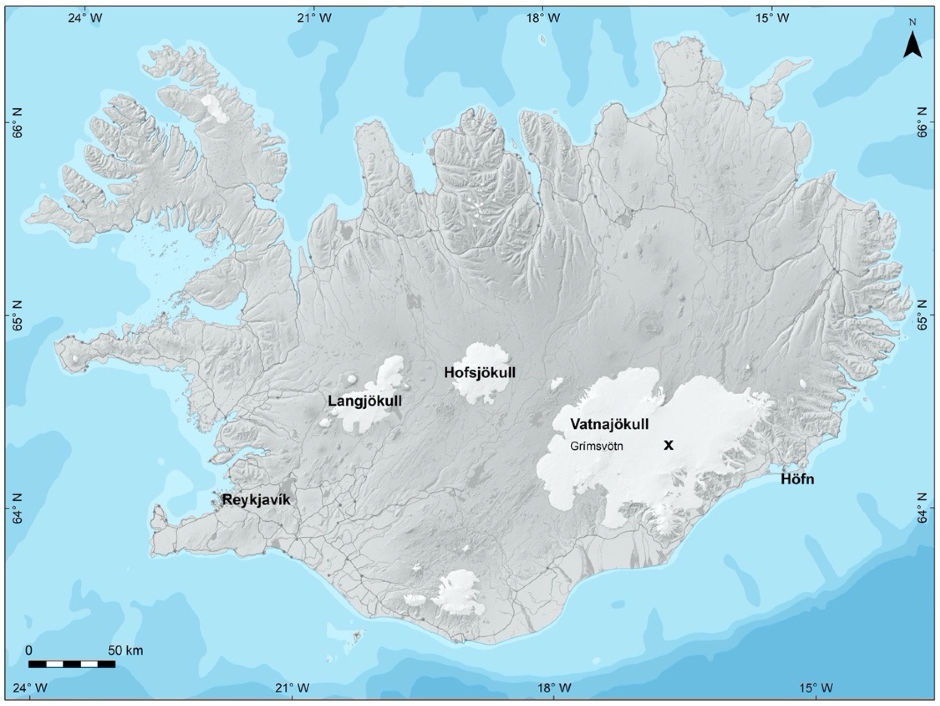
Samples were taken from the central part of the Vatnajökull ice cap in southeast Iceland (see Figure 1). The sampling site coordinates are 64.43415° N and 16.43670° W. Vatnajökull is the largest glacier in Iceland, covering around 8% of the country [35], and by volume, it is the largest glacier in Europe. The study location was chosen in a remote area in a central part of the ice cap. The direct distance to the nearest urban area is around 65 km, a village called Höfn, with a population ~1800 (Figure 1). The shortest distance to sea is around 45 km. Furthermore, the sample location is rarely visited by tourists or other travellers. The elevation of the site of sampling is >1400 m above sea level. Around 30 km west of the location is the active volcano Grímsvötn.
2.1.2. Collection of Samples
The samples were collected during an excursion to Vatnajökull in June 2020. A PICO hand coring drill/auger [36] was used to retrieve two snow cores, each 3 m long. The uppermost 50 cm of snow was removed to avoid contamination from approaching the samples. The sample cores only contained snow, i.e., no glacier ice, as the winter layer of snow is, on average, 5 m at this location [37]. Given that winter snow accumulates usually from October until June, the 3 m long cores represent four to five months of snow accumulation. The ablation season had not started at the time the samples were collected. Each sample was divided into an upper and lower 1.5 m long core. The cores were carefully packed into aluminium foil, which was sealed using electrical tape. The cores were transported in well-isolated and -protected boxes made of polystyrene plastic, filled with snow before the cores were placed in the boxes. The samples were kept frozen until they were melted in the laboratory.
2.2. Methods for Processing and Analysing the Samples
2.2.1. Melting and Sieving of the Samples
Samples that were continuously kept frozen during transport were melted and filled into clean glass containers. The next step of the process was the filtration of the ~5 L of melted samples into three size fractions of >800 µm, 800–180 µm, and 180–50 µm. The sieves used consist of stainless steel mesh (150 mm diameter) and a wooden frame. Special effort was made during the melting and filtration process to avoid any potential laboratory contamination.
2.2.2. Optical Microscopy and Raman Spectroscopy
The detection of microplastics from the sieves was made in two steps. The first step was to distinguish between potential plastic particles and rock fragments, i.e., volcanic ashes, using a standard optical polarising microscope (Leica DMLP). µ-Raman spectroscopy analysis was then used to characterise potential plastic particles. µ-Raman spectroscopy has not been much used in microplastics research in the past, but has recently received increased attention due to its capabilities for identifying particles, films, and fibres as plastic and classifying the type of plastic. It can be used to characterise and quantify microplastics down to 1 µm and to visualise different microplastics simultaneously with minimal interference from water, organic matter, and fluorescence background signals. Therefore, Raman spectroscopy requires minimal preparation of samples, does not destroy the samples, and even allows the analysis of microplastics directly on the filters without first sorting visually [20,38].
Raman measurements were carried out at room temperature using a Horiba (Jobin Yvon) LabRam HR Evolution at the Earth and Environmental Science Department of the University of Gothenburg, Sweden. The samples were exited with air-cooled intra-cavity 785 nm and 532 nm laser diodes utilising an Olympus 50× objective (numerical aperture = 0.9; Table 1). The lateral resolution of the unpolarised confocal laser beam was on the order of 1 μm. Spectra were generated in the range of 100–4000 cm−1 utilising a 600 grooves/cm grating and a thermoelectric cooling electron multiplier CCD, including a front illuminated 1600 × 200 pixel chip. The spectral resolution on the sample was in the order of 1 cm−1. The wavenumber calibration was performed using the 520.7 cm−1 Raman band on a polished silicon wafer with a wavenumber accuracy usually better than 0.5 cm−1. Raman spectra were collected with different laser intensities, acquisition times, and cycles, and partly included a baseline correction of the spectra (see Table 1 for more information).
The measured spectrum of the identified particles was cross-correlated against validated spectra from a reference library database. The reference libraries used in this study were the SLoPP and SLoPP-E Raman spectral libraries for microplastics research. The SLoPP library contains 148 reference spectra from microplastic particles of different types, colours, and morphologies. The SLoPP-E, on the contrary, contains 113 spectra from microplastic particles, which have been identified in environmental samples and have aged in the environment, also of different types, colours, and morphologies [39]. In addition to the specific microplastic spectral libraries, other libraries, such as parts of the Raman–Forensic–HORIBA (RHX) library from Wiley Science Solutions, were used to identify non-plastic microparticles.
The similarity of each prospective match from the reference libraries was quantified by the Hit Quality Index (HQI), ranging between 0 and 1, or scaled between 0 and 100, where the upper level indicates a complete match. The spectrum from the reference database with the highest HQI value was chosen as the most likely material of the identified microplastic particles in the samples from the Vatnajökull glacier.
3. First Results from the Vatnajökull Ice Cap
Microplastic particles were identified in the snow cores from the remote location in the central Vatnajökull ice cap. Several different types of microplastic particles with various morphologies were identified in the samples. Four plastic fragments ranged in equal-area diameter (EAD) from ~30 to 3000 µm, and two plastic fibres were 1300 µm and 3000 µm long (Table 1). The types of plastic materials identified included polyurethane (PU; samples VN01, VN05, and VN06; Table 1), polyvinyl chloride (PVC; sample VN03), polyamide (PA; sample VN04), and acrylonitrile butadiene styrene (ABS; sample VN02). The results, along with the Raman setup, for the analysed particles are summarised in Table 1.
Photomicrographs of the identified microplastic particles, together with the correlation of measured and reference Raman spectra, are shown in Figure 2, Figure 3, Figure 4, Figure 5, Figure 6 and Figure 7. In the charts, the black line represents the signal from the analysed particle and the red line the signal from the reference material. In Figure 2, Figure 3, Figure 4, Figure 5, Figure 6 and Figure 7, we also provide the value of the HQI index for the level of similarity with the known spectra from the reference library databases.
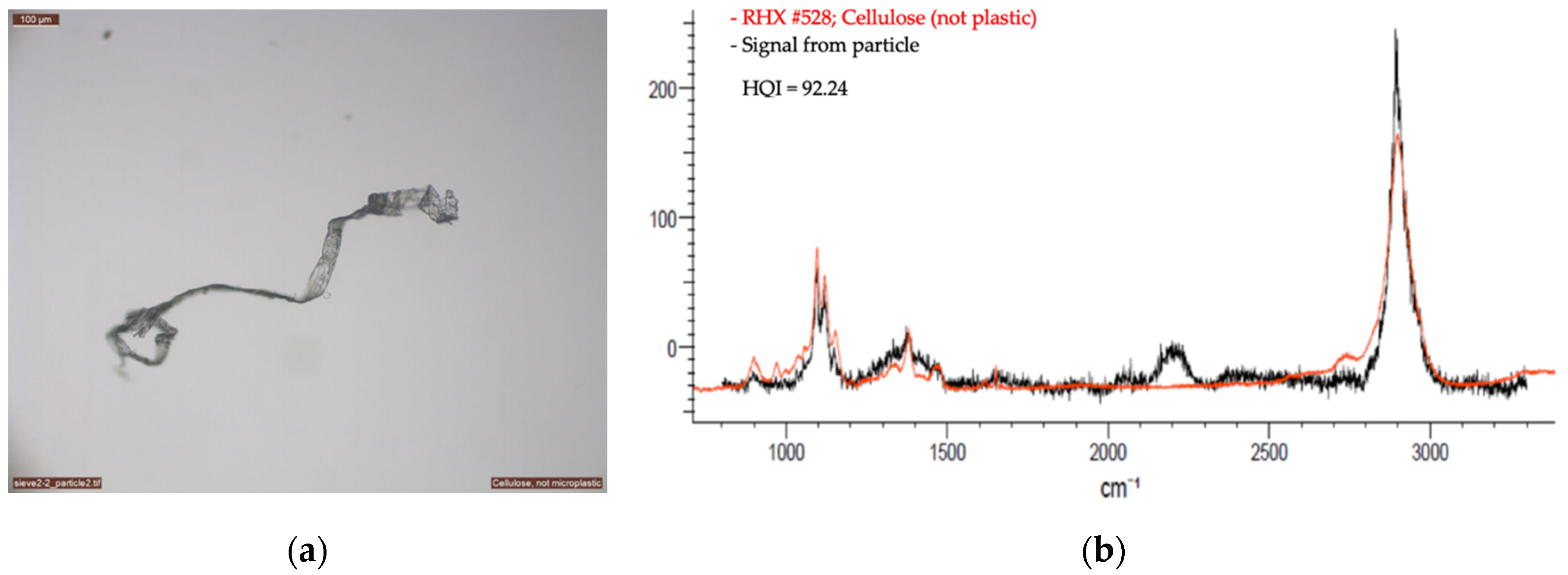
With µ-Raman analysis, it was also possible to distinguish between microplastics and microparticles made of different materials but looking identical under a polarising microscope. One example of such particles is given in Figure 8.
4. Discussion
Despite the lack of research on microplastic contamination, glaciers are of great importance for the overall water-cycle, cover a considerable share of the Earth’s land-surface, and are the largest freshwater reservoirs. Microplastics are a second-phase material inside the glacier. Several studies on other second-phase materials such as minerals, ash/tephra, or other rock fragments have shown that second-phase materials do have an influence on glaciers’ behaviour, such as the change in the light absorbance, structural, and general rheological properties of glaciers [40,41], and hereby, potentially contribute to the melting of glaciers, which is the largest source of rising sea levels [34]. To which extent and at which concentration microplastics increase these effects is not known yet, but microplastics will likely act similar to other second-phase materials.
The particles identified in our study were similar in morphology and size and were made of similar materials as reported in the other published literature on microplastic findings in glaciers [31,32,33]. Unpublished data from the Hofsjökull ice cap in Iceland further indicate the wide-spread presence of microplastic particles in glaciers in Iceland [42].
The location of the sampling site on Vatnajökull is remote, hence direct contamination is unlikely. Due to their small sizes, the most likely pathway for the microplastic particles to the sampling site is through atmospheric transport [20]. The morphology of the identified particles, fragments, and fibres further supports the hypothesis of atmospheric transport [20]. Atmospheric transport of microplastics has not yet been extensively studied and is not fully understood [20,21,43]. Further research is needed on the transport pathways from sources to sinks and how they are affected by meteorological and geographical conditions, and also by the density and types of particles. In the case of the Vatnajökull ice cap, the pathways for the microplastics may either be wind or precipitation. The microplastic particles may either be directly released from urban or industrial areas or indirectly from the sea, lakes, or other natural systems where they have accumulated [44]. The potential sources of microplastics in the case of the Vatnajökull ice cap include small villages, the closest one being Höfn, located 65 km from the sampling site, and larger towns, including Reykjavík, which is likely the largest plastic polluter in Iceland, located 270 km west of the sampling site (see Figure 1). The direct distance to the sea is 45 km, which is also a potential source [45]. Furthermore, the microplastic particles may also be carried over a long distance with trade winds from sources far away, such as in Northern America and Europe. An air mass trajectory analysis can be used to study the atmospheric transport of microplastics [30,46]. Tools such as the HYSPLIT model [47] have been successfully used for estimating the diffusion of various air pollutants and for back trajectory analysis to establish source–sink relationships for known events. In the case of atmospheric transport of microplastics, there are, however, still many unknowns as previously described, and therefore great uncertainty regarding many of the required modelling parameters [20]. Identifying microplastic particles in snow and ice on remote pristine glaciers, such as the Vatnajökull ice cap, can help understand the airborne distribution of microplastics and will be studied further.
Even though an effort was made to avoid contamination during sampling and analysis of the samples, it is still difficult to fully exclude the possibility of contamination of the analysed samples. Three main sources of potential contamination are identified as (i) the coring equipment, which is partially made of fibreglass (glass-reinforced plastic (GRP)), (ii) the black plastic tape, which is composed of polyvinyl chloride (PVC) backing and a non-corrosive rubber-based adhesive, and (iii) the boxes made of polystyrene plastic. Samples were taken from these potential sources of plastic contamination and used as a reference for the analysis of particles. The Raman spectroscopy procedure we employed for analysing the samples was very effective in identifying microplastic particles. Nevertheless, we experienced some difficulties, mostly since the microplastic particles are often weathered, and therefore the Raman signal can be different from unweathered plastic. Other reasons for the relative differences between the samples and the reference standards could be caused by differences in colour or the presence of degradation products or contaminations. However, it was useful to have both the SLoPP and the SLoPPE microplastic particle spectral libraries, as the latter contains weathered samples from natural environments. Furthermore, the colour of microplastics often dominates the Raman spectrum, which can make the identification more difficult. For example, this was a problem when the particles shown in Figure 6 and Figure 7 were analysed and could partly explain the low HQI values obtained. Finally, the microplastic particles can be easily mixed up with cotton or cellulose, which are the most common sources of possible contamination during sampling. Figure 8a is an example of a cellulose fibre that is visually similar to a plastic particle. It is therefore of great importance for spectroscopic analysis of microplastic particles to have extensive libraries of known reference materials.
The preliminary investigation performed in this work only collected and analysed a single set of snow cores from the Vatnajökull ice cap. It is of great interest to collect samples from other locations on the ice cap. Comparing the potential microplastic content in such samples will help correlate the microplastic quantities with atmospheric and geographical conditions and hopefully contribute to an improved understanding of the atmospheric transport of plastic particles.
5. Conclusions
Microplastic pollution is an increasing threat to the biosphere, including the food chain and human health. Furthermore, microplastic particles, as a second-phase material in the ice, may contribute to the effect such particles have on the melting and rheological behaviour of glaciers, and thus play a crucial role for future meltwater contribution to the oceans. Plastic particles degrade slowly in the cold glacier environment and can persist in glaciers for a long time, from which they are eventually released, which can lead to pollution in rivers and marine environments. Hence, it is important to map and understand the presence and dispersal of microplastics on a global scale. In this work, we identified microplastic particles in snow cores collected in a remote and pristine location on the Vatnajökull ice cap in Iceland. Utilising optical microscopy and µ-Raman spectroscopy, we visualised and identified microplastic particles of various sizes and materials. Our findings support that atmospheric transport of microplastic particles is one of the important pathways for microplastic pollution. However, further research is needed on the transport pathways and how they are affected by the conditions and types of particles.
This study demonstrated the presence of microplastic particles on the Vatnajökull ice cap, but a systematic sampling program is needed to better quantify the microplastic contamination in this remote area of the North Atlantic. In the long-term, such work will contribute to the currently limited existing knowledge on microplastic contamination of glaciers and the potential changes such contamination can cause, and will improve the understanding of the atmospheric transport of microplastics.
Author Contributions
Conceptualisation, H.S., M.P., M.K.-S., H.H., E.J.Á., and E.S.; methodology, M.P. and M.K.-S.; validation, H.S., M.P., M.K.-S., H.H., E.J.Á., and E.S.; formal analysis, M.P. and M.K.-S.; investigation, H.S., M.P., M.K.-S., H.H., E.J.Á., and E.S.; writing—original draft preparation, H.S.; writing—review and editing, H.S., M.P., M.K.-S., H.H., E.J.Á., and E.S.; visualisation, H.S., M.P., M.K.-S., and H.H. All authors have read and agreed to the published version of the manuscript.
Funding
This research received no external funding.
Institutional Review Board Statement
Not applicable.
Informed Consent Statement
Not applicable.
Data Availability Statement
The data presented in this study are available on request from the corresponding author.
Acknowledgments
The authors acknowledge the field work assistance of Vilhjálmur Kjartansson during the Vatnajökull expedition in June.
Conflicts of Interest
The authors declare no conflict of interest.
References
- Ritchie, H.; Roser, M. Plastic Pollution. In Our World Data; Oxford Martin School: Oxford, UK, 2018. [Google Scholar]
- Hartmann, N.B.; Hüffer, T.; Thompson, R.C.; Hassellöv, M.; Verschoor, A.; Daugaard, A.E.; Rist, S.; Karlsson, T.; Brennholt, N.; Cole, M. Are We Speaking the Same Language? Recommendations for a Definition and Categorization Framework for Plastic Debris; ACS Publications: Washington, DC, USA, 2019; ISBN 0013-936X. [Google Scholar]
- Wang, Y.-L.; Lee, Y.-H.; Chiu, I.-J.; Lin, Y.-F.; Chiu, H.-W. Potent Impact of Plastic Nanomaterials and Micromaterials on the Food Chain and Human Health. Int. J. Mol. Sci. 2020, 21, 1727. [Google Scholar] [CrossRef] [Green Version]
- Kershaw, P.; Rochman, C. Sources, Fate and Effects of Microplastics in the Marine Environment: Part 2 of a Global Assessment; GESAMP: London, UK, 2015. [Google Scholar]
- Dris, R.; Gasperi, J.; Mirande, C.; Mandin, C.; Guerrouache, M.; Langlois, V.; Tassin, B. A First Overview of Textile Fibers, Including Microplastics, in Indoor and Outdoor Environments. Environ. Pollut. 2017, 221, 453–458. [Google Scholar] [CrossRef] [PubMed] [Green Version]
- Gasperi, J.; Wright, S.L.; Dris, R.; Collard, F.; Mandin, C.; Guerrouache, M.; Langlois, V.; Kelly, F.J.; Tassin, B. Microplastics in Air: Are We Breathing It in? Curr. Opin. Environ. Sci. Health 2018, 1, 1–5. [Google Scholar] [CrossRef] [Green Version]
- Lehner, R.; Weder, C.; Petri-Fink, A.; Rothen-Rutishauser, B. Emergence of Nanoplastic in the Environment and Possible Impact on Human Health. Environ. Sci. Technol. 2019, 53, 1748–1765. [Google Scholar] [CrossRef] [PubMed]
- Prüst, M.; Meijer, J.; Westerink, R.H. The Plastic Brain: Neurotoxicity of Micro-and Nanoplastics. Part. Fibre Toxicol. 2020, 17, 1–16. [Google Scholar] [CrossRef]
- Van Raamsdonk, L.W.; van der Zande, M.; Koelmans, A.A.; Hoogenboom, R.L.; Peters, R.J.; Groot, M.J.; Peijnenburg, A.A.; Weesepoel, Y.J. Current Insights into Monitoring, Bioaccumulation, and Potential Health Effects of Microplastics Present in the Food Chain. Foods 2020, 9, 72. [Google Scholar] [CrossRef] [PubMed] [Green Version]
- Prata, J.C.; da Costa, J.P.; Lopes, I.; Duarte, A.C.; Rocha-Santos, T. Environmental Exposure to Microplastics: An Overview on Possible Human Health Effects. Sci. Total Environ. 2020, 702, 134455. [Google Scholar] [CrossRef] [PubMed]
- Andrady, A.L. Microplastics in the Marine Environment. Mar. Pollut. Bull. 2011, 62, 1596–1605. [Google Scholar] [CrossRef]
- Law, K.L.; Thompson, R.C. Microplastics in the Seas. Science 2014, 345, 144–145. [Google Scholar] [CrossRef]
- Wang, C.; Zhao, J.; Xing, B. Environmental Source, Fate, and Toxicity of Microplastics. J. Hazard. Mater. 2020, 2020, 124357. [Google Scholar] [CrossRef]
- Rochman, C.M. Microplastics Research—From Sink to Source. Science 2018, 360, 28–29. [Google Scholar] [CrossRef]
- Novotna, K.; Cermakova, L.; Pivokonska, L.; Cajthaml, T.; Pivokonsky, M. Microplastics in Drinking Water Treatment—Current Knowledge and Research Needs. Sci. Total Environ. 2019, 667, 730–740. [Google Scholar] [CrossRef]
- Setälä, O.; Fleming-Lehtinen, V.; Lehtiniemi, M. Ingestion and Transfer of Microplastics in the Planktonic Food Web. Environ. Pollut. 2014, 185, 77–83. [Google Scholar] [CrossRef]
- Schwabl, P.; Köppel, S.; Königshofer, P.; Bucsics, T.; Trauner, M.; Reiberger, T.; Liebmann, B. Detection of Various Microplastics in Human Stool. Ann. Intern. Med. 2019, 171, 453–457. [Google Scholar] [CrossRef]
- Evangeliou, N.; Grythe, H.; Klimont, Z.; Heyes, C.; Eckhardt, S.; Lopez-Aparicio, S.; Stohl, A. Atmospheric Transport Is a Major Pathway of Microplastics to Remote Regions. Nat. Commun. 2020, 11, 3381. [Google Scholar] [CrossRef]
- Zhang, Y.; Gao, T.; Kang, S.; Sillanpää, M. Importance of Atmospheric Transport for Microplastics Deposited in Remote Areas. Environ. Pollut. 2019, 254, 112953. [Google Scholar] [CrossRef]
- Zhang, Y.; Kang, S.; Allen, S.; Allen, D.; Gao, T.; Sillanpää, M. Atmospheric Microplastics: A Review on Current Status and Perspectives. Earth Sci. Rev. 2020, 203, 103118. [Google Scholar] [CrossRef]
- Allen, S.; Allen, D.; Phoenix, V.R.; Le Roux, G.; Durántez Jiménez, P.; Simonneau, A.; Binet, S.; Galop, D. Atmospheric Transport and Deposition of Microplastics in a Remote Mountain Catchment. Nat. Geosci. 2019, 12, 339–344. [Google Scholar] [CrossRef]
- Cózar, A.; Martí, E.; Duarte, C.M.; García-de-Lomas, J.; Van Sebille, E.; Ballatore, T.J.; Eguíluz, V.M.; González-Gordillo, J.I.; Pedrotti, M.L.; Echevarría, F. The Arctic Ocean as a Dead End for Floating Plastics in the North Atlantic Branch of the Thermohaline Circulation. Sci. Adv. 2017, 3, e1600582. [Google Scholar] [CrossRef] [PubMed] [Green Version]
- La Daana, K.K.; Gårdfeldt, K.; Lyashevska, O.; Hassellöv, M.; Thompson, R.C.; O’Connor, I. Microplastics in Sub-Surface Waters of the Arctic Central Basin. Mar. Pollut. Bull. 2018, 130, 8–18. [Google Scholar]
- La Daana, K.K.; Gardfeldt, K.; Krumpen, T.; Thompson, R.C.; O’Connor, I. Microplastics in Sea Ice and Seawater beneath Ice Floes from the Arctic Ocean. Sci. Rep. 2020, 10, 1–11. [Google Scholar] [CrossRef] [Green Version]
- Lusher, A.L.; Tirelli, V.; O’Connor, I.; Officer, R. Microplastics in Arctic Polar Waters: The First Reported Values of Particles in Surface and Sub-Surface Samples. Sci. Rep. 2015, 5, 14947. [Google Scholar] [CrossRef] [PubMed] [Green Version]
- Munari, C.; Infantini, V.; Scoponi, M.; Rastelli, E.; Corinaldesi, C.; Mistri, M. Microplastics in the Sediments of Terra Nova Bay (Ross Sea, Antarctica). Mar. Pollut. Bull. 2017, 122, 161–165. [Google Scholar] [CrossRef] [PubMed]
- Obbard, R.W.; Sadri, S.; Wong, Y.Q.; Khitun, A.A.; Baker, I.; Thompson, R.C. Global Warming Releases Microplastic Legacy Frozen in Arctic Sea Ice. Earths Future 2014, 2, 315–320. [Google Scholar] [CrossRef]
- Tirelli, V.; Suaria, G.; Lusher, A.L. Microplastics in Polar Samples. In Handbook of Microplastics in the Environment; Springer: Cham, Switzerland, 2020; pp. 1–42. [Google Scholar]
- Cuffey, K.M.; Paterson, W.S.B. The Physics of Glaciers; Academic Press: Cambridge, MA, USA, 2010; ISBN 0-08-091912-X. [Google Scholar]
- Allen, D.; Allen, S.; Sonke, J.; Phoenix, V. Atmospheric Transport and Our Planetary Boundary Layer as an Environmental Compartment of Microplastics Pollution. In Proceedings of the International Conference on Microplastic Pollution Inthe Mediterranean Sea. Springer Water; Springer: Cham, Switzerland, 2019; pp. 1–8. [Google Scholar]
- Ambrosini, R.; Azzoni, R.S.; Pittino, F.; Diolaiuti, G.; Franzetti, A.; Parolini, M. First Evidence of Microplastic Contamination in the Supraglacial Debris of an Alpine Glacier. Environ. Pollut. 2019, 253, 297–301. [Google Scholar] [CrossRef] [PubMed]
- Bergmann, M.; Mützel, S.; Primpke, S.; Tekman, M.B.; Trachsel, J.; Gerdts, G. White and Wonderful? Microplastics Prevail in Snow from the Alps to the Arctic. Sci. Adv. 2019, 5, eaax1157. [Google Scholar] [CrossRef] [Green Version]
- Cabrera, M.; Valencia, B.G.; Lucas-Solis, O.; Calero, J.L.; Maisincho, L.; Conicelli, B.; Moulatlet, G.M.; Capparelli, M.V. A New Method for Microplastic Sampling and Isolation in Mountain Glaciers: A Case Study of One Antisana Glacier, Ecuadorian Andes. Case Stud. Chem. Environ. Eng. 2020, 2, 100051. [Google Scholar] [CrossRef]
- Chen, X.; Zhang, X.; Church, J.A.; Watson, C.S.; King, M.A.; Monselesan, D.; Legresy, B.; Harig, C. The Increasing Rate of Global Mean Sea-Level Rise during 1993–2014. Nat. Clim. Chang. 2017, 7, 492–495. [Google Scholar] [CrossRef]
- Aðalgeirsdóttir, G.; Pálsson, F.; Thorsteinsson, T.; Magnússon, E.; Belart, J.; Jóhannesson, T.; Hannesdóttir, H.; Sigurðsson, O.; Gunnarsson, A.; Einarsson, B. Glacier Changes in Iceland from ~1890 to 2019. Front. Earth Sci. 2020, 8, 520. [Google Scholar] [CrossRef]
- Talalay, P.G. (Ed.) Hand- and Power-Driven Portable Drills. In Mechanical Ice Drilling Technology; Springer Geophysics; Springer: Singapore, 2016; pp. 27–52. ISBN 978-981-10-0560-2. [Google Scholar]
- Pálsson, F.; Gunnarsson, A.; Skagfjörð Pálsson, H.; Steinþórsson, S. Vatnajökull: Mass Balance, Meltwater Drainage and Surface Velocity of the Glacial Year 2019–20; Landsvirkjun: Reykjavik, Iceland, 2021. [Google Scholar]
- Sobhani, Z.; Zhang, X.; Gibson, C.; Naidu, R.; Mallavarapu, M.; Fang, C. Identification and Visualisation of Microplastics/Nanoplastics by Raman Imaging (i): Down to 100 Nm. Water Res. 2020, 174, 115658. [Google Scholar] [CrossRef]
- Munno, K.; De Frond, H.; O’Donnell, B.; Rochman, C.M. Increasing the Accessibility for Characterizing Microplastics: Introducing New Application-Based and Spectral Libraries of Plastic Particles (SLoPP and SLoPP-E). Anal. Chem. 2020, 92, 2443–2451. [Google Scholar] [CrossRef]
- Peternell, M.; Wilson, C.J. Effect of Strain Rate Cycling on Microstructures and Crystallographic Preferred Orientation during High-Temperature Creep. Geology 2016, 44, 279–282. [Google Scholar] [CrossRef]
- Wilson, C.J.; Hunter, N.J.; Luzin, V.; Peternell, M.; Piazolo, S. The Influence of Strain Rate and Presence of Dispersed Second Phases on the Deformation Behaviour of Polycrystalline D2O Ice. J. Glaciol. 2019, 65, 101–122. [Google Scholar] [CrossRef] [Green Version]
- Gomiero, A.; Ásmundsdóttir, Á.M.; Þorsteinsson, Þ.; Oysæd, K.B. Preliminary Occurrence of Microplastics in a Remote Icelandic Glacier. 2021. Available online: https://pame.is/projects/arctic-marine-pollution (accessed on 3 March 2021).
- Dris, R.; Gasperi, J.; Saad, M.; Mirande, C.; Tassin, B. Synthetic Fibers in Atmospheric Fallout: A Source of Microplastics in the Environment? Mar. Pollut. Bull. 2016, 104, 290–293. [Google Scholar] [CrossRef]
- Bianco, A.; Passananti, M. Atmospheric Micro and Nanoplastics: An Enormous Microscopic Problem. Sustainability 2020, 12, 7327. [Google Scholar] [CrossRef]
- Allen, S.; Allen, D.; Moss, K.; Le Roux, G.; Phoenix, V.R.; Sonke, J.E. Examination of the Ocean as a Source for Atmospheric Microplastics. PLoS ONE 2020, 15, e0232746. [Google Scholar] [CrossRef]
- Liu, K.; Wu, T.; Wang, X.; Song, Z.; Zong, C.; Wei, N.; Li, D. Consistent Transport of Terrestrial Microplastics to the Ocean through Atmosphere. Environ. Sci. Technol. 2019, 53, 10612–10619. [Google Scholar] [CrossRef]
- Draxler, R.; Rolph, G. HYSPLIT (HYbrid Single-Particle Lagrangian Integrated Trajectory) Model Access via NOAA ARL READY Website; NOAA Air Resources Laboratory: Silver Spring, MD, USA, 2010.
Figure 1.
The remote sampling location on the Vatnajökull ice cap in Iceland is marked with an “X”. The coordinates of the location are 64.43415° N and 16.43670° W.
Figure 1.
The remote sampling location on the Vatnajökull ice cap in Iceland is marked with an “X”. The coordinates of the location are 64.43415° N and 16.43670° W.

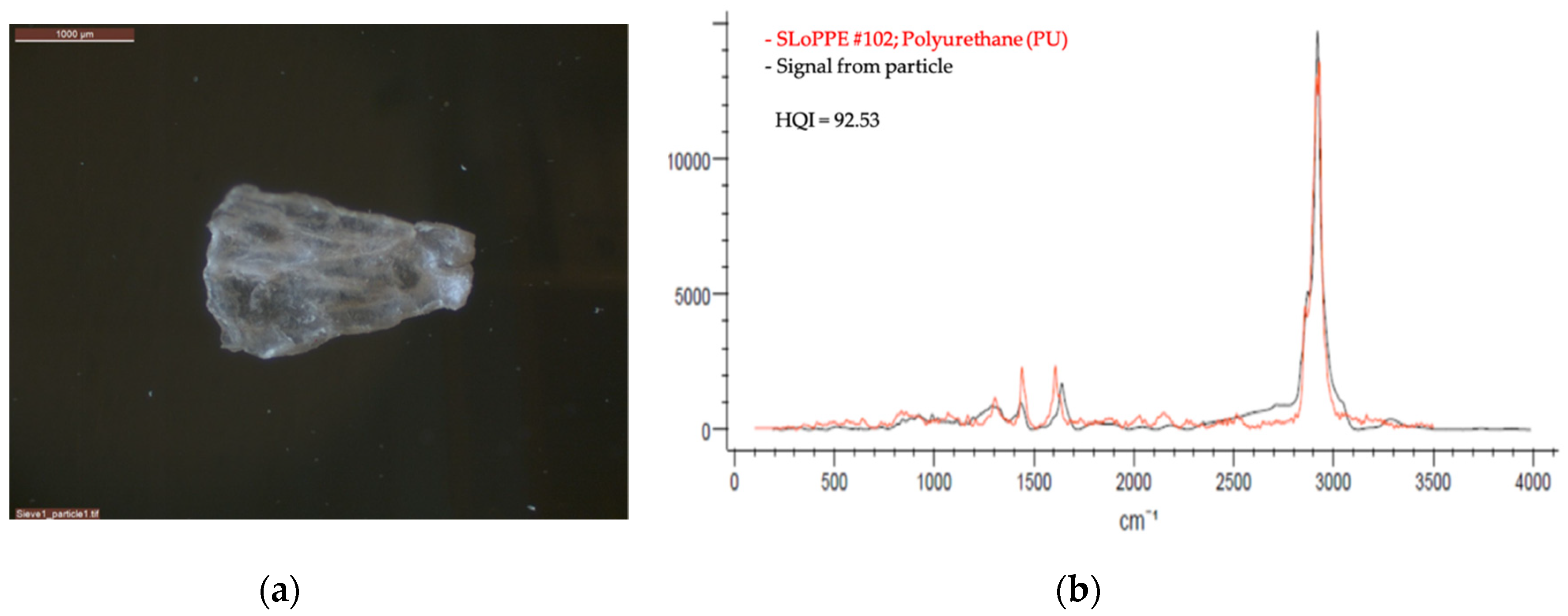
Figure 2.
Sample VN01: (a) The figure shows an example of a clear polyurethane (PU) microplastic fragment. The equal-area diameter (EAD) of the fragment was ~3000 µm. (b) The chart shows the spectrum of the identified fragment as a black line, and the spectrum of the reference material as a red line. The Hit Quality Index (HQI) value of the best match was 92.53, which was obtained when compared to particle #102, polyurethane white fibre, from the SLoPPE reference library.

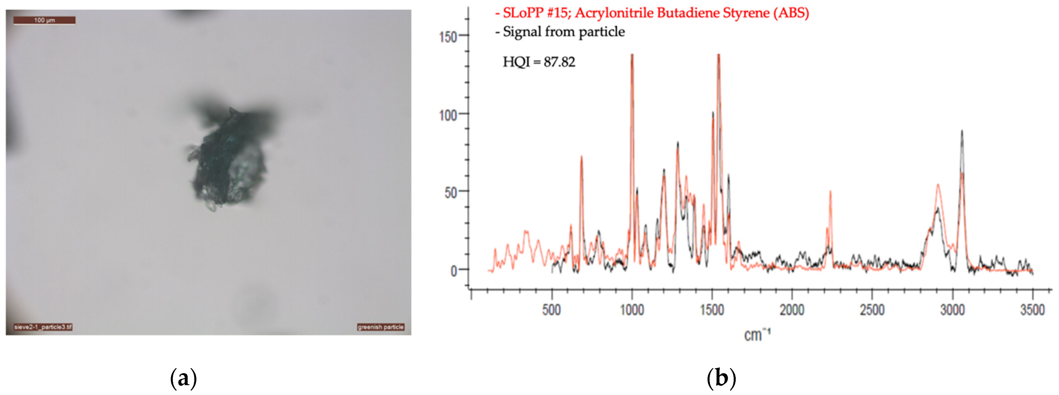
Figure 3.
Sample VN02: (a) The figure shows an example of a greenish acrylonitrile butadiene styrene (ABS) microplastic fragment. The EAD of the fragment was ~230 µm. (b) The chart shows the spectrum of the identified fragment as a black line, and the spectrum of the reference material as a red line. The HQI value of the best match was 87.82, which was obtained when compared to particle #15, acrylonitrile butadiene styrene, “6. dark green LEGO,” from the SLoPP reference library.

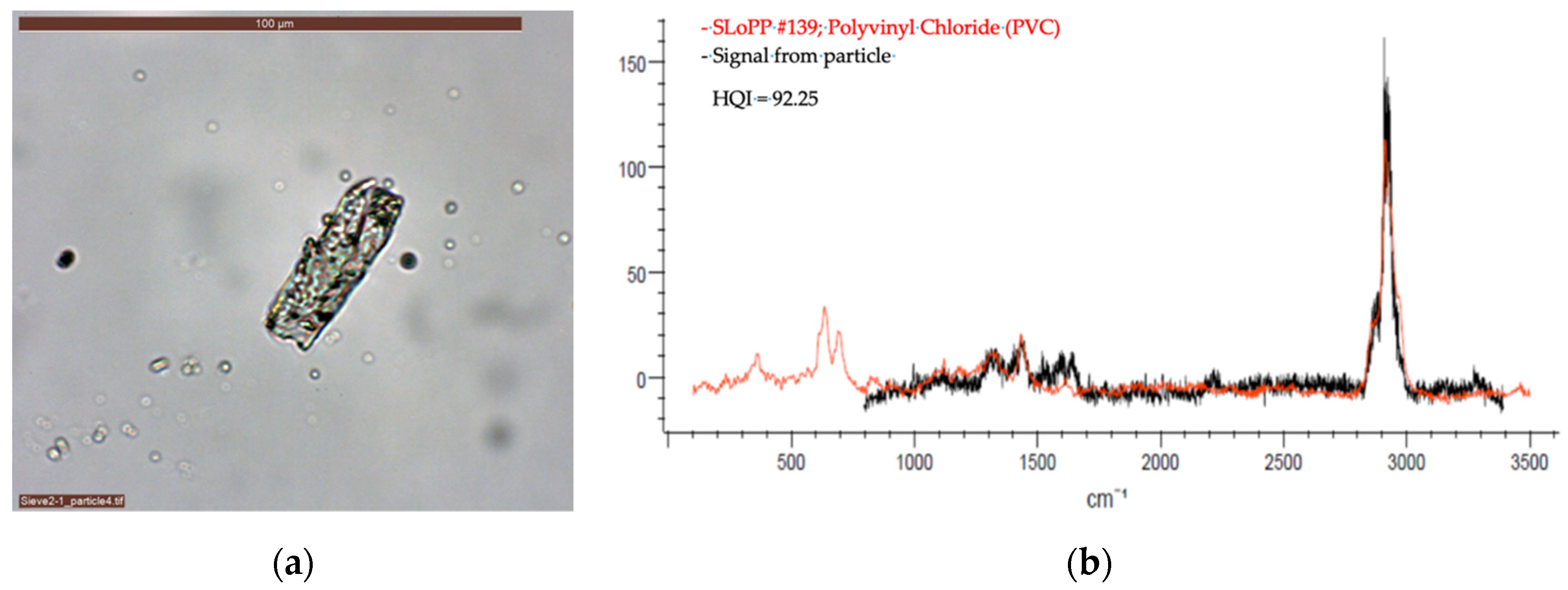
Figure 4.
Sample VN03: (a) The figure shows an example of a clear polyvinyl chloride (PVC) microplastic fragment. The EAD of the fragment was ~40 µm. (b) The chart shows the spectrum of the identified fragment as a black line, and the spectrum of the reference material as a red line. The HQI value of the best match was 92.25, which was obtained when compared to particle #139, polyvinyl chloride, “5. clear cling wrap,” from the SLoPP reference library.

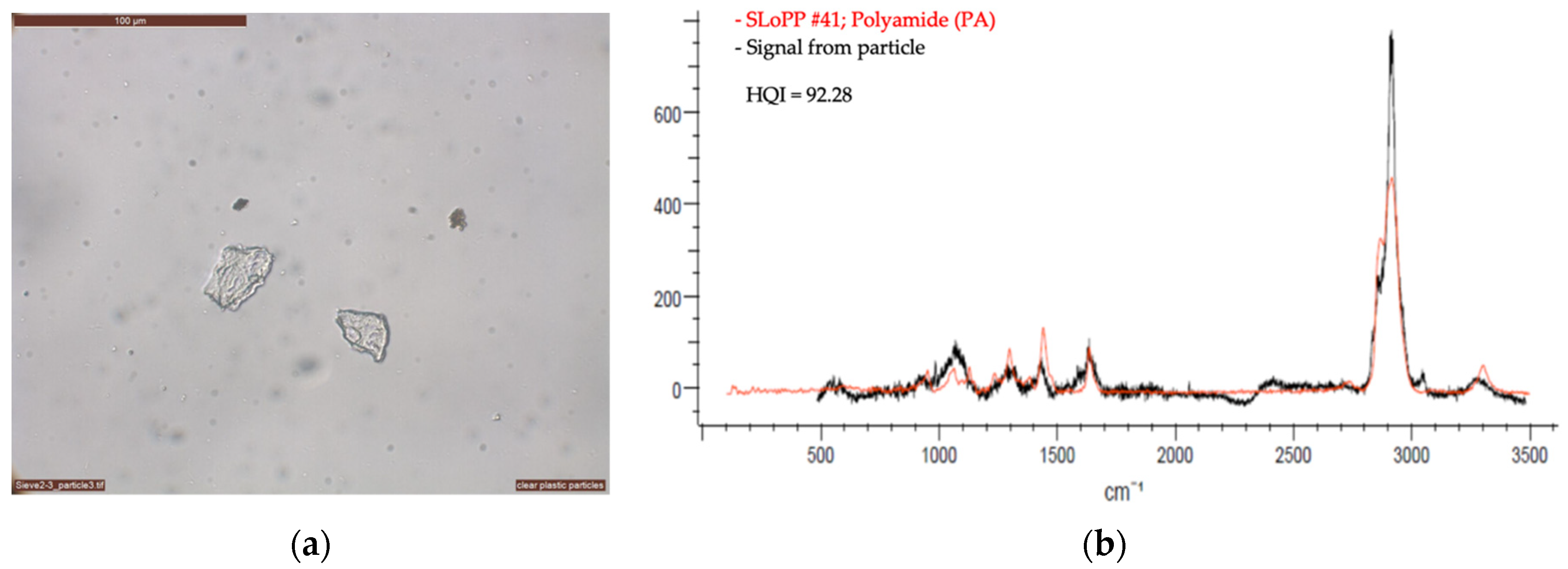
Figure 5.
Sample VN04: (a) The figure shows an example of clear polyamide (PA) microplastic fragments. The EAD of the fragments was ~30 µm. (b) The chart shows the spectrum of the identified fragment as a black line, and the spectrum of the reference material as a red line. The HQI value of the best match was 92.88, which was obtained when compared to particle #41, polyamide, “1. white cable tie,” from the SLoPP reference library.

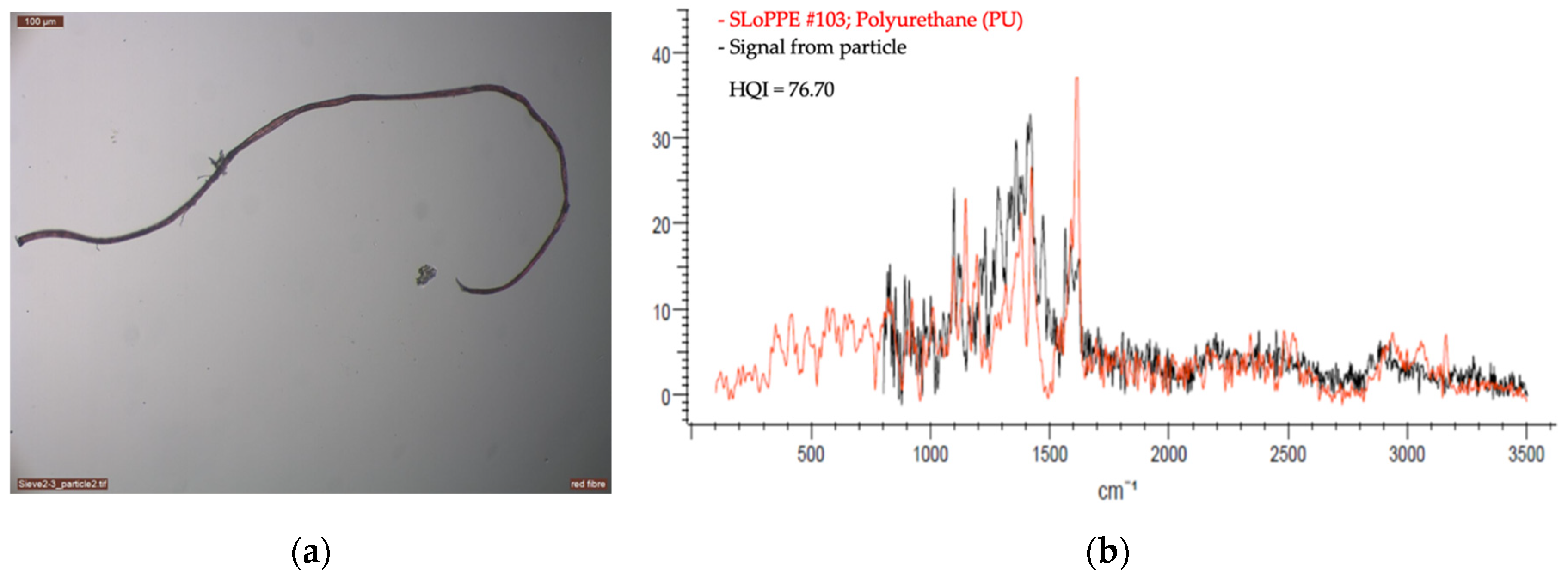
Figure 6.
Sample VN05: (a) The figure shows an example of a red microplastic fibre, likely made of polyurethane (PU). The length of the fibre was ~3000 µm. (b) The chart shows the spectrum of the identified fragment as a black line, and the spectrum of the reference material as a red line. The HQI value of the best match was only 76.70, which was obtained when compared to particle #103, polyurethane, “4. pink fragment,” from the SLoPPE reference library.

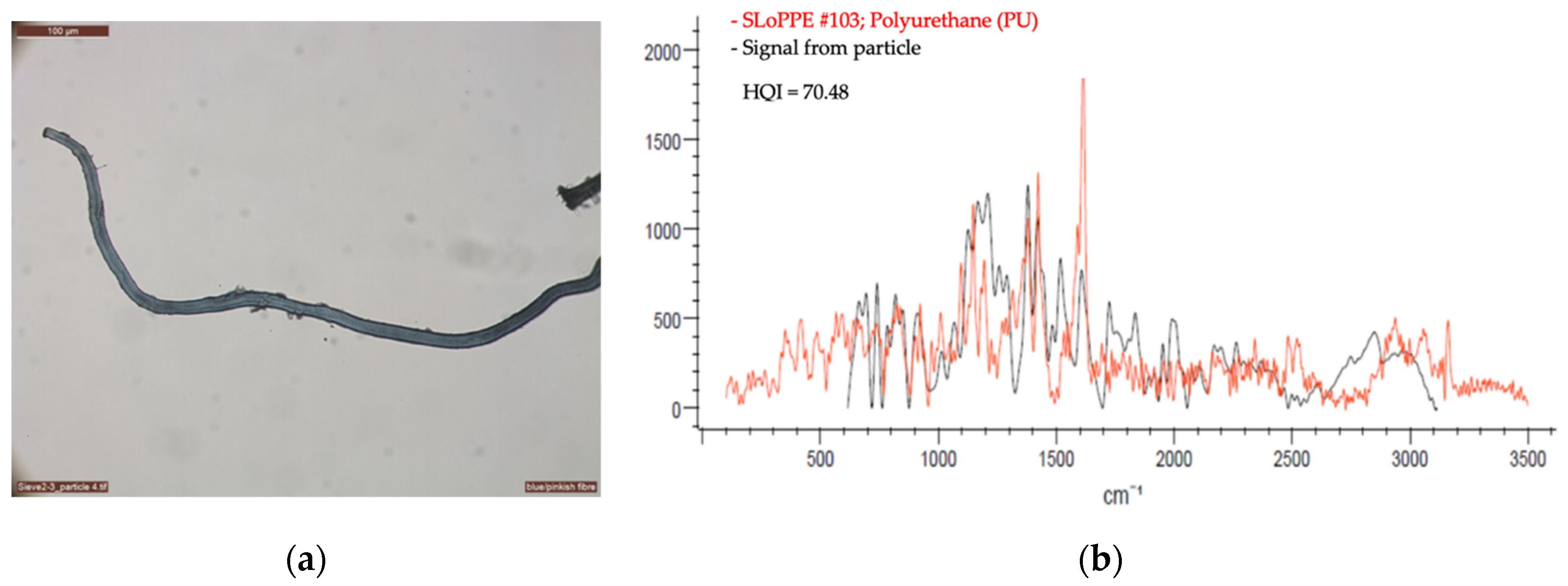
Figure 7.
Sample VN06: (a) The figure shows an example of a blue/pinkish microplastic fibre, likely made of polyurethane (PU). The length of the fibre was ~1300 µm. (b) The chart shows the spectrum of the identified fragment as a black line, and the spectrum of the reference material as a red line. The HQI value of the best match was only 70.48, which was obtained when compared to particle #103, polyurethane, “4. Pink fragment,” from the SLoPPE reference library.

Figure 8.
Sample VN07: (a) The figure shows a microparticle that is made of cellulose, i.e., not made of plastic. The length of the fibre was ~2400 µm. (b) The chart shows the spectrum of the identified fragment as a black line, and the spectrum of the reference material as a red line. The HQI value of the best match was 92.24, which was obtained when compared to particle #528, cellulose, from the Raman–Forensic–HORIBA (RHX) reference library.


Table 1.
Particle characteristics and Raman setup for the analysed particles.
| Sample | Material | Equal Area Diameter, Particle Size (µm) | Laser (nm) | Laser Intensity (%) | Acquisition | Baseline Correction | Number of Measurement Per Sample | |
|---|---|---|---|---|---|---|---|---|
| Time | Cycles | |||||||
| VN01 | Polyurethane | 3000 | 785 | 10 | 120 | 10 | Yes | 5 |
| VN02 | Acrylonitrile butadiene styrene | 230 | 785 | 5 | 30 | 10 | Yes | 3 |
| VN03 | Polyvinyl chloride | 40 | 785 | 5 | 60 | 5 | Yes | 2 |
| VN04 | Clear polyamide | 30 | 785 | 10 | 60 | 3 | No | 3 |
| VN05 | Polyurethane fibre | 3000 | 532 | 100 | 60 | 3 | Yes | 5 |
| VN06 | Polyurethane fibre | 1300 | 532 | 100 | 240 | 3 | No | 4 |
| VN07 | Cellulose | 2400 | 785 | 5 | 30 | 5 | Yes | 2 |
Publisher’s Note: MDPI stays neutral with regard to jurisdictional claims in published maps and institutional affiliations. |
© 2021 by the authors. Licensee MDPI, Basel, Switzerland. This article is an open access article distributed under the terms and conditions of the Creative Commons Attribution (CC BY) license (https://creativecommons.org/licenses/by/4.0/).
Share and Cite
MDPI and ACS Style
Stefánsson, H.; Peternell, M.; Konrad-Schmolke, M.; Hannesdóttir, H.; Ásbjörnsson, E.J.; Sturkell, E. Microplastics in Glaciers: First Results from the Vatnajökull Ice Cap. Sustainability 2021, 13, 4183. https://doi.org/10.3390/su13084183
AMA Style
Stefánsson H, Peternell M, Konrad-Schmolke M, Hannesdóttir H, Ásbjörnsson EJ, Sturkell E. Microplastics in Glaciers: First Results from the Vatnajökull Ice Cap. Sustainability. 2021; 13(8):4183. https://doi.org/10.3390/su13084183
Chicago/Turabian StyleStefánsson, Hlynur, Mark Peternell, Matthias Konrad-Schmolke, Hrafnhildur Hannesdóttir, Einar Jón Ásbjörnsson, and Erik Sturkell. 2021. "Microplastics in Glaciers: First Results from the Vatnajökull Ice Cap" Sustainability 13, no. 8: 4183. https://doi.org/10.3390/su13084183
Note that from the first issue of 2016, this journal uses article numbers instead of page numbers. See further details here.