Article

Isolation and Characterization of a Double Stranded DNA Megavirus Infecting the Toxin-Producing Haptophyte Prymnesium parvum

1
Department of Biological Chemistry, John Innes Centre, Norwich Research Park, Norwich NR4 7UH, UK
2
Marine Biological Association of the UK, Plymouth PL1 2PB, UK
3
Centre for Ocean and Atmospheric Studies, School of Environmental Sciences, University of East Anglia, Norwich Research Park, Norwich NR4 7TJ, UK
*
Author to whom correspondence should be addressed.
Viruses 2017, 9(3), 40; https://doi.org/10.3390/v9030040
Submission received: 29 January 2017 / Revised: 22 February 2017 / Accepted: 27 February 2017 / Published: 9 March 2017
(This article belongs to the Special Issue Marine Viruses 2016)

Abstract

:
Prymnesium parvum is a toxin-producing haptophyte that causes harmful algal blooms globally, leading to large-scale fish kills that have severe ecological and economic implications. For the model haptophyte, Emiliania huxleyi, it has been shown that large dsDNA viruses play an important role in regulating blooms and therefore biogeochemical cycling, but much less work has been done looking at viruses that infect P. parvum, or the role that these viruses may play in regulating harmful algal blooms. In this study, we report the isolation and characterization of a lytic nucleo-cytoplasmic large DNA virus (NCLDV) collected from the site of a harmful P. parvum bloom. In subsequent experiments, this virus was shown to infect cultures of Prymnesium sp. and showed phylogenetic similarity to the extended Megaviridae family of algal viruses.

1. Introduction

The last two decades have seen a boom in the study of marine viruses and the role that they play in regulating both bacterial and unicellular eukaryote bloom dynamics [1,2]. Although phages and the bacteria that they infect have been studied for many years, the more recently discovered Acanthamoeba polyphaga mimivirus (APMV) and its Megaviridae relatives have brought about a new age in photosynthetic protist virology. It has recently been shown that dsDNA viruses infecting algae do not form monophyletic lineages [3], with divergence occurring even within the host division. A good example of this evolutionary divergence can be found in viruses that infect the coccolithophore Emiliania huxleyi (EhV) [4,5] and the prymnesiophyte Phaeocystis globosa (PgV) [6], which along with other algal viruses have been proposed to form an extended branch of the Megaviridae [7]. It is widely accepted that these viruses not only play a crucial role in ecosystem dynamics [8,9], but also contribute significantly to biogeochemical cycles [10,11]. A lesser studied impact, however, lies in the role that such viruses may play in the termination of toxic eukaryotic algal blooms. Lytic viruses that infect the toxic raphidophyte Heterosigma akashiwo have been extensively studied [12,13,14,15,16,17,18,19] but, because of the elusive nature of H. akashwio toxicity to fish, none of these studies sought to investigate the role of viral infection on levels of algal toxicity.
The toxin-producing haptophyte Prymnesium parvum forms dense blooms in marine, brackish and inland waters, devastating fish populations through the release of natural product toxins [20,21]. The haptophytes are a diverse division of microalgae that include the bloom-forming Emiliania huxleyi and Phaeocystis globosa, both of which play crucial roles in oceanic carbon and sulfur cycles [22,23]. Virus infection of these organisms has been studied in some detail, with the genome of the dsDNA Phaeocystis globosa virus (PgV-16T) being recently described [3]. From a metabolomics perspective, Phaeocystis pouchetti lysis by a strain-specific virus has been shown to cause substantial release of dimethyl sulphide and its major precursor dimethylsulphoniopropionate [24], an action that is believed to contribute significantly to the global sulfur cycle. Although much effort has gone into studying the relationship between E. huxleyi and its infecting viruses, viruses infecting toxin-producing algal species within the haptophyte family are much less well studied. These include the euryhaline species Prymnesium spp. and Chrysochromulina spp., whose blooms can often result in severe economic damage through loss of fish stocks [25,26]. Viruses that infect the non-toxic P. kappa have recently been described, but to date no viruses have been isolated and characterized that infect the toxin-producing P. parvum species, even though Schwierzke et al. have previously suggested a role for viruses in regulating natural P. parvum populations [27].
In this study, we isolated a novel lytic virus of P. parvum 946/6, Prymnesium parvum DNA virus BW1 (henceforth referred to as PpDNAV), from the site of a recent harmful bloom event of this species in Norfolk, England. We show that the virus has a typical narrow host range; using morphological characterisation and phylogenetics, we also show that the virus lies in the recently described clade of algal megaviruses.

2. Materials and Methods

2.1. Prymnesium parvum Culture Conditions

For choice of host cell, P. parvum 946/6 was obtained from the Culture Collection of Algae and Protozoa (CCAP—www.ccap.ac.uk). The additional 14 strains used for host range screening were obtained from the Marine Biological Association Culture Collection (https://www.mba.ac.uk/culture-collection/). Batch cultures were maintained at 22 °C on a 14:10 light cycle at 100 µmol·photons·m−2·s−1. Cultures were grown in f/2–Si medium at a salinity of 7–8 practical salinity unit (PSU). Under these conditions, cell densities of ~3 × 10⁶ cells·mL−1 could be achieved after 12–16 days of growth.

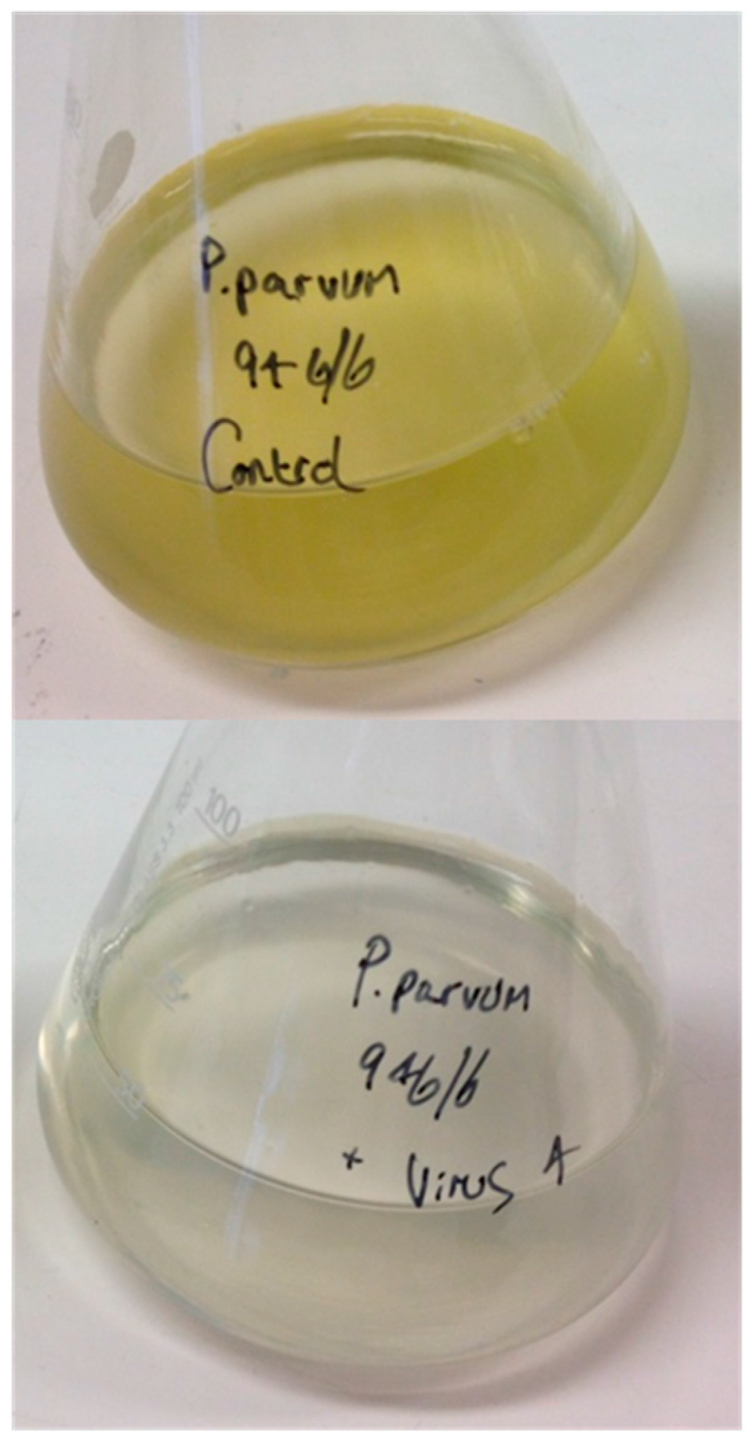
2.2. Isolation of Lytic Virus Particles

PpDNAV was isolated from surface water samples taken at various locations on Hickling Broad, Norfolk, England on 9 February 2016. In brief, 4 × 100 mL water samples from various locations around the Broad were centrifuged at 3000× g and the supernatant subsequently filtered through 0.45 µm pore-size filters (Sartorius AG, Goettingen, Germany). The resulting solutions were then concentrated 100- to 200-fold using 100 kDa MW cut off spin filters (Amicon Ultra 15, Merck Millipore, Watford, UK) to give 0.5 to 1 mL of viral concentrate, which was stored at 4 °C in the dark until use. Small volumes (0.2 mL) of concentrate from each location were added to 1.8 mL of exponentially growing cultures of P. parvum 946/6. Blank culture medium was used as a control. Cultures were visually inspected for signs of cell lysis (culture clearing) after 7–10 days where the control cultures continued to grow. Culture clearing was then followed up by Transmission Electron Microscopy (TEM) analysis of the culture lysates. Clonal populations of PpDNAV were obtained by taking the supernatant of a lysed culture, and exhaustively diluting with media. These diluted samples (0.2 mL) were added to 1.8 mL of an exponentially growing culture of P. parvum 946/6. The highest dilution that still produced cell lysis after seven days was taken through to the next round. This was repeated at least three times and resulted in a population of PpDNAV free of morphologically different viruses, as judged by TEM.

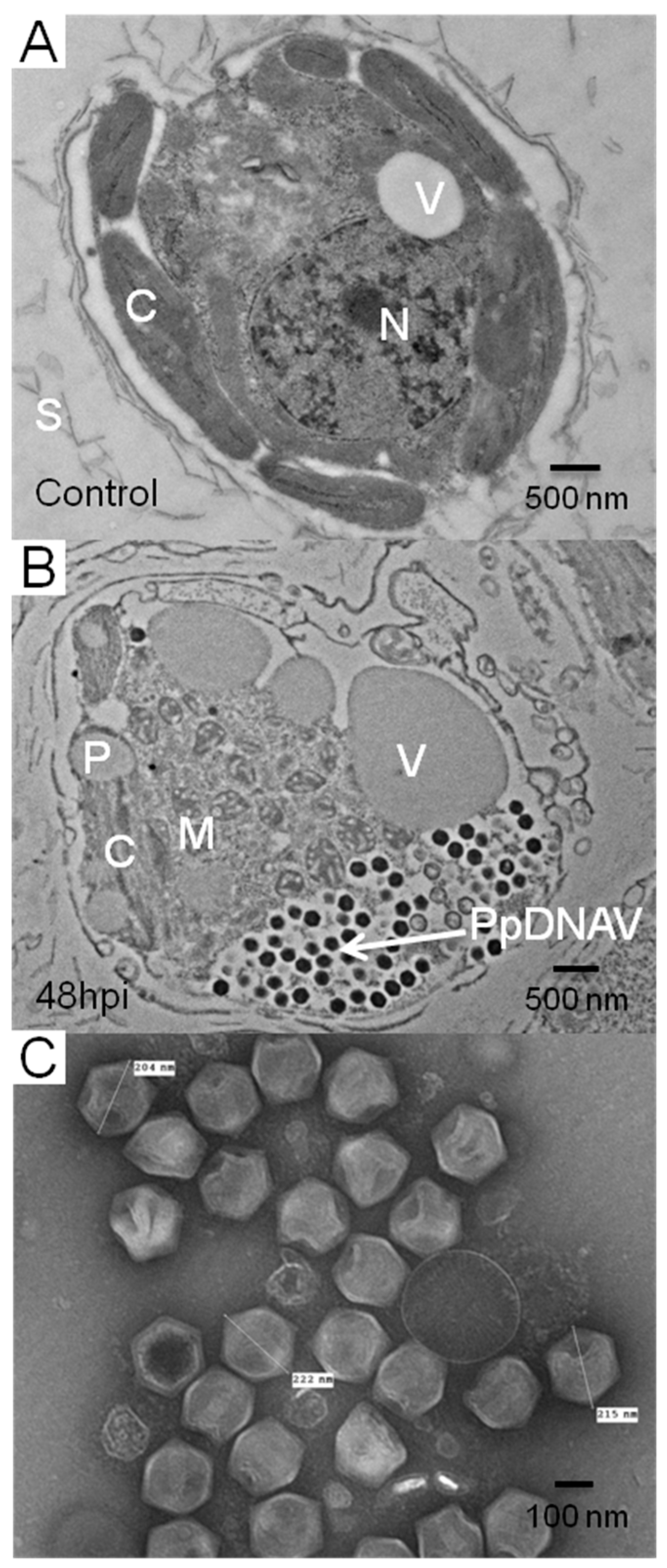
2.3. Transmission Electron Microscopy

For TEM analysis of virus-like particles (VLPs) in culture supernatant, 2 mL of a virus-lysed culture was filtered through 0.45 µm filters and 10 µL of the filtrate was adsorbed onto a 400 mesh copper palladium grid with a carbon-coated pyroxylin support film before being negatively stained with 2% aqueous uranyl acetate [28]. The grids were viewed in a FEI Tecnai 20 transmission electron microscope (Eindhoven, The Netherlands) at 200 kV and digital TIFF images were taken with an AMT XR60B digital camera (Deben, Bury St Edmunds, UK).
For analysing intracellular VLPs, 1 mL of infected cultures of P. parvum 946/6 was taken at 24 and 48 h post-infection (p.i.). These were centrifuged at 3000× g to pellet algal cells and the supernatant was discarded. The pellet was washed twice with sterile medium. The pelleted cells were then resuspended in 2.5% (v/v) aqueous glutaraldehyde solution and left overnight. This suspension was then centrifuged at 3000× g to pellet the algal cells. Half the volume of the supernatant was then discarded and an equal volume of warm (60 °C) low gelling temperature agarose (Sigma Aldrich, Haverhill, UK) was added, before resuspension of the cells and placing on ice to solidify. The solidified samples were then put into 2.5% (v/v) glutaraldehyde with 0.05 M sodium cacodylate, pH 7.3 [29] and left overnight. Using a Leica EM TP machine (Leica Microsystems, Cambridge, UK), the samples were washed in 0.05 M sodium cacodylate and then post-fixed with 1% (w/v) OsO4 in 0.05 M sodium cacodylate for 60 min at room temperature. After washing and dehydration with ethanol, the samples were gradually infiltrated with LR White resin (London Resin Company, London, UK) according to the manufacturer’s instructions. After polymerization, the resulting material was sectioned with a diamond knife using a Leica EM UC6 ultramicrotome (Leica Microsystems). Ultrathin sections of approximately 90 nm were picked up on 200 mesh gold grids that had been coated in pyroxylin and carbon. The grids were then contrast-stained with 2% (w/v) uranyl acetate for 1 h and 1% (w/v) lead citrate for 1 min, washed in distilled water and air-dried. The grids were then viewed with a FEI Tecnai 20 transmission electron microscope (Eindhoven, The Netherlands) at 200 kV and digital TIFF images were produced.

2.4. Host Specificity

Fifteen different strains of Prymnesium were tested in triplicate for signs of cell lysis by PpDNAV using the infection methodology described above. Cell lysis, as observed by culture clearing, was noted for five of the 15 strains tested (Table 1).

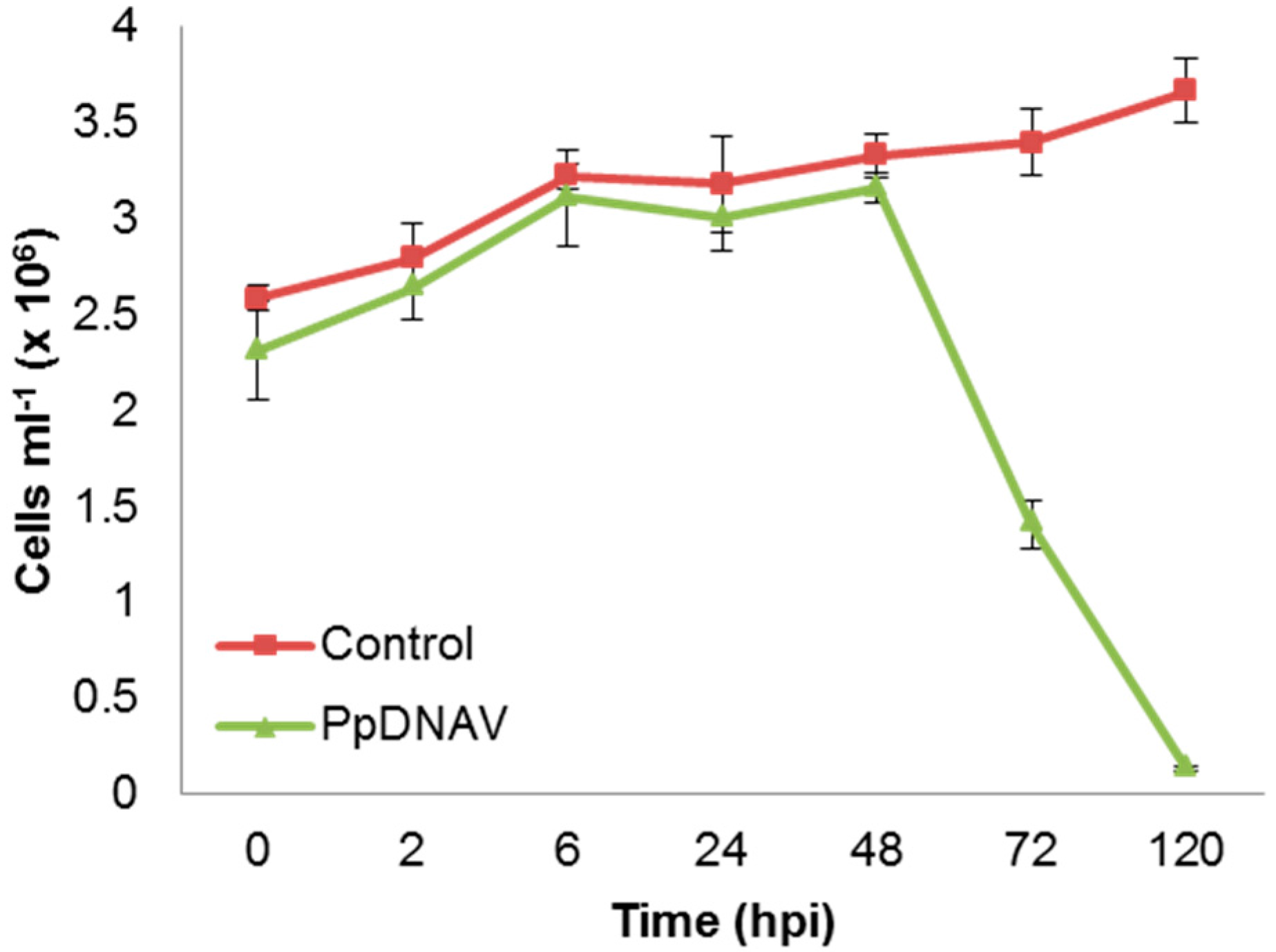
2.5. Infection Cycle

The virus–algae lytic cycle was investigated by accurately recording algal cell abundance during an infection cycle. A late-log phase culture of P. parvum 946/6 was infected with PpDNAV (0.1% v/v) and triplicate 2 mL aliquots were taken at various time points post infection (p.i.). These were diluted with 0.2 µm filtered seawater prior to counting using a Multisizer 3 Analyser (Beckman Coulter, High Wycombe, UK) fitted with a 100 µm aperture tube. The control culture continued to grow throughout the experiment, whilst the infected algal culture was lysed rapidly after 48 h.

2.6. Chloroform Sensitivity

To test the virus sensitivity to chloroform, an adaptation of the method of Martínez Martínez et al. was employed [30]. Briefly, 1 mL of 0.45 µm-filtered PpDNAV was added to an equivalent volume of chloroform and shaken vigorously for 5 min. The resulting mixture was then centrifuged at 4000× g in a benchtop centrifuge for 5 min to separate the organic and polar layers. The aqueous phase was transferred by pipetting to a clean microcentrifuge tube and incubated at 37 °C for 1 h to remove residual chloroform. As a control, 1 mL of chloroform was added to 1 mL of f/2 medium. Chloroform-treated PpDNAV, chloroform-treated medium and untreated PpDNAV were added to P. parvum 946/6 as described above in the infectivity experiment protocol; signs of lysis, as judged by culture clearing, were recorded after one week.

2.7. Viral DNA Extraction, Sequencing, and Phylogenetic Analyses

For DNA extraction, 1 L of late log phase P. parvum 946/6 was infected with axenic PpDNAV (0.1% v/v). Lysis was allowed to occur over a period of five days, by which point almost all cells had been lysed. The culture was centrifuged at 6500× g to pellet cell debris, before being filtered through 0.22 µm filters to remove remaining cell debris or contaminating bacteria. The filtrate was incubated for 72 h with 100 μg/mL carbenicillin before being concentrated to 30 mL using 100 kDa mw cut-off spin filters. Ultracentrifugation at 150,000× g was used to pellet viral particles, and these were re-suspended in 2 mL of ρ = 1.4 CsCl and layered onto a CsCl gradient which was resolved at 150,000× g for 18 h. Fractions from ρ = 1.3 to ρ = 1.4 were pooled and DNA extracted using a PureLink Viral RNA/DNA Kit, according to the manufacturer’s protocol.
An amount of 1 µg of purified viral DNA was then sent to The Earlham Institute, UK, for Illumina MiSeq sequencing (Illumina, Inc., San Diego, CA, USA) and assembly. The initial assembly was then analysed using GeneMarkS [31] which identified 332 protein-coding sequences. BLASTp analysis was then performed against the National Center for Biotechnology Information (NCBI) GenBank nonredundant (nr) protein sequence database [32] to identify major capsid protein and DNA Pol B candidates. Nucleic acid and amino acid sequences for the major capsid protein (MCP) and DNA Polymerase B (DNA polB) were submitted to Genbank with the accession codes KY509047 and KY509048, respectively.
Phylogenetic analysis was performed using the obtained sequences for MCP and DNA polB, as well as other related sequences from previously discovered algal viruses, identified using BLASTp. These sequences were aligned using the default settings of multiple sequence alignment software version 7 (MAFFT) [33], and trees were constructed from the neighbour-joining method [34] (midpoint-rooted) using Molecular Evolutionary Genetics Analysis version 7.0 (MEGA7) [35].

3. Results

3.1. Isolation of Lytic Virus Particles

PpDNAV isolation was conducted from water samples collected at Hickling Broad, Norfolk, England. Among four water samples from which viral lysates were prepared, lysis of P. parvum 946/6 occurred with three samples (Figure 1). Transmission electron micrographs of the viral lysates showed that icosahedral VLPs were present in all three samples, but samples 1 and 2 also contained significant levels of phage-like particles; we suspect that these were a result of infection with the low levels of bacteria that were present in the non-axenic P. parvum 946/6 cultures. To avoid further downstream separation of viruses, we chose to continue working with sample 4 only (sourced at—52°44′19.12″ N, Long—1°34′39.49″ E), which appeared by TEM to be free of phages. After a triplicate dilution series, the resulting monoclonal viral lysate still lysed the host cells and TEM of thin-sectioned cells confirmed the presence of VLPs (Figure 2A,B); thereby fulfilling Koch’s postulates.

3.2. Virus Morphology, Host Range, and Infectious Properties

Transmission electron microscopy of isolated and intracellular (thin sectioned) viruses revealed an icosahedral capsid with an average diameter of 221 nm (n = 71) (Figure 2). Although no external viral lipid membrane was evident, some viral particles showed an internal white ‘halo’ between the capsid and the DNA of PpDNAV, suggestive of the virus having an internal membrane. The presence of a viral factory or viroplasm [36] in the host cytoplasm, and in some cases an imperfect vertex or a tail-like structure were also observed (Figures S1 and S2). As seen in Figure 2B, establishment of a viral factory in the host cytoplasm also results in a loss of the nuclear envelope and therefore loss of the nucleus. This was observed in the majority of infected cells examined at 48 h p.i.
Fifteen different strains of Prymnesium were screened for sensitivity to PpDNAV (Table 1). PpDNAV was found to be sensitive to chloroform, whereby the chloroform-treated virus no longer caused lysis of P. parvum 946/6 (Figure S3). This supports the notion of a viral membrane in this system.
The lytic cycle of the virus was explored to determine both the incubation period and eclipse period (Figure 3). At 48 h p.i., the cells had clearly lost mobility and sedimented at the base of the culture flask. Re-suspension of the cells by shaking led to similar cell counts as seen at 24 h p.i., as determined by Coulter counting. The time before symptoms of viral infection, the incubation period, was therefore judged to be 24 h. The eclipse period reflects the time between infection and appearance of mature virus particles within the host; as new mature virions were first observed 48 h p.i., the eclipse period was judged to be 24–48 h. At 72 h p.i., the onset of cell lysis had occurred. PpDNAV appeared to lyse >95% of host cells by 120 h p.i., whilst uninfected control cultures continued to grow over the full course of the experiment.

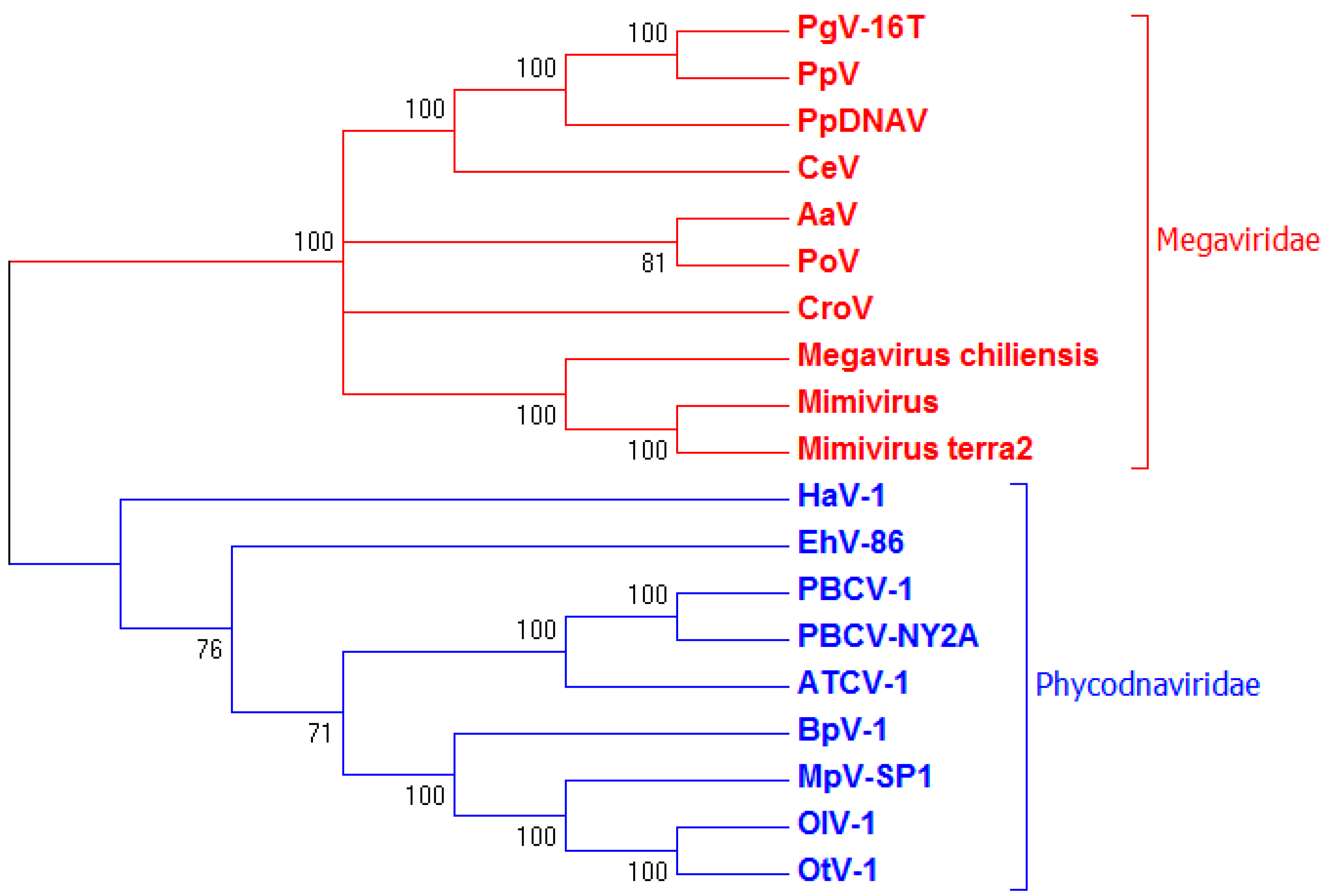
3.3. Genome Sequencing and Phylogenetic Analysis

Predicted proteins from the initial genome assembly included the MCP1 protein (KY509047) and DNA polB (KY509048) which were used for phylogenetic analysis. The 525 aa sequence for MCP1 was found to have 91% sequence similarity to the major capsid protein 1 of Phaeocystis globosa virus (YP_008052475.1) and 84% similarity to MCP1 of Organic Lake Phycodnavirus 2 (ADX06358.1) with E-values of 0.0 in each case. This alignment allowed construction of a phylogenetic tree (Figure S4) that shows clustering with other megaviruses, including PgV-16T.
For DNA polB (KY509048), the 1281 aa sequence displayed 77% sequence similarity to DNA polB of PgV-16T (YP_008052566.1) and 64% similarity to DNA polB of Organic Lake phycodnavirus 2 (ADX06483.1). The resulting phylogenetic tree (Figure 4) shows a similar clustering of PpDNAV to the algal Megaviridae family, but also illustrates an obvious divergence between algal viruses that fall within the Megaviridae family and those that do not; with EhV-86 and Heterosigma akashiwo virus (HaV)-1 rightfully placed outside of the Megaviridae clade.

4. Discussion

Haptophytes are abundant in marine waters but can also thrive in brackish inland waters. Whilst a significant amount of work has been done on the marine dwelling coccolithophore Emiliania huxleyi and its associated viruses, little work has looked at the toxin-producing members of the haptophytes. In the present study, we isolated and characterized a novel megavirus, PpDNAV, from brackish inland waters where harmful blooms of Prymnesium parvum frequently occur [37]. We showed that this lytic virus was able to infect P. parvum 946/6, later expanded to five out of 15 Prymnesium strains tested. Morphological and phylogenetic analysis of two core dsDNA virus conserved genes suggests that this virus belongs to the extended Megaviridae family of algal-infecting viruses.
Transmission electron microscopy of negatively stained virus particles from a lysed culture supernatant revealed icosahedral capsids with an average diameter of 221 nm. Many particles appeared to have one imperfect vertex, with some showing material protruding from what appeared to be a stargate [38] (Figure S1). These likely represent particles in an advanced stage of packing or unpacking genetic material [38], and suggested early on that PpDNAV lies in the extended Megaviridae branch of algal viruses. Thin sections of infected P. parvum 946/6 cells showed evidence for a viroplasm as the site of replication, where empty capsids could be seen closer to the centre of the viroplasm (Figure S2). This further supported the inclusion of PpDNAV in the extended Megaviridae family [39]. The infectivity of PpDNAV is chloroform sensitive (Figure S3), and the lack of an obvious external lipid membrane observed by TEM may suggest that internal membrane/s are present; although chloroform sensitivity cannot always be used to confirm lipid membrane presence [40].
New mature virions were first observed by electron microscopy at 48 h p.i., so the eclipse period of the virus in infected algal cells was estimated to be 24–48 h. At 48 h, the algal cell count as recorded by Coulter counting was still the same as at 24 h, but a complete sedimentation of cells had occurred, suggesting a loss in motility and a likely shutdown of important cellular processes. By 72 h, a rapid decline in cell abundance could be observed, showing that the loss of motility precedes the host lysis event, as is seen for some other flagellated algae [18]. In its natural environment, this may lead to accumulation of viral particles at the sediment surface rather than dispersed in the water column.
The algal host species specificity of PpDNAV was assessed against P. parvum 946/6, which had been kept in 7–8 PSU f/2 medium for two years, and 14 other Prymnesium strains which had been maintained in a full strength seawater medium. Initially, PpDNAV only infected P. parvum 946/6, but after ~6 months of sub-culturing of the other 14 strains in 7–8 PSU f/2 medium, the host range broadened to five out of the 15 strains. We speculate that the change in salinity contributed to the change in sensitivity to PpDNAV; recent work by Nedbalová et al. [41] suggests that a change in membrane lipid composition in different salinities may account for this situation. This somewhat less restricted host range is similar to that found for Haptolina ericina virus (HeV RF02), Prymnesium kappa virus (PkV RF01) and Prymnesium kappa virus (PkV RF02) [42]. Taken together, this suggested that PpDNAV was a member of the algal Megavirus family [43].
Phylogenetic analysis using sequences for MCP1 and DNA polB of PpDNAV confirmed morphological findings, showing that PpDNAV clusters amongst the algal viruses belonging to the Megaviridae family, such as PgV-16T [3,6], Chrysochromulina ericina virus (CeV) [44], Pyramimonas orientalis virus (PoV) [45] and the recently reassigned Aureococcus anophagefferens virus (AaV) [46]. With the exception of Emliania huxleyi virus (EhV-86), which appears to branch independently, the close clustering of viruses infecting haptophytes, as well as the chlorella viruses clustering together, supports the notion that viruses co-evolve with their hosts [6,46,47].
Of the algal viruses compared in this study, only HaV-1 is known to infect a toxin producing host [12,13,14]. However, the toxic metabolites responsible for bloom toxicity are not established in Heterosigma akashiwo, making studies of viral impact on toxicity difficult. On the other hand, reports of toxic P. parvum metabolites are numerous and include fatty acids [48], glycerolipids [49] and very large ladder-frame polyether toxins, known collectively as the prymnesins [50,51,52]. Reports of cases of toxic and non-toxic blooms of Prymnesium and other harmful algal species [37] has led to speculation that an ecological trigger exists for toxicity. While efforts have been made to associate nutrients, pH and other conditions to bloom toxicity [21], the identity of the full spectrum of toxicity-causing agents remains to be establshed; there may conceivably be a role for viral infection in Prymnesium cell lysis and hence toxin release. We now have the opportunity to use this algae–virus system in clearing up some of these unanswered questions. Further studies into the effect of viral infection and host algal cell lysis on toxic bloom events need to be explored in order to fully understand the underlying mechanisms behind production and release of toxins from Prymnesium. In addition, as further sequences of algal viruses become available, new opportunities will open up for accurate monitoring of viral population fluctuations with respect their host. Furthermore, the increase in characterized viruses will provide more information when analysing metagenomic data sets such as those generated by the Tara Oceans expedition [53,54]. Hence the discovery and characterization of PpDNAV in this study will aid this burgeoning field of scientific endeavour.

Supplementary Materials

The following are available online at www.mdpi.com/1999-4915/9/3/40/s1. Figure S1: Electron micrographs showing PpDNAV particles with an imperfect vertex; Figure S2: Electron micrographs showing both mature and immature virions close to a viroplasm; Figure S3: Chloroform sensitivity as judged by culture clearing; Figure S4: Neighbor-joining phylogenetic tree of MCPs.

Acknowledgments

These studies were supported by the BBSRC Institute Strategic Programme on Understanding and Exploiting Metabolism (MET) [BB/j004561/1] and the John Innes Foundation. B.A.W. was supported by a BBSRC industrial CASE PhD studentship supported by Environment Agency. We thank Willie Wilson for helpful advice of viral morphology. We also thank Steve Lane, Andy Hindes, John Currie, Jenny Pratscher and Colin Murrell for their ongoing support.

Author Contributions

B.A.W. and R.A.F. conceived and designed the experiments; B.A.W. performed the experiments, with help from I.C.V. for chloroform sensitivity and culture maintenance, and J.E.B. for thin-section preparation and EM imaging; D.C.S. provided 14 Prymnesium strains for host range screening and reviewed the manuscript; G.M. also reviewed the manuscript; B.A.W., G.M. and R.A.F. analysed the data; B.A.W. and R.A.F. wrote the manuscript.

Conflicts of Interest

The authors declare no conflict of interest.

References

  1. Thingstad, T.F. Elements of a theory for the mechanisms controlling abundance, diversity, and biogeochemical role of lytic bacterial viruses in aquatic systems. Limnol. Oceanogr. 2000, 45, 1320–1328. [Google Scholar] [CrossRef]
  2. Fuhrman, J.A. Marine viruses and their biogeochemical and ecological effects. Nature 1999, 399, 541–548. [Google Scholar] [CrossRef] [PubMed]
  3. Santini, S.; Jeudy, S.; Bartoli, J.; Poirot, O.; Lescot, M.; Abergel, C.; Barbe, V.; Wommack, K.E.; Noordeloos, A.A.; Brussaard, C.P.; et al. Genome of Phaeocystis globosa virus PgV-16T highlights the common ancestry of the largest known DNA viruses infecting eukaryotes. Proc. Natl. Acad. Sci. USA 2013, 110, 10800–10805. [Google Scholar] [CrossRef] [PubMed]
  4. Wilson, W.H.; Tarran, G.A.; Schroeder, D.; Cox, M.; Oke, J.; Malin, G. Isolation of viruses responsible for the demise of an Emiliania huxleyi bloom in the English Channel. J. Mar. Biol. Assoc. UK 2002, 82, 369–377. [Google Scholar] [CrossRef]
  5. Schroeder, D.C.; Oke, J.; Malin, G.; Wilson, W.H. Coccolithovirus (Phycodnaviridae): Characterisation of a new large dsDNA algal virus that infects Emiliana huxleyi. Arch. Virol. 2002, 147, 1685–1698. [Google Scholar] [CrossRef] [PubMed]
  6. Brussaard, C.P.D.; Short, S.M.; Frederickson, C.M.; Suttle, C.A. Isolation and Phylogenetic Analysis of Novel Viruses Infecting the Phytoplankton Phaeocystis globosa (Prymnesiophyceae). Appl. Environ. Microbiol. 2004, 70, 3700–3705. [Google Scholar] [CrossRef] [PubMed]
  7. Moniruzzaman, M.; Gann, E.R.; LeCleir, G.R.; Kang, Y.; Gobler, C.J.; Wilhelm, S.W. Diversity and dynamics of algal Megaviridae members during a harmful brown tide caused by the pelagophyte, Aureococcus anophagefferens. FEMS Microbiol. Ecol. 2016, 92, fiw058. [Google Scholar] [CrossRef] [PubMed]
  8. Short, S.M. The ecology of viruses that infect eukaryotic algae. Environ. Microbiol. 2012, 14, 2253–2271. [Google Scholar] [CrossRef] [PubMed]
  9. Brussaard, C.P.D. Viral Control of Phytoplankton Populations—A Review. J. Eukaryot. Microbiol. 2004, 51, 125–138. [Google Scholar] [CrossRef] [PubMed]
  10. Suttle, C.A. Marine viruses—Major players in the global ecosystem. Nat. Rev. Microl. 2007, 5, 801–812. [Google Scholar] [CrossRef] [PubMed]
  11. Wilhelm, S.W.; Suttle, C.A. Viruses and Nutrient Cycles in the Sea: Viruses play critical roles in the structure and function of aquatic food webs. Bioscience 1999, 49, 781–788. [Google Scholar] [CrossRef]
  12. Nagasaki, K.; Yamaguchi, M. Isolation of a virus infectious to the harmful bloom causing microalga Heterosigma akashiwo (Raphidophyceae). Aquat. Microb. Ecol. 1997, 13, 135–140. [Google Scholar] [CrossRef]
  13. Keizo, N.; Mineo, Y. Intra-species host specificity of HaV (Heterosigma akashiwo virus) clones. Aquat. Microb. Ecol. 1998, 14, 109–112. [Google Scholar] [CrossRef]
  14. Keizo, N.; Mineo, Y. Effect of temperature on the algicidal activity and the stability of HaV (Heterosigma akashiwo virus). Aquat. Microb. Ecol. 1998, 15, 211–216. [Google Scholar] [CrossRef]
  15. Lawrence, J.E.; Chan, A.M.; Suttle, C.A. A novel virus (HaNIV) causes lysis of the toxic bloom-forming alga Heterosigma akashiwo (Raphidophyceae). J. Phycol. 2001, 37, 216–222. [Google Scholar] [CrossRef]
  16. Lawrence, J.E.; Chan, A.M.; Suttle, C.A. Viruses causing lysis of the toxic bloom-forming alga Heterosigma akashiwo (Raphidophyceae) are widespread in coastal sediments of British Columbia, Canada. Limnol. Oceanogr. 2002, 47, 545–550. [Google Scholar] [CrossRef]
  17. Tai, V.; Lawrence, J.E.; Lang, A.S.; Chan, A.M.; Culley, A.I.; Suttle, C.A. Characterization of HaRNAV, a single-stranded RNA virus causing lysis of Heterosigma akashiwo (Raphidophyceae). J. Phycol. 2003, 39, 343–352. [Google Scholar] [CrossRef]
  18. Janice, E.L.; Curtis, A.S. Effect of viral infection on sinking rates of Heterosigma akashiwo and its implications for bloom termination. Aquat. Microb. Ecol. 2004, 37, 1–7. [Google Scholar] [CrossRef]
  19. Lawrence, J.E.; Brussaard, C.P.D.; Suttle, C.A. Virus-Specific Responses of Heterosigma akashiwo to Infection. Appl. Environ. Microbiol. 2006, 72, 7829–7834. [Google Scholar] [CrossRef] [PubMed]
  20. Granéli, E.; Edvardsen, B.; Roelke, D.L.; Hagström, J.A. The ecophysiology and bloom dynamics of Prymnesium spp. Harmful Algae 2012, 14, 260–270. [Google Scholar] [CrossRef]
  21. Manning, S.R.; La Claire, J.W. Prymnesins: Toxic Metabolites of the Golden Alga, Prymnesium parvum Carter (Haptophyta). Mar. Drugs 2010, 8, 678–704. [Google Scholar] [CrossRef] [PubMed]
  22. Schoemann, V.; Becquevort, S.; Stefels, J.; Rousseau, V.; Lancelot, C. Phaeocystis blooms in the global ocean and their controlling mechanisms: A review. J. Sea Res. 2005, 53, 43–66. [Google Scholar] [CrossRef]
  23. Leblanc, K.; Hare, C.E.; Feng, Y.; Berg, G.M.; DiTullio, G.R.; Neeley, A.; Benner, I.; Sprengel, C.; Beck, A.; Sanudo-Wilhelmy, S.A.; et al. Distribution of calcifying and silicifying phytoplankton in relation to environmental and biogeochemical parameters during the late stages of the 2005 North East Atlantic Spring Bloom. Biogeosciences 2009, 6, 2155–2179. [Google Scholar] [CrossRef]
  24. Malin, G.; Wilson, W.H.; Bratbak, G.; Liss, P.S.; Mann, N.H. Elevated production of dimethylsulfide resulting from viral infection of cultures of Phaeocystis pouchetii. Limnol. Oceanogr. 1998, 43, 1389–1393. [Google Scholar] [CrossRef]
  25. Edvardsen, B.; Paasche, E. Bloom Dynamics and Physiology of Prymnesium and Chrysochromulina in Physiological Ecology of Harmful Algal Blooms; Springer-Verlag: Berlin/Heidelberg, Germany, 1998. [Google Scholar]
  26. Roelke, D.L.; Barkoh, A.; Brooks, B.W.; Grover, J.P.; Hambright, K.D.; LaClaire, J.W.; Moeller, P.D.R.; Patino, R. A chronicle of a killer alga in the west: Ecology, assessment, and management of Prymnesium parvum blooms. Hydrobiologia 2016, 764, 29–50. [Google Scholar] [CrossRef]
  27. Schwierzke, L.; Roelke, D.L.; Brooks, B.W.; Grover, J.P.; Valenti, T.W.; Lahousse, M.; Miller, C.J.; Pinckney, J.L. Prymnesium parvum Population Dynamics During Bloom Development: A Role Assessment of Grazers and Virus. J. Am. Water. Resour. Assoc. 2010, 46, 63–75. [Google Scholar] [CrossRef]
  28. Brenner, S.; Horne, R.W. A negative staining method for high resolution electron microscopy of viruses. Biochim. Biophys. Acta 1959, 34, 103–110. [Google Scholar] [CrossRef]
  29. Gordon, G.B.; Miller, L.R.; Bensch, K.G. Fixation of tissue culture cells for ultrastructural cytochemistry. Exp. Cell Res. 1963, 31, 440–443. [Google Scholar] [CrossRef]
  30. Martínez Martínez, J.; Boere, A.; Gilg, I.; van Lent, J.W.M.; Witte, H.J.; van Bleijswijk, J.D.L.; Brussaard, C.P.D. New lipid envelope-containing dsDNA virus isolates infecting Micromonas pusilla reveal a separate phylogenetic group. Aquat. Microb. Ecol. 2015, 74, 17–28. [Google Scholar] [CrossRef]
  31. Besemer, J.; Lomsadze, A.; Borodovsky, M. GeneMarkS: A self-training method for prediction of gene starts in microbial genomes. Implications for finding sequence motifs in regulatory regions. Nucleic Acids Res. 2001, 29, 2607–2618. [Google Scholar] [CrossRef] [PubMed]
  32. Altschul, S.F.; Gish, W.; Miller, W.; Myers, E.W.; Lipman, D.J. Basic local alignment search tool. J. Mol. Biol. 1990, 215, 403–410. [Google Scholar] [CrossRef]
  33. Katoh, K.; Toh, H. Recent developments in the MAFFT multiple sequence alignment program. Brief. Bioinform. 2008, 9, 286–298. [Google Scholar] [CrossRef] [PubMed]
  34. Saitou, N.; Nei, M. The neighbor-joining method: A new method for reconstructing phylogenetic trees. Mol. Biol. Evol. 1987, 4, 406–425. [Google Scholar] [CrossRef] [PubMed]
  35. Kumar, S.; Stecher, G.; Tamura, K. MEGA7: Molecular Evolutionary Genetics Analysis version 7.0 for bigger datasets. Mol. Biol. Evol. 2016, 33, 1870–1874. [Google Scholar] [CrossRef] [PubMed]
  36. Den Boon, J.A.; Diaz, A.; Ahlquist, P. Cytoplasmic Viral Replication Complexes. Cell Host Microbe 2010, 8, 77–85. [Google Scholar] [CrossRef] [PubMed]
  37. Holdway, P.A.; Watson, R.A.; Moss, B. Aspects of the ecology of Prymnesium parvum (Haptophyta) and water chemistry in the Norfolk Broads, England. Freshwat. Biol. 1978, 8, 295–311. [Google Scholar] [CrossRef]
  38. Zauberman, N.; Mutsafi, Y.; Halevy, D.B.; Shimoni, E.; Klein, E.; Xiao, C.; Sun, S.; Minsky, A. Distinct DNA Exit and Packaging Portals in the Virus Acanthamoeba polyphaga mimivirus. PLoS Biol. 2008, 6, e114. [Google Scholar] [CrossRef] [PubMed]
  39. Mutsafi, Y.; Fridmann-Sirkis, Y.; Milrot, E.; Hevroni, L.; Minsky, A. Infection cycles of large DNA viruses: Emerging themes and underlying questions. Virology 2014, 466–467, 3–14. [Google Scholar] [CrossRef] [PubMed]
  40. Feldman, H.A.; Wang, S.S. Sensitivity of various viruses to chloroform. Proc. Soc. Exp. Biol. Med. 1961, 106, 736–738. [Google Scholar] [CrossRef] [PubMed]
  41. Nedbalová, L.; Střížek, A.; Sigler, K.; Řezanka, T. Effect of salinity on the fatty acid and triacylglycerol composition of five haptophyte algae from the genera Coccolithophora, Isochrysis and Prymnesium determined by LC-MS/APCI. Phytochemistry 2016, 130, 64–76. [Google Scholar] [CrossRef] [PubMed]
  42. Johannessen, T.V.; Bratbak, G.; Larsen, A.; Ogata, H.; Egge, E.S.; Edvardsen, B.; Eikrem, W.; Sandaa, R.-A. Characterisation of three novel giant viruses reveals huge diversity among viruses infecting Prymnesiales (Haptophyta). Virology 2015, 476, 180–188. [Google Scholar] [CrossRef] [PubMed]
  43. Wilhelm, S.W.; Coy, S.R.; Gann, E.R.; Moniruzzaman, M.; Stough, J.M.A. Standing on the Shoulders of Giant Viruses: Five Lessons Learned about Large Viruses Infecting Small Eukaryotes and the Opportunities They Create. PLoS Pathog. 2016, 12, e1005752. [Google Scholar] [CrossRef] [PubMed]
  44. Gallot-Lavallée, L.; Pagarete, A.; Legendre, M.; Santini, S.; Sandaa, R.-A.; Himmelbauer, H.; Ogata, H.; Bratbak, G.; Claverie, J.-M. The 474-Kilobase-Pair Complete Genome Sequence of CeV-01B, a Virus Infecting Haptolina (Chrysochromulina) ericina (Prymnesiophyceae). Genome Announc. 2015, 3, e01413–e01415. [Google Scholar] [CrossRef]
  45. Sandaa, R.-A.; Heldal, M.; Castberg, T.; Thyrhaug, R.; Bratbak, G. Isolation and Characterization of Two Viruses with Large Genome Size Infecting Chrysochromulina ericina (Prymnesiophyceae) and Pyramimonas orientalis (Prasinophyceae). Virology 2001, 290, 272–280. [Google Scholar] [CrossRef] [PubMed]
  46. Moniruzzaman, M.; LeCleir, G.R.; Brown, C.M.; Gobler, C.J.; Bidle, K.D.; Wilson, W.H.; Wilhelm, S.W. Genome of brown tide virus (AaV), the little giant of the Megaviridae, elucidates NCLDV genome expansion and host-virus coevolution. Virology 2014, 466–467, 60–70. [Google Scholar] [CrossRef] [PubMed]
  47. Mirza, S.F.; Staniewski, M.A.; Short, C.M.; Long, A.M.; Chaban, Y.V.; Short, S.M. Isolation and characterization of a virus infecting the freshwater algae Chrysochromulina parva. Virology 2015, 486, 105–115. [Google Scholar] [CrossRef] [PubMed]
  48. Henrikson, J.C.; Gharfeh, M.S.; Easton, A.C.; Easton, J.D.; Glenn, K.L.; Shadfan, M.; Mooberry, S.L.; Hambright, K.D.; Cichewicz, R.H. Reassessing the ichthyotoxin profile of cultured Prymnesium parvum (golden algae) and comparing it to samples collected from recent freshwater bloom and fish kill events in North America. Toxicon 2010, 55, 1396–1404. [Google Scholar] [CrossRef] [PubMed]
  49. Kozakai, H.; Oshima, Y.; Yasumoto, T. Isolation and Structural Elucidation of Hemolysin from the Phytoflagellate Prymnesium parvum. Agric. Biol. Chem. 1982, 46, 233–236. [Google Scholar] [CrossRef]
  50. Igarashi, T.; Satake, M.; Yasumoto, T. Prymnesin-2: A Potent Ichthyotoxic and Hemolytic Glycoside Isolated from the Red Tide Alga Prymnesium parvum. J. Am. Chem. Soc. 1996, 118, 479–480. [Google Scholar] [CrossRef]
  51. Igarashi, T.; Satake, M.; Yasumoto, T. Structures and Partial Stereochemical Assignments for Prymnesin-1 and Prymnesin-2: Potent Hemolytic and Ichthyotoxic Glycosides Isolated from the Red Tide Alga Prymnesium parvum. J. Am. Chem. Soc. 1999, 121, 8499–8511. [Google Scholar] [CrossRef]
  52. Rasmussen, S.A.; Meier, S.; Andersen, N.G.; Blossom, H.E.; Duus, J.Ø.; Nielsen, K.F.; Hansen, P.J.; Larsen, T.O. Chemodiversity of Ladder-Frame Prymnesin Polyethers in Prymnesium parvum. J. Nat. Prod. 2016, 79, 2250–2256. [Google Scholar] [CrossRef] [PubMed]
  53. Roux, S.; Brum, J.R.; Dutilh, B.E.; Sunagawa, S.; Duhaime, M.B.; Loy, A.; Poulos, B.T.; Solonenko, N.; Lara, E.; Poulain, J.; et al. Ecogenomics and potential biogeochemical impacts of globally abundant ocean viruses. Nature 2016, 537, 689–693. [Google Scholar] [CrossRef] [PubMed]
  54. Karsenti, E.; Acinas, S.G.; Bork, P.; Bowler, C.; De Vargas, C.; Raes, J.; Sullivan, M.; Arendt, D.; Benzoni, F.; Claverie, J.-M.; et al. A Holistic Approach to Marine Eco-Systems Biology. PLoS Biol. 2011, 9, e1001177. [Google Scholar] [CrossRef] [PubMed] [Green Version]
Figure 1. (Top) control culture; (Bottom) ‘Cleared’ culture 96 h post viral infection.
Viruses 09 00040 g001
Figure 2. (A) Thin-sections of healthy P. parvum 946/6 cells; (B) Thin-sections of P. parvum 946/6 48 h post infection. (C) Free Prymnesium parvum DNA virus (PpDNAV) particles in culture supernatant 72 h post infection. C: chloroplast; V: contractile vacuole; N: nucleus; S: scales; M: mitochondria, P: pyrenoid.
Viruses 09 00040 g002
Figure 3. PpDNAV infection cycle propagated on P. parvum 946/6. Graph shows the average number of algal cells in control cultures (squares) and PpDNAV infected cultures (circles). Error bars represent the standard error for triplicate cultures.
Viruses 09 00040 g003
Figure 4. Phylogenetic clustering of PpDNAV with other large algal Megaviridae. Alignment was performed using the default settings of multiple sequence alignment software version 7 (MAFFT) [33], and the neighbour-joining method (midpoint-rooted) [34] was used to construct a tree from 19 viral DNA Polymerase Beta (polB) sequences using Molecular Evolutionary Genetics Analysis version 7.0 (MEGA7) [35]. The final tree was based on 630 ungapped positions, 500 resampling permutations, and was collapsed for bootstrap values <50. The tree shows that PpDNAV clusters with the well-defined clade of Megaviridae and the algal-infecting Megaviridae (red), and not with the Phycodnaviridae (blue).
Viruses 09 00040 g004
Table 1. Host range of PpDNAV. + lysed culture, − culture not lysed.
Genus/SpeciesStrain CodeLysis with PpDNAV
Prymnesium parvum946/6+
Prymnesium parvum94A-
Prymnesium parvum94C+
Prymnesium parvum579-
Prymnesium patelliferum527A+
Prymnesium patelliferum527C+
Prymnesium patelliferum527D-
Prymnesium sp.522-
Prymnesium sp.569-
Prymnesium sp.592+
Prymnesium sp.593-
Prymnesium sp.595-
Prymnesium sp.596-
Prymnesium sp.597-
Prymnesium sp.598-

